A NEW LOOK
BENEFITS OF CONTRAST-ENHANCED BREAST IMAGING


PAGE 36
METROPOLIS INTERNATIONAL COMPANY SHOWCASE
PAGE 28
PRODUCT FOCUS
WOMEN'S HEALTH
PAGE 31


BENEFITS OF CONTRAST-ENHANCED BREAST IMAGING


PAGE 36
METROPOLIS INTERNATIONAL COMPANY SHOWCASE
PAGE 28
PRODUCT FOCUS
WOMEN'S HEALTH
PAGE 31


• Our team of expert instructors has decades of experience
• Our courses are all designed to engage students through live presentations, lectures, discussions, peer learning sessions, and labs
• All of our courses are AAMI accredited
• Our instructors are here to mentor you and answer your questions after training has been completed
• Courses covering entry-level to experienced engineers in MRI, CT, Angio, Rad Fluoro and Nuclear Medicine
TAKE ADVANTAGE OF OUR YEAR-END COURSES AND FALL INTO SAVINGS!
COMING SOON!
Our 2024 training course schedule will be released soon! For details on courses and upcoming course dates, check out our website www.technicalprospects.com
Contact our team for discounted pricing available on these 2023 year-end courses.
SOMATOM
October 16-27, 2023
NEW! Our Sonatom Go course is 60-hours (virtual and in-person) centered on the core principles of operation, configuration, troubleshooting and repair.
• Identify, configure, troubleshoot, and diagnose componets of the Somatom Go
» Gantry and Table systems
» Power Distribution
» Air-Cooling System
» Image Control and Reconstruction systems
• Relationships and interaction of all system componets
• syngro Native software interface
• Configuration and adjustments via service software
• Troubleshoot and diagnose system problems based on error codes
ARTIS Q
NEW! Our Artis Q course is 60-hours (virtual and inperson) centered on the core principles of operation, configuration, troubleshooting and repair.
• Identify, configure, troubleshoot, and diagnose componets of the Artis Q Zen:
» Stand
» Table
» Computer system and software interface
» Imaging systems
» Generator systems (Polydoros A100G generators)
• Functional use of the system
• Relationships and interactions of all system componets
• Interact with and navigate the system’s syngo software interface
• Configuration and adjustments via service software
YSIO/YSIO MAX
Course is 60-hours (virtual and in-person) which covers servicing the high-end Ysio and Ysio Max.
• Identify, configure, troubleshoot, and diagnose componets
» Table and overhead tube cranes, associated componets
» Polydoros generators - F80, R80, F55
» Siemens FD detector
» The RADIS imaging station and software interface
• Relationships and interactions of all system componets
• Configuration and adjustments via service software
• Troubleshoot and diagnose generator, table, and tube componets based on error code
• Preventative maintenence







Proof that some of the best ideas come as a response to a stressful situation.
COVER STORY
CEM has emerged as a diagnostic tool that adds functional information with which clinicians may identify more quickly whether an abnormality in breast tissue is likely cancerous.


Justin Kern is a multi-modality technologist at St. Joseph Medical Center in Houston, Texas.

IMAGING NEWS

Keep up with the latest news from the diagnostic imaging world.
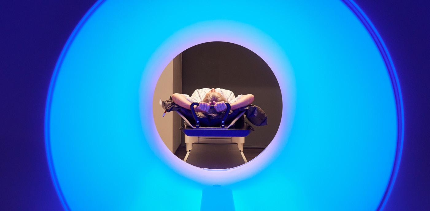
PRODUCT FOCUS
Explore the latest women’s imaging devices.

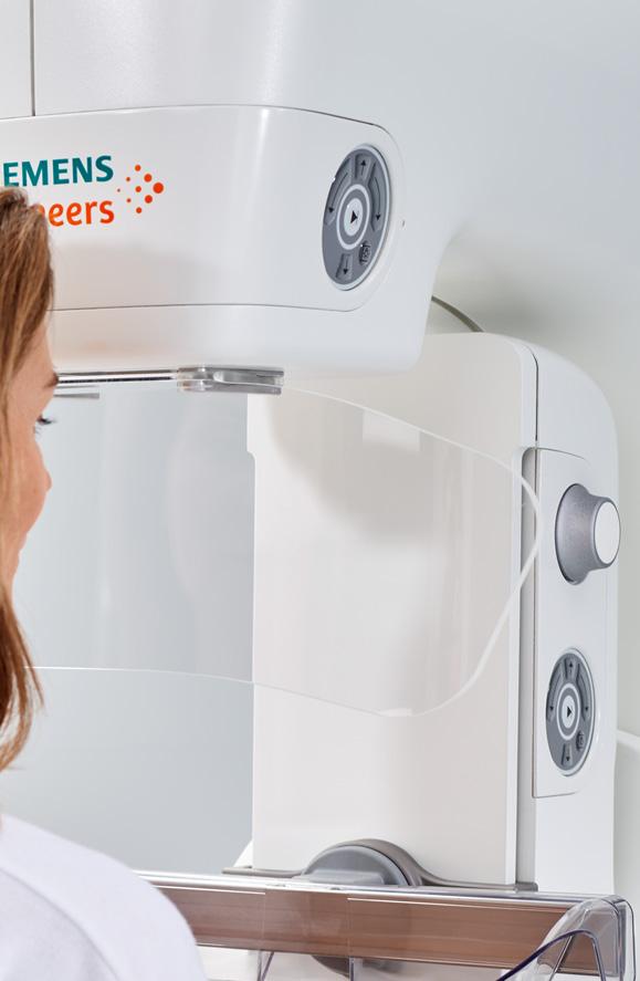
EMOTIONAL INTELLIGENCE
Research shows that people working in small teams to tackle or remove obstacles are more likely to ask questions and brainstorm solutions.

Jamie Coder, RT(R), MHA, CRA, recently explained how a high school injury eventually led to a career in imaging.
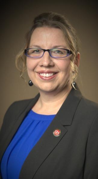
“I tore my ACL in high school and going through diagnostic testing, surgery and rehab led me to wanting to be in health care. I chose imaging because it was a multifaceted field and there is never a dull moment,” Coder says. “I always say that imaging allows you to be there for patients and families and make a difference in a short amount of time.”
Looking back, she is grateful for the help she has received along the way.
“I want to thank North Kansas Hospital for giving my first leadership job in radiology and all the employees that I have had the pleasure of leading, as well as the other supervisors by my side, that guided me to the leader I am today,” Coder says. “I am not perfect and have made mistakes during my career journey. I sometimes even still get in my own way when it comes to my future goals. People outside of radiology don’t always understand the complexities of
all the modalities in radiology. From a young, immature X-ray tech to a career in leadership, I can’t say that I would have changed anything.”
When asked about her greatest accomplishment, she says that she is fortunate to be in a good place.
“I am lucky to have multiple accomplishments in my professional career,” Coder says. “My first was Employee of the Year for a 400-plus bed trauma 2 hospital, but I think the one that felt the most special, was after only 3 months of working with my current staff, I was nominated and awarded the AHRA Award of Excellence in Leadership.”
The work that made the award possible no doubt benefits from the fact that she loves her job.
“I have always loved radiology as it is a close family no matter where you work,” Coder explains. “My first job at Mayo Clinic was operations manager of radiology in La Crosse, Wisconsin. I moved my family seven hours away from our hometown to gain more experience and to reach my ultimate goal to become a radiology director. But Mayo Clinic has shown me that if you can lead in radiology, you can gain skills that can take you anywhere. This is not always the case at
other organizations.”
“Almost two years ago, I was given the opportunity to move outside of my comfort zone and try something new but with some familiarity,” she continues. “I was hired as the operations manager of enterprise radiation safety for Mayo Clinic (this includes all sites in Minnesota, Wisconsin, Florida and Arizona). I have found a position that I absolutely love. The intelligence of my staff amazes me everyday. The direct focus on one very important subject has allowed me to focus on supporting my staff. Oftentimes, in other positions, I felt pulled in many directions and felt like I was not giving one my full attention. I love my job as I can now focus to lead my staff and department into the bright future, without feeling overwhelmed. I can focus on each one individually and supporting all aspects of radiation safety throughout Mayo Clinic.”
Listening to her, it is obvious that there is a sense of caring and an urge to develop talent that comes out in her leadership.
“I lead with my heart. But after 20 years in leadership, I have learned to balance the emotional leadership with the business aspect. I challenge status quo and value the intelligence and visions of those around me,” Coder says. “I also value work/life balance without supporting the human side of your employee, you lose their drive and passion for the organization. We have to support our staff, especially in health care, to make sure they are not giving their all without a solid return on their emotional investment.”
Coder says mentors have helped her grow and advance through the imaging realm.
“Many leaders I can credit for their mentorship throughout my 23 years in imaging and now safety career,” she explains.
She said the two that stand out are the following:
• Matt Foresman, vice president of professional services from North Kansas City Hospital. “He taught me the value of being human regardless of title. He taught me to be present. He was a very busy man but always found time to round each of his departments twice a day. After every conversation I felt empowered and had a smile on my face.”
• Jason Newmark with AHRA. “Along with Bill Algee and many others, Jason was among the first group of people I met at my first annual AHRA conference in 2013 that welcomed me and encouraged me to volunteer. After leaving NKCH for my new role in La Crosse, Jason was always just one call away. He has helped me with resume prep, interviews and ultimately navigating through tough career decisions ever since I met him.”
“The lessons I have learned include: be patient, be kind and never give up. Another takeaway is to ‘listen to learn, not to respond,’” she says.
Coder is paying it forward by providing direction to the next generation of imaging stars.
“Mentorship has always been a passion of mine. I have mentored several new imaging leaders throughout my career and
the focus of that mentorship was guiding my mentee through their passions, hardships and future goals,” she says. “It is not about showing them the path but it is about helping them unleash their own self drive to go out in this big scary world and fight for what they believe in and never look back. Your past teaches you but does not define your future, only you can do that.”
What is more important than her job and colleagues?
“My family is the most important part of me. I have a husband, Ralph, (my high school sweetheart), a 20-year-old son, Robert, and a 17-year-old daughter, Zoe. My parents, in-laws, and cousins mean the world to me,” Coder says. “Like all families, we have faced many adversities. The one word to describe us is resilant. My son, at a young age, was diagnosed with Autism and my very independent, sassy daughter at 16 was involved in a serious car accident that had her fighting for her life, in a minimally conscious state for more than three months. She is now considered a survivor of a traumatic brain injury and fights everyday to find that independence and sassiness again. My son is thriving with our advocacy and his determination, and it is safe to say that my daughter found her sass and is the strongest person I know. I don’t tell this story to have people feel sorry for us, I share to validate that we are humans and life is messy and I have the best support system in my family, friends and the Mayo Clinic family.” •
1. What is the last book you read? The Prey Series (currently on Righteous Prey) by John Sanford
2. Favorite movie? Guardian of the Galaxy movies
3. What is something most of your coworkers don’t know about you? Not much, I am an open book
4. Who is your mentor? Jason Newmark- most recent. As a new leader- Matt Foresman
5. What is one thing you do every morning to start your day? Have a chai tea latte
6. Best advice you ever received? Live for today (You can’t change the past and if you focus too much on the future you will miss out on so much today)
7. Who has had the biggest influence on your life? My Grandpa Bob. He taught me to love unconditionally, always help people in need and resiliency.
8. What would your superpower be? See the future
9. What are your hobbies? Spending time with my family and dog, Callie, shopping, anything in the water, listening to my murder mystery ebooks
10. What is your perfect meal? Surf and Turf
Interventional radiology is a rapidly evolving field with increasingly more complex and technical life-saving procedures that previously could only be performed in the operating room. These procedures require much time and resource which can strain an already full schedule. The challenges of getting procedures on the IR schedule in a timely fashion compete with the overall volume, presenting a unique barrier to reducing lead times.
The decision was made to trial a Saturday clinic with a goal to reduce lead times. The thyroid fine needle aspiration (FNA) for biopsy was determined to be the focus procedure for this trial. Thyroid FNA is a low-risk, non-sedated option that could turn over a number of cases in a short time.

Planning for this clinic required conversations with various stakeholders to determine the availability of resources.
• The interventional radiologist was supportive and agreed to come in on a Saturday.
• Radiology RNs, specimen collector and sonographer were also recruited with incentive pay.
• Pathology determined the method of specimen preservation appropriate that would last over the weekend until specimens could be processed on Monday.
• Pathology also coordinated with the system lab to ensure everyone knew about the influx of specimens.
• Patient Access Services confirmed a registrar available to register patients and a workflow was developed with PAS to alert the team when the patients were ready to be taken for the procedure.
• Patients were scheduled every half hour to allow for registration time and no patients with more than two sites were scheduled during this clinic. At this point, finding an appropriate space was our next
step. The recovery unit attached to the cath lab was secured, an ideal area with nine bays in a contained area and a centralized nursing station. A cart was stocked with all needed procedure supplies. A checklist was made for the team from which to base their process on the day of the clinic.
On the clinic day, the following workflow was followed:
The team arrived 30 minutes prior to the start of the clinic to set up the space. On arrival the patients were registered and the registrar called the team. The RN retrieved the patient, brought them to a bay, did an initial set of vital signs and labeled the consent form. The specimen collector prepared the labels for the specimens. The ultrasound technologist did the preliminary scout scan. The physician arrived, viewed the images and performed the consent. The procedure began with a time out conducted by the RN. As the procedure ended the RN did discharge teaching and recovered the patient. Once parameters were met the patient was walked out by their RN. The room was turned over by the RN or the specimen collector depending on who was available.
As this procedure was occurring, the second RN was ready to accept the next patient and take them through the same process. The RNs alternated taking the patients and kept the schedule moving. Our thyroid clinic proved beneficial:
• The lead time for thyroid biopsies was cut by half.
• Time was open on the schedule during the week for more complex cases that needed more resources to perform.
• Patient satisfaction was clear – uniformly patients were pleased to reduce their wait for an appointment and had the added benefit of not needing to take off work.
While the financial return on thyroid FNA is fairly low, the difference is made up in the open slots created in the schedule for more complex and higher revenue cases. •
Cap your imaging parts expenses.
WHAT IS A CAPITATED PARTS COVERAGE (CPC)?
• Unlimited coverage of all parts and X-ray tubes
• Certified Pre-Owned Glassware
• Includes Technical Support and System Training
• Forward Stocking
WHAT ARE THE BENEFITS TO OUR CUSTOMERS?
• Cap risk on parts expenses
• Improved access to parts & tubes
• Faster access to critical parts with our forward stocking locations
Under our unique Capitated Parts Coverage (CPC), you will have unlimited parts and glass coverage for your imaging equipment. You will also receive premium access to our technical support team and technical training classes.
Call us today to learn more.
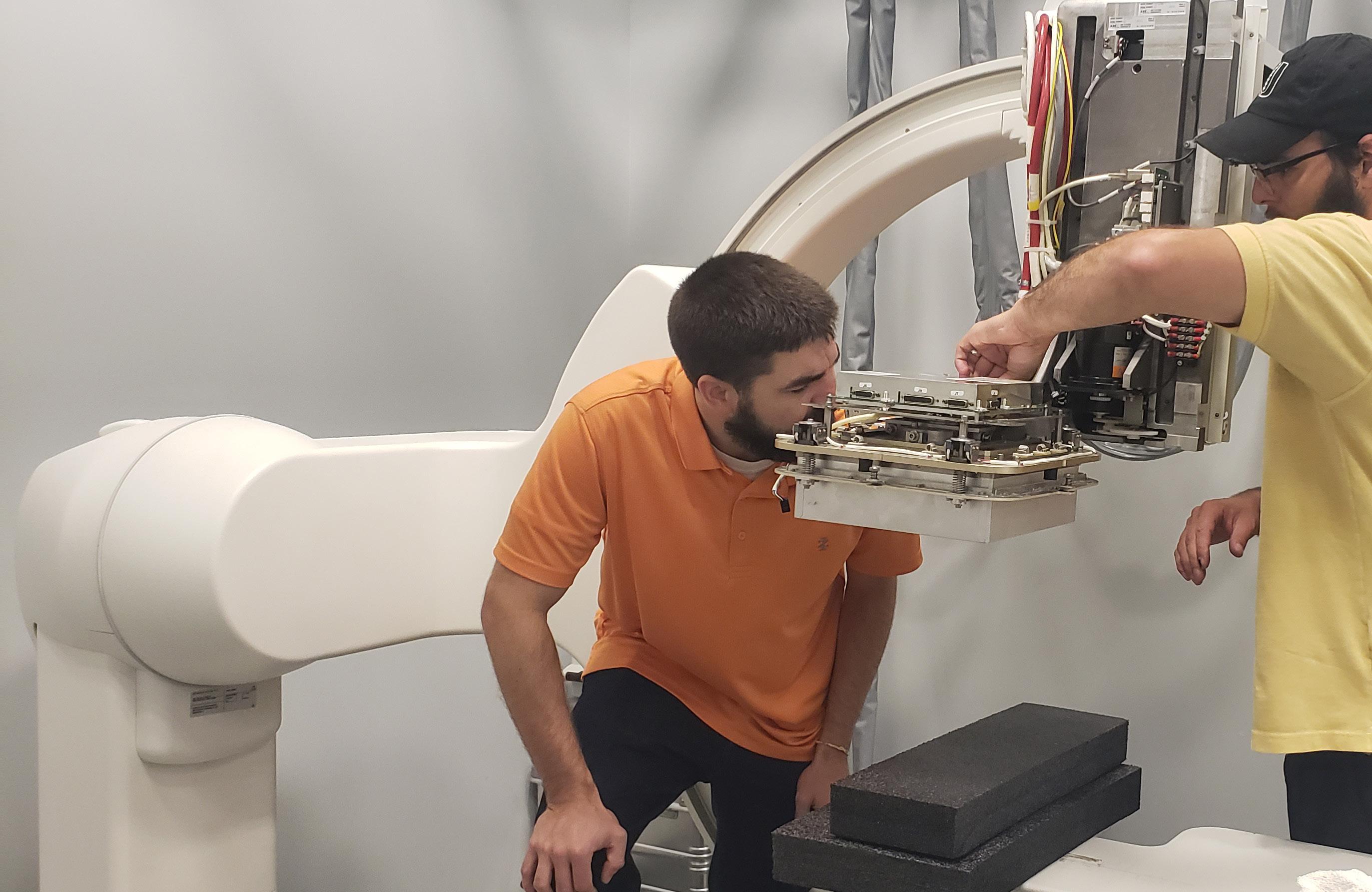
Justin Kern, RT (R,CT), is a multi-modality technologist at St. Joseph Medical Center in Houston, Texas. He earned an associate degree in radiology from Houston Community College before beginning his imaging career. ICE magazine recently found out more about this rising star.

Q: WHERE DID YOU GROW UP?
A: I’m from St. Charles Illinois, a suburb of Chicago.
Q: WHERE DID YOU RECEIVE YOUR IMAGING TRAINING/ EDUCATION?
A: I received my associate degree in radiology from Houston Community College. As a student, I was fortunate to rotate through several large hospitals in the medical center here in Houston. I have a CT certification from ARRT’s post-primary eligibility pathway.
Q: HOW DID YOU FIRST DECIDE TO START WORKING IN IMAGING?
A: I was originally an English and history major at Ohio State University. I eventually decided I wanted a career in health care. I think it was because of my own experience in a hospital during a surgery I underwent in my early 20s. I looked at the
Justin Kern is a history major turned imaging technologist.
hospital environment and thought, “I want to work here.” Imaging was the open road I saw that would take me into the hospital setting professionally, and it has opened a lot of doors for me.
Q: WHAT DO YOU LIKE MOST ABOUT YOUR POSITION?
A: I work in the emergency room for a hospital in downtown Houston. I never know what to expect – every day brings surprises and challenges. I like that.
Q: WHAT IS THE MOST REWARDING ASPECT OF YOUR JOB?
A: I help with the treatment of patients every day who are not only ill or injured, but who are on the fringes of society, for one reason or another. I go home every day knowing I am helping people who truly are in need, and that just feels good. I don’t have to wonder whether my job or my life is making the world a better place. I know it is, at least a little bit.
Q: WHAT INTERESTS YOU THE MOST ABOUT THE IMAGING FIELD?
A: Imaging is a profession that is accessible to anybody with motivation, and which opens up all kinds of different upward paths and opportunities.
Q: WHAT HAS BEEN YOUR GREATEST ACCOMPLISHMENT IN YOUR FIELD THUS FAR?
A: Making it through the COVID-19 pandemic. The whole year of 2020, from February to September in particular, was just a very difficult, frightening and confusing time. Every suspected case, nearly every patient, required a chest X-ray. I was up to my neck in COVID for a while, although I managed to avoid getting sick myself. I’m glad that the worst of that seems to be behind us.
Q: WHAT GOALS DO YOU HAVE FOR YOURSELF IN THE NEXT 5 YEARS?
A: Sometimes I wonder about unconventional imaging jobs, like an imaging tech on a cruise ship, or an imaging tech at NASA. I wonder how someone ends up in those kinds of positions. I think it might be interesting to do something like that some day. •
FAVORITE HOBBY: Piano
FAVORITE FOOD: Indian
FAVORITE VACATION SPOT: Japan
1 THING ON YOUR BUCKET LIST: Learn Cantonese
SOMETHING YOUR COWORKERS DON’T KNOW ABOUT YOU: I can read some Chinese.
As a teenager picking out his first few chords on an old guitar to the sound of the radio, David Jordan finally believed that music could be fun. In school, he’d played clarinet and saxophone, but that was work, not recreation. Plunking along to the death throes of hair metal and the nursery cries of grunge was finally “something I was interested in,” Jordan said; “something my friends think is cool.” In the days when the M in MTV still stood for “music,” videos by bands like Guns N’ Roses and Pearl Jam offered some of the only clues for aspiring rockers like him to get a handle on how some of his favorite songs were performed.
“It was really hard to figure out,” Jordan said. “What is that guitar that you always see Slash play? Those two guys in Pearl Jam have totally different guitars. What are those? I was trying to figure that out.”
Mom and Dad were less impressed at the notion of shelling out for an upgrade to the hand-me-down instrument their boy was starting on. They believed their duties had been discharged in covering the cost of school instruments, and the electric guitar was little more than a toy. So Jordan marshaled the money he’d saved up from a summer of painting decks, and headed down to the shops in his Cleveland hometown. His budget was somewhere between a little and not much, but the clerks recognized his interest level, and set him up with a black Gibson Les Paul knockoff manufactured by Hohner.
“At the time you could get a cheap Strat for ninety-nine bucks,” Jordan said. “This thing cost me like $300. They said, ‘It’s not quite a beginner instrument, but by the time you outgrow it, you’ll be ready for something else.’ I played the heck out of it for four years, and by the time I could have had it fixed, it would have cost more than it was worth.”
With four more years of chops under his belt, Jordan was
no longer the novice who’d started out on the Hohner. One night when he went into the buy-sell-trade shop, the owner was hot to show him its latest acquisition: a single-cutaway Yamaha Weddington Classic. It looked like a Les Paul, and played like a Paul Reed Smith; overengineered for the typical Yamaha customer, the line was only manufactured from 1990 to 1995. But Jordan’s friend knew the instrument could punch above its weight class, especially at the $500 price tag for which he had it on the rack.
“He said, ‘You have to buy this guitar,’” Jordan remembered. “I said, ‘I will find a way.’ I still have it. It’s my number-one favorite go-to.”
Beyond learning his instrument, Jordan was also getting more comfortable as a singer. In the time when popular music was celebrating stripped-down arrangements of songs by bands that were known for electrified performances, “guys with acoustic guitars who can manage a passable Alice in Chains harmony” were everywhere at open mic nights and coffeehouses, Jordan said.

“We would just go in and play a song, and we kind of did that until college,” he said. “Along the way, as I ran across people, it would just come up that I played or that I sing, and I would get in and out of different bands. It was always different stuff, but to me, it was always more fun to plug into an amp and crank it way up.”
In college, Jordan played in a few bands that gigged frequently; after school, he moved to Atlanta, and kept up the pace. Inevitably, his performing career “just kind of fell by the wayside because of life transitions.”
“You run into that tension of trying to find people who hit the sweet spot,” Jordan said. “You’re always going to have folks who are more interested in playing shows, and then you have the folks who want to write and record, and for them doing the shows is a distraction.”
Jordan’s interest in music indirectly led him into his career in medical physics. He’d originally planned to pursue an engineering career that could allow him to focus on the construction
of sound equipment — just as the whole of the music industry began pivoting from analog to digital infrastructure. Even after switching to nuclear physics, “it was always in my head,” Jordan said.
“I’d be sitting in these classes, they’d be talking about linear and non-linear systems, and the problem I’d be thinking about is, ‘How do you digitize the vacuum tube?’ ” he said.
These days, Jordan’s dance card is full, with roles including Chief Medical Physicist of Radiation Safety and Senior Staff Scientist of Radiology at University Hospitals Cleveland Medical Center, Chief Diagnostic Medical Physicist at the Louis Stokes Cleveland VA Medical Center, and professor of radiology at Case Western Reserve University. In addition to those, his children, 8 and 6, keep him busy enough to put music “on hold until it makes sense.”
“It’s not as regular a thing as it used to be for me, but it’s
still there,” Jordan said.
He does still have moments and memories from the road as good as any teenaged rocker could hope to be telling as an adult, including one evening at a live-band karaoke bar in Atlanta, in which he busted out a rendition of Skid Row’s “18 and Life,” accompanied by the band’s bassist and songwriter, Rachel Bolan.


“We do the song; he says, ‘Hey man, that was pretty good!’ ”Jordan said. “Two weeks later, I’m at the same bar, and he comes over and says, ‘Did you sign up [to sing]?’ ” Bolan then introduced him to Skid Row drummer Dave Gara, and the three of them took the stage together with the house guitarist for an encore performance.
“So, for two songs, I was a quarter, and then a half, of Skid Row,” Jordan laughed. •

Family (21/31/4100, 3XX/5XX/6XX)
Innova/IGS/Optima Family (21/31/4100, 3XX/5XX/6XX)
Allura FD Family (FD10/FD20)
Multicare Platinum Breast Biopsy System
Digital Mammography (1-week ESSENTIAL only)
Selenia Digital Mammography
Affirm, SecurView, R2, ATEC Sapphire
Inspiration or Novation
Bone Densitometry
REGISTER ONLINE OR CALL 1.833.229.7784
ISO 9001:2015 CERTIFIED (IQC CERTIFICATE NO. Q-1158); STATE OF OHIO REG. NO. 93-09-1377T
FUJIFILM Healthcare Americas Corporation recently announced FDA 510(k) clearance of its new 1.5 Tesla magnetic resonance imaging (MRI) system, the ECHELON Synergy. The system employs Synergy DLR, Fujifilm’s proprietary Deep Learning Reconstruction (DLR) technology powered by artificial intelligence (AI), to enhance the sharpness of images and acquire scans faster, contributing to higher throughput, image quality and patient satisfaction.
A recent study conducted by NYU Grossman School of Medicine and Meta AI Research indicates that reconstructing MRI images with AI delivers scans four times faster than standard scans, which could expand MRI access to more patients and reduce wait times for appointments.
“Fujifilm is committed to delivering medical advancements focused on enhancing the patient’s experience while streamlining the workflow for today’s busy imaging providers,” said Shawn Etheridge, executive director, modality solutions, FUJIFILM Healthcare Americas Corporation.
“ECHELON Synergy is designed to facilitate optimal comfort and help alleviate anxiety with its large bore and wide patient table. Alleviating any patient anxiety not only helps to enhance the patient experience, but could also improve a radiologist’s workflow, as the less nervous a patient, the more efficient and quickly the scan will get done.”

In addition to Fujifilm’s sophisticated DLR technology, ECHELON Synergy’s unique AutoExam One Touch operation provides technologists with an automated workflow for brain and knee exams. AutoExam One Touch operation automatically positions the patient and starts the exam after
the scan room door is closed, to speed technologist workflow and exam times for patients.
Designed to enhance patient comfort, ECHELON Synergy features a wide, 70 centimeter-wide bore and 62 centimeter-wide table accommodating patients up to 550 pounds. SoftSound gradient technology reduces acoustic noise, further enhancing the patient experience.
To help reduce health care associated infections (HAIs), Fujifilm’s exclusive Hydro AG antibacterial coating is embedded onto the system’s high touch surfaces. The antibacterial coating provides a layer of added protection to suppress growth of various types of bacteria, microorganisms and mold on the surfaces, and is 99.99% effective against most common bacteria.
Phillip Kilgore and Keyvan Shahrdar have created an ICD code reference powered by artificial intelligence to help medical professionals sift more quickly through the approximately 9,000 International Classification of Diseases (ICD) codes that describe everything from heart attacks to impacted teeth.
“People usually go through an insane amount of training just to learn the codes, and then you have these lists you’re referring back to,” said Kilgore, a machine learning expert and a former LSUS computer science instructor. “Take something like cancer – there are potentially hundreds of ICD codes for cancer.”
“But with this tool, you can describe the symptoms, and the bot will produce a list of codes that are related to what you’re describing. This is especially helpful when a patient might have comorbidities that impact the primary diagnosis. Our goal with this software is to provide a reliable tool that enables medical professionals to rapidly access accurate ICD codes, freeing up more time for patient care.”
The code list can be viewed digitally on the Discord server or imported into an Excel spreadsheet for download.
Shahrdar, an LSUS computer science instructor and CEO of Shahrdar Enterprises, believes this technology can improve patient outcomes by reducing coding errors and enhancing efficiency.
“This is the first of its kind Discord server that’s able to use a chatbot to return ICD codes,” Shahrdar said. “As an educator and entrepreneur, I’m thrilled to introduce this advanced AI tool to the health care community.”
“The intersection of technology and health care holds immense potential, and we’re proud to contribute to this evolving landscape. We believe this software will not only support medical professionals but will also inspire students and researchers with its innovative application of AI in health care.”
The medical coding chatbot is built upon the ChatBotCPA framework, a sophisticated conversational process automation (CPA) platform.
Medical professionals wanting to test the medical coding chatbot can visit discord.gg/x5BXTPqG7U
For more information, visit ChatBotCPA.com.
Royal Philips recently highlighted how it integrates AI in cardiac ultrasound and across cardiac care to help improve clinical confidence and increase efficiency. On center-stage at the European Society of Cardiology (ESC, Aug. 25-28, Amsterdam) Congress was the portable Philips Ultrasound Compact System 5500 CV, which includes an AI-powered automation tool (the automated strain quantification) to assess the function of the heart’s left ventricle, a key indicator of heart health.
“The demands on cardiology departments have never been higher, driving clinicians to balance the delivery of high-quality care for a growing volume of complex patients with pressures to improve departmental efficiency,” said Bert van Meurs, executive vice president and chief business leader of image guided therapy and precision diagnosis at Philips.
Ultrasound is one of the most used imaging modalities as the first line of diagnosis for cardiac patients. A shortage of operators dealing with heavy workloads and growing numbers of complex cases continues to challenge health systems across the globe. Using AI to help streamline workflows and enhance diagnostic confidence is core to Philips cardiac ultrasound technology. Each year, Philips’ ultrasound systems support the diagnosis and treatment of more than 240 million patients.
Philips is a leader in integrating AI-powered technology across its cardiac ultrasound portfolio, including full 3D quantification and modeling of the heart, automated 2D Doppler and length measurements, reproducible 2D strain quantification and dynamic analysis of the mitral valve, according to a press release. The company is now integrating AI-powered automated analysis and reporting.
Individually, Philips’ solutions can help solve cardiology’s daily challenges. Together, they create a powerful ecosystem that helps clinicians realize better cardiac care with greater efficiency. At ESC, Philips also highlighted its Advanced Visualization Workspace, which integrates AI-enabled algorithms and workflows into a single workspace. The integration of Cardiac MR and CT on Philips Advanced Visualization Workspace has lowered overall analysis time by 20-30 percent, according to the press release.
Philips monitors 1.2 million patients every year, with 4 billion heartbeat data transmissions daily, the release adds. Philips cardiology management solutions include remote cardiac monitoring innovations from Philips ECG Solutions, a leading AI-powered remote ambulatory cardiac monitoring provider, to better manage heart patients by extending physicians’ diagnostics and cardiac monitoring from home to hospital – and hospital to home.
Combining artificial intelligence (AI) systems for short- and long-term breast cancer risk results in an improved cancer risk assessment, according to a study published in Radiology, a journal of the Radiological Society of North America (RSNA).
Most breast cancer screening programs take a one-sizefits-all approach and follow the same protocols when it comes to determining a woman’s lifetime risk of developing breast cancer. Using mammography-based deep learning models may improve the accuracy of breast cancer risk assessment and can also lead to earlier diagnoses.
“About 1 in 10 women develop breast cancer throughout their lifetime,” said study author Andreas D. Lauritzen, Ph.D., from the department of computer science at the University of Copenhagen in Denmark. “In recent years, AI has been studied for the purpose of diagnosing breast cancer earlier by automatically detecting breast cancers in mammograms and measuring the risk of future breast cancer.”
A variety of AI tools exist to aid in detecting cancer risk. Diagnostic AI models are trained to detect suspicious lesions on mammograms and are well suited to estimate short-term breast cancer risk.
More suitable for long-term breast cancer risk are texture AI models, capable of identifying breast density. Women with dense breast tissue are at higher risk of developing breast cancer and may benefit from supplemental MRI screening.
“It is important to enable reliable and robust assessment of breast cancer risk using information from the screening mammogram,” Lauritzen said.

For this study, Lauritzen and his research team sought to identify whether a commercially available diagnostic AI tool and an AI texture model, trained separately and then subsequently combined, may improve breast cancer risk assessment.
The researchers used the diagnostic AI tool Transpara and a texture model that was developed by the researchers. A Dutch training set of over 39,000 exams was used to train the models. The short- and long-term risk models were combined using a three-layer neural network.
The combined AI model was tested on a study group of more than 119,000 women who were included in a breast cancer screening program in the Capital Region of Denmark between November 2012 and December 2015. The average age of the women was 59 years.
Compared to the diagnostic and texture models alone, the combined AI model showed an overall improved risk assessment for both interval and long-term cancer detection. Interval cancers are those that are found between routine screenings.
The model also enabled identification of women at high risk for breast cancer. Women identified by the combined model as having the 10% highest combined risk accounted for 44.1% of interval cancers and 33.7% of long-term cancers.
Using AI to identify a women’s breast cancer risk from a single mammogram will not only result in earlier cancer detection but can also improve the strain on the health care system due to the worldwide shortage of specialized breast radiologists.
“Current state-of-the-art clinical risk models require multiple tests such as blood work, genetic testing, mammogram and filling out extensive questionnaires, all of which would substantially increase the workload in the screening clinic,” Lauritzen said. “Using our model, risk can be assessed with the same performance as the clinical risk models but within seconds from screening and without introducing overhead in the clinic.”
RefleXion Medical, a therapeutic oncology company, recently announced that the first patient has completed treatment with SCINTIX biology-guided radiotherapy on the RefleXion X1 machine at the Stanford Medicine Cancer Center. SCINTIX therapy is a cutting-edge radiopharmaceutical-directed treatment applicable for early and late-stage cancers that uses the individual biology of each tumor to drive its own treatment.
“We are elated that Stanford Medicine is the first health care provider to deliver SCINTIX treatment, marking the first delivery of autonomous radiotherapy,” said Sam Mazin, Ph.D., founder and CTO of RefleXion. “We have been working toward this milestone for over a decade, and on behalf of everyone at RefleXion, I want to thank our early adopter clinical sites, numerous advisors and our investors for working alongside us to develop and bring to market this new option for patients with all stages of solid tumors.”
The FDA cleared SCINTIX biology-guided radiotherapy to treat patients with lung and bone tumors. These tumors may arise from primary cancers or from metastatic lesions spread from other cancers in the body. Prior to this, SCINTIX therapy was also designated as a Breakthrough Device for treating lung tumors due to its potential to precisely manage tumor motion.
“This first SCINTIX treatment is a watershed moment for physicians and patients alike because finally, we have a technology with the potential to meet the challenges of treating meta-
static disease head on,” said Todd Powell, president and CEO of RefleXion. “Clinical literature and emerging practice guidelines are beginning to incorporate the importance of treating all visible disease in certain cancers. SCINTIX was conceived with that end in mind, and to work in combination with drug therapies to improve outcomes, especially for patients with advanced-stage cancers.”
Over the next several weeks, multiple cancer centers in the United States, including City of Hope Comprehensive Cancer Center in Southern California, and sites in Pennsylvania and Texas will begin offering SCINTIX therapy. By the end of 2023, SCINTIX therapy will also be available in New Jersey, Connecticut and Oregon.
Overcoming the hurdles of targeting and motion management, especially for multiple tumors, is the main challenge preventing external-beam radiotherapy from efficiently treating metastatic cancer. SCINTIX therapy uses emissions from cancer cells created by injecting the patient with a radiopharmaceutical to deliver a radiation dose that continuously and autonomously targets the cancer itself. Moreover, this fundamental capability simultaneously lights up all indicated cancer targets in the body during treatment. The X1 with SCINTIX is the only dual-treatment modality radiotherapy platform in the world that can treat patients with indicated solid tumors of any stage, according to a news release.


Come experience everything Arizona and Colorado have to offer. Life doesn’t get much better than 300 days of sunshine, hiking, biking, lakes, rivers, and so much more. We invite you to our out patient imaging centers where your feedback and life work balance are valued.


Rad AI, a radiologist-led AI company, has announced the launch of Rad AI Omni Reporting, its next-generation intelligent radiology reporting software. Omni Reporting is in use at several of the largest radiology practices across the country, and Rad AI will make Omni Reporting commercially available across the United States at this year’s Radiological Society of North America (RSNA) Annual Meeting.
“We are thrilled to announce our launch day event for Omni Reporting, our transformative reporting software. On September 27, 2023, we’ll showcase how our pioneering work in generative AI led us to create a reporting solution that radiologists will love,” said Dr. Jeff Chang, ER radiologist and co-founder of Rad AI. “Being a radiologist-led and radiologist-focused company, we understand the importance of AI solutions that save radiologists time, reduce fatigue and burnout, and radically improve our daily experience.”
Rad AI Omni Impressions automatically generates a customized radiology report impression from the findings and clinical indication dictated by the radiologist, using the most advanced neural networks, according to a press release.
“It automatically customizes according to each individual radiologist’s exact language preferences by learning from their historical reports, to create an impression that the radiologist can simply review and finalize,” the report states.
“Rad AI Continuity closes the loop on follow-up recom-
mendations for significant incidental findings in radiology reports. Using AI-driven automation, Continuity ensures that appropriate patient follow-up is communicated and completed. This improves patient outcomes, reduces health system liability, and drives new financial value for health systems and radiology practices,” it reads.
Rad AI Nexus is an AI-enabled dynamic worklist built from the ground up by radiologists which prioritizes performance, minimal workflow interruption, and AI-driven optimal case assignment, ensuring that each radiologist gets the right study to read at the right time. Radiology practices using Nexus have experienced 15% productivity gains and see it as the better, faster, smarter worklist.
“Rad AI is uniquely positioned to transform the radiology reporting landscape,” said CEO Doktor Gurson. “Having been the pioneer in introducing large language models (LLMs) to radiology in 2018 with our Rad AI Omni Impression solution, we’re now expanding the use of our technology across various facets of the radiology reporting workflow and are harnessing the power of generative AI in truly novel and transformative ways to empower radiologists. By ushering in the next generation of radiology reporting and taking radiology reporting from stagnation to innovation, we will improve the lives of radiologists and patients across the globe.”

Axial3D has announced that it is the first to receive FDA clearance for an automated, AI-driven, cloud-based segmentation platform for orthopedic trauma, orthopedic, maxillofacial and cardiovascular applications. This achievement is the second FDA clearance Axial3D has received for its segmentation platform, INSIGHT, and a significant milestone for the health care industry in its embrace of automation and artificial intelligence to personalize patient care. The FDA clearance is expected to provide a major boost towards scaling up production processes particularly by medical device companies.
Differentiated by its AWS cloud-based infrastructure, AI algorithms and advanced machine learning techniques, Axial3D INSIGHT automates the conversion process of 2D DICOM images such as CT and MRI scans into accurate 3D visualizations, 3D print-ready files, 3D mesh files, or
3D printed anatomical models, made with Stratasys print technology. By automating the traditionally arduous and time-consuming task of manual or semi-manual image segmentation, Axial3D enables health care providers and medical device companies to address more applications while saving hours or days per case and reducing capital expenditures.
The Axial INSIGHT platform also allows medical device companies the ability to accelerate their patient-specific programs quickly by being able to process more patient data with the same resources. This 3D data can then be used to design personalized devices and surgical kits that include surgical plans, models, and surgical guides, that can be 3D printed on a variety of printers including Stratasys systems from Digital Anatomy 3D printers to production-scale additive manufacturing systems. •






The recent ICE webinar presentation “Don’t L et Revenue Opportunities Slip Through Your Fingers” was eligible for 1.0 ARRT Category A CE credit by the AHRA.
Melody W. Mulaik, MSHS, president of Revenue Cycle Coding Strategies LLC, reviewed potential revenue opportunities in diagnostic and interventional radiology. She also shared how to best approach potential opportunities from a clinical and operational approach. Attendees left this webinar with specific takeaways to implement within their organization.
Revenue Cycle Coding Strategies LLC is a national consulting firm that serves health care systems, physician practices, billing companies and other industry stakeholders. Mulaik is a frequent speaker and author for nationally recognized professional organizations and publications. Her areas of expertise include coding and compliance, management engineering, and operations improvement. She is nationally recognized for her extensive compliance expertise.
The session included 33 attendees and a recording of the webinar is available for on-demand viewing at ICEwebinars.live. The webinar received a great rating of 4.4 out of 5 from attendees.
Mulaik opened the floor to questions after her presentation. One attendee asked, “Do you have any strategies to get the radiologist’s attention regarding denials?” Another attendee wanted to know, “Where do you typically see the biggest opportunities to
build up to bill EM services?” The next question asked, “How do organizations deal with getting E/M visit notes to their billing company?” The complete Q&A is included in the on-demand recording available at ICEwebinars.live.
Attendees provided feedback about the webinar via a survey that asked, “Why do you join ICE webinars?”
“Hoping to learn more and understand the complete revenue cycle. To stay connected and gain insight to help make a difference in my organization,” said Brenda Gould, Charge Analyst, Sarasota Memorial Hospital.
“ICE always has great presentations that are convenient to my work schedule,” shared Nancy Godby, Director of Radiology, Cabell Huntington Hospital, Inc.
“To get relevant information about imaging departments and procedures as well as technology,” stated Christine Drath, Clinical Coordinator, Marion Technical College.
“To stay abreast of the latest trends and information in imaging services,” Jeff Kennelly, Director of Radiology, Arizona Spine and Joint Hospital.
Approximately 500 registrations have been recorded for ICE webinars thus far in 2023. Generous sponsors have allowed imaging professionals to login and view the webinars at no cost while also earning continuing education credits.
Email Megan Cabot (megan@mdpublishing.com) for information about sponsoring a webinar or visit ICEwebinars.live. •

Metropolis International has been in business for almost 20 now, while it’s president, Leon Gugel, has been in the industry for 25 years. Through the years, Metropolis has established an excellent reputation of being an honest and reliable company.
Located in the heart of the Big Apple, Metropolis International is the only stocking dealer based out of New York City. Being in the city has many positives, but nothing is as powerful as the firm’s years of positive interactions with customers. From the brokers and dealers they work with, to the end users they contract with, Metropolis is a force in the industry, as well as here on the East Coast and the NY Metro area. The facility is a combination of an office and warehouse geographically located in the center of New York City so that it can cover the entire metro area within 20 minutes of travel. It is located on the ground floor of a building in a great neighborhood just two blocks from the Ed Koch bridge, overlooking Manhattan.
“We’ve built a solid reputation of knowing the market for various systems as well as having the technical knowledge to assist customers,” Gugel shares. “Many others claim this too, but very few can match us.”
“Being based in New York City, we truly are in the capital of the world,” he adds “Even though there are a lot of things that happen, because of our longevity, we are the first to know about systems coming out, what hospital is selling it’s older systems, and pricing.”
“But aside of that, what truly makes us different – and better – is that we truly have that personalized approach to doing business. It is more real, not just a text or an email. We also pride ourselves on learning the technical aspects of the systems we sell,” he continues. “We’ve also built a base of knowledge and approach of being able to acquire the systems directly, thus offering better prices for our customers.” Regardless of if they are brokers, dealers, or the actual end-users.
Metropolis International is also FDA registered to go along with its stellar reputation.
“Especially with the new rules in the modern environment, more and more facilities require third-party companies to be FDA registered in order to do business,” Gugel explains.
The FDA itself noted almost a decade ago, that one cannot sell, service or propagate radiological devices unless you are registered. As such, because we are also FDA registered, that has given us a great advantage compared to other companies
Being FDA registered and dealing with a lot of OEMs and large hospital chains requires good record keeping – something Metropolis International has a long track record of doing. A good portion of dealers out there, who are not registered, do not have the logistical capability to deliver quality service to the end-user and OEM partners.
The company’s years of success did not come without challenges. It is how a business can pivot and overcome obstacles that leads to continued solutions for customers and partners. Some challenges over the last 5 to7 years have been a lack of consistency in the market.
“Pricing and purchasing trends have been all over the place. Some modalities flourished, others not so much. As well as the disappearance of many smaller players from the industry as a result of lingering effects of ACA rules, regulations and the way the market has shifted over the years,” Gugel says. “We were able to overcome that by simply going back to the basics of reaching out to customers personally even more. In the end, the relationships we’ve built have been solidified even more.”
And with COVID-19 playing havoc with supply chains and delivery schedules in recent years, it has been another challenge the industry as a whole is facing.
More recently, inflation has been a challenge.
“Challenges have included inflation and a very uncertain economic outlook that affects everyone,” Gugel says. “We overcome this by providing perfect pricing for both customers and vendors.”
Metropolis International’s signature approach to business and customer service continue to serve it well.
“Our core competencies are our ability to locate, source and purchase 95% of our systems directly from medical facilities. As a result, our prices are lower, more in line with modern spending trends for both wholesale as well as retail customers,” Gugel says. “Because we also have a real warehouse and do not subcontract with anyone else for that service, we are able to stock parts, systems and a bevy of components that customers routinely look for. And, of
course, we are based in the capital of the world!”
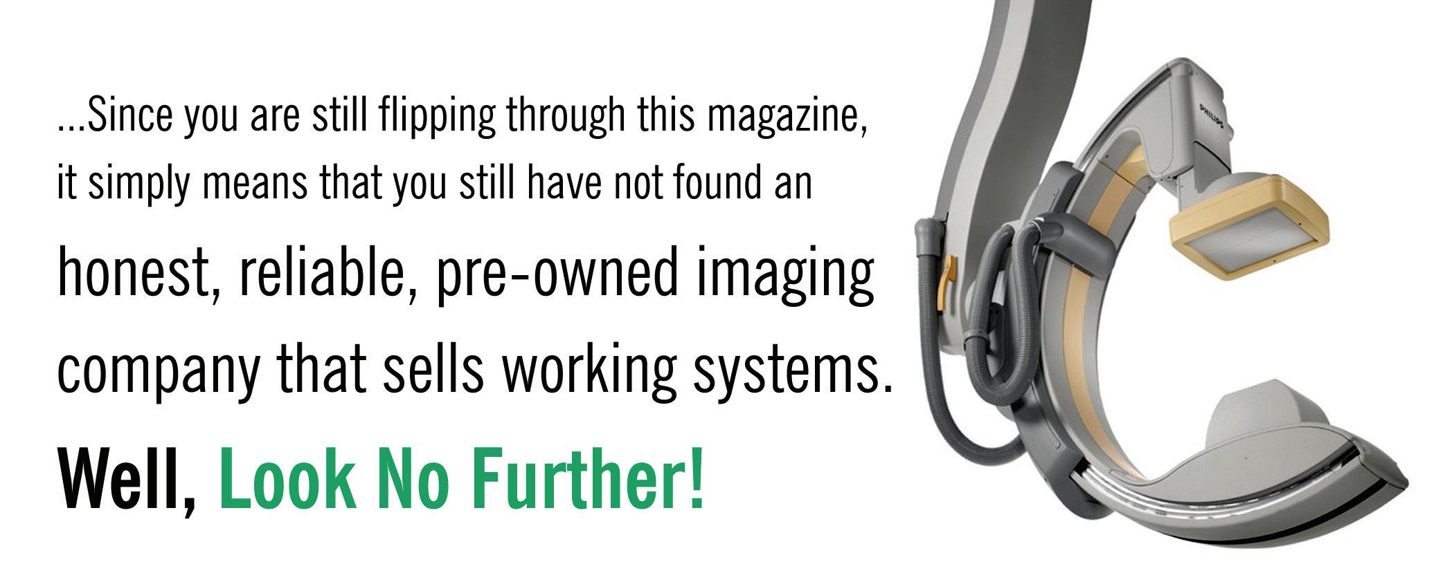
“We sell all imaging modalities, but more so, with each passing year, we have demonstrated a continued ability to work with OEMs and hospitals alike in acquiring systems direct – helping both the OEMs and their respective customers in the process,” he adds. “And, we are still excited about the fact that we still sell more C-arm systems than anyone else in the industry – worldwide!” Metropolis has been expanding it’s Mammography, CT and Ultrasound sales as well.

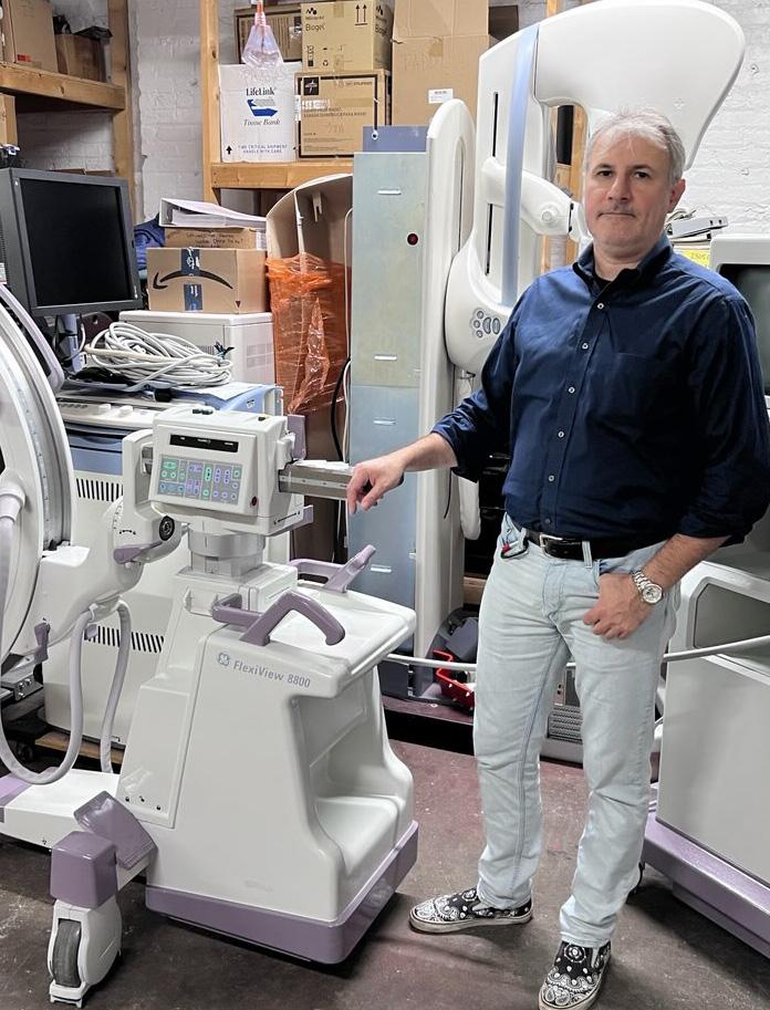
Metropolis International will continue to expand its servicing capabilities and refurbishment of systems in the future.
“The future will always remain uncertain, but it is something that we can make for ourselves! Every day is a gift, so we always do the right thing by all of our customers and vendors,” Gugel says. “The future for Metropolis will be full of opportunities as we expand more into the retail side, as well as increase our service capabilities. Metropolis is also exploring partnerships to expand into selling some new systems, as well as keeping our pre-owned and refurbished side moving.”
Other positive changes have also been in the works.
“The major changes that Metropolis has implemented, include a new and revamped website. Metropolis International is expanding its e-commerce presence,” Gugel explains. “We have an amazing marketing person who is adjusting how Metropolis reaches new customers and sells to new customers around the world. We have shown incredible results in being able to reach a broader swath of customers with a new marketing and advertising approach. As technology advances, so does the industry and us as individuals. We have to adapt to

the changing times to be successful, as well as competitive.”
One thing that will never change is the impact that those behind the scenes play in the company’s continued success delivering outstanding customer support.
“The employees at Metropolis are all great people and all contribute to our future and our success. We are all humbled to be part of something greater and to be able to help small clinics and private practices in extending patient care,” Gugel says.
In closing, the company president wanted to share a message with readers.
“Again, Metropolis truly strives to leave a legacy for the next 100 years – in the world and in this industry. To build a company that can rival GE or Philips or anyone else out there so that doctors and dealers in the future can recognize the value of working with a company with a personal touch like Metropolis,” Gugel says. “Metropolis is evolving and is more responsive and is able to bring more systems into stock to be able to offer more choices to customers that want a real product.” •
For more information, visit metropolismedical.com.
ResearchAndMarkets.com recently issued a report on the global women’s health diagnostics market from 2022 to 2028.
The global women’s health diagnostics market is anticipated to grow at a substantial CAGR of 7.9% during the forecast period, according to the report.
“The major factor propelling the growth of the market includes the rapidly rising rate of obesity across the globe, especially among women. Women suffer from certain diseases and disorders mostly related to menstrual disorders, osteoporosis, obesity, ovarian cancer, breast cancer, pregnancy and childbirth, and mammography,” the report states.
The key factors that are contributing to the growth of the global women’s health diagnostics market include numerous government initiatives to promote women’s health awareness, increasing adoption of diagnostics imaging procedures and demand for point-of-care diagnostic testing, according to ResearchAndMarkets.com.
“However, the high cost of a dearth of skilled professionals and diagnostics tests acts as a restraint in the growth of the global women’s health diagnostics market,” the report adds.
Grand View Research recently issued a report on the global breast imaging market and predicts continued growth.
The global breast imaging market size was valued at $4.7 billion in 2022 and is projected to expand at a compound annual growth rate (CAGR) of 8.6% from 2023 to 2030, according to Grand View Research.

“Factors such as the rising prevalence of breast cancer, technological breakthroughs in the domain of breast imaging, and investment from several organizations in breast cancer screening programs drive the market,” the
report states. “According to the World Cancer Research Fund (WCRF) by 2030, the number of cases of breast cancer is likely to reach about 2.1 million globally. Also, as per the World Health Organization report on breast cancer released in March 2021, over 2.3 million women globally were diagnosed with the disease in 2020, with approximately 685,000 dying as a result of its severity. Breast imaging is the primary method of diagnosis for a vast majority of breast cancer patients globally. This aspect is expected to contribute to the growth of the market during the forecast period.”
The increased frequency of breast cancer in recent years along with its associated mortality at a young age, and its sometimes delayed presentation, has encouraged many women to find medical attention sooner rather than later in life.
However, Grand View Research points out that many breast cancer cases in developing countries are discovered at a late stage due to a lack of information about early warning symptoms and screening methods. As a result, numerous organizations all around the world are implementing breast cancer awareness initiatives and campaigns.
“These screening programs have been shown to save lives compared to unscreened populations,” Grand View Research shares. “Thus, rising screening programs and government initiatives are anticipated to upsurge the awareness associated with the diagnosis and treatment of breast cancer and thus might eventually aid the breast imaging market growth. Widespread research in the development of hybrid imaging systems provides high growth opportunities and also aids market growth. Recent developments in imaging technology have completely revolutionized the nature of treatment and the way it is approached. To view the metabolic data and anatomical characteristics of the organs, it has become more crucial to combine imaging methods like CT scans, MRIs, PET and others.” •
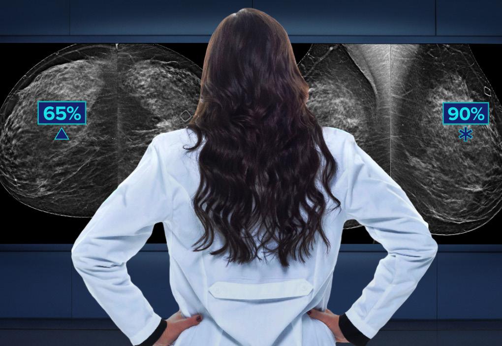
Hologic’s 3Dimensions system is designed to provide high quality 3D images for radiologists, an enhanced workflow for technologists1, and a more comfortable patient experience.2 The system offers the unrivaled performance of the Genius 3D Mammography exam, the only mammogram that’s FDA approved as superior for women with dense breasts compared to 2D alone.3,4 The exam detects 20%-65% more invasive breast cancers and is more accurate than conventional 2D exams.3 With a 70 micron pixel size, the Hologic Clarity HD high-resolution 3D imaging technology quickly captures and generates high resolution 3D images.1 Additionally, it enables options such as more natural Intelligent 2D synthesized images and workflow advantages with 3DQuorum technology – reducing file size and interpretation time. The system also includes the SmartCurve breast stabilization system to provide patients with a more comfortable mammogram without compromising on image quality, exam time, dose or workflow.2
References
1. Data on file and from public sources, 2017
2 . Smith, A. Improving Patient Comfort in Mammography. Hologic WP-00119 Rev 003 (2017).
3 . Results from Friedewald, SM, et al. “Breast cancer screening using tomosynthesis in combination with digital mammography.” JAMA 311.24 (2014): 2499-2507; a multi-site (13), non-randomized, historical control study of 454,000 screening mammograms investigating the initial impact the introduction of the Hologic Selenia® Dimensions® on screening outcomes. Individual results may vary. The study found an average 41% (95% CI: 20-65%) increase and that 1.2 (95% CI: 0.8-1.6) additional invasive breast cancers per 1000 screening exams were found in women receiving combined 2D FFDM and 3D™ mammograms acquired with the Hologic 3D Mammography™ System versus women receiving 2D FFDM mammograms only.
4. FDA submissions P080003, P080003/S001, P080003/S004, P080003/S005, P080003/S006
*Disclaimer: Products are listed in no particular order.
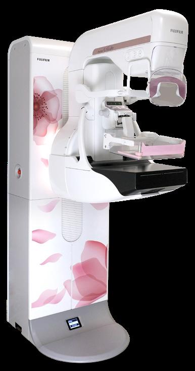
Fujifilm’s ASPIRE Cristalle is engineered with an innovative image capture design and advanced image processing technologies to produce exceptional image quality for all breast types at gentle dose. Fujifilm’s contrast enhanced digital mammography (CEDM) offering uses a dual-energy technique performed after the IV administration of iodinated contrast agent to identify abnormalities on the basis of angiogenesis, as well as morphologic features and density. Patient experience enhancements such as its patented Comfort Paddles and new Comfort Comp feature are designed to make mammograms noticeably more comfortable for the patient. The Comfort Comp feature minimizes compression prior to the exposure, and the Comfort Paddle’s soft edges and four-way pivot contours to all breast shapes and sizes, applying the right compression more comfortably.
Carestream’s DRYVIEW DVM+ Mammography

Laser Imaging Film is optimized for mammography applications. It has high maximum density so that subtle image differences are visible. The laser imaging film is available in 8x10 in. (20x25cm); 10x12 in. (25x30 cm); and 11x14 in. (28x35 cm) sizes. Each cartridge holds 125 film sheets. DRYVIEW Film cartridges are simple to load in room light in DRYVIEW Laser Imagers. Its long shelf life enables DRYVIEW Film to be stored and used for many months, simplifying inventory control and creating a potential for cost savings.
Choosing a new women’s health care ultrasound system is all about balance. With the Affiniti OB/GYN ultrasound system you have the latest technology, out of the box usability, accurate diagnostic information quickly, a simplified yet intuitive user interface, and easy access to the critical features to produce the results you need. All in an ergonomic design to let you work with less reach and fewer steps.
The MAMMOMAT Revelation mammography platform from Siemens Healthineers is engineered to make a difference in early breast cancer detection, workflow, dose and patient comfort. HD Breast Tomosynthesis offers the widest image acquisition angle available at 50 degrees, resulting in the industry’s highest in-plane resolution for improved separation of overlapping breast tissue, and 3D image-quality, improving diagnostic confidence and aiding in earlier cancer detection. HD Breast Biopsy enables one-click targeting of suspicious areas with a +/- 1mm accuracy. The InSpect integrated specimen imaging tool permits imaging and real-time review of biopsy samples at the technologist workstation to improve biopsy workflow, shorten compression time, and reduce patient discomfort. The MAMMOMAT Revelation is the first platform to offer automated breast density measurement during a mammogram for immediate, personalized risk stratification and more personalized care. Standard comfort features include personalized soft compression technology, which prevents unnecessary pain by adjusting to the anatomy of each woman’s breast, and soft-edged paddles with a breast-optimized shape. Also, the ambient MoodLight feature bathes the examination area in a soft, comforting glow. The system’s VC20 software includes efficiency improvements, reduced system calibration times, and an accelerated booting time, in addition to accelerated exam times.


• 300+ open opportunities throughout the United States.

» A variety of posting options ranging from single-job postings to 12-month unlimited memberships.

“My HR department advertised on various government sites and our web site but we did not get a single applicant in over 120 days. Fairbanks Alaska is hard to recruit for but I took out an ad on HTM Jobs and got two good applicants in less than 30 days. I am hiring them both. Thanks HTM Jobs.”


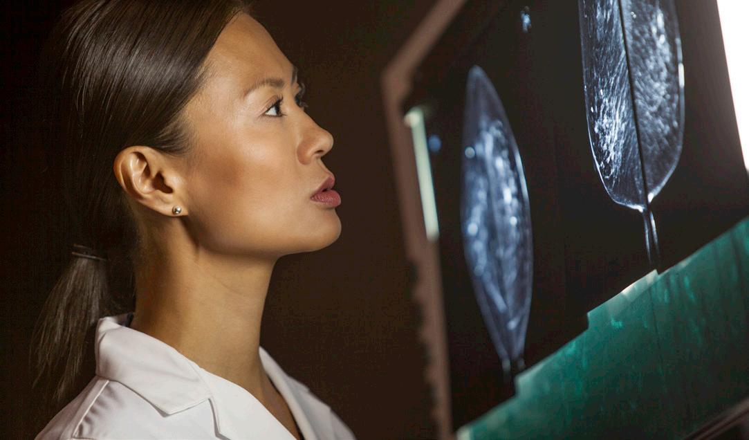
For decades, X-ray-based mammography has been regarded as the gold-standard imaging modality for breast cancer screening. Since 2011, when the U.S. Food and Drug Administration approved contrast-enhanced mammography (CEM) as a clinical adjunct to mammography, CEM has emerged as a diagnostic tool that adds functional information with which clinicians may identify more quickly whether an abnormality in breast tissue is likely cancerous.
“Mammography and tomosynthesis are the standard of care for breast cancer screening, showing morphological information vital for detecting any changes in the breast,” said Abby Weldon, senior director of women’s health for Siemens Healthineers North America of Malvern, Pennsylvania. “Adding contrast to mammography can provide functional information like blood flow and oxygenation. This can provide a quick diagnostic advantage by visualizing atypical physiological processes.”
In addition to delivering diagnostic information faster and with a high degree of confidence, another principal benefit of CEM is as an alternative to magnetic resonance imaging (MRI), a diagnostic modality typically offered to women with dense breast tissue, or who might rely upon a scanner with higher-sensitivity capabilities. As Weldon points out, however, breast MRI is not as ubiquitous as mammography; neither is MRI purpose-built for breast imaging, as mammography machines are. And for women who can’t undergo an MRI for any number of reasons – an implanted medical device; an allergy to the gadolinium contrast agent upon which breast imaging with contrast relies; an inability to lie prone for the study – contrast-enhanced mammography can be a cost-effective alternative that delivers functional information earlier in the diagnostic process. Most importantly, Weldon said, CEM can help provide more women access to this functional technique and possibly help avoid unnecessary biopsies.
“Injection of contrast only needs to be done one to two minutes before the mammogram, and it’s a lot quicker than an actual MRI would be,” she said. “CEM functionality can be added to a normal mammography unit — which doesn’t have to be used just for contrast-enhanced mammography — and clinicians also can perform screening and biopsies on the same unit.”
In addition to the relatively rapid nature of a contrast-enhanced mammogram – the contrast agent decays within 10 minutes, and image acquisition typically only takes six to eight minutes – CEM costs about a quarter of what a breast MRI does. In a webinar prepared for Siemens Healthineers, Dr. Noel Bergquist, an assistant professor at the University of Tennessee at Knoxville Breast Center, said that women in her care have opted for CEM owing to its lesser expense and greater accessibility.
“A lot of my patients that are reluctant to pay for an MRI are happy to pay for this,” she said. “We’ve had no difficulties with insurance companies covering this modality.”
Additionally, Bergquist pointed out the high negative predictive value (NPV) of CEM: at 94 to 100 percent, the modality is effective at not returning

false-negative results, and patients can be confident in the results of their study.
However, CEM isn’t without its drawbacks, among which Bergquist enumerated adverse reactions to the contrast agent, the potential for false-positive study results, and the possibly limited sensitivity of CEM given its ability to enhance images of normally functioning breast tissue, also known as background enhancement. Neither is CEM recommended for pregnant or breastfeeding women, those who are too young for a mammogram, women in renal failure, and, often, women with breast implants.
“Sometimes you are going to have to go to MRI, or you will have to biopsy based on landmarks,” Bergquist said. “But we have had great success at our center, and these challenges are minimal at most.”
Bergquist described finding the most effective uses of CEM in annual screenings of high-risk patients, for complicated cases, as a tertiary modality, and as an MRI alternative after neoadjuvant cancer treatment therapies already have been administered. It’s also useful in helping to determine the extent of disease in pre-surgical approaches to cancer treatment, as well as in cases of contralateral disease, in which breast masses are identified six months after detection of breast cancer in the opposite breast.
Siemens Healthineers’ chief platform for delivering CEM is the MAMMOMAT Revelation, which can be used to perform 3D mammography as well as titanium contrast-enhanced mammography, or TICEM, whereby a titanium filter helps alleviate the heat associated with dual-energy imaging. The Revelation also includes a feature that calculates the patient’s breast density after the first image is captured, which Weldon described as useful in assessing whether supplementary imaging is necessary.
“Any tools we can offer as vendors just to aid the facilities in helping inform women of their breast density are great,” she said, “but in the end, the only FDA-approved method for breast density assessment, or cancer diagnosis, is the radiologist’s assessment.”
Weldon said the install base for CEM is “totally mixed” across a variety of settings, from university to community hospitals. Currently, it’s less prevalently found at ambulatory clinics, but Weldon believes there’s opportunity for growth of the modality outside of hospital environments: in imaging centers, outpatient facilities, breast cancer centers and women’s health clinics.
“We do expect the utilization of CEM to grow,” Weldon said. “We’ve used MRI to identify breast cancer
in a diagnostic setting or possibly for very high-risk women in a screening setting. However, MRI is not available everywhere due to a variety of reasons, including budget and space requirements. Although MRI is an extremely sensitive modality, CEM is expected to be a great alternative – especially for intermediate risk patients.”
Weldon also believes that the presence of a variety of medical imaging modalities reinforces clinical efforts to deliver personalized medicine for women with different types of breast health challenges, the better to tailor treatment appropriate for their distinct needs.
“Largely, we’re following the guidance of having a mammogram at the age of 40,” she said. “Care providers have urged for a more personalized approach, and for women to start to understand their potential risk earlier; to advocate for a modality that’s more in line with what their risk profile is. I think contrast-enhanced mammography offers a useful option to help enable risk-based screening.”
Scott Pohlman, director of outcomes research for Hologic Inc. of Marlborough, Massachusetts, also points out that contrast-enhanced breast imaging, whether mammography or MRI, can offer potential benefits “at various steps along the continuum of care.”
“At this time, exactly which women benefit from supplemental screening – and which type of supplemental screening is most appropriate – is not standardized, and depends on a variety of factors like a facility’s resources, a woman’s breast density, and insurance coverage, or if the patient has any health conditions that might eliminate certain modalities as an option,” Pohlman said.
“No patient is the same, and we need to tailor the breast cancer journey to fit each individual’s needs,” he said.
Despite the lack of consensus on the differences in sensitivity between CEM and MRI, Pohlman also pointed out that research into the use of both modalities indicates that each provides a higher sensitivity of image capture than mammography alone, as well as the capability of detecting breast lesions with a high degree of sensitivity.
“In a general sense, both CEM and contrast MRI depict the same fundamental physiological process – the accumulation of vascular contrast agent caused by the formation of new blood vessels (angiogenesis), which is associated with tumor growth,” he said. “As a result, there is a physiological reason to believe that they should both be able to detect malignant lesions that are not well visualized by more traditional anatomic modalities, such as mammography and ultrasound. However, CEM and MRI differ enough, particularly because acquir-
ing multiple MRI images provides dynamic information about the rate of contrast enhancement, that some difference in performance should be expected.”
Ultimately, Pohlman concluded that the best way to more precisely measure the comparative effectiveness of CEM and MRI will require additional, large-scale studies of their relative performance in specific patient populations, because those studies undertaken to date have been too widely varied or too limited in scope. However, he also noted that feedback on patient experiences with CEM versus breast MRI patients has emphasized “faster procedure time, greater comfort, no issues with claustrophobia, lower anxiety and lower noise level,” as per a 2015 study published in the Journal of Medical Imaging and Radiation Oncology . Beyond that, however, there’s plenty to be said for the straightforward fulfillment of annual breast cancer screenings for women aged 40 and older.
“Something the medical community does agree on is that early detection and improved survival rates all start with regular breast screening beginning at age 40,” Pohlman noted. “In my opinion, the most impactful overall measure is that all women should be screened on digital breast tomosynthesis (DBT) – also known as 3D mammography – systems as a first step of the screening process, if they have the opportunity.”
DBT provides specific advantages for women with dense breast tissue as compared with 2D mammography alone, Pohlman notes; because dense breast tissue can mask small cancers, as both present similarly on imaging studies, which can potentially delay confirmation of a cancer diagnosis. As nearly half of all women in the United States may have dense breast tissue, DBT systems can help identify lesions that might be embedded beneath dense tissue by examining the breast, layer by layer.
“What is exciting is that the FDA recently released new data that showed DBT gantries now account for 46 percent of U.S. mammography imaging systems, with experts anticipating that DBT will replace the vast majority of full-field digital mammography (FFDM) systems in the next decade,” Pohlman said.
“Around the world, DBT is becoming the standard for breast imaging,” he continued. “In December 2022, the European Council updated the Article 168 recom -
mendations to encourage the use of DBT for all breast cancer screenings. Additionally, in 2021, the European Commission Initiative on Breast Cancer (ECIBC) recommended for the first time that women with dense breast tissue could benefit from the use of DBT. These recommendations are monumental, as it creates an opportunity for more women to access DBT during their screenings.”
Pohlman also remarked on the significance of breast density inform laws in the United States as having an outsized impact on breast cancer detection. He believes that the continued advancement of legislative reforms related to health care screenings can only further those advancements.
“At Hologic, we applaud the FDA for their recent updates to the Mammography Quality Standards Act (MQSA) that require patient notification of breast density,” Pohlman said. “Prior to this, only 38 states and the District of Columbia mandated that facilities notify patients of their breast density. In addition to making this mandatory across the United States, it also creates a national standard for communicating about breast density.”
“However, there is still significant progress that needs to be made to address health care laws around screenings,” he said. “While the Patient Protection and Affordable Care Act (ACA) requires most health insurers to cover annual mammograms with no outof-pocket expenses for women ages 40 years or older, this may not extend to supplemental screenings. For women with dense breast tissue, their health care providers may recommend supplementing mammography with whole breast ultrasound, CEM or MRI if their screenings are inconclusive.
“This leaves women with dense breast tissue to face additional barriers to care even though research shows they are at greater risk of breast cancer,” Pohlman said. “While many individual states have passed legislation mandating coverage for supplemental screening, inconsistencies remain between the states when it comes to which specific populations and modalities are covered, and whether the patients may be responsible for out-of-pocket costs associated with supplemental screening.” •
“No patient is the same, and we need to tailor the breast cancer journey to fit each individual’s needs.”
- Scott Pohlman

Some of the best ideas come as a response to a stressful situation. Banner Imaging has 27 separate outpatient facilities of various sizes and design. There are no code buttons to alert a team to come to the rescue like there is in an acute setting. When we have an emergency, we respond to the situation with our emergency supplies and an AED while having someone call 911 immediately.
Many of our offices were designed years ago. Some have secluded hallways or modalities that are away from the rest of our team. We are open extended hours and may have limited staff on site.
We had a critical situation in CT at one of our offices. Fortunately, everything turned out fine. Our team acted quickly and appropriately, and our patient recovered. At the moment that the CT tech needed immediate assistance, there just happened to be no one in the vicinity. She continued to yell for help while attending to a patient who suddenly became unresponsive. A team member did finally hear her and gathered more people to assist. EMS arrived extremely quickly and took over. Through this real-life scenario, we learned that we needed a better way to alert the rest of our team that someone needed help RIGHT NOW. Time moves very slowly when it appears no one is coming to assist.
We implemented the use of a very loud whistle. This isn’t a plastic toy whistle. This is a very loud emergency whistle and is Banner Imaging branded to identify it as a medical tool. Every employee was given a whistle to keep in their pocket or attach to their security badge. We removed the metal rings used to attach the whistle to a lanyard or badge for those working in MRI and found a plastic alternative.
We came up with a code of 3 short bursts on the whistle to alert the entire team that help is needed. Any available team member is trained to follow the sound of the whistle to render assistance. The team member in need continues
to blow the short bursts, three at a time, until help arrives.
We tested these whistles and found they could be heard through the leaded doors and from isolated rooms. Our plan was recently put into action when a 90-year-old patient could not sit on the DXA table, so instead she just sat down on the floor. The technologist was unable to help the patient back up, so she blew her whistle in three short bursts. Three team members ran to help. Within a few minutes the patient was able to be helped to the table. The technologist did not have to leave the patient to find assistance. After seeing this in action, other team members were excited to get their whistles.
This has been a low-tech, low-cost solution that was able to be implemented quickly and easily. Our team has been very accepting of the new workflow, even though it has been in place for a short time. We have heard positive comments about how it provides a sense of support, since help is a whistle away. At first this idea sounded a little bit too simple, but the amount of time and expense it would take to install an emergency alert system in every room in every office is not reasonable.
This did not take long to organize and facilitate. Each new team member receives a whistle when they first report to their office as part of our onboarding process. This also should be uncomplicated to maintain. The whistles are also incorporated into our simulated code training. We remind our teams to use the whistle in the event of an emergency on our daily safety huddle and encourage our team members to always keep the whistle on them.
With all the sophisticated technology that we use every day, the emergence of AI and even communication devices introduced to us by the Jetsons, it is amazing that this solution would have worked in Wilhelm Roentgen’s X-ray lab.
Sometimes simple is best.
Thanks for all you do. •
Beth Allen, CRA, RT(R)(CT), is the director of clinical operations at Banner Imaging.A COOL SERIES FOR HOT TOPICS
ICE Magazine connects with industry leaders to provide top-quality educational opportunities through its monthly webinar series. Reach key decision makers and imaging leaders with your company's message, while collecting important data for sales.


For more information, visit icewebinars.live.
“The content is always relevant and timely, very professional presentations.”
The content is always relevant and timely, very professional presentations.
– N. Godby, Director of Radiology, Cabell Huntington Hospital Inc.
– N. Godby, Director of Radiology, Cabell Huntington Hospital Inc.
“Learn about best practices and newer technology.”
Learn about best practices and newer technology.
– K. Stich, Radiology Director,East Market, University Hospitals.
– K.Stich, Radiology Director,East Market, University Hospitals.
I love that ICE webinars help me gain my CEUs, it also helps that they have some of the best presenters/lecturers!
- J.Sturm, Nuclear Medicine Technologist, Novant Health Brunswick Medical Center.
“I love that ICE webinars help me gain my CEUs, it also helps that they have some of the best presenters/lecturers!”
- J. Sturm, Nuclear Medicine Technologist, Novant Health Brunswick Medical Center.BY SCOTT TREFNY
Throughout my career as an ultrasound field service engineer, I’ve often encountered the question among friends and family, “What is your profession?” Naturally, upon sharing my role, I’ve been met with expressions of confusion or the common response, “What does that involve?” Typically, most people associate ultrasound in health care with its applications in pregnancy – assessing the health of the baby and the mother. This usage appears to be the most widely recognized, with many individuals who have had children having experienced 3D/4D scans, resulting in images featuring profiles of their baby’s face or 2D scans reminiscent of old black and white television pictures.

Obstetrics, primarily focused on pregnancy and childbirth, tends to be a familiar application of ultrasound. However, various health fields, including cardiology, general radiology, breast and cancer detection, and the assessment of muscle and joint concerns, frequently find more extensive use, particularly within women’s health. The versatility of ultrasound systems, coupled with advancing technology, has
led to the design of systems tailored to these applications. This aims to optimize treatments and simplify diagnostic decision-making more effectively than in previous years.
Breast ultrasound has experienced remarkable technological progress, expanding its applications as a safer yet equally effective alternative to X-ray-based mammography. Unlike X-ray systems, ultrasound doesn’t emit potentially harmful radiation, making it particularly valuable for patients at elevated risk of exposure.
BreastCancer.org conducted a case study that demonstrated comparable detection rates between ultrasound and mammography. Additionally, in procedures involving biopsies, ultrasound is the technology employed for pinpointing needle locations and providing real-time visualization during the biopsy process.
Another prevalent use of ultrasound in women’s health, aside from obstetrics, is found within gynecology practices. The integration of 3D ultrasound can significantly enhance the diagnosis and treatment of various concerns, including uterine adhesions, IUD placement and subsequent follow-up examinations, as well as the identification and pinpointing of fibroids and polyps. By having ultrasound services readily accessible within the same site or office, health care providers can achieve
faster assessments and diagnoses of these issues, eliminating the need to refer patients to external facilities or recommend alternative imaging modalities. This approach substantially bolsters the availability of treatment options, leads to more favorable outcomes and improves patient care.
As ultrasound gains prominence as a preferred method for detection and diagnosis, manufacturers are increasingly tailoring systems for specific applications. Concurrently, advancements in technology are resulting in smaller and more portable ultrasound systems, which not only reduces equipment procurement expenses but also lowers the costs associated with servicing these systems and probes. While repair or replacement costs for specialty probes designed for specific applications might initially appear high, their availability is on the rise, potentially contributing to cost reduction for these specialized components. Among the most expensive items to service, besides the system itself, are 3D/4D mechanical probes. However, due to the evolving technology and enhanced repair capabilities, many service providers, like Avante Health Solutions, have managed to offer more cost-effective repairs for these probes.
Over the years, while servicing ultrasound systems, I’ve found it truly exciting to witness the evolution of technology and its expanding role within various practices and women’s health centers, extending well beyond traditional X-ray applications. Engaging in conversations with users and health care providers, I’ve had the privilege of gaining insights into the positive impact of these advancements, particularly from individuals with extensive experience in the field. The reach of ultrasound is continually broadening, and it’s no longer limited solely to expectant mothers; it’s becoming increasingly embraced across the women’s health sector.
For more information about Avante’s ultrasound service and repair capabilities, call one of our experts at 800-979-6742 or visit avantehs.com. •
Alameda Health is a level one trauma center in the heart of Oakland. They have more work than they can handle! As a safety net hospital, they treat all in need. This workload was documented in the documentary “The Waiting Room.”
The emergency room provides for all levels of acuity. This open-to-all approach leads to stress on the resources. They need to improve efficiency to meet the demands. The CNO called a meeting and asked for suggestions to improve the patient throughput in the emergency room. Alameda Health is a teaching hospital with employed physicians. They do not have a profit motive and do not get paid to do more work. The use of voice recognition AI and effective PACS was already provided. The only improvement they could come up with was to have the radiologist work with the emergency room to reduce unnecessary imaging exams. This is a common problem in health care nationally.
Radiologists play a crucial role in helping reduce the number of unnecessary imaging studies ordered by emergency room (ER) doctors. Here are some brainstormed ways in which radiologists can contribute to this goal:
• Implement clinical decision support systems: Radiologists can work with hospital administrators and IT departments to implement clinical decision support systems. These systems provide evidence-based guidelines and recommendations for ordering appropriate imaging studies, helping ER doctors make more informed decisions.

• Provide educational resources: Radiologists can develop educational materials, such as guidelines,
protocols, and case studies, to educate ER doctors about appropriate imaging utilization. This can include highlighting situations where imaging may not be necessary or suggesting alternative diagnostic approaches.
• Develop evidence-based imaging guidelines: Collaborating with other health care professionals, radiologists can participate in the development of evidence-based imaging guidelines specific to the ER setting. These guidelines can help standardize imaging practices and reduce unnecessary studies.
• Act as consultants: Radiologists can make themselves available as consultants to ER doctors, providing expertise in imaging utilization. They can review clinical histories, discuss appropriate imaging options, and help determine the most effective imaging modalities for specific clinical scenarios.
• Establish clinical pathways: Working with ER physicians, radiologists can establish clinical pathways or protocols for specific clinical conditions. These pathways outline the appropriate use of imaging studies based on best practices and evidence, reducing unwarranted variations in ordering practices.
• Provide real-time feedback: Radiologists can provide real-time feedback to ER doctors regarding the appropriateness of imaging studies. This feedback can be incorporated into quality improvement initiatives and can help ER physicians better understand the impact of their ordering patterns.
• Participate in multidisciplinary conferences: Radiologists can actively participate in multidisciplinary conferences where patient cases are discussed. By contributing their expertise, they can help guide discussions regarding the neces -
sity of imaging studies, fostering a collaborative approach to patient care.

• Develop imaging utilization dashboards: Radiologists can collaborate with hospital administrators and data analysts to develop imaging utilization dashboards. These dashboards can provide ER doctors with information on their own ordering patterns compared to benchmarks and peer groups, promoting awareness and self-reflection.
• Offer second opinions: Radiologists can offer second opinions on imaging requests from ER doctors. By providing an alternative perspective, they can help ensure that imaging studies are appropriate and necessary, potentially avoiding unnecessary tests.
• Conduct research on imaging utilization: Radiologists can contribute to research efforts focused on
imaging utilization in the emergency room setting. By identifying trends, best practices, and areas of improvement, research findings can inform strategies to reduce unnecessary imaging studies.
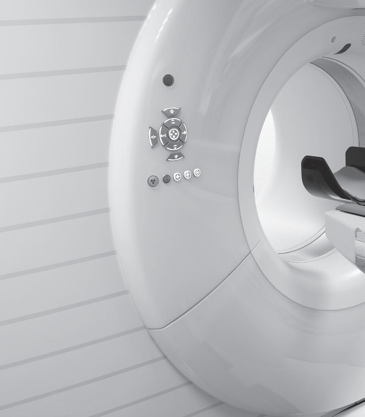
It’s important to note that effective collaboration and communication between radiologists and ER physicians are essential for achieving the goal of reducing unnecessary imaging studies. By working together, they can ensure that patients receive appropriate care while minimizing unnecessary radiation exposure and health care costs. I welcome your feedback and suggestions you have found successful in your emergency departments. •
Mark Watts is an experienced imaging professional who founded an AI company called Zenlike.ai.
Every organization worth its salt strives for continuous improvement. It’s great when an organization has this mindset, but when it comes to problem-solving, that’s almost always done best with small teams.
Research shows that people working in small teams to tackle or remove obstacles are more likely to ask questions and brainstorm solutions. On smaller teams, groupthink gets minimized and so does fear of getting mocked for offering an idea that someone else thinks is dumb.
It’s been said that sometimes the problem with problem-solving is the way we look at the problem. With that in mind, let’s go through a few basic suggestions. After all, problem-solving is a critical skill for organizational success.
I’ve heard horror stories of bosses who discount solutions because they’re not coming from the “right” person. Then, after a few days of exploration, it turns out “that” person’s solution worked best. That’s a lot of wasted time and also damaged morale.

We should be open to considering all input and all solutions, even if ideas are coming from someone who’s not the company rock star. In fact, not only should we be open to it, we should invite it. The objective in any problem-solving endeavor is to solve a problem in the best way possible, not for the right person to have the solution.
Along with that idea, we should also seek input from those who aren’t on the problem-solving team. You’ve probably heard the story about the different blind people being asked to stand next to an elephant and describe it. One, touching the leg, says an elephant is like a tree trunk. The blind person touching the tail says an elephant is a like a rope. The person touching the animal’s side says an elephant is like a wall. None of them are 100 percent correct, but none of them are wrong, either.
Similarly, if we actively seek input from people who see the problem from different angles, you get a bigger and better understanding and can often arrive at a more comprehensive solution.
This guideline complements being open-minded, in that it means being cognizant of the fact that neither we nor the people are our teams know all there is to know. Oftentimes people say things with confidence when what they’re really offering is conjecture. If we’re going to be factual and truthful, then we need to be able to test what we’re discussing.
Put another way, sometimes things are said without concrete evidence. A couple of well-phrased questions asked with respectful voice tones can help us explore and work through unverified statements. Underscore that part about using respectful voice tones, but one question might be, “What evidence exists for that?”
The key here is to inquire so that assumptions can be explored, but done in way that allows people to save face if needed. It’s OK to brainstorm, but sometimes we need to clarify where things are coming from so we remain on a solid foundation.
Familiar environments are usually comfortable in that they are predictable. We tend to meet in the same offices or in the same conference rooms. It’s not against the law to get together for a problem-solving session at an unusual venue. One gentleman I know took his team out for pizza whenever they got stuck solving a problem. He believed a different setting and trying different types of pizza got people thinking differently.
This is similar to Agent K in the third “Men in Black” movie. Agent K’s grandfather always said that if you have problems you can’t solve, go have some pie. The idea in all of this is a different atmosphere helps to clear one’s mind which leads to fresh perspectives.
Oftentimes people place limitations on their ideas before those ideas can be spoken and explored for their potential. Those self-limiting roadblocks are usually in place because people don’t want to be scoffed at for making suggestions seen as unrealistic. The problem with these self-imposed limitations is that many fantastic ideas go unspoken.
One method I recommend to overcome this is called, “Disney’s Creative Strategy.” It was a brainstorming techniques that had three distinct phases: the Dreamer phase, the Realist phase and the Spoiler phase.
In the Dreamer phase, teams were encouraged to dream up the most fantastic and absurd ideas for solving a problem. They were to place no filters or limitations on their imagination, such as no cost constraints nor manpower constraints.
In the Realist phase, ideas were re-examined and re-worked into something more practical. The focus was on how something could be done with the resources actually available instead of why something couldn’t be done.
The Spoiler phase was reserved for Walt Disney himself. He would carefully examine the ideas and look for flaws. The purpose wasn’t to criticize, it was to figure out what needed to be done to ensure an idea’s success.
Teams tasked with solving big problems might find Disney’s technique useful.

Several different techniques exist for getting to the root cause of a problem. I won’t take up this space to explain each one, as instructions for using them can be found online. These are not tools for deciding which caterer to use for the next company gathering, but rather problems that have multiple causes and can’t be resolved with just one action.
Look up Fishbone (or Ishikawa) diagrams, Five Whys, and Failure Mode and Analysis (FMEA). Each has pros and cons, and some are better suited to solving one type of problem over another. I suggest these tools because they use a systematic, analytical approach, and some people are much more comfortable with such methods.
Bottom line, problems, or obstacles, appear in every organization. Successful organizations are those with teams that can solve those problems. I’ll assume you’re in such an organization. If not –now’s a good time to move in that direction. •
Daniel Bobinski, who has a doctorate in theology, is a best-selling author and a popular speaker at conferences and retreats. For more than 30 years he’s been working with teams and individuals (1:1 coaching) to help them achieve excellence. He was also teaching Emotional Intelligence since before it was a thing. Reach him by email at DanielBobinski@protonmail.com or 208-375-7606.
Whether or not you can bill for post procedure mammograms is a topic that has waxed and waned over the years. Changing payer guidelines and pending regulations changes from Mammography Quality Standards Act (MQSA) has this topic back in the spotlight. From a broad coding guidance standpoint there has not been a real change since 2017, but it is important to dive into the details to ensure compliance with payer guidelines.
Before I get into the details, let me first say that it is important to review your current protocols. If your breast biopsies are performed utilizing stereotactic equipment and you never move the patient to another machine for any type of imaging, you will likely never be billing any payer for a post procedure mammogram. The documented report should clearly define everything that is being done for the patient but understanding what is occurring and why helps enlighten the coding and billing process.
Now to the details …
On the surface it seems straightforward. If you perform a post percutaneous procedure mammogram to confirm removal of calcifications and/or to check position of the localization clip, and there is supporting documentation in the radiology report, then it is appropriate for both the facility and the radiologist to bill for this service; unless the procedure was performed under mammographic guidance. The National Correct Coding Initiative (NCCI) Policy Manual states that the mammogram is bundled only if the procedure was performed with mammographic guidance. 1
Unfortunately, it is anything but straightforward.
For example, the authoritative guidance differs for hospitals and physicians for MR guided biopsies. For hospitals, the American Hospital Association (AHA) guidance differs from NCCI guidance. According to AHA Coding Clinic for HCPCS (First Quarter 2022), it is not appropriate to assign 77065 for a post-procedure mammography following placement of a breast localization device represented by CPT code 19085 to confirm placement of the localization device post procedure. This mammogram is not considered a diagnostic study and therefore cannot be represented by CPT 77065. The AHA guidance applies to all outpatient hospital facilities so hospitals should not be charging for a post-procedure mammogram following an MRI guided breast biopsy.
For physician billing the last published guidance specifically for MR was included in Clinical Examples in Radiology (Spring 2018), “CPT code 77065 is reported for the craniocaudal and true lateral diagnostic mammogram views of the left breast performed in the mammography department after the MRI procedure to verify the appropriate position of the localization wires and for surgical guidance.”
Individual payer guidelines, including Medicare Administrative Contractors (MACs) must be reviewed to ensure compliance with their guidelines. For example, Noridian Healthcare Solutions states that “It is Noridian’s interpretation that a follow-up mammogram performed post tomosynthesis-guided breast biopsy will be considered part of the procedure and not separately payable, regardless of whether the patient is brought to a different room and/or unit for the mammography … ” The policy goes on to provide specific details as to what can and cannot be billed together.
Some commercial payer policies have published guidance that addresses how the post procedure
mammogram is obtained when defining coverage. For example, one Blue Cross Blue Shield payer has a policy, “Coding and Billing Guidelines for Breast Biopsies,” 2 that states the following: “If a combination stereotactic-tomosynthesis guided biopsy is performed using a separate piece of equipment (such as a prone table) and the patient is moved to another unit for a post-procedure mammogram, it is appropriate to report the post-procedure mammogram separately. If the combination stereotactic–tomosynthesis guided biopsy is performed using a standard digital breast tomosynthesis mammography unit on which the post-procedure mammogram is also obtained, it is not appropriate to report the post-procedure mammogram separately.”
And then there is the new information from MQSA. The Final Rule, published on March 10, 2023, outlined new guidelines for “Post-Procedure Mammogram for Marker Placement” that will be effective September 10, 2024. The MQSA requirements language for reporting is being revised to specifically state “As FDA described in approval of the alternative standard, if a facility makes the post-procedure examination part of the interventional procedure instead of a separately charged examination, then the examination is not
subject to the MQSA quality standard requirement and need not receive an assessment (Ref. 24). Nor would it require any report separate from the report of the interventional procedure. However, when the post-procedure mammogram is logged or charged separately from the interventional procedure, this mammogram is a separate examination and requires a separate report.”
This requirement is an important distinction since many practices include the post procedure diagnostic mammogram interpretation in the body of the biopsy procedure. This new regulation will require potential changes to workflow and documentation practices.
So, in summary – can you bill for a post procedure mammogram? As with many things in coding, it depends … what modality was utilized for the biopsy? What are your payer guidelines? This is something that should be continually monitored to ensure compliance and correct billing practices. •
References
1 https://www.cms.gov/files/document/medicare-ncci-policy-manual-2023-chapter-3.pdf, page 11
2 https://www.bcbsnd.com/providers/policies-precertification/reimbursement-policy/coding-and-billing-guidelines-for-breast-biopsies
Imaging Service Solutions


global provider of OEM quality pre-owned and refurbished medical imaging parts. ISS specializes in supplying parts for Philips and Siemens MRI and CT scanners.

Established in 2009 in Baltimore, MD, we proudly work closely with hospitals, service providers, and manufacturers globally.
SELL US YOUR MRI AND CT SYSTEMS!
We purchase installed systems at fair market value and provide quality replacement parts for your equipment at a great value. We can remove purchased equipment at no cost as well.






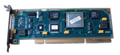
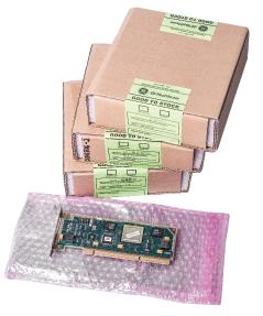


In the August issue of ICE magazine, I wrote “Expert Body Language” about being surprised at an individual because of his lack of discomfort during an interview. He was being interviewed regarding his firsthand knowledge of, and interaction with, extraterrestrial (ET) beings. I surmised that he was telling the truth, he actually believed it or had extraordinary nonverbal control. I also said, “Some say that within five minutes you can evaluate people with 70% accuracy, which means that the other 30% can get you in much trouble.”

As I was searching for a topic for this month’s column, I came across an old commentary I wrote a long time ago. In a previous life, 10 or so years ago, I wrote comments on stuff which I called “Manny’s Moans.” (I searched for the title and found a couple of stragglers, if you have interest … https://1technation.com/ search/Manny’s/.)
The Moan was regarding a topic similar to the August column mentioned above. The title of the Moan was “Fresh Squeezed People.” Here it is:
“When you squeeze an orange, what do you expect to get? ‘Why, orange juice, of course,’ you say. That is absolutely correct. If you got hummus from squeezing an orange, you would be taken by surprise. Since you know what oranges are, it is easy to predict what will come out of a good squeeze. Oranges don’t get the option to change their appearance, such as painting themselves yellow like a lemon.
People, on the other hand, are different. People can, sometimes too easily, change their appearance. They can and do change what they project to the world. It is difficult to know what is truly inside people. Is there good, bad, honor, integrity, greed, cowardice, courage, etc.? Under normal circumstances, people can ooze whatever they wish to present to you. It is fairly easy to ooze friendship, honor, even
love or whatever will create the desired perception in others. Oozing is slow so it can be controlled.
Only when someone is squeezed does the real inside stuff come out. Under the pressure of a squeeze, the stuff must come out without having the benefit of being controlled and modified. The squeeze could come from a significant emotional event such as a truly unexpected, and threatening event. A true surprise may also provide a good squeeze.
You want to know what’s inside a person? Watch what happens when the pressure is on. Stand back. Often what comes out will be smelly, slippery and brown.”
Regarding the August article about the ET guy, I may have been duped by his apparent veracity. The interviewer was very supportive and encouraging. She also believed, so there existed a mutually sympathetic encounter. Therefore, it is quite possible that the “softball” interview and the camera angles provided no threats to the ET guy. He was never given a squeeze. He was allowed to ooze whatever he wanted to present to make his case. Also, the expected audience would have been predisposed to belief in the subject matter.
So, the takeaway from this should be that if the presenter is not given the occasional squeeze, credibility might be suspect. If you want the truth, you have to squeeze. How is that accomplished? It is more than merely asking questions. You must dig deep for additional information. A good presenter will have anticipated and prepared to answer a minimum of 10 questions. A few prepared and unexpected questions will provide squeezes that will allow you to see beyond the words. Get in the habit of squeezing people and your life will be easier, more lonely, but easier with a mixture of brown and smelly. •
There’s more to maintaining a healthy heart than just eating right and exercising regularly. While these practices play an important role in both cardiovascular and overall health and well-being, getting a good night’s sleep is also key.

However, more than 1 in 3 adults in the United States are not getting the recommended 7-9 hours of sleep per night, according to the Centers for Disease Control and Prevention (CDC). In addition to increasing risk for cardiovascular conditions like high blood pressure, heart disease, heart attack and stroke, lack of sleep may also put people at risk of depression, cognitive decline, diabetes and obesity.
“We know that people who get adequate sleep manage other health factors better as well, such as weight, blood sugar and blood pressure,” said Donald M. LloydJones, M.D., Sc.M., FAHA, past volunteer president of the American Heart Association and chair of the department of preventive medicine, the Eileen M. Foell Professor of Heart Research and professor of preventive medicine, medicine and pediatrics at Northwestern University’s Feinberg School of Medicine. “The American Heart Association added sleep to the list of factors that support optimal cardiovascular health. We call these Life’s Essential 8, and they include: eating a healthy diet, not smoking or vaping, being physically active and getting adequate sleep along with controlling your blood pressure and maintaining healthy levels of cholesterol and lipids, healthy blood sugar levels and a healthy weight.”
Education about healthy heart habits from the American Heart Association is nationally supported by Elevance Health Foundation. Some practices to improve sleep health and impact heart health include:
To help reduce sleep disruptions caused by food, avoid
late dinners and minimize fatty and spicy foods. Similarly, keep an eye on caffeine intake and avoid it later in the day.
Physical activity can have a noticeable impact on overall health and wellness but can also make it easier to sleep at night. However, exercising too close to bedtime may hinder your body’s ability to settle; aim to have your workout complete at least four hours before bed.
Getting a good night’s rest often requires getting into a routine. Start by setting an alarm to indicate it’s time to start winding down. Rather than heading straight to bed, create a to-do list for the following day and knock out a few small chores. Then consider implementing a calming activity like meditating, journaling or reading (not on a tablet or smartphone) before drifting off to sleep. Also set an alarm to wake each morning, even on weekends, and avoid hitting the snooze button.
The ideal space for sleeping is dark, quiet and a comfortable temperature, typically around 65 F depending on the individual. Use room-darkening curtains or a sleep mask to block light and ear plugs, a fan or a white noise machine to help drown out distracting noises. Remember, using your bed only for sleep and sex can help establish a strong mental association between your bed and sleep.
The bright light of televisions, computers and smartphones can mess with your Circadian rhythm and keep you alert when you should be winding down. Try logging off electronic devices at least one hour before bedtime and use the “do not disturb” function to avoid waking up to your phone throughout the night. Better yet, charge devices away from your bed or in another room entirely. •
Find more tips to create healthy sleep habits at Heart.org.


“Good health and good sense are two of life’s greatest blessings.”
– Publilius Syrus
ICE Webinars p. 41

626 Holdings p. 5
Advanced Health Education Center p. 25 AllParts Medical p. 45

Banner Imaging p. 23


Brandywine Imaging p. 43
Imaging Academy p. 2
PM Imaging Management p. 50

CM Parts Plus


p. 47
Engineering Services p. 3
Imaging Service Solutions p. 49 Innovatus Imaging p. 9


KEI Medical Imaging


p. 43
Maull Biomedical p. 25

Metropolis International p. 26

Radon Medical LLC p. 26
Ray-Pac® Ray-Pac p. BC
Radiological Service Training Institute p. 18
Technical Prospects p. 4
SOLUTIONS
TriImaging Solutions
p. 13
W7 Global, LLC. p. 50

HTMJobs.com p. 34
MW Imaging Corp. p. 50



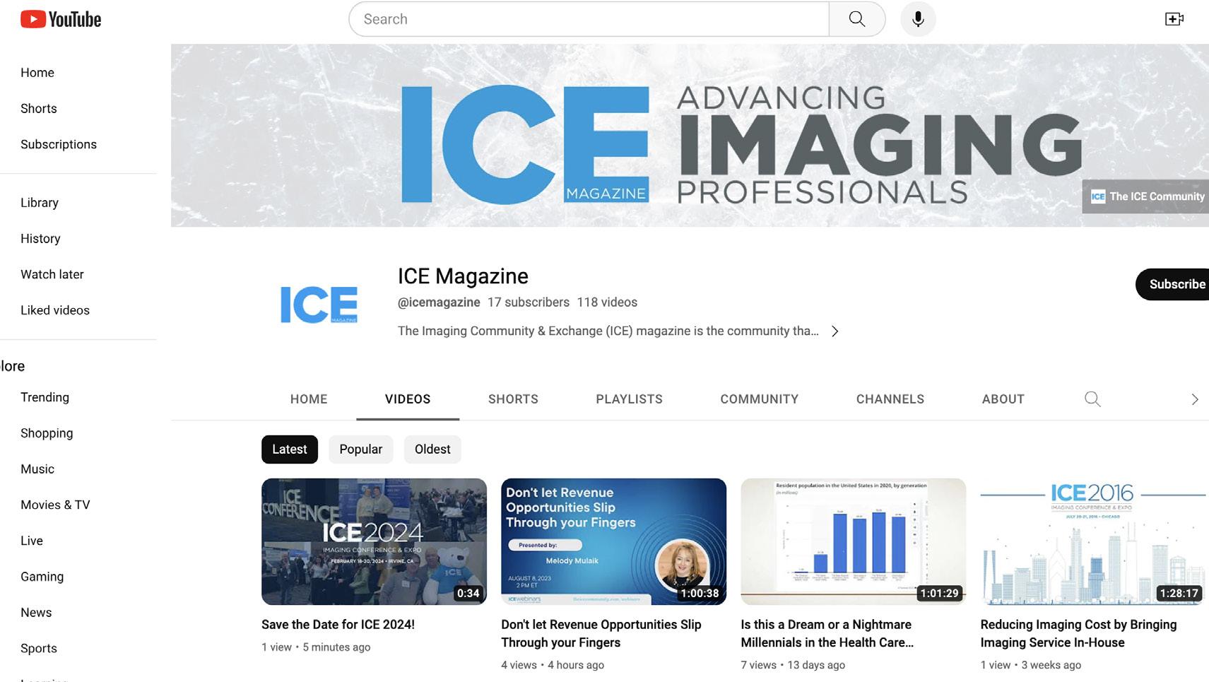
E7254 with RAD-21 and RAD-60 options now available. E7252 with RAD-14 options.
Plus Replacements for: E7239, E7242, E7252, DRX3724
