Published by
Matt
Gloria


Published by
Matt
Gloria
by Matt Herman
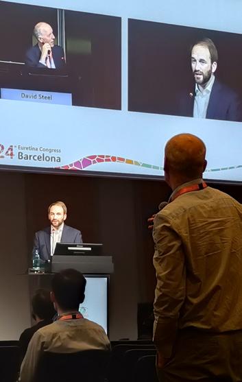
After years of speculation and research on its massive potential, artificial intelligence is finally making its way into ophthalmology clinics around the world. Experts at the 24th Congress of the European Society of Retina Specialists (EURETINA 2024) spoke about the developments making an instant impact on retinal clinical practices.
Artificial intelligence’s moment has officially arrived in ophthalmology, and a panel of world-renowned experts at a EURETINA 2024 Day 4 session shared how retinal specialists can—and should—get on board.
In a symposium chaired by EURETINA Board Member Prof. Martin Zinkernagel (Switzerland) and artificial intelligence luminary Prof. Pearse Keane (UK), some of ophthalmology’s leading minds in the subject talked about how AI is ready for the everyday clinic—and why the time is now.
AI’s ophthalmic roots are in retinal image analysis—and so it is only fitting that this is
now one of the most accessible entryways to integrating the technology into your clinic.
Data scientist Dr. Mathias Gallardo (France) surveyed the current opportunities for integrating AI image analysis into your clinic—but also took stock of the many pitfalls that come without thoughtful adoption.
“The science of AI is an opportunity for ophthalmology and retinal imaging,” said Dr. Gallardo. “AI models are fast, can perform both simple and complex tasks, handle huge amounts of data, and be tailored at any time,” he said.
The impacts that this could have are profound. Dr. Gallardo listed enhanced diagnostic accuracy, accessibility to care for
Thank you for visiting the BAYER-sponsored symposium and our booth in Barcelona.
We hope you found the information interesting and informative. See you next year in Paris!
>> Cont. from Page 1
patients in remote areas, personalized medicine, time efficiency, and cost savings as possible consequences of the opportunities of AI.
Pioneering initiatives like those from RetInSight (Vienna, Austria) and EviRed (Paris, France) and their work in fluid monitoring in age-related macular degeneration (AMD) and diabetic retinopathy (DR), respectively, are already taking advantage—and are finding their way into clinics.
But there are pitfalls, warned Dr. Gallardo, and this should inform the way forward. Image quality and its potential to complicate analysis is one. Others include a lack of standardization, prohibitive difficulties in translating gains in machine learning to the clinic, and the consequences of over-reliance on AI, including expertise erosion.
“AI should be used as an assistive tool, with doctors retaining the final decision,” Dr. Gallardo concluded. “A balanced approach, considering the opportunities and pitfalls of this technology, will ensure that AI models enhance, rather than replace, human skill.”
AI’s massive promise is now going well beyond its familiar territory of image analysis in exudative retinal disease and DR, and two panelists spoke on these new horizons.
Presenting for Prof. Raphael Sznitman who was unable to make it, Prof. Zinkernagel spoke about the potential of machine learning in surgical applications.
Screening for possible surgical interventions is one, and Prof. Zinkernagel presented the analysis of ultra wide field fundus imaging as one possible avenue. “We’re trying to create a model to detect changes in fundus images to guide the diagnosis of surgical pathologies,” he said.
He pointed out one image where the experimental model detected
breaks that marked a detachment. “It really helps you to plan your surgery when you go into the theater to do vitrectomies,” he said.
Beside this, he also looked at novel work on OCT biomarkers that could correlate to postoperative functional outcomes. Hyperrreflective foci was one that he described as potent for rhegmatogenous retinal detachments, helping to predict visual acuity improvement after procedures.
Another novel avenue for AI is in the area of inherited retinal diseases, according to Dr. Alexandra Miere (France).
Classification and segmentation of disease using color fundus photography, optical coherence tomography and fundus autofluorescence—as well as genotype-phenotype correlation, function prediction, and the generation of synthetic data is where AI for IRDs can shine, said Dr. Miere.
After introducing a series of studies, including much of her own work on this relatively novel topic, Dr. Miere reached her ultimate conclusion. 1-7
“The question is, does this [AI in IRDs] matter in clinical practice, and how does it impact the management of diseases?” she asked.
“The answer is yes. It will improve diagnostic accuracy, facilitate screening, enhance genotypephenotype correlations by providing new insights, new correlations and predicting disease severity. And, of course, it will provide future therapies that we hope will be made available for inherited retinal disease.”
With all of these current and prospective applications at our fingertips, the question of implementation becomes a practical one.
Amajor obstacle, according to presenter Dr. Clara Sanchez (Netherlands), is trust. In her presentation, Dr. Sanchez turned to
the reality of reliability—or perceived lack thereof—of some of these AI models, and how physicians can overcome it to realize AI’s full potential.
Though the performance of AI in many of the tasks described in the other presentations, adaptation is still scarce, observed Dr. Sanchez. “There’s a clear gap between what has been proposed in AI and what is really being adopted. So what’s happening?” she asked.
AI models, she proposed, are facing a credibility crisis.
“What we have to do is evaluate AI along different dimensions,” said Dr. Sanchez, naming accuracy, reliability, repeatability, resilience and safety as these dimensions.
Other less obvious areas must also be improved, she said, including defining a model’s intended use, standardizing data collection and labeling and making it transparent, regulating models and providing for both prospective and retrospective validation.
The best way to ensure this, she said in conclusion is a multi stakeholder approach to make implementation a reality—a reality much hoped for, and perhaps closer than ever as talks like this become increasingly common at marquee ophthalmic congresses like EURETINA 2024.
1. Miere A, Le Meur T, Bitton K, et al. Deep Learning-Based Classification of Inherited Retinal Diseases Using Fundus Autofluorescence. J Clin Med. 2020;9(10):3303.
2. Miere A, Zambrowski O, Kessler A, et al. Deep Learning to Distinguish ABCA4-Related Stargardt Disease from PRPH2-Related Pseudo-Stargardt Pattern Dystrophy. J Clin Med. 2021;10(24):5742.
3. Crincoli E, Zhao Z, Querques G, et al. Deep learning to distinguish Best vitelliform macular dystrophy (BVMD) from adult-onset vitelliform macular degeneration (AVMD). Sci Rep. 2022;12(1):12745.
4. Fujinami-Yokokawa Y, Pontikos N, Yang L, et al.; Japan Eye Genetics Consortium OBO. Prediction of Causative Genes in Inherited Retinal Disorders from Spectral-Domain Optical Coherence Tomography Utilizing Deep Learning Techniques. J Ophthalmol. 2019;2019:1691064.
5. Pontikos N, Woof W, Krawitz P, et al. Eye2Gene: prediction of causal inherited retinal disease gene from multimodal imaging using AI. Invest. Ophthalmol. Vis. Sci. 2022;63(7):1161.
6. Müller PL, Treis T, Odainic A, et al. Prediction of Function in ABCA4-Related Retinopathy Using Ensemble Machine Learning. J Clin Med. 2020;9(8):2428.
7. Veturi YA, Woof W, Lazebnik T, et al. SynthEye: Investigating the Impact of Synthetic Data on Artificial Intelligence-assisted Gene Diagnosis of Inherited Retinal Disease. Ophthalmol Sci. 2022;3(2):100258.

“I remember when Pepsi used the slogan, The Choice of a New Generation, and that is what EURETINA feels like now. The new generation of retinal specialists and industry is sophisticated yet suave, experienced yet eager. It’s all very uptempo, and is exactly my eye care vibe.”
- Matt Young


by Diana Truong
Research met recognition on Day 4 of the 24th Congress of the European Society of Retina Specialists (EURETINA 2024) as recipients of the prestigious RMCR Awards shared their groundbreaking work, shedding light on the future of retinal medicine with projects that push the boundaries of how we understand and treat retinal diseases.
At EURETINA 2024, the spotlight was on more than just cuttingedge research; it was also a celebration of the innovators shaping the future of retinal medicine.
The Retinal Medicine Clinical Research Grant (RMCR Award) took center stage, offering a vital boost to independent researchers dedicated to advancing our understanding and treatment of retinal diseases.
With up to €300,000 in funding available, this prestigious award
empowers clinicians and researchers across Europe and beyond to explore groundbreaking clinical studies. From observational research to genomic studies, the RMCR Award not only fuels scientific discovery but also fosters collaboration, pushing the boundaries of patient care.
Prof. Dr. Robert Finger (Germany) and Prof. Dominik Fischer (United Kingdom), previous recipients of the RMCR Award, presented their preliminary findings, while PD Dr. med. Maximilian Pfau (Switzerland) and
Dr. Javier Zarranz-Ventura (Spain), winners of this year’s grants, gave a sneak peek into the scope of their research endeavors.
Microbiome and metabolome in AMD
Funded by a 2021 RCMR Award, Prof. Dr. Finger’s research has focused on the interplay between the microbiome, metabolome and complement activation in age-related macular degeneration (AMD).
“There’s ongoing evidence that metabolic alterations are associated with complement occlusion in AMD, and that microbiome alterations are also linked to the disease.”
- Prof. Dr. Robert Finger
“There’s ongoing evidence that metabolic alterations are associated with complement occlusion in AMD,
and that microbiome alterations are also linked to the disease,” Prof. Dr. Finger explained. His study is aiming to connect these factors, providing a comprehensive analysis of intermediate and late AMD patients compared to healthy controls.
While still in progress, the research has already revealed subtle differences in systemic complement activation levels. “We’ve found some differences in metabolomics,” he noted, referring to untargeted analyses conducted on patient samples. However, he emphasized that the synthesis of these datasets, using advanced techniques like multi-omics factor analysis (MOFA), is ongoing.
The hope is that this work will identify key biomarkers or therapeutic targets within the metabolome and microbiome, paving the way for earlier detection and more personalized treatments for AMD. “More to come next year,” Prof. Finger teased, as the project heads toward its final phase.
Gene therapy, as Prof. Fischer noted, “is not only a virus when we inject, but a product,” comprised of various elements, such as full or empty capsids and host cell DNA, which can elicit immune responses.
His ongoing RMCR-funded research is investigating the immune responses to retinal gene therapy, with a focus on understanding the risks of gene therapy-associated uveitis (GTAU) in patients receiving adeno-associated virus (AAV) treatment. While efforts have been made to understand AAV’s immunogenic potential, particularly its adaptive immune response, the study reveals gaps in correlating clinical observations with biomarkers.
The team has collected samples from patients both before and after gene therapy. Notably, preliminary results show that cytokine levels remain elevated long after gene therapy, despite low clinical signs of inflammation.
“Clinical quantification of GTAU does not closely reflect cytokine levels,” Prof. Fischer shared. He speculated that the persistent immune response might play a role in unexplained post-therapy retinal changes, such as chorioretinal atrophy.
The study is far from complete, but its early findings underscore the need for more sensitive diagnostic tools beyond slit-lamp evaluations to fully grasp the immune complexities in retinal gene therapy.
Dr. Pfau, one of this year’s RMCR award recipients, will use the funds towards his study on enhanced vision and imaging tests for detecting early and intermediate AMD, focusing on the limitations of traditional vision tests like best-corrected visual acuity (BCVA) and aiming to revolutionize how AMD is diagnosed and tracked.
Dr. Pfau highlighted the insufficiency of chart-based tests, noting that “you only can uncover about 10% of AMD patients” in its early stages. This gap has led him to develop a novel approach using fundus-tracked dark adaptometry, which measures how the eye adapts to darkness—an early marker of AMD. His method tests closer to the fovea, where vision deterioration begins, making it more precise than previous tools.
This technique holds promise as a predictive biomarker for early AMD, potentially even before clinical symptoms arise. Dr. Pfau explained, “Dark adaptation at 2° eccentricity is much more altered than at 4°, 6°, and 8°,” an observation that may lead to earlier interventions and better treatment outcomes.
With this new test, he envisions a future where clinicians can catch AMD at its inception, offering hope for those at risk of devastating vision loss.
Dr. Zarranz-Ventura ended the session with an introduction of the groundbreaking ALMA project—an artificial intelligence (AI)-driven tool designed to predict treatment needs in neovascular AMD.
“This is ALMA,” Dr. Zarranz-Ventura explained, noting that the name fittingly means ‘soul’ in Spanish. The project aims to harness the power of AI by integrating clinical and OCT data to optimize patient outcomes.
By leveraging Spain’s national registry, which now includes data from over 8,000 eyes, ALMA’s potential lies in its ability to predict treatment intervals, injection frequency and visual outcomes. “We need to look at the previous data that we have to be able to make predictions for the future,” Dr. Zarranz-Ventura explained.
The tool will not only quantify retinal data but also provide personalized treatment predictions, such as whether a patient will need fewer than seven injections or experience significant visual improvement.
As Dr. Zarranz-Ventura emphasized, “With this, we will run the algorithms to try to do the predictions based on these previous studies.” The ALMA project, poised for its next phase of validation in 2025, promises to enhance precision medicine in the treatment of neovascular AMD, bringing new hope to patients and clinicians alike.
by Tan Sher Lynn
The EURETINA database has been a groundbreaking tool for vitreoretinal surgeons, allowing for anonymous, accessible, and robust data collection, significantly improving clinical outcomes for conditions such as retinal detachment and macular holes.
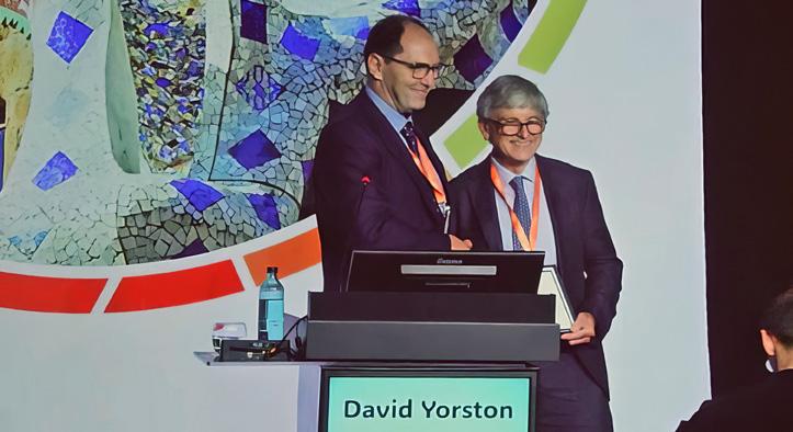
Dr. David Yorston (UK) delivered the esteemed Gisbert Richard Lecture, reflecting on the development and impact of the EURETINA database over the last 10 years.
Initiated by Bill Aylward during his presidency, the EURETINA database aims to improve how vitreoretinal surgeons audit their outcomes.
Recognizing the inadequacies in existing methods, he formed working groups to develop standard datasets for retinal detachment and macular hole surgeries. The database now holds data on 15,600 retinal detachments and 5,300 macular hole surgeries, noted Dr. Yorston.
He emphasized that the database’s guiding principles include complete anonymity for both surgeons and patients, ensuring data accessibility for users to analyze their own outcomes at any time. It employs software for accurate data collection and is classified as a service evaluation, thus negating the need for ethics approval.
According to Dr. Yorston, the EURETINA database has revealed that
the failure rate of retinal detachment surgery in patients over 80 is twice that of middle-aged patients. It was previously assumed that older patients had a worse prognosis due to difficulty positioning or non-ophthalmic comorbidity.
However, the data indicates that older patients have a worse prognosis to start with, being more likely to have a total detachment and have proliferative vitreoretinopathy (PVR), he noted.
Recent findings reveal important trends and improvements in the management of retinal conditions. Notably, “women are more likely to have larger full thickness macular holes with poor prognosis,” Dr. Yorston said. “Additionally, older patients and pseudophakic eyes are more likely to present with complex detachments that may require further surgery.”
Other key results have also come out of this treasure trove of data. The definition of a large macular hole, for example, has been refined to 500 microns or greater—and it has been shown that surgery for recent onset macular holes should be performed urgently.
The data also indicates that inferior breaks and inferior detachments correlate with an increased risk of surgical failure. Using chromatin gas during surgery and instilling a bubble with sponges while releasing subretinal fluid may enhance outcomes, Dr. Yorston suggested.
He also stressed that early surgical intervention, ideally within the first 72 hours, is also crucial for better visual results in cases of macular detachments.
Research coming out of the database has also demonstrated that an eyelid flap is superior to eyelid peels in large holes, and denser gas yields better visual and anatomical outcomes than lighter gas in eyes with predominantly inferior morphology.
The impact of these strategies is evident in the data: the median duration of retinal detachment in macular off patients has decreased from five days to four days, reflecting a significant change in practice.
In concluding his lecture, Dr. Yorston reflected on the ongoing advancements made possible by the Euretina database. “We live in a world characterized by increasing polarization and division. The retina database reminds us of how much we can achieve when we work collaboratively together,” he said.
Editor’s Note:
A version of this article was first published on piemagazine.org.
by Matt Herman
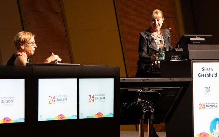
Acclaimed author and researcher Baroness Susan Greenfield delivered the Diversity Keynote at the 24th Congress of the European Society of Retina Specialists (EURETINA 2024). As one of the planet’s foremost neuroscientists, she delivered an address on human individuality, how to nurture it—and the existential threat that modern society poses to it.
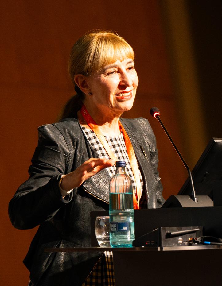
One of the marquee sessions of EURETINA 2024 was far removed from the world of retinal scholarship and innovation.
Baroness Susan Greenfield, author of several bestselling books and a powerful voice in the neuroscience world, delivered the EURETINA 2024 Diversity Keynote—and by the end of the session, attendees in Rooms 116/117 of CCIB Barcelona were lining up to join the discussion.
“For a
young child and someone with dementia, they
don’t
have the privilege of this carefully curated human brain, personalized to give you your unique cognitive take on the world and look beyond the sensory press. Connections liberate you from that.”
The topic at hand was the neuroscientific basis of individuality. “How is it that for the hundreds of thousands of years we’ve stalked this
planet, there’s never been anyone like you again?” she asked.
The simple answer, according to Baroness Greenfield, is plasticity—the creation of uncountable neurons via branching as we interact with the world around us.
From London black cab drivers whose hippocampi are enlarged through intensive memorization of the city’s labyrinthine road network, to the differences in the brains of rats in radically different stimulatory environments, Baroness Greenfield argued for experience and the bestowal of arbitrary meaning on objects and ideas as the basis of human individuality.
“Forming more connections enables you to give life some kind of significance over time,” she summarized. “One can say that this is the transition from a sensory evaluation of the world to a cognitive one, where you have meaning that is not apparent from pure sensation.”
“This has an impact on thought in ways that we don’t fully understand, but computers don’t touch you and you don’t touch them and they don’t touch each other—and this is an illustrative aspect of our uniqueness.”
For Baroness Greenfield, this is the essential crux of our individuality—the divergent development of our cognitive selves from pure sensation.
To highlight this, she pointed out the parallels between young children and adults with dementia.
Both have comparatively fewer neural connections, and experience is relegated purely to the world of the sensory—either because plasticity has not developed with time yet,
or because neural connections are degenerating with age and disease.
“For a young child and someone with dementia, they don’t have the privilege of this carefully curated human brain, personalized to give you your unique cognitive take on the world and look beyond the sensory press. Connections liberate you from that,” she illustrated.
For all of the hope and wonder inspired by this take on the nature of human individuality, pitfalls and threats to it abound.
Baroness Greenfield illuminated the menace that modern society poses to our ability to escape the realm of pure sensation. Above all else, she believes that the greatest threat to the unique self lies in the warped ways we interact with and interpret certain features of modern technological advancements.
The first of threats, she explained, is a growing tendency to ignore the uniqueness of the brain in a world full of silicon rivals to it.
The human brain, she argued, is capable of utilizing both crystallized and fluid intelligence. Humans possess a memory of stored experience—our crystallized intelligence—that allows us to interpret and place meaning on the world around us, instead of
just viewing reality as a junkheap of disparate pieces of information.
“I think we need to distinguish that information and knowledge are not the same thing,” she said. “The mind is therefore beyond just the mere complexity that gets everyone so excited. It’s non computational,” she said.
This qualitative character of the brain has its root in its chemistry, she said, and the fact that there’s a sensing body attached. Touch, for example, stimulates endorphins.
“This has an impact on thought in ways that we don’t fully understand, but computers don’t touch you and you don’t touch them and they don’t touch each other—and this is an illustrative aspect of our uniqueness.”
Machines, she continued, don’t have endocrine and immune systems. They don’t have a lifetime of unique experiences and they don’t have an inner subjective state of agency. They don’t possess varying degrees of consciousness like we do. And it is therefore fallacious, according to Baroness Greenfield, to believe that the human self is not something apart from the silicon brains we surround ourselves with.
For Baroness Greenfield, it is ultimately the complex interaction of these three components—the personalized mind, the brain and
its plasticity, and the subjective experience of consciousness and its varying degrees—that separates us from our modern machines. To lose sight of this uniqueness in the face of technological marvels like artificial intelligence is to lose touch with the roots of our individuality, she said.
The final portion of Baroness Greenfield’s talk was devoted to the threat of technology itself on individuality—and the opportunities we have to liberate ourselves from this threat through creativity and the connection to our cognitive selves.
Returning to themes from the beginning of her talk, Baroness Greenfield encouraged the audience to see potentially harmful technologies like social media as part of a larger trend in human societies towards external stimulation.
“This
is opposed to thinking, where our prefrontal cortex is now fully operational. We call it the cognitive take on the world. We have a past, present and future, and internal stimuli are now dominant. Meaning is personalized, you have a strong sense of who you are—your identity and your individuality— and a clear time space reference.”
This can be seen as a tendency to return to a state of purely sensory existence—in many ways the opposite of a cognitive state and a step away from individuality. Boredom, she said, is a consequence of reliance on such sensory stimulation.
“This is a world driven by external stimulation, but what’s happening in
the brain? What happens when you’re just in the present?” she asked.
The answer lies in the prefrontal cortex, whose underdevelopment is shared by children and teenagers and sufferers of schizophrenia.
Excessive external stimulation leads to excessive levels of dopamine, which dampens down the prefrontal cortex. The result is an existence dominated by arousal, reward and addiction. Baroness Greenfield described humans, then as divided between two different states.
“I think we can think of the human brain normally as having two modes. One is feeling, with the prefrontal cortex underfunctioning and we are driven by raw senses, living in the here and now. External stimuli are dominant. The world has little meaning,” she said.
“This is opposed to thinking, where our prefrontal cortex is now fully operational. We call it the cognitive take on the world. We have a past, present and future, and internal stimuli are now dominant. Meaning is personalized, you have a strong sense of who you are—your identity and your individuality—and a clear time space reference.”
The key, Baroness Greenfield argued, is not to overemphasize our cognitive side. It’s not that we should ignore the human need for external stimulation, and eschew living in the sensory moment for asceticism. But we should be aware that technology is pulling us strongly in one direction—and do something about it.
“I want to argue that the way around this, if you want to be fully fledged and fulfilled, is to develop an inner narrative,” she said, nearing the end of her time at the podium. This is what creativity is in her conception, and she commented on the power it has.
“When you’re young, you know that magic invitation—let’s make up a game. And when you’re doing that, you’re empowering yourself, because
you’re developing an inner narrative. You are in control and anything is possible. You’re not limited by some web designer or by someone else’s plot or driven by options or menus,” she said.
“I
think we can think of the human brain normally as having two modes. One is feeling, with the prefrontal cortex underfunctioning and we are driven by raw senses, living in the here and now. External stimuli are dominant. The world has little meaning.”
There are many ways to engage this inner narrative. She emphasized linear activities that have a past, present and future—or a beginning, middle and end—like making music or sharing stories or exercising.
Thought is movement confined to the brain, she said, paraphrasing Parkinson’s researcher Oleh Hornykiewicz—and without thinking and its sequential structure, we are trapped in a world full of sensory information with no meaning.
To summarize, Baroness Greenfield brought things back to a final purpose. “So what is the ultimate goal? It’s to make unprecedented neural connections, but those that have a significance. You suddenly see the world in a different way, and this is the most exciting and fulfilling and amazing thing—to do something for the first time that no one else has done.
“That, I would say, is the true fulfillment of individuality. I think that how we can make the most of being diverse and being true individuals is to have life stories that actually reflect your identity, because that’s your individual mind. And only when you have an individual mind can you be truly creative. And this will give you a wondrous sense of uniqueness.”