
21 minute read
Face Blindness


Prof. Richard Muscat
Tutor
Advertisement
Dr. Christian Zammit M.D.,M.Sc.
Overview on Face Blindness Facial recognition is a complex task, often done immediately and readily, involving discrimination of subtle differences in facial structures with differences in facial expressions, ageing, perspectives and lighting. Facial recognition requires fast identification of stimuli which are then correlated against reservoirs of faces which are accumulated throughout life (Barton and Corrow, 2016). The facial recognition system is extremely complex, and if impaired, cannot be fully remedied by other areas of the brain. When such injury occurs early on in life, juvenile brain plasticity has been shown to be potentially inadequate to restore facial recognition functions, thereby suggesting that such an impairment can have severe, permanent implications, even at an early age (Barton et al., 2003) Damage to any part of the facial recognition mechanism may result in the development of face blindness. Such dysfunction results in the development of selective face-recognition and visual learning deficits, a condition called prosopagnosia. Prosopagnosia can be either acquired or congenital. The acquired form of prosopagnosia is considered to be a rare consequence of occipital or temporal lobe damage, possibly due to stroke or lesions occurring in adulthood. Congenital prosopagnosia, on the other hand, is usually not found associated with any gross abnormalities, and no clear underlying causative agent is found to be associated with the development of the disease (Grüter et al., 2008). Nevertheless, face blindness in children may also be associated with inherited or acquired brain lesions, and may not be exclusively of a congenital/hereditary aetiology. Moreover, prosopagnosia can also occur in association with other disorders, which may be psychiatric, developmental or associated with multiple types of visual impairment (Watson et al., 2016). Impact on Social Behaviours The impact of face recognition is of crucial importance in human social interaction. Face recognition allows the determination of identity of third parties and self, and permits the gathering of information on age, health status, gender and mood of an individual. Facial features are a considerable aspect in sexual attraction, and also play a vital role in the interpretation of speech via the observation of lip movements and in determination of direction of gaze (Grüter et al., 2008). The discovery of neurons associated with facial recognition in primates suggests that this process has evolutionary implications (Tsao et al., 2006). Prosopagnosia may sometimes be associated with autism spectrum disorders (ASD). The link between autism and prosopagnosia may be two-fold. The lack of interest in social interaction characteristic of autism may result in a deficit in the development of facial recognition. In this case, the anatomical and functional features of facial recognition would be working normally, but prosopagnosia would ensue due to the lack of facial recognition
experience (Grüter et al., 2008). On the other hand, dysfunctions in the amygdala and the fusiform face area may result in an anatomical basis for the development of autism and ensuing prosopagnosia (Sasson, 2006). Both instances may in turn lead to a detrimental impact on social development and interaction.
However, a direct correlation between ASD and facial recognition dysfunction is still to be determined (Grüter et al., 2008). The causative agents in the development of ASD (Santangelo and Tsatsanis, 2005), and therefore, of associated symptoms of prosopagnosia remain unclear (Wang et al., 2015).
Different degrees of severity of prosopagnosia may lead to social and occupational deficits, together with problems in daily functioning. These may include an inability to recognize self, mistaking familiar faces such as family members as strangers when there are changes in hairstyles and an excessive reliance on verbal cues for identification (Barton and Corrow, 2016). explain the association between prosopagnosia and colour blindness, as well as other visual field defects including quadrantinopias or hemianopias (Bouvier and Engel, 2006). Emotion recognition can also be impaired in acquired prosopagnosia, but was thought to be absent in hereditary face blindness; however, recent data has proven otherwise. Acquired prosopagnosia is usually characterized by a loss of familiarity, whilst hereditary prosopagnosia usually is associated with a loss of confidence in the feeling of familiarity, brought about by a generalized visual impairment which mostly affects the facial recognition process (Grüter et al., 2008).
Incidence, Prevalence and Aetiology of Face Blindness Recent data has shown a considerable prevalence of congenital prosopagnosia, with levels comparable to those of dyslexia and dyscalculia (Kennerchnecht et al., 2006), affecting 2.5-2.9% of the Caucasian population (Bowles et al., 2009). On the other hand, although purely acquired prosopagnosia is a rare condition, many individuals suffering from brain lesions tend to suffer from ranging degrees of visual impairments affecting facial recognition together with other cognitive disorders (Bate and Bennetts, 2014).
Acquired prosopagnosia is usually the result of localized tissue damage usually to the occipito-temporal lobe, whereas congenital prosopagnosia is due to a problem in neural development. In acquired prosopagnosia, together with an occipito-temporal lesion, adjacent areas of the cortex may also be involved in the damage. This could potentially
Facial Recognition Process Facial recognition and memory involves multiple areas of the brain, which include the middle temporal lobe, amygdala, inferior frontal and parahippocampal gyri, the hippocampus and orbitofrontal cortex. The process of facial recognition is dependent on a number of factors, including orientation, attention, age and emotional demeanour. Throughout the years, research has shown that the facial recognition mechanism is brought about by a specialized neuronal network, rather than forming part of a larger recognition process (Grüter et al., 2008).
The normal face recognition process involves multiple stages. Primarily, the face is recognized as being a face. Following the initial detection phase, generalized facial information is gathered, including the gender, age, health status, and emotional demeanour. This process involves a correlation of the facial information with stored images. The original model of facial
recognition by Bruce and Young (1986) [Figure 1] does not include cortical involvement in the process (Grüter et al., 2008). A later model developed by Ellis and Lewis suggests the parallel involvement of facial recognition and cortical involvement in the process of familiarity (Ellis & Lewis, 2001). Disconnection between the two systems could potentially result in a feeling of unfamiliarity (Grüter et al., 2008).
Any damage to these three areas of the cortex may result in the development of prosopagnosia. Damage to the occipital face area may result in a deficit in the obtaining of facial information. A deficit in the fusiform face area may result in a reduced adaptation to familiar faces. Lesions to the dorsal superior temporal gyrus may lead to reduced processing of dynamic facial data (Grüter et al., 2008).

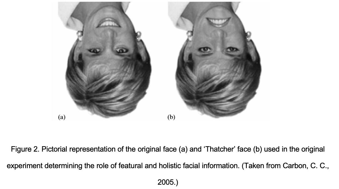
A functional model of facial recognition was developed in association with specific areas of the cortex. This model suggests the presence of a core component which involves the occipital face area in the inferior occipital lobe, the fusiform face area in the fusiform gyrus (also called the extended system) and the face area in the dorsal superior temporal gyrus (Grüter et al., 2008; Towler et al., 2017). The dorsal superior temporal gyrus is thought to be involved in dynamic facial information, whereas the occipital face area and the fusiform face area is thought to be associated with constant facial features in facial recognition (Grüter et al., 2008). It has been proposed that facial recognition is initiated by the occipital face area and is then transmitted to the other two areas of the cortex. On the contrary, the extended system is thought to be involved in the emotional response and the information perceived by the individual. A correlation between facial and vocal recognition has also been determined via an association between the fusiform face area and the dorsal superior temporal gyrus (Gobbini and Haxby, 2007). Electrical current stimulation in the occipital face area of pre-surgical epileptic patients resulted in the development of temporary prosopagnosia, where the patients failed to recognize famous faces (Jonas et al., 2012) Stimulation of the fusiform face area resulted in perceptual facial disorders, whereby facial features appeared to be moving, and experienced changes in facial identity from a third-party individual to another. These results suggest a confirmatory link between these areas of the cortex and facial processing (Rangarajan et al., 2014). Facial recognition is present in neonates; however, it is finessed over the years. There are a number of strategies utilized in the recognition and learning of faces. Faces may be viewed as whole or as the identification of individual facial features, the latter being more prominent in upright faces (Grüter et al., 2008). The process of configural recognition, namely the piecing together of individual features to make up a whole face is lost when faces are upside down (Carbon and Leder, 2006). This is evident when selective facial features such as the eyes and mouth are turned upside down in an upright and inverted picture. The change is immediately noticed in the upright picture but is lost in the inverted one. This has led to a hypothesis that faces are stored and pieced together against a reservoir of what counts as a typical face to the individual (Valentine, 1991). When faces are rotated from their normal, upright orientation, holistic processing, that is, the piecing together of facial features against a whole face, is diminished, resulting in the aforementioned
This phenomenon could also potentially explain the ‘other-race’ effect, whereby an individual is more confident in recognizing faces of his or her own race. This could possibly occur because the reservoir of faces mostly stored by an individual against which a new face is compared to are those of own-race (Grüter et al., 2008). When an individual stores more otherrace faces, this discrepancy eventually lessens considerably (Sporer, 2001).
A recent study has shown that prosopagnosia can occur due to damage on both sides of the brain, with a prevalence on right-sided damage. This suggests that there is a right lateralization of the process of face recognition after birth which persists. Although damage to the rightside of the brain can be compensated for, conspicuous damage to this area may lead to permanent deficits in face recognition (Watson et al., 2016).
The exact pathophysiology of this condition is still unclear to date. However, Harris et al., 2005 proposed that patients suffering from prosopagnosia were found to have an abnormality in the N170 wave on electroencephalography (EEG). This wave is considered to be the hallmark of face processing in the temporal lobe and functional MRI scans have associated this wave abnormality with potential damage to the occipital face are and smaller anterior fusiform gyri (Grüter et al., 2008; Towler et al., 2017).
Acquired Prosopagnosia Acquired prosopagnosia may be considered as a lingering condition following brain tissue damage. The presentation of the disorder may be extremely varied, depending on the extent and location of damage. This condition may be viewed as a deficit in image and object recognition which leaves a detrimental effect on the facial recognition process.
Acquired prosopagnosia is an extremely rare disorder, which in most cases is not present in patients with occipito-temporal lesions. When present, it is mostly associated with patients who have other severe symptoms, including hemi-spatial neglect and visual field defects. The varied presentation of the disorder adds another level of complexity to the diagnosis of the disorder (Grüter et al., 2008).
There are three major variants of acquired prosopagnosia. Appreciative prosopagnosia involves the loss of ability to derive sufficient data about a face from visual cues results, and is characterized by a lack of activation of the familiarity signal. In associative prosopagnosia, one loses the ability to associate the acquired facial information to the reservoir of stored facial images (Davies-Thompson et al., 2014). In cases where the reservoir of stored images is lost, and the individual cannot correlate new images to stored ones, the amnesic form of the disorder, termed amnesic prosopagnosia follows (Damasio et al., 1990).

Congenital Prosopagnosia Congenital prosopagnosia is characterized as a selective facial processing deficit in the absence of any intellectual disability or brain lesion (Towler et al., 2017). Usually, individuals with this type of prosopagnosia first report any
symptoms during adolescence or adulthood, potentially due to the increased social demands during these life-stages (Towler and Eimer, 2012; Susilo and Duchaine, 2013). In cases where prosopagnosia is not associated with any brain lesion, and manifests itself early in life, a familial, and hereditary component of this condition has been reported. A study of pedigrees has shown a characteristic autosomal dominant manner of inheritance, potentially due to point mutation in a single or multiple genes. This could also suggest that within the context of a single family, the causal defect could potentially by the same mutation (Grüter et al., 2008). However, this does not exclude the development of prosopagnosia due to uterine environment or de novo mutations (Barton & Corrow, 2016). Contrary to acquired prosopagnosia, congenital prosopagnosia has a number of analogous and common symptoms. The diagnostic hallmark present in all patients with congenital prosopagnosia reported during a study was found to be a lack of confidence about facial familiarity (Kennerknecht et al., 2006). Rather than the inability of recognizing faces, it was the determination of familiar faces which was found to be dysfunctional. This could in turn translate in unfamiliar responses to familiar faces, and hyper-familiarity to strangers (Grüter et al., 2008). Other features of congenital prosopagnosia are prolonged recognition of familiar faces and learning of new ones. A modified pattern of scanning facial features was found in congenital prosopagnosia. Congenital prosopagnosia also allows more time for coping mechanisms. Individuals with the disorder usually behave in a preventative, apologetic and compensating manner to avoid any unfavourable situations (Grüter et al., 2008). A recent study has also shown that contrary to popular perception, congenital prosopagnosics also display emotion recognition deficits, when asked to follow changes in emotional display from whole-face, or ocular regions. This further highlights the heterogeneity present in this condition (Biotti and Cook, 2016). Diagnosis The diagnosis of facial recognition impairment is challenging. To date, there is no test which can accurately determine any dysfunction of the system. Moreover, there are currently no set standards for the levels of facial recognition which should be reached by an individual at specific ages, and skills associated with facial recognition are not taught in the education system (Grüter et al., 2008). The current process of diagnosis of facial blindness is the use of behavioural test questionnaires. This may result in problems with the diagnosis of specific conditions, since it simply elucidates a score which is below a set criterion, thereby indicating the presence of a problem with facial recognition. Diagnosis is usually established via the utilization of neural, biochemical and genetic markers which may contribute in an additive manner to the results of behavioural test questionnaires (Barton & Corrow, 2016). To add another level of complexity to the diagnosis of the condition, there is a range of abilities that even healthy individuals have in the recognition of faces (Zhu et al., 2010). Healthy individuals may either never forget a face or may find problems in remembering faces (Russel et al, 2009). This in turn translates to a difficulty in the determination of the definition of prosopagnosia, and what segment of the population actually suffers from the condition. Moreover, certain behavioural mechanisms adopted by prosopagnosics may also hinder the determination of the severity of the disorder (Barton and Corrow, 2016). Several markers are usually used to dissociate between individuals suffering from prosopagnosia and individuals who are at the lower end of the spectrum with respect to facial recognition. These include an absence of face-inversion effect (Behrmann et al., 2005),
absence of holistic dispensation (Avidan et al., 2011), irregular scanning of the face (Schwarzer et al., 2007) and a paradoxical better processing of the buccal than ocular regions (DeGutis et al., 2012). These tests usually show a reduced rather than absent features, and may be solely indicative of a potential problem rather than a hallmark diagnosis (Barton and Corrow, 2016).
The diagnosis of acquired prosopagnosia could potentially be easier than that for congenital prosopagnosia, primarily because the individual can recognize a discrepancy and a decline in the ability to recognize faces, and secondarily because this decline in facial recognition can be correlated to a condition, such as a stroke or trauma (Barton and Corrow, 2016).
Treatment and Coping Mechanisms A number of individuals with developmental prosopagnosia tend to develop individual coping mechanisms to allow them to identify the people around them, mostly via non-facial cues such as voice, clothing, gait and hairstyle recognition (DeGutis et al., 2014, Bate and Bennetts, 2014).
The paucity of data surrounding effective remedies for prosopagnosia could suggest an important, and currently missing link on how to better enhance knowledge on the disorder both on a theoretical and practical levels (Bate and Bennetts, 2014).
The general consensus on the development of facial recognition throughout the lifespan is that the general holistic skills on facial recognition are generally developed at a very early age, with no considerable qualitative changes beyond the ages of 4-5 years. This in turn implies limited plasticity beyond early childhood with respect to face processing, and that rehabilitation of such patients may be difficult (Nelson, 2001). However, the mechanisms involved in face recognition may be finessed and transformed beyond childhood, possibly even during adulthood (Bate and Bennetts, 2014). The determination of the best mode of rehabilitation of prosopagnosia requires the determination of the timing, location and extent of injury, if present. The locus of the injury, that is, whether the manifestation of the disorder is perceptual or semantic should be determined, and the rehabilitation tailored for the specific case (Bate and Bennetts, 2014).
Two types of rehabilitation methods may be used: compensatory and remedial. The compensatory model enhances the behaviours which allow coping with the condition. The remedial one focuses on reinstating normal facial recognition behaviours. Due to the lack of data available, and the variable manifestations of the disease, no rehabilitation data has yet proven to be more efficient than the other, but possibly depend on the parameters of individual cases (Bate and Bennetts, 2014).
Spontaneous recovery and neuronal remodelling have been reported in some cases of acquired prosopagnosia (Bate and Bennetts, 2014; DeGutis et al., 2014). This could suggest a potential niche for treatments and rehabilitation modalities to considerably improve facial processing.
Conclusion Facial recognition is an essential process in functional social development and interaction. It involves multiple areas of the brain, and involves a complex mechanism involved in the acquiring of multitude information gathered from facial features. Specific areas of the brain have been found to be associated with facial recognition. Any lesions in these areas of the brain may lead to acquired prosopagnosia, a rare condition which causes a deficit in facial recognition. Another form of prosopagnosia, not associated with any gross brain abnormalities, with a much higher prevalence is congenital prosopagnosia, which usually manifests early in life and follows an autosomal dominant mode of inheritance.
Although the data on prosopagnosia and its potential treatment or rehabilitation is
very scarce, there have been some potential improvements in the rehabilitation of the disorder. This could in turn translate in application of treatment for other populations with face processing and cognitive deficits.
Despite being acknowledged as a neurological disorder, prosopagnosia has received little attention within the clinical field. This could be potentially due to a lack of formal diagnostic standards, lack of awareness about the condition and considerable difficulties in the diagnosis of such patients. Increased interdisciplinary awareness of the condition, and introduction of formal, standardized diagnostic criteria could potentially improve the current situation considerably, thereby improving efficacy in managing and treating patients with this condition and other related conditions.
References
1. Avidan, G., Tanzer, M., & Behrmann, M. (2011). Impaired holistic processing in congenital prosopagnosia. Neuropsychologia, 49(9), 2541-2552. 2. Barton, J. J., Cherkasova, M. V., Press, D. Z., Intriligator, J. M., & O’Connor, M. (2003). Developmental prosopagnosia: A study of three patients. Brain and cognition, 51(1), 12-30. 3. Barton, J. J., & Corrow, S. L. (2016). The problem of being bad at faces. Neuropsychologia, 89, 119-124. 4. Bate, S., & Bennetts, R. J. (2014). The rehabilitation of face recognition impairments: a critical review and future directions. Frontiers in human neuroscience, 8, 491. 5. Bate, S., & Bennetts, R. (2015). The independence of expression and identity in faceprocessing: evidence from neuropsychological case studies. Frontiers in psychology, 6. 6. Behrmann, M., Avidan, G., Marotta, J. J., & Kimchi, R. (2005). Detailed exploration of face-related processing in congenital prosopagnosia: 1. Behavioral findings. Journal of Cognitive Neuroscience, 17(7), 1130-1149. 7. Biotti, F., & Cook, R. (2016). Impaired perception of facial emotion in developmental prosopagnosia. Cortex, 81, 126-136. 8. Bouvier, S. E., & Engel, S. A. (2006). Behavioral deficits and cortical damage loci in cerebral achromatopsia. Cerebral Cortex, 16(2), 183-191. 9. Bowles, D. C., McKone, E., Dawel, A., Duchaine, B., Palermo, R., Schmalzl, L., ... & Yovel, G. (2009). Diagnosing prosopagnosia: Effects of ageing, sex, and participant–stimulus ethnic match on the Cambridge Face Memory Test and Cambridge Face Perception Test. Cognitive Neuropsychology, 26(5), 423-455. 10. Bruce, V., & Young, A. (1986). Understanding face recognition. British journal of psychology, 77(3), 305-327. 11. Carbon, C. C., & Leder, H. (2005). When feature information comes first! Early processing of inverted faces. Perception, 34(9), 1117-1134. 12. Carbon, C. C., & Leder, H. (2006). When faces are heads: View-dependent recognition of faces altered relationally or componentially. Swiss Journal of Psychology, 65(4), 245-252. 13. Damasio, A. R., Tranel, D., & Damasio, H. (1990). Face agnosia and the neural substrates of memory. Annual review of neuroscience, 13(1), 89-109. 14. Davies-Thompson, J., Pancaroglu, R., & Barton, J. (2014). Acquired prosopagnosia: structural basis and processing impairments. Front Biosci (Elite Ed), 6, 159-174. 15. DeGutis, J., Cohan, S., Mercado, R. J., Wilmer, J., & Nakayama, K. (2012). Holistic processing of the mouth but not the eyes in developmental prosopagnosia. Cognitive Neuropsychology, 29(5-6), 419-446. 16. DeGutis, J. M., Chiu, C., Grosso, M. E., & Cohan, S. (2014). Face processing improvements in prosopagnosia: successes and failures over the last 50 years. Frontiers in human neuroscience, 8, 561. 17. Gobbini, M. I., & Haxby, J. V. (2007). Neural systems for recognition of familiar faces. Neuropsychologia, 45(1), 32-41. 18. Grüter, T., Grüter, M., & Carbon, C. C. (2008). Neural and genetic foundations of face recognition and prosopagnosia. Journal of Neuropsychology, 2(1), 79-97. 19. Ellis, H. D., & Lewis, M. B. (2001). Capgras delusion: a window on face recognition. Trends in cognitive sciences, 5(4), 149-156. 20. Harris, A. M., Duchaine, B. C., & Nakayama, K. (2005). Normal and abnormal face selectivity of the M170 response in developmental prosopagnosics. Neuropsychologia, 43(14), 2125-2136. 21. Jonas, J., Descoins, M., Koessler, L., Colnat-Coulbois, S., Sauvée, M., Guye, M., ... & Maillard, L. (2012). Focal electrical intracerebral stimulation of a face-sensitive area causes transient prosopagnosia. Neuroscience, 222, 281-288. 22. Kennerknecht, I., Grueter, T., Welling, B., Wentzek, S., Horst, J., Edwards, S., & Grueter, M. (2006). First report of prevalence of non‐syndromic hereditary prosopagnosia (HPA). American Journal of Medical Genetics Part A, 140(15), 1617-1622. 23. Nelson, C. A. (2001). The development and neural bases of face recognition. Infant and child development, 10(1‐2), 3-18. 24. Rangarajan, V., Hermes, D., Foster, B. L., Weiner, K. S., Jacques, C., Grill-Spector, K., & Parvizi, J. (2014). Electrical stimulation of the left and right human fusiform gyrus causes different effects in conscious face perception. Journal of Neuroscience, 34(38), 12828- 12836. 25. Russell, R., Duchaine, B., & Nakayama, K. (2009). Super-recognizers: People with extraordinary face recognition ability. Psychonomic bulletin & review, 16(2), 252-257. 26. Santangelo, S. L., & Tsatsanis, K. (2005). What is known about autism. American Journal of Pharmacogenomics, 5(2), 71-92. 27. Sasson, N. J. (2006). The development of face processing in autism. Journal of autism and developmental disorders, 36(3), 381-394. 28. Schwarzer, G., Huber, S., Grüter, M., Grüter, T., Groß, C., Hipfel, M., & Kennerknecht, I. (2007). Gaze behaviour in hereditary prosopagnosia. Psychological research, 71(5), 583-590. 29. Sporer, S. L. (2001). Recognizing faces of other ethnic groups: An integration of theories. Psychology, Public Policy, and Law, 7(1), 36. 30. Susilo, T., & Duchaine, B. (2013). Advances in developmental prosopagnosia research. Current opinion in neurobiology, 23(3), 423-429. 31. Towler, J., & Eimer, M. (2012). Electrophysiological studies of face processing in developmental prosopagnosia: neuropsychological and neurodevelopmental perspectives. Cognitive Neuropsychology, 29(5-6), 503-529. 32. Towler, J., Fisher, K., & Eimer, M. (2017). The cognitive and neural basis of developmental prosopagnosia. The Quarterly Journal of Experimental Psychology, 70(2), 316-344. 33. Tsao, D. Y., Freiwald, W. A., Tootell, R. B., & Livingstone, M. S. (2006). A cortical region consisting entirely of face-selective cells. Science, 311(5761), 670-674. 34. Valentine, T. (1991). A unified account of the effects of distinctiveness, inversion, and race in face recognition. The Quarterly Journal of Experimental Psychology, 43(2), 161-204. 35. Wang, R., Liu, L., & Liu, J. (2015). A new approach to the diagnosis of deficits in processing faces: Potential application in autism research. Science China. Life Sciences, 58(10), 1024. 36. Watson, R., & Huis, E. M. (2016). The Neural Basis of Individual Face and Object Perception. Frontiers in human neuroscience, 10. 37. Zhu, Q., Song, Y., Hu, S., Li, X., Tian, M., Zhen, Z., ... & Liu, J. (2010). Heritability of the specific cognitive ability of face perception. Current Biology, 20(2), 137-142.

Charmaine Cordina 5th Year Medical Student at the Faculty of Medicine & Surgery, University of Malta.


