8 minute read
Acute case of Expressive Aphasia
An Acute Case of Expressive Aphasia Following Ischaemic Stroke
External Reviewer Dr. Christian Zammit M.D.,M.Sc.
Advertisement
Tutor
Dr. Darlene Mercieca Balbi
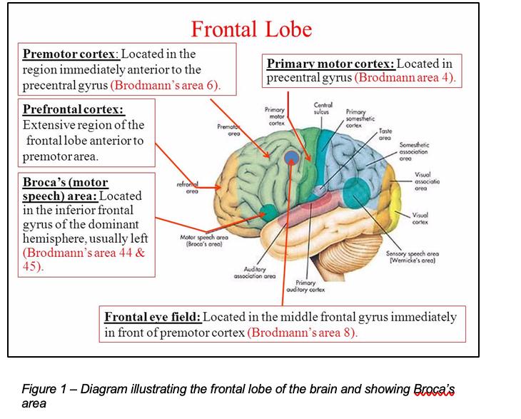
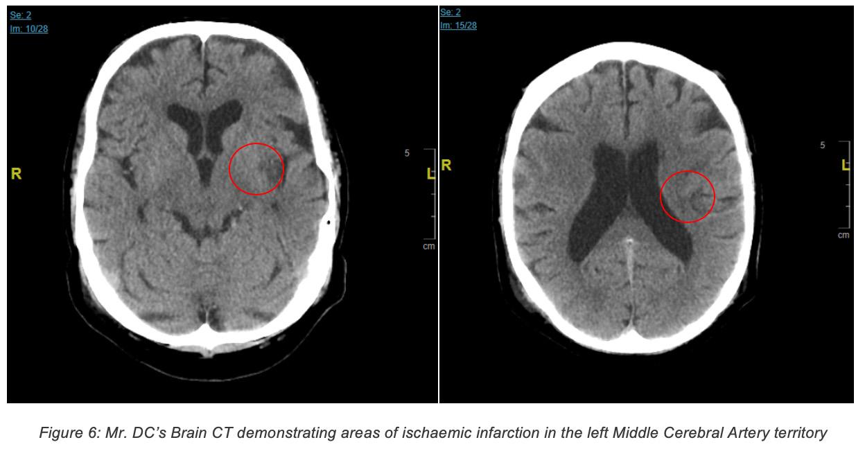
Mr. D.C. is a 75-year old gentleman brought to casualty after a neighbour found him unable to speak. Since he was severely aphasic, he was unable to give a proper history. The patient was able to understand commands, making this a pure expressive aphasia. He was also noted to have right hemiparesis and right facial weakness. On examination, Mr D.C. was found to have 0/5 power on the right half of his body and 3/5 power on the left side of his body using the MRC muscle power assessment scale. He was noted to have right facial weakness in an upper motor neurone lesion pattern. He was urgently admitted to a medical ward and a CT scan was requested. The scan confirmed a left middle cerebral artery (MCA) ischaemic infarct affecting the basal ganglia and Broca’s Area in the inferior frontal lobe. Fact file on Expressive Aphasia Expressive aphasia, also known as Broca’s aphasia or non-fluent aphasia, is a condition which is characterized by a loss of the ability to produce both spoken and written language. Although patients are unable to communicate properly, their understanding of speech is mostly intact and much better than when compared to their speech. The most common cause of Broca’s aphasia is a left middle cerebral artery thrombus or embolus causing a stroke which affects the third frontal convolution of the frontal lobe, which is also known as Broca’s area (Figure 1), and extending into the white matter. However, it may also be caused by any disease or injury which might affect Broca’s area. The fact that the area of this lesion is anterior to the inferior part of the precentral gyrus, which is responsible for executing voluntary motor movements, explains why there is also associated weakness in the right upper extremity in this condition (“Stroke Rehabilitation: A Function-Based Approach,” 2000). In contrast to Broca’s area, Wernicke’s area is important in the understanding of speech not the production of speech. Damage to this area of the brain results in impaired comprehension of speech as well as generation of speech which may contain paraphrase and neologisms, often resulting in a word salad. It is located in the sensory area of the posterior superior temporal lobe in the dominant cerebral hemisphere, close to the lateral sulcus. Infarction of the Middle Cerebral Artery can result in both Broca’s aphasia and Wernicke’s aphasia. The superior division of the middle cerebral artery supplies Broca’s area, therefore decreased perfusion of this vessel or more proximal to it will cause Broca’s aphasia. Wernicke’s area is supplied by the inferior division of the MCA. Strokes of the inferior MCA hence result in Wernicke’s aphasia (‘’Broca’s, Wernicke’s and Conduction Aphasias,’’ 2006). Patients suffering from Broca’s aphasia have a dominant feature of agrammatism in their speech and thus find it hard to form full sentences even though content words like verbs and nouns are preserved. Patients therefore end up producing speech which is described as
‘telegraphic speech’. Patients’ ability to repeat words and sentences is also very poor (“Broca’s Aphasia,” n.d.). There is currently no treatment for aphasia. The approach to facilitating the patient’s quality of life is a multidisciplinary approach involving speech therapy, physiotherapy, neurologists and psychologists. Recovery of language function peaks within two to six months, after which progress decreases, however the patient is still encouraged to keep on working on speech production as improvement can be seen long after the stroke (Acharya and Dulebohn, 2017).

This condition is named after Paul Pierre Broca (Figure 2), a French general surgeon who contributed greatly to the field of anatomy. His most important contribution was his discovery of the specific region in the brain that is responsible for speech, Broca’s area, in 1861 (Joynt, 1964). Data on the incidence of expressive aphasia is quite limited, however it is estimated that 11,400 people in Britain become aphasic every year following stroke (Wade et al., 1986)
Presenting Complaint: Mr. DC, a 75-year old male, was found by his neighbour expressively aphasic and suffering from right hemiparesis and right facial weakness. No other symptoms were able to be elicited in the history other than what was clearly visible.
Past Medical/ Surgical History, Family History, Drug History and Social History: Since the patient was severely aphasic it was not possible to obtain a thorough history from him.
Systemic Inquiry: Nil of note.
Physical Examination and Preliminary Investigations: A full neurological examination was carried out.
On examination: • Right upper limb power: 0/5 (flaccid) • Right lower limb power: 0/5 • Right facial nerve palsy with upper motor neuron lesion patterns • Left upper limb power: 3/5 • Left lower limb power: 3/5
Power was assessed using the MRC power scale (Table 1).

• Chest was clear • Abdomen was soft and non-tender • Heart sounds S1 and S2 were intact with no added heart sounds • Tachycardic with a rate of 115 beats per minute • Blood pressure was found to be 117/77 • Oxygen saturations: 99% SaO2 • Afebrile
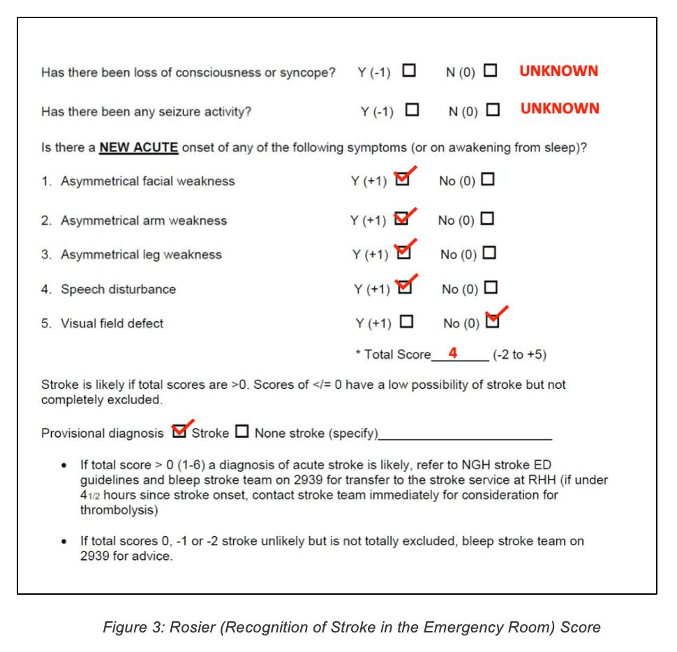
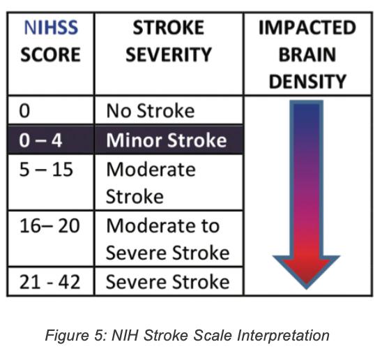
Various stroke screening techniques were used. The results are shown in the following figures:

Investigations ordered: 1. ECG – To identify any predisposing conditions which may have resulted in the stroke e.g.: Atrial fibrillation/flutter, acute myocardial infarction, infective endocarditis etc. In case any are identified, further prophylactic treatment may be indicated. 2. Routine bloods (Complete Blood Count, ESR, CRP, Urea and Electrolytes, Blood Glucose Levels etc) – to rule out underlying conditions such as sepsis, renal disease and diabetes. Inflammatory markers are often raised in ischaemic stroke. In case any are identified, further treatment may be indicated. 3. CT brain – To determine whether the stroke was ischaemic or haemorrhagic in nature, to properly locate the lesion and to rule out any possible differentials such as space occupying lesions. 4. Echocardiogram – To rule out any underlying structural heart disease which may have caused the stroke e.g.: Valvular heart disease, patent foramen ovale, atrial septal aneurysm etc. In case any are identified further prophylactic treatment may be indicated. 5. Carotid doppler ultrasound – To identify any possible stenosis or plaque build-up in the carotid arteries which may predispose to recurrent strokes. If stenosis is greater than 50%, carotid endarterectomy may be indicated. 6. Geriatric review 7. Stroke rehabilitation including outreach to physiotherapy, occupational therapy and
Findings: • The CT brain revealed an ischaemic stroke of the middle cerebral artery territory. CT brain results are shown below:
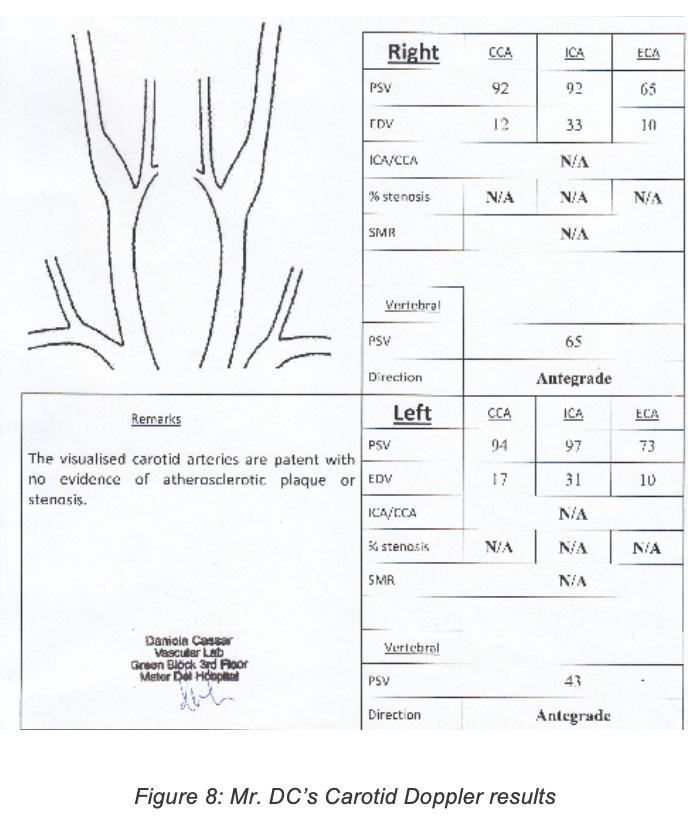
Mr DC’s ECG revealed atrial flutter, a possible source of emboli which may have resulted in the stroke. C-Reactive Protein was found to be 51.9 mg/L (normal range <3.0 mg/L). This is common in stroke patients since acute ischaemic stroke may trigger an inflammatory response in the surrounding tissues. No abnormalities seen on the echocardiogram. Carotid doppler showed no stenosis or atherosclerosis of the carotid arteries. Carotid doppler results are shown below:

Pharmacological Therapy: Since the CT showed that the stroke was ischaemic rather than haemorrhagic, pharmacological intervention was indicated rather than a surgical approach.
The patient was outside the therapeutic window for administration of Tissue plasminogen activator (tPA) which is 3-4.5 hours, and was therefore started on aspirin 75mg daily, Dipyridamole 100mg tds and Simvastatin 20mg daily. The patient was also started on omeprazole 20mg once daily due to the potential side effect of gastrointestinal bleeding associated with aspirin.
The antiplatelet (Dipyridamole) is to be switched to warfarin after 4-6 weeks. 4 weeks are allowed before switching in order to prevent haemorrhagic conversion i.e. the transformation of an ischaemic stroke into a haemorrhagic stroke. Mr DC was also started on Perindopril 2mg once daily.
Follow Up: Mr. DC was monitored and followed up closely during the week he spent at Mater Dei Hospital recovering from the stroke. The following is a summary of his recovery:
Day 1 Post Stroke:
• Patient aphasic but able to communicate with hand gestures and by writing on paper • Patient found to have good gait pattern • No dyspraxia Right sided facial weakness persistent but able to swallow.
Day 2 Post Stroke:
Blood pressure increased to 120/80 IVI stopped and cannula removed
Day 3 Post Stroke: Day 7 Post Stroke:
No further physiotherapy or speech therapy needed Patient to be discharged home to be supported by wife
References: 1. Acharya, A.B., Dulebohn, S.C., 2017. Aphasia, Broca, StatPearls. 2. Broca’s Aphasia [WWW Document], n.d. URL https://www.asha.org/Glossary/BrocasAphasia/ 3. Joynt, R.J., 1964. Paul Pierre Broca: His Contribution to the Knowledge of Aphasia. Cortex. https://doi.org/10.1016/S0010-9452(64)80022-8 4. Broca’s, Wernicke’s and Conduction Aphasias [WWW Document], 2006. . Case West. Reserv. Univ. Sch. Med. URL http://casemed.case.edu/clerkships/neurology/ NeurLrngObjectives/Brocas.htm 5. Stroke Rehabilitation: A Function-Based Approach, 2000. . Am. J. Occup. Ther. https:// doi.org/10.5014/ajot.54.3.349 6. Wade, D.T., Hewer, R.L., David, R.M., Enderby, P., 1986. Aphasia after stroke: Natural history and associated deficits. J. Neurol. Neurosurg. Psychiatry. https://doi.org/10.1136/ jnnp.49.1.11
Patient noted to have increased vocabulary – vocalised “thank you” Range of movement of the right upper limb noted to have increased substantially Able to mobilize with minimal help Activities of daily living require help
Day 4 Post Stroke:
Ambulation and balance good Improvement in speech - “I don’t like it” Hot cold sensation intact Communication impairment prevented full interpretation of sensory testing
Day 5 Post Stroke:
Patient managing to carry out all activities of daily living independently Only remaining issue is speech but it is improving, according to wife Good gait and balance
Day 6 Post Stroke:
Upper limb sensory testing repeated - patient noted to have decreased sensation mostly in sharp/dull discrimination. Patient was able to identify light touch Able to form sentences but speech intelligibility remains poor Written word only contains minor spelling errors which were self-corrected
Hannah Xuereb 4th Year Medical Student at the Faculty of Medicine & Surgery, University of Malta.


32 Adam Al Gededi 4th Year Medical Student at the Faculty of Medicine & Surgery, University of Malta.


