10 minute read
NSTEMI
NSTEMI in the Context of Cardiovascular Risk Factors and Co-morbidities External Reviewer Dr. Alexander Borg M.D.,M.R.C.P.(Lond.),F.R.C.P.(E din.),M.D.(Manc.),F.E.S.C.
Tutor
Advertisement
Dr Maryanne Caruana M.D. (Melit.) Ph.D.(Melit.)FRCP (Edin) FESC
Mr A.N., a 59-year-old male, was referred to casualty by a health centre physician, following the complaint of two episodes of worsening exertional chest pain. He is a known case of hypertension, non-insulin-dependent diabetes mellitus and smokes 1 pack of cigarettes daily.
Following appropriate investigations such as electrocardiogram (ECG), cardiac biomarkers and echocardiography, the patient was diagnosed with non-ST elevation myocardial infarction (NSTEMI). The patient is now stable on treatment; however, his overall cardiovascular risk remains high due to factors such as smoking, uncontrolled type 2 diabetes, hypertension and hypercholesterolaemia. This case highlights the importance of measuring global cardiovascular risk since this has important treatment and follow-up implications; importantly, ischaemic heart disease can be primarily or secondarily prevented through nonpharmacological or pharmacological alteration of the cardiovascular risk factors.
Fact file on NSTE-ACS, Atherosclerosis and Cardiovascular Risk Factors Acute coronary syndromes (ACS) affect around 1 million patients annually in the USA, and around 75% of these patients are diagnosed with non-ST elevation-ACS: unstable angina or NSTEMI. (Hedayati et al., 2018). These two conditions share a similar pathophysiology but NSTEMI is differentiated from unstable angina since in NSTEMI, the levels of cardiac troponin are higher than the 99th percentile of the upper reference limit for the normal range of the assay. (ESC, Fourth universal definition of myocardial infarction (2018).
This case presents a very important dilemma for a casualty physician: determining the cause of chest pain. The determination of aetiology was complicated by the fact that the patient also suffered from gastro-oesophageal reflux disease (GORD). Due to the fact that visceral pain pathways of the stomach/oesophagus and the heart are shared, the referred pain from these organs can give very similar symptoms.
The pathophysiology of NSTEMI entails mismatch between myocardial oxygen supply and demand: there is subendocardial ischaemia where the atherothrombosis is causing partial blood flow obstruction. As with the other acute coronary syndromes, NSTEMI is the result of the inflammatory process of atherosclerosis and occurs most commonly due to plaque erosion with an intact fibrous cap (Basit et al., 2019; Puymirat et al., 2019).
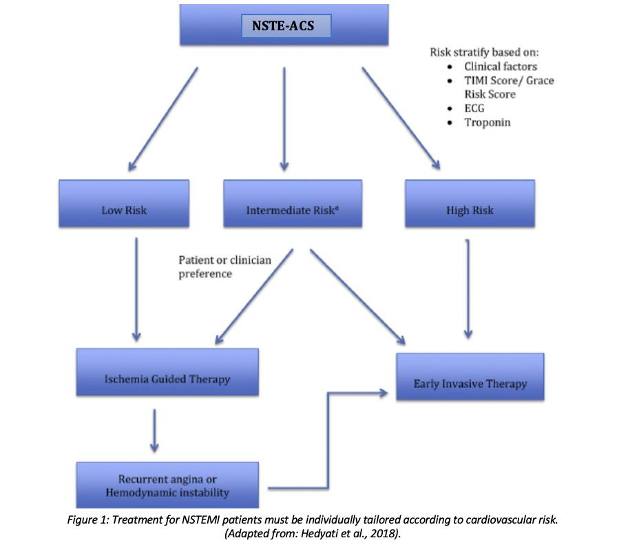
The other major important clinical point pertaining to this case is the presence of cardiovascular risk factors which determine treatment in the form of early invasive therapy. This patient had a HEART score of 8, indicating high risk and a need for urgent invasive intervention. The HEART score table (Hedayati et al., 2018) shown in figure 2 is a useful tool in stratification and management of patients with ACS.

Presenting Complaint The patient was referred to A&E after he experienced two episodes of diffuse chest pain of an intermittent nature.
History of Presenting Complaint The first episode occurred when the patient was at work: he had exerted himself by climbing some stairs and a ladder when he experienced a deep compressive pain in his chest which lasted 10 minutes. The second episode occurred three days later and lasted for around 15 minutes, and was described as centrally compressive chest pain associated with diaphoresis. There was no vomiting or loss of consciousness during either episode, but the patient complained of sensations of nausea. The patient was unable to pinpoint the precise site of the pain: the pain was of a diffuse nature throughout his chest. The patient had been suffering from chest pains of varying nature for 10 years; these have been found to be a mixture of stable angina and pain due to GORD. However, these two particular episodes of chest pain were different from the other chronic episodes since the character of the pain was more compressive, caused more shortness of breath and was of higher severity. The patient denied any radiation of the pain to the left arm, right arm, jaw or neck. This pain was associated with sweating and nausea but not fever. In both episodes, the pain lasted for 15 minutes or less and was intermittent. Cold temperatures and exertion exacerbated the severity of the pain, which the patient grades as 7 out of 10.
Past Medical and Surgical History The patient is a known case of type II diabetes mellitus and hypercholesterolaemia. During the last year his diabetes has not been wellcontrolled and the patient admits to not taking medications for his elevated cholesterol levels. Coupled with other non-modifiable risk factors such as genetic predisposition, age and male gender, these modifiable risk factors contributed to this acute coronary syndrome.
The patient also has a 10-year history of GORD which he states was mainly triggered by stress. The patient had a minor operation to remove a benign skin lesion 10 years ago. Importantly, the patient had experienced a variety of chest pains throughout the last 10 years: these may be of cardiac origin or due to GORD. The patient had never had an acute coronary event, coronary artery bypass grafting (CABG) or percutaneous coronary intervention (PCI) before.

Drug History and Allergies Patient has no known drug allergies. His drug history is listed in table 1. An important factor, as previously discussed, is that the patient was poorly compliant to prescribed medications.
Family History The patient’s mother had a history of type II diabetes mellitus and hypertension, but did not die due to a cardiovascular event. His father had a history of heart failure and died aged 82. None of his siblings have experienced an acute cardiovascular event.
Social History The patient is married and lives at home with his wife and daughter. He is independent and his occupation entails a certain degree of manual labour. His condition might now restrict him from continuing his usual work.
Importantly, given that smoking is an independent risk factor for cardiovascular disease, the patient has smoked 10-15 cigarettes per day since around the age of 20, which equates to 39 pack years.The patient only drinks alcohol socially.
Systemic Enquiry • General health: overall the patient is healthy, however he has a number of cardiovascular risk factors and is also quite stressed • Cardiovascular system: hypercholesterolaemia, hypertension, no palpitations • Respiratory system: no shortness of breath (SOB) at rest, SOB occurred during episodes of chest pain • Gastrointestinal tract: dyspepsia • Genitourinary system: pt has no problems passing urine, feels no burning sensation • Musculoskeletal system: nil to note • Endocrine system: nil to note
Differential Diagnosis 1. ACS (ECG points towards NSTEMI, diagnosis pending troponin result) 2. Chest pain due to severe GORD (unlikely) 3. Musculoskeletal pain e.g. costochondritis (highly unlikely) 4. Pulmonary embolism due to the presence of right bundle branch block on the ECG. There were no older ECGs to compare with and check whether this is an old or new right bundle branch block. (Nature of the pain meant that pericarditis was very unlikely).
Diagnostic Investigations 1. Investigation: 12-lead ECG 2. Justification: ECG is essential in diagnosis of any ACS since it distinguishes STEMI from NSTEMI, and identifies any arrhythmias which the patient may have developed 3. Result & conclusion: the patient’s ECG showed right bundle branch block: QRS complex >120ms and RSR’ pattern in the anterior precordial leads V1-V3. In V1-V3, one can observe reciprocal ST segment depressions and T wave inversion, while there was no ST elevation in any of the leads (refer to figure 3). The clinical presentation and the raised troponins combined with these ECG changes led to the diagnosis of NSTEMI.
1. Investigation: bloods: troponins, CBC, U&E, ABGs, lipid profile, glucose levels, liver function tests 2. Justification: CBC is done to rule out any anaemia or infective cause, troponins are used as biomarkers of myocardial injury, ABGs are done in casualty since the patient was short of breath, lipid profile and HGTs aid assessment of cardiovascular risk factors such as hypercholesterolaemia and diabetes mellitus, U&E and liver function tests provide baseline organ function levels which must be considered when choosing pharmacological therapy 3. Result & conclusion: elevated initial troponins and an increase of >20% in the 3-hr repeated troponins together with the typical chest pain and lack of ST elevation on ECG indicated that the patient had NSTEMI, while HGTs and lipid profile confirmed uncontrolled diabetes mellitus and hypercholesterolaemia. The other tests showed no remarkable findings: the patient has adequate liver function and does not have chronic kidney disease.
1. Investigation: transthoracic echocardiography
2. Justification: assessment of any hypokinetic or akinetic areas of myocardium, assessment of LV ejection fraction: it has been found that reduced ejection fraction in NSTEMI patients is associated with greater mortality (Siddiqui and Holzmann, 2019). 3. Result & conclusion: mild concentric LVH indicative of long-standing hypertension, ejection fraction EF>55%, good LV systolic function, reversed mitral inflow pattern in keeping with impaired LV relaxation. Normal RV size and systolic function. The aortic and mitral valves showed no stenosis or regurgitation No specific regional wall motion abnormality was reported.

Management 1. Pharmacological Therapy Following investigations, the patient was started on the following:

3. Follow-up After NSTEMI, the patient is prescribed one year of dual antiplatelet therapy: aspirin (75mg once daily) and clopidogrel (75mg once daily). Risk factor modification is another important issue: patient is given advice to stop smoking, control his diabetes, hypertension control and keeping LDL below 1.4mmol/L. The patient recovered well following PCI, and was discharged two days later with an outpatients appointment 3 months later.
References 1. Basit H, Malik A & Huecker MR (2019). Non-ST Segment Elevation (NSTEMI) Myocardial Infarction. In StatPearlsNon ST Segment Elevation (NSTEMI) Myocardial Infarction. StatPearls Publishing LLC, Treasure Island (FL). 2. Brown MD, Wolf SJ, Byyny R, Diercks DB, Gemme SR, Gerardo CJ, Godwin SA, Hahn SA, Harrison NE & Hatten BW (2018). Clinical Policy: Critical Issues in the Evaluation and Management of Emergency Department Patients With Suspected Non–ST-Elevation Acute Coronary Syndromes. Ann Emerg Med 72, e65-e106. 3. Puymirat E, Cayla G, Cottin Y, Elbaz M, Henry P, Gerbaud E, Lemesle G, Popovic B, Labèque J & Roubille F (2019). Twenty-year trends in profile, management and outcomes of patients with ST-segment elevation myocardial infarction according to use of reperfusion therapy: Data from the FAST-MI program 1995-2015. Am Heart J 214, 97-106. 4. Siddiqui AJ & Holzmann MJ (2019). Association between reduced left ventricular ejection fraction following non-ST-segment elevation myocardial infarction and long-term mortality in patients of advanced age. Int J Cardiol 296, 15-20. 5. Hedayati, T., Yadav, N. and Khanagavi, J. (2018). Non–ST-Segment Acute Coronary Syndromes. Cardiology Clinics, 36(1), pp.37-52.
6. ESC, Fourth universal definition of myocardial infarction (2018)
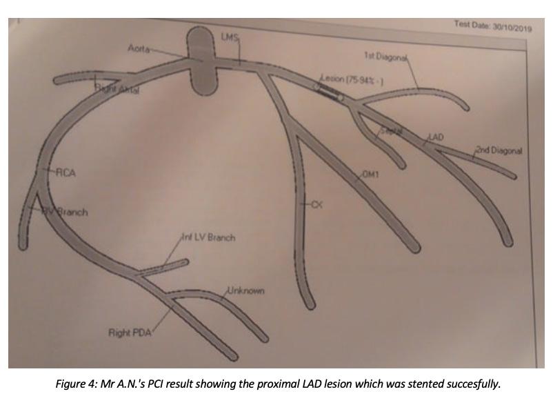
2. PCl The patient’s cardiovascular risk factors (stage 2 hypertension, smoking, diabetes mellitus, hypercholesterolaemia) meant that early invasive therapy was chosen as the preferred treatment pathway. (Brown et al., 2018). As shown in figure 4, a very tight stenosis was identified in the proximal LAD, and PCI was done on this lesion. The area was predilated with a 3.5x15mm balloon and stented with a 4x18mm DES (drug-eluting stent).
Mark Miruzzi 3rd Year Medical Student at the Faculty of Medicine & Surgery, University of Malta.

Magazine Designers

Jade Borg Public Relations Officer 2019-2020
Owen Cachia Public Relations Coordinator 2019-2020 Minima Medica Layout Designer




Thank You to Our Sponsors!

