8 minute read
Skeletal anatomy


Integumentary System The tenth and final body system is the integumentary (in-teg˝-umen′ -tar-e) system, which is composed of the skin and all structures derived from the skin. These derived structures include hair, nails, and sweat and oil glands.
The skin is an organ that is essential to life. The skin is the largest organ of the body, covering a surface area of approximately 7620 cm2 and constituting 8% of total body mass in the average adult.
Advertisement
The five functions of the integumentary system are as follows: 1. Regulate body temperature 2. Protect the body, within limits, against microbial invasion and mechanical, chemical, and ultraviolet (UV) radiation damage 3. Eliminate waste products through perspiration 4. Receive certain stimuli such as temperature, pressure, and pain 5. Synthesize certain vitamins and biochemicals such as vitamin D
Skeletal Anatomy
Because a large part of general diagnostic radiography involves examination of the bones and joints, osteology (os˝-te-ol′ -o-je) (the study of bones) and arthrology (ar-throl′ -o-je) (the study of joints) are important subjects for the technologist.
osTeology The adult skeletal system is composed of 206 separate bones, which form the framework of the entire body. Certain cartilages, such as those at the ends of long bones, are included in the skeletal system. These bones and cartilages are united by ligaments and provide surfaces to which the muscles attach. Because muscles and bones must combine to allow body movement, these two systems sometimes are collectively referred to as the locomotor system.
The adult human skeleton is divided into the axial skeleton and the appendicular skeleton.
Axial Skeleton The axial (ak′ -se-al) skeleton includes all bones that lie on or near the central axis of the body. The adult axial skeleton consists of 80 bones and includes the skull, vertebral column, ribs, and sternum (the dark-shaded regions of the body skeleton in Fig. 1-12).
ADUlT AXiAl sKeleTon
Skull
Hyoid Auditory ossicles (small bones in each ear) Vertebral column
Thorax Cranium 8 Facial bones 14 1
Cervical Thoracic Lumbar Sacral Coccyx Sternum Ribs
6 7 12 5 1 1 1 24 Fig. 1-11 Integumentary system.
Fig. 1-12 Axial skeleton—80 bones.
Total bones in adult axial skeleton 80
Appendicular Skeleton The second division of the skeleton is the appendicular (ap˝-endik′ -u-lar) portion. This division consists of all bones of the upper and lower limbs (extremities) and the shoulder and pelvic girdles (the dark-shaded regions in Fig. 1-13). The appendicular skeleton attaches to the axial skeleton. The adult appendicular skeleton comprises 126 separate bones.
ADUlT APPenDiCUlAR sKeleTon
Shoulder girdles Clavicles Scapula (scapulae)
Upper limbs Humerus (humeri) Ulna (ulnae) Radius (radii)
Pelvic girdle Lower limbs
Carpals Metacarpals Phalanges Hip bones (innominate bones) Femur (femora) Tibia Fibula (fibulae) Patella (patellae) Tarsals Metatarsals Phalanges Total bones in adult appendicular skeleton Entire adult skeleton—206 separate bones*
*This includes the 2 sesamoid bones at the knees: the right and left patellae.
2 2 2 2 2 16 10 28 2 2 2 2 2 14 10 28
126
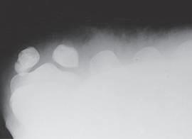
Sesamoid Bones A sesamoid bone is a special type of small, oval-shaped bone that is embedded in certain tendons (most often near joints). Although sesamoid bones are present even in a developing fetus, they are not counted as part of the normal axial or appendicular skeleton except for the two patellae, the largest sesamoid bones. The other most common sesamoid bones are located in the posterior foot at the base of the first toe (Figs. 1-14 and 1-15).
In the upper limb, sesamoid bones are found most commonly in tendons near the anterior (palmar) surface of the hand at the base of the thumb. Others may be found in tendons of other upper or lower limb joints.
Any sesamoid bone can be fractured by trauma; this may have to be demonstrated radiographically or by CT (computed tomography).
ClAssiFiCATion oF Bones Each of the 206 bones of the body can be classified according to shape as follows: • Long bones • Short bones • Flat bones • Irregular bones
Long Bones Long bones consist of a body and two ends or extremities. Long bones are found only in the appendicular skeleton. (Fig. 1-16 is a radiograph of a humerus, a typical long bone of the upper arm.)
Fig. 1-13 Appendicular skeleton—126 bones.
Fig. 1-14 Sesamoid bones on the posterior base of the first toe. Fig. 1-15 Sesamoid bones. Tangential projection (base of first toe).
Extremity
Body
Extremity
Composition The outer shell of most bones is composed of hard or dense bone tissue known as compact bone, or cortex, meaning an external layer. Compact bone has few intercellular empty spaces and serves to protect and support the entire bone.
The body (older term is shaft) contains a thicker layer of compact bone than is found at the ends, to help resist the stress of the weight placed on them.
Inside the shell of compact bone and especially at both ends of each long bone is found spongy, or cancellous, bone. Cancellous bone is highly porous and usually contains red bone marrow, which is responsible for the production of red blood cells.
The body of a long bone is hollow. This hollow portion is known as the medullary (med′ -u-lar˝-e) cavity. In adults, the medullary cavity usually contains fatty yellow marrow. A dense fibrous membrane, the periosteum (per˝-e-os′ -te-am), covers bone except at the articulating surfaces. The articulating surfaces are covered by a layer of hyaline cartilage.
Hyaline (hi′ -ah-lin), meaning glassy or clear, is a common type of cartilage or connecting tissue that is also known as “gristle.” Its name comes from the fact that it is not visible with ordinary staining techniques, and it appears “clear” or glassy in laboratory studies. It is present in many places, including within the covering over ends of bones, where it is called articular cartilage.
The periosteum is essential for bone growth, repair, and nutrition. Bones are richly supplied with blood vessels that pass into them from the periosteum. Near the center of the body of long bones, a nutrient artery passes obliquely through the compact bone via a nutrient foramen into the medullary cavity.
Short Bones Short bones are roughly cuboidal and are found only in the wrists and ankles. Short bones consist mainly of cancellous tissue with a thin outer covering of compact bone. The eight carpal bones of each wrist and the seven tarsal bones of each foot are short bones.
Flat Bones Flat bones consist of two plates of compact bone with cancellous bone and marrow between them. Examples of flat bones are the bones that make up the calvaria (skull cap), sternum, ribs, and scapulae.
The narrow space between the inner and the outer table of flat bones within the cranium is known as the diploë (dip′ -lo-e). Flat bones provide protection for interior contents and broad surfaces for muscle attachment.
Irregular Bones Bones that have peculiar shapes are lumped into one final category— irregular bones. Vertebrae, facial bones, bones of the base of the cranium, and bones of the pelvis are examples of irregular bones.
Compact bone Medullary cavity
Body Spongy or cancellous bone (contains red marrow)
Periosteum
Nutrient foramen
Nutrient artery
Fig. 1-17 Long bone.
Fig. 1-18 Short bones (carpals). Fig. 1-19 Flat bones (calvaria).

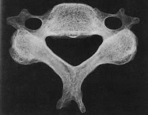
Fig. 1-20 Irregular bone (vertebra).
DeVeloPMenT oF Bones The process by which bones form within the body is known as ossification (os˝-i-fi-ka′-shun). The embryonic skeleton is composed of fibrous membranes and hyaline cartilage. Ossification begins at about the sixth embryonic week and continues until adulthood.
Blood Cell Production In adults, red blood cells (RBCs) are produced by the red bone marrow of certain flat and irregular bones such as the sternum, ribs, vertebrae, and pelvis as well as the ends of the long bones.
Primary center: Diaphysis (body) Secondary centers: Metaphysis Epiphyseal plate
Epiphyses
Bone Formation Two types of bone formation are known. When bone replaces membranes, the ossification is called intramembranous (in˝-trahmem′-brah-nus). When bone replaces cartilage, the result is endochondral (en˝-do-kon′-dral) (intracartilaginous) ossification.
Intramembranous ossification Intramembranous ossification occurs rapidly and takes place in bones that are needed for protection, such as sutures of the flat bones of the skullcap, which are centers of growth in early bone development.
Endochondral ossification Endochondral ossification, which is much slower than intramembranous ossification, occurs in most parts of the skeleton, especially in the long bones.
Primary and Secondary Centers of Endochondral Ossification The first center of ossification, which is called the primary center, occurs in the midbody area. This primary center of ossification in growing bones is called the diaphysis (di-af′-i-sis). This becomes the body in a fully developed bone. secondary centers of ossification appear near the ends of the limbs of long bones. Most secondary centers appear after birth, whereas most primary centers appear before birth. Each secondary center of ossification is called an epiphysis (e-pif′-i-sis). Epiphyses of the distal femur and the proximal tibia are the first to appear and may be present at birth in a term newborn. Cartilaginous plates, called epiphyseal plates, are found between the metaphysis and each epiphysis until skeletal growth is complete. The metaphysis is the wider portion of a long bone adjacent to the epiphyseal plate. The metaphysis is the area where bone growth in length occurs. Growth in the length of bones results from a longitudinal increase in these epiphyseal cartilaginous plates. This is followed by progressive ossification through endochondral bone development until all the cartilage has been replaced by bone, at which time growth to the skeleton is complete. This process of epiphyseal fusion of the long bones occurs progressively from the age of puberty to full maturity, which occurs at about 25 years of age. However, the time for each bone to complete growth varies for different regions of the body. On average, the female skeleton matures more quickly than the male skeleton. Extensive charts that list the normal growth patterns of the skeleton are available.
Radiograph Demonstrating Bone Growth Fig. 1-22 shows a radiograph of the knee region of a 6-year-old child. Primary and secondary centers of endochondral ossification or bone growth are well demonstrated and labeled.
Fig. 1-21 Endochondral ossification.
Primary center: Diaphysis (body)
Secondary centers: Metaphysis Epiphyseal plate
Epiphyses
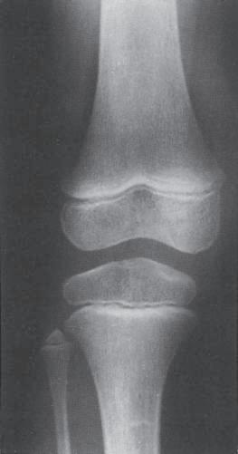
Fig. 1-22 Knee region (6-year-old child).