7 minute read
Special projection terms
Following are some additional terms that are commonly used to describe projections. These terms, as shown by their definitions, also refer to the path or projection of the CR and are projections rather than positions.
Axial projection Axial (ak′-se-al) refers to the long axis of a structure or part (around which a rotating body turns or is arranged). Special application—AP or PA axial: In radiographic positioning, the term axial has been used to describe any angle of the CR of 10° or more along the long axis of the body or body part.*
Advertisement
However, in a true sense, an axial projection would be directed along, or parallel to, the long axis of the body or part. The term semiaxial, or “partly” axial, more accurately describes any angle along the axis that is not truly along or parallel to the long axis.
However, for the sake of consistency with other references, the term axial projection is used throughout this text to describe both axial and semiaxial projections as defined earlier and as illustrated in Figs. 1-66 through 1-68.
Inferosuperior and superoinferior axial projections inferosuperior axial projections are frequently performed for the shoulder and hip, where the CR enters below or inferiorly and exits above or superiorly (Fig. 1-68). The opposite of this is the superoinferior axial projection, such as a special nasal bone projection (Fig. 1-66).

Fig. 1-66 Axial (superoinferior) projection.
CR
37
Tangential projection Tangential (tan˝-jen′-shal) means touching a curve or surface at only one point. This is a special use of the term projection to describe a projection that merely skims a body part to project that part into profile and away from other body structures.
Examples Following are two examples or applications of the term tangential projection: • Zygomatic arch projection (Fig. 1-69) • Tangential projection of patella (Fig. 1-70)
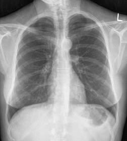
AP axial projection—lordotic position This is a specific AP chest projection for demonstrating the apices of the lungs. It also is sometimes called the apical lordotic projection. In this case, the long axis of the body rather than the CR is angled. The term lordotic comes from lordosis, a term that denotes curvature of the cervical and lumbar spine (see Chapters 8 and 9).
As the patient assumes this position (Fig. 1-71), the lumbar lordotic curvature is exaggerated, making this a descriptive term for this special chest projection.
*Frank E, Long B, Smith B: Merrill’s atlas of radiographic positioning & procedures, ed 12, vol 1, St. Louis, 2012, Mosby.
CR
CR Fig. 1-67 AP axial (semiaxial) projection (CR 37° caudal).


Fig. 1-68 Inferosuperior axial projection.

Transthoracic lateral projection (right lateral position) A lateral projection through the thorax. Requires a qualifying positioning term (right or left lateral position) to indicate which shoulder is closest to the IR and is being examined (Fig. 1-72).
noTe: This is a special adaptation of the projection term, indicating that the CR passes through the thorax even though it does not include an entrance or exit site. In practice, this is a common lateral shoulder projection and is referred to as a right or left transthoracic lateral shoulder.
Dorsoplantar and plantodorsal projections These are secondary terms for AP or PA projections of the foot. Dorsoplantar (DP) describes the path of the CR from the dorsal (anterior) surface to the plantar (posterior) surface of the foot (Fig. 1-73). A special plantodorsal projection of the heel bone (calcaneus) is called an axial plantodorsal projection (PD) because the angled CR enters the plantar surface of the foot and exits the dorsal surface (Fig. 1-74).
noTe: The term dorsum for the foot refers to the anterior surface, dorsum pedis (Fig. 1-42).
Parietoacanthial and acanthioparietal projections The CR enters at the cranial parietal bone and exits at the acanthion (junction of nose and upper lip) for the parietoacanthial projection (Fig. 1-75). The opposite CR direction would describe the acanthioparietal projection (Fig. 1-76). These are also known as PA Waters and AP reverse Waters methods and are used to visualize the facial bones.
Submentovertex (SMV) and verticosubmental (VSM) projections These projections are used for the skull and mandible. CR enters below the chin, or mentum, and exits at the vertex or top of the skull for the submentovertex (sMV) projection (Fig. 1-77). The less common, opposite projection of this would be the verticosubmental (VsM) projection, entering at the top of the skull and exiting below the mandible (not shown).

Fig. 1-72 Transthoracic lateral shoulder projection (R lateral shoulder position). Fig. 1-73 AP or dorsoplantar (DP) projection of foot. Fig. 1-74 Axial plantodorsal (PD) projection of calcaneus. Fig. 1-75 Parietoacanthial projection (Waters position).


Fig. 1-76 Acanthioparietal projection.

Fig. 1-77 Submentovertex (SMV) projection.


Following are paired positioning or anatomic terms that are used to describe relationships to parts of the body with opposite meanings.
Medial versus lateral Medial (me′-de-al) versus lateral refers to toward versus away from the center, or median plane. In the anatomic position, the medial aspect of any body part is the
“inside” part closest to the median plane, and the lateral part is away from the center, or away from the median plane or midline of the body.
Examples In the anatomic position, the thumb is on the lateral aspect of the hand. The lateral part of the abdomen and thorax is the part away from the median plane.
Medial plane
Lateral abdomen
Lateral arm
Medial arm Proximal
Proximal versus distal Proximal (prok′-si-mal) is near the source or beginning, and distal (dis′-tal) is away from. In regard to the upper and lower limbs, proximal and distal would be the part closest to or away from the trunk, the source or beginning of that limb.
Examples The elbow is proximal to the wrist. The finger joint closest to the palm of the hand is called the proximal interphalangeal (PIP) joint, and the joint near the distal end of the finger is the distal interphalangeal (DIP) joint (see Chapter 4).
Cephalad versus caudad Cephalad (sef′-ah-lad) means toward the head end of the body, whereas caudad (kaw′-dad) means away from the head end of the body. A cephalad angle is any angle toward the head end of the body (Figs. 1-79 and 1-81). (Cephalad, or cephalic, literally means
“head” or “toward the head.”) A caudad angle is any angle toward the feet or away from the head end (Fig. 1-80). (Caudad or caudal comes from cauda, literally meaning “tail.”) In human anatomy, cephalad and caudad also can be described as superior (toward the head) or inferior (toward the feet).
noTe: As is shown in Figs. 1-79, 1-80, and 1-81, these terms are correctly used to describe the direction of the CR angle for all axial projections along the entire length of the body, not just projections of the head.
Lateral hand
Distal
Fig. 1-78 Medial vs. lateral, proximal vs. distal.

Fig. 1-79 Cephalad CR angle (toward head). Fig. 1-80 Caudad CR angle (away from head).

Interior (internal, inside) versus exterior (external, outer) interior is inside of something, nearer to the center, and exterior is situated on or near the outside. The prefix intra- means within or inside (e.g., intravenous: inside a vein). The prefix inter- means situated between things (e.g., intercostal: located between the ribs). The prefix exo- means outside or outward (e.g., exocardial: something that develops or is situated outside the heart).
Superficial versus deep superficial is nearer the skin surface; deep is farther away.
Example The cross-sectional drawing in Fig. 1-82 shows that the humerus is deep compared with the skin of the arm.
Another example would be a superficial tumor or lesion, which is located near the surface, compared with a deep tumor or lesion, which is located deeper within the body or part.
Ipsilateral versus contralateral Ipsilateral (ip˝-si-lat′-er-al) is on the same side of the body or part; contralateral (kon˝-trah-lat′-er-al) is on the opposite side.
Cephalad (superior) Caudad Caudad (inferior) (inferior)
Fig. 1-81 Cephalic angle (AP axial projection of sacrum).
Skin (superficial)
Humerus (deep)


