3 minute read
Exposure factors


PART TWO: IMAGING PRINCIPLES IMAGE QUALITY IN FILM-SCREEN (ANALOG) RADIOGRAPHY
ANALOG IMAGES
Advertisement
Since the discovery of x-rays in 1895, methods of acquiring and storing x-ray images have evolved. Conventional film-screen technology with the associated chemical processing and film libraries is being replaced rapidly by digital technology. Digital technology uses computers and x-ray receptors to acquire and process images; specialized digital communication networks are used to transmit and store the x-ray images.
This period of technologic transition necessitates that students have an understanding of all image acquisition technologies because they will find themselves working in imaging departments that acquire images by using only digital technology, only filmscreen technology, or a combination of both.
This part provides an introduction to radiographic technique and image quality for both film-screen imaging and digital imaging. The study of radiographic technique and image quality includes factors that determine the accuracy with which structures that are being imaged are reproduced in the image. Each of these factors has a specific effect on the final image, and the technologist must strive to maximize these factors to produce the best image possible at the lowest achievable dose.
This part also describes methods of digital image acquisition, discusses the application of digital imaging, and provides an introduction to the important principles of radiation safety.
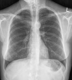
Analog (film) images provide a two-dimensional image of anatomic structures. The image acquisition device is a film-screen system that consists of a pair of intensifying screens with a film between them. The screens and film are housed in an x-ray cassette that protects the film from light and ensures that screens are in close contact with the film. When screens receive the remnant radiation from the patient, they fluoresce; this light exposes the film, which must be chemically processed so the image can be viewed. Chemical processing includes several steps (developing, fixing, washing, and drying) and typically takes 60 to 90 seconds.
The film image (radiograph), which actually is composed of a deposit of metallic silver on a polyester base, is permanent; it cannot be altered. The various shades of gray displayed on the image are representative of the densities and atomic numbers of the tissues being examined. The film image is often referred to as a hard-copy image.
Analog image receptors are best described as self-regulating systems with a limited dynamic range. Analog image receptors are also described using the term exposure latitude. Exposure latitude is the range of exposure over which a film produces an acceptable image. An image produced with a level of exposure outside of the exposure latitude is an unacceptable image. Figs. 1-118 and 1-119 illustrate the dynamic range and exposure latitude of an analog IR. Note the impact of doubling the mAs on the diagnostic quality of the images of the elbow. Analog images have relatively narrow exposure latitude.
Screen/film
Exposure Factors for Analog (Film-Screen) Imaging
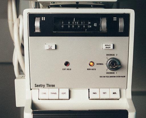
For each radiographic image obtained, the radiographer must select exposure factors on the control panel of the imaging equipment. The exposure factors required for each examination are determined by numerous variables, including the density/atomic number and thickness of the anatomic part, any pathology present, and image acquisition technology.
Exposure factors, sometimes referred to as technique factors, include the following: • Kilovoltage (kV)—controls the energy (penetrating power) of the x-ray beam • Milliamperage (mA)—controls the quantity or number of x-rays produced • exposure time (ms)—controls the duration of the exposure, usually expressed in milliseconds
Each of these exposure factors has a specific effect on the quality of the radiographic image. When performing radiographic procedures, technologists must apply their knowledge of exposure factors and imaging principles to ensure that images obtained are of the highest quality possible, while exposing patients to the lowest radiation dose possible.
R e s p o n s e
0 Exposure latitude
Dynamic range