RESURRECTING THE TASMANIAN TIGER


Faster processing with Box-To-Bench™ workflow Your bioreactor is ready for operation straight from the box.

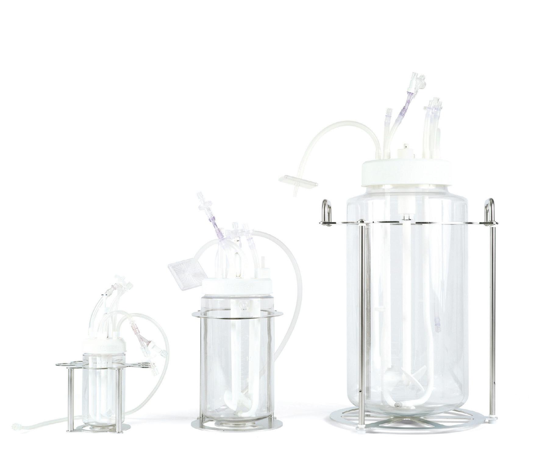
Flexible design Using 3D printing technology, we are able to optimise the bioreactor to match your process and application.
Optimised efficiency Using single-use bioreactors allows for a higher throughput thanks to an easier set-up and operation.
2022 was the Year of the Tiger, according to the Chinese zodiac, so it is appropriate that research efforts over the past 12 months have shone a light on the Australian thylacine.


Exposure to certain environmental risk factors during embryonic development can cause changes to a key gene, leading to alterations in social behaviour that are similar to those found in individuals who have autism.
With the right post-market surveillance processes in place, manufacturers are in a better position to facilitate compliance with regulatory requirements as well as to identify any early issues.
Adopting automation in academic laboratories could ease the translation from research to commercialisation, improve reproducibility and see researcher efficiency soar.

Labs are among the most programmatically complex environments to plan, design and engineer, with right-sizing and flexibility sitting at the heart of laboratory planning and design outcomes.
A Melbourne-led team has shown that 800,000 brain cells living in a dish can perform goal-directed tasks — in this case, the simple tennis-like computer game ‘Pong’.
Second chances are always nice to have, but they’re far from inevitable. This is certainly the case when it comes to the state of the climate, and our ever-dwindling opportunities to limit global warming to under 2°C compared to pre-industrial levels — not exactly something that can be reversed once we approach it, particularly if our politicians decide not to act until the last possible moment.
But sometimes we can surprise ourselves, and an avenue that we thought closed will suddenly reopen. For example, The University of Queensland (UQ) recently revealed it is taking its second-generation ‘molecular clamp’ vaccine to a proof-of-concept human trial, after the Coalition for Epidemic Preparedness Innovation (CEPI) said it would fund the further development of the technology for potential use in the global response to future disease outbreaks.
In 2019, CEPI entered into a partnering agreement with UQ to fund the development of a molecular clamp platform that would enable targeted and rapid vaccine production against multiple viral pathogens; the technology works by ‘locking’ viral

proteins, involved with infection and cell entry, into a shape that allows for an optimal immune response. In 2020 the UQ team started testing the technology against SARS-CoV-2, at which point they discovered that a constituent of their vaccine candidate also resulted in diagnostic interference with some HIV tests — an unforeseen cross-reaction — and regrettably decided to halt its development.
CEPI continued to support the UQ team so that the platform could be developed for use against other diseases in the future, potentially within 100 days of a new virus emerging. Now the team has successfully re-engineered the technology and demonstrated its promise and safety in laboratory testing, with none of the previous diagnostic issues. They have even designed a second-generation version of their SARSCoV-2 vaccine using this upgraded technology, and will soon begin Phase I testing to demonstrate safety and benchmark immunogenicity.
a decent share of scepticism, but regardless of the eventual outcome, it is sure to result in some novel research that could very well have other, potentially more easily achievable applications.
Other highlights this issue include a look at how our genes are important for the development of basic social behaviours (page 13); how brain cells living in a dish can learn to play video games (page 30); and an insight into the science of lab design (page 27). I hope you’ll find it an interesting mix of stories to wrap up 2022, and I look forward to coming back for more news and breakthroughs in 2023.
Regards, Lauren Davis LLS@wfmedia.com.auIn another story of second chances, the muchmissed Tasmanian tiger (or thylacine) has been in the news lately as researchers at The University of Melbourne are looking to develop technologies that could achieve the de-extinction of the animal as well as the conservation of currently threatened species (see our article on page 6). It’s fair to say that the project has received a lot of hype, as well as Lauren Davis

With 2022 being the Year of the Tiger according to the Chinese zodiac, it is appropriate that research efforts over the past 12 months have shone a light on the Australian thylacine, also known as the Tasmanian tiger. In some cases this was a literal light, with ABC journalists discovering that thylacine fur glows under black (UV) light. Yet this focus on the thylacine is rather remarkable given that the apex predator has been extinct for close to a century — although some researchers are doing their utmost to change that.
The thylacine, a unique marsupial carnivore, was once widespread in Australia but was confined to Tasmania by the time Europeans arrived in the 18th century. It was soon hunted to extinction by colonists, with the last known animal, made famous through photographs and film footage, dying at Hobart’s Beaumaris Zoo in 1936. But it was recently found that this particular thylacine was in fact outlived by an older female, captured by trapper Elias Churchill and sold to the zoo in May 1936.
“The sale was not recorded or publicised by the zoo because, at the time, ground-based snaring was illegal and Churchill could have been fined,”
said Robert Paddle, a comparative psychologist from the Australian Catholic University.
When the female died — with no one realising it was the last known thylacine in captivity — its body was transferred to the Tasmanian Museum and Art Gallery (TMAG). But with no record of thylacine material from 1936 ever filed or found in the museum’s zoological collection, it was assumed that the body had been discarded. It was only the detection of an unpublished museum taxidermist’s report dated 1936/37, mentioning a thylacine among the list of specimens worked on during the year, which led to a review of all the thylacine skins and skeletons in the TMAG

collection — and the subsequent discovery of the missing specimen in a cupboard in the museum’s education department.
It turned out that the body had been prepared by the taxidermist not as a research specimen — which is why it wasn’t recorded –but as an education specimen. “The thylacine body had been skinned, and the disarticulated skeleton was positioned on a series of five cards to be included in the [museum’s] newly formed education collection,” explained Kathryn Medlock, Honorary Curator of Vertebrate Zoology at TMAG. Indeed, the skin and skeleton were included in an educational program that travelled
from school to school, teaching students about the anatomy of Tasmanian marsupials, until they were eventually stored away in the 1980s — all without anyone knowing the significance of what they had been handling.
Paddle and Medlock’s paper on their discovery will soon be available to view in the journal Australian Zoologist , while the last thylacine’s skin and skeleton — still attached to the five cards created for the education collection — are now on display at TMAG for any curious visitors to see. But if evolutionary biologist Professor Andrew Pask has his way, it won’t be the last thylacine for long…
The idea of bringing the thylacine back from extinction is ambitious, to say the least, but what would be the benefit? According to some researchers, resurrecting the species has the potential to rebalance the Tasmanian and broader Australian ecosystems, which have suffered biodiversity loss and ecosystem degradation ever since the animal went extinct. The apex predator played a critical role in regulating the ecosystem by hunting non-native mesopredators, which prey on native herbivores — so rewilding the thylacine to select areas could help return them to their natural state, as has been the case with the reintroduction of wolves to Yellowstone National Park, for example.
Back in March, The University of Melbourne received a $5 million philanthropic gift to establish a world-class research lab for de-extinction and marsupial conservation science, aptly named the Thylacine Integrated Genetic Restoration Research (TIGRR) Lab. Helmed by Pask, the lab would develop technologies that could achieve

Resurrecting the species has the potential to rebalance the Tasmanian and broader Australian ecosystems, which have suffered biodiversity loss and ecosystem degradation
Skull of the last thylacine that died at Beaumaris Zoo on 7 September 1936. Collection: Tasmanian Museum and Art Gallery

de-extinction of the thylacine and provide crucial tools for threatened species conservation.
Pask and his team had already succeeded in sequencing the thylacine genome, having previously extracted DNA from the soft tissue of a 108-year-old, alcohol-preserved thylacine pouch young specimen from Museums Victoria. With the genome providing “a complete blueprint on how to essentially build a thylacine”, according to Pask, the philanthropic donation allowed the TIGRR team to move forward, improving their understanding of the thylacine genome and comparing it to that of the thylacine’s closest living relatives — such as the fat-tailed dunnart or ‘marsupial mouse’ — in the hope of using CRISPR gene-editing technology to make the latter genome more closely resemble the former. Indeed, the researchers have already figured out how to reprogram dunnart skin cells into stem cells, which they hope to use to create a gene-edited living embryo.

Some months after receiving the donation, it was announced that the TIGRR Lab was being joined in its quest by Colossal Biosciences, a Texas-based genetic engineering and de-extinction company that had previously announced its own plans to de-extinct the woolly mammoth and restore the species to the Arctic tundra. The collaboration saw Pask join Colossal’s scientific advisory board, bringing a wealth of knowledge to the company around marsupial gestation and evolution.
Pask said at the time that the partnership was the most significant contribution to marsupial conservation research in Australia to date, unlocking access to a consortium of scientists and resources for the thylacine de-extinction effort. It would see Colossal’s resources and expertise in
CRISPR gene editing paired with TIGGR’s genome sequencing and identification of marsupials such as the dunnart to provide living cells and a template genome, to be edited to recreate a thylacine genome.
“A lot of the challenges with our efforts can be overcome by an army of scientists working on the same problems simultaneously, conducting and collaborating on the many experiments to accelerate discoveries,” Pask said. “With this partnership, we will now have the army we need to make this happen.”
The TIGGR Lab is also looking to establish reproductive technologies tailored to Australian marsupials, as the successful birth of the thylacine requires the advancement of current marsupial assisted reproductive technology. Pask said the TIGRR Lab has been pursuing growing marsupials from conception to birth in a test tube without a surrogate — which is conceivable given infant marsupials’ short gestation period and their small size — and that the team is close to producing the first lab-created embryos from Australian marsupial sperm and eggs.
According to Pask, the partnership is set to accelerate de-extinction efforts to the extent that the first living baby thylacine could be a reality in as little as 10 years’ time. Furthermore, the partnership is expected to produce technology and knowledge to influence the next generation of Australia’s marsupial conservation efforts and combat increasing extinction events.
“Our efforts to protect the endangered northern quoll — long threatened by the invasive cane toad native to South and Central America — will also be aided by this partnership, as we could produce northern quolls with a slight genome-edit making them resistant to cane toads,” Pask said.
“While our ultimate goal is to bring back the thylacine, we will immediately apply our advances to conservation science, particularly our work with stem cells, gene editing and surrogacy, to assist with breeding programs to prevent other marsupials from suffering the same fate as the Tassie tiger.
“This is a landmark moment for marsupial research and we’re proud to team up with Colossal to make this dream a reality.”
In 2020, researchers from Northland College in the United States shone a black light — a type of ultraviolet light — on a preserved platypus pelt and were surprised to find that the fur glowed. Researchers at the Western Australian Museum, including Curator of Mammalogy Dr Kenny Travouillon, later replicated the finding, discovering biofluorescence was also possible in marsupial species including bilbies and wombats. This led ABC journalists Zoe Kean and Lucie Cutting to wonder if thylacines would glow as well.
Having approached TMAG about the possibility of examining some thylacine pelts, Kean and Cutting found their request rejected, as they were informed that black light is damaging to museum specimens. But the museum passed on the contact details of conservator David Thurrowgood, who had previously seen the story about biofluorescent platypuses and set out to source a fluorescence microscope to apply the same test to a thylacine sample in his care, comprising a small tuft of fur and half a whisker. Kean and Cutting joined Thurrowgood for his test, and together they found that the sample did indeed glow.
Travouillon has also been able to confirm biofluorescence in thylacines, having conducted what he claims to be the “biggest study” ever of mammals that glow under black light. At the time of writing his findings have not yet been published, but Travouillon has revealed that the thylacine’s living relatives — marsupial carnivores such as quolls and Tasmanian devils — also glow under black light. He also noted that many animals can see into the UV spectrum and so would be able to see these colours, suggesting the fur may help members of the same species to spot each other from a distance; further research is of course required.
Researchers from the Leibniz Institute for Food Systems Biology at the Technical University of Munich (LSB) have automated an established method for the gentle, artefact-avoiding isolation of volatile food ingredients. As the team’s comparative study shows, automated solventassisted flavour evaporation (aSAFE) offers significant advantages over the manual process, achieving higher yields on average and reducing the risk of contamination by non-volatile substances.
The optimised method is particularly important for odorant analysis. Odorants contribute significantly to the sensory profile of food and have a major influence on eating pleasure. Knowing the key odorants that shape the aroma of a food is therefore of interest both for analytical quality control and for targeted product development in the food industry.
Isolating volatile compounds from food is not trivial. Many established methods lead to losses of labile odorants as well as to odour-active artefacts and are therefore unsuitable for odorant research. The manual SAFE technique developed in 1999 made it possible for the first time to easily isolate even thermally labile odorants from food without artefact formation.
“This is an important prerequisite for using further analytical methods to identify the key odorants,” said doctoral student Philipp Schlumpberger, who contributed to the study.
Today, manual SAFE is established worldwide as a standard procedure in aroma research. Nevertheless, the research team saw a need for optimisation in terms of ease of use, yields achieved and reducing the risk of transferring non-volatile material, which can significantly interfere with subsequent analytical steps.
“As we discovered, the problems are mainly associated with the manual operation of the valve on the dropping funnel,” said Martin Steinhaus, section and working group leader at LSB. “Therefore, we replaced it with an electronically controlled pneumatic valve. To fully automate the SAFE apparatus, we optionally extended it with an automatic liquid nitrogen refill system as well as an endpoint detection and shutdown system.”
The team’s study, published in the journal European Food Research and Technology, shows that the installation of the automatic valve increased yields, particularly for lipid-rich food extracts and for odorants with comparatively high boiling points. In addition, operator errors, which can lead to contamination of isolates with non-volatile substances in the manual version, are eliminated with the automated SAFE.
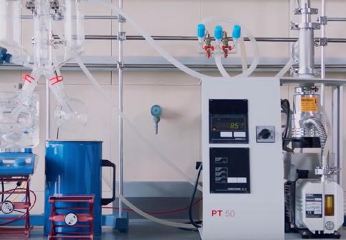
“Automated SAFE has replaced the manual variant in our laboratories,” said principal investigator Martin Steinhaus. “Other academic and industrial research groups are already following our example.”
reduces the need for complex and invasive intracytoplasmic sperm injection procedures (injecting a single sperm into an egg), in favour of artificial insemination directly into the uterus. Supervising researcher Dr Resa Nosrati said the innovation would ultimately result in higher success rates for couples seeking fertility treatment, at lower cost.
“Sperm selection is a crucial part of infertility treatment, but the conventional clinical methods for sperm selection haven’t changed over the past 30 years,” Nosrati said. Due to this lack of technological developments, the success rate of treatment methods has stagnated at 35% per cycle.

Bioengineering researchers from Monash University have developed a syringe that uses a 3D filter to detect and isolate viable sperm in less than 15 minutes. The breakthrough harnesses simple plastic syringe technology that can be readily mass produced, bringing hope and cheaper treatment solutions to 180 million people affected by infertility worldwide.
As described in the journal Advanced Materials Technologies, the syringe works by drawing 1.5 mL of semen into a chamber that then passes through a network of 560 parallel microchannels (tiny cylinders). The quality sperm swim through the microchannels into the selection chamber, where they can be extracted, leaving the poor-quality sperm behind. This process takes less than 15 minutes and is able to retrieve more than 41% of healthy sperm from the sample.
Using this technique, researchers can improve the quality of sperm selection by more than 65% compared to conventional methods. This
“Using the sperm syringe we can select sperm with over 65% improvement in DNA integrity and morphology (make-up), and since DNA quality is directly linked with fertilisation success, we expect to improve assisted reproductive technology (ART) outcomes. This technology can help to standardise and streamline the sperm selection process in fertility clinics.”
The research was led by PhD candidate Farin Yazdan Parast, who explained, “The sperm syringe provides a high-throughput device for one-step semen purification and sperm selection. The 3D sorting platform provides the maximum contact area between the semen sample and selection events by arranging microchannels in a 3D structure to enable highly parallelised and rapid sorting.
“Due to this considerably high surface-to-volume ratio, active sperm can easily find and enter the microchannels, leaving the raw sample behind. This provides an effective selection mechanism that outperforms both conventional clinical methods and other more recent sperm selection technologies.”

An analysis based on thousands of human genomes by Queensland University of Technology (QUT) researchers has found shared genetic factors that contribute to the risk of severe COVID-19 and blood analyte (component) levels, suggesting new targets for better screening, prognosis and treatment of COVID-19.
QUT’s Professor Dale Nyholt said a key finding of the study, which was published in the journal Cell Reports, was that one of the 344 studied blood analytes had widespread shared genetic influences with COVID-19, causing an increased risk of severe COVID-19.
“Our genetic causality analyses found that higher levels of triglycerides, a type of fat that is a cardiovascular disease biomarker, was strongly linked to increased risk of severe COVID-19 disease,” Nyholt said.
“The high genetic causality proportion of 0.82 indicates that increased triglyceride levels are causal for severe COVID-19 disease.
“This fits with the observation that hospitalised patients who died or were in ICU had significantly higher levels of triglycerides compared to those who were discharged or had a mild case.
“Our finding provides a genetic explanation for the greater severity of disease for people with higher triglycerides and supports the use of lipidlowering drugs such as statins and fibrates against severe COVID-19.”
PhD candidate Hamzeh Mesrian Tanha said the team used genome-wide association studies (GWAS) data to search for shared genetic influences between severe COVID-19 and blood analytes at the levels of the genome, gene and differences in a single DNA base. GWAS enable screening of the genome of many thousands of people to look for associations between millions of genetic variants and different diseases, with the goal of identifying genetic factors underlying disease conditions.
“Our analyses genetically linked blood levels of 71 analytes to severe COVID-19 in at least one of the three levels of investigation, suggesting common biological mechanisms or causal relationships,” Tanha said.
“Of those 71 analytes, we found six that showed evidence of shared influence with severe COVID-19 at all three levels, among these only triglycerides showed causality.”
Tanha said a recent study of COVID-19 patients in hospital treated with statins had fewer deaths compared with a group of those who did not receive this treatment. “However, retrospective studies have produced conflicting results on the protective effect of the prior use of statins. This could be partially explained by the presence of other medical conditions in statin users,” he said.
“Therefore, our results provide important clarity and support targeted reduction of triglycerides to help prevent severe COVID-19.”
Scientists analysing the effects of an organic compound on drugresistant bacteria have discovered how it can inhibit and kill a germ that causes serious illness or in some cases death.


Pseudomonas aeruginosa is a type of bacteria, often found in hospital patients, which can lead to infections in the blood, lungs (pneumonia) or other parts of the body after surgery. Hydroquinine, an organic compound found in the bark of some trees that is already known to be an effective agent against malaria in humans, was recently found to have bacterial-killing activity against the germ and several other clinically important bacteria, including Staphylococcus aureus, Escherichia coli and Klebsiella pneumoniae.
The team behind the discovery, from the University of Portsmouth, Naresuan University and Pibulsongkram Rajabhat University, have now explored the molecular responses of Pseudomonas aeruginosa strains to hydroquinine. They did this by looking at which genes were switched on and which were switched off in response to the drug.
Their study, published in the journal Antibiotics, revealed hydroquinine significantly alters the expression levels of virulence factors of Pseudomonas aeruginosa. It also suggests the compound interferes with the assembly and movement of the bacteria.
“There’s quite a long list of antibiotics that don’t work on Pseudomonas aeruginosa, but our experiments found some of the genes governing the motility of the bacterium were quite drastically switched off by hydroquinine,” said Dr Robert Baldock from the University of Portsmouth. “Biofilm formation and the swarming and swimming of the germ were significantly reduced.
“If we know that this drug is working in a really unique or different way then it firstly explains why it’s active on these drug-resistant cells, but it also means that you can potentially look at combining it with other existing antibiotics to make them more effective.”
Dr Jirapas Jongjitwimol, from Naresuan University, added, “Antimicrobial resistance has become one of the greatest threats to public health globally, so to discover an organic compound has the potential to be used as an effective weapon in the fight is very exciting.
“We now need to look at how the compound works against a wider variety of bacterial strains so that we better understand why some germs are affected or not affected by it.”

Little is known about how social behaviour develops in the earliest stages of life, but most animals (including humans) are born with an innate ability to interact socially or form bonds with others. Now, a new study led by the University of Utah has pointed to a gene that is important for the earliest development of basic social behaviours.
exposed to fluoroquinolones were more likely to swim closer to other fish, demonstrating that the drug helped restore sociability. They saw similar results with other drugs known to inhibit TOP2a.
The work, published in the journal Science Advances, suggests that exposure to certain drugs and environmental risk factors during embryonic development can cause changes to this gene, leading to alterations in social behaviour that are similar to those found in individuals who have autism. The study authors also found they could reverse some of the effects using an experimental drug.
Scientists suspect that many social traits are determined before birth, but the precise mechanisms involved in this process remain murky. One promising area of research suggests that social behaviour and other characteristics and traits are influenced not only by our genetic make-up but also by how and where we live.
To test this model, US scientists evaluated whether environmental exposures during embryonic development could influence social behaviour. Corresponding author Dr Randall T Peterson and colleagues exposed zebrafish embryos to more than 1100 known drugs — one drug per 20 embryos — for 72 hours beginning three days after conception.
The researchers determined that four of the 1120 tested drugs significantly reduced sociability among the zebrafish, with fish exposed to these
drugs less likely to interact with other fish. It turned out that the four medications all belonged to the same class of antibiotics, called fluoroquinolones. These drugs are used to treat upper and lower respiratory tract infections in people.
When the scientists gave a related drug to pregnant mice, the offspring behaved differently when they became adults. Even though they appeared normal, they communicated less with other mice and engaged in more repetitive acts — like repeatedly poking their head in the same hole — than other rodents.
Digging deeper, the researchers found that the drugs suppressed a gene called TOP2a, which, in turn, acted on a cluster of genes that are known to be involved in autism in humans. They also found that the cluster of autism-associated genes shared another thing in common — a higher than usual tendency to bind a group of proteins called the PRC2. The researchers hypothesised that TOP2a and the PRC2 work together to control the production of many autism-associated genes, with the former potentially serving as a link between genetic and environmental factors that contribute to onset of autism.
To determine whether the antisocial behaviours could be reversed, the research team gave embryonic and young zebrafish an experimental drug called UNC1999, which is known to inhibit the PRC2. After treatment with the drug, fish
“That really surprised me, because I would’ve thought disrupting brain development when you’re an embryo would be irreversible,” Peterson said. “If you don’t develop sociality as an embryo, you’ve missed the window. But this study suggests that even in those individuals later in life, you can still come in and inhibit this pathway and restore sociality.”
Although the scientists only found four compounds that are TOP2a inhibitors, evidence suggests hundreds of other drugs and naturally occurring compounds in our environment can inhibit its activity. Peter noted, “It’s possible that these four compounds are just the tip of the iceberg in terms of substances that could be problematic for embryonic exposure.”
Peterson acknowledged that the study was conducted in animals, and more research needs to be done before any of its results can be confirmed in humans. Therefore, he cautions against drawing conclusions about real-world applications.
“We have no evidence that fluoroquinolones or any other antibiotic causes autism in humans,” he said. “So there is no reason to stop using antibiotics. What this paper does identify is a new molecular pathway that appears to control social development and is worthy of further exploration.
“This study helps us understand at the molecular level why sociability is disrupted during the very earliest stages of life,” Peterson concluded. “It also gives us an opportunity to explore potential treatments that could restore sociability in these animals and, perhaps in time, eventually in humans as well.”
Manufacturers of medical devices must put in place a wide range of processes and documentation to meet post-market surveillance requirements. While this means more work, these requirements do give manufacturers better insight into the performance of their devices.

After some notable medical device recalls and compliance issues — such as silicone breast implants and metal-on-metal hip implants — regulators increased their scrutiny of post-market surveillance processes and activities. Regulatory authorities globally have put in place stricter postmarket requirements, particularly in the EU, with the Medical Device Regulation (MDR) and the In Vitro Diagnostic Medical Devices Regulation (IVDR). The hope, and expectation, is that these processes will reduce risk from the outset; however, it’s inevitable that issues will occur with medical devices once they are on the market, particularly in the case of high-risk and innovative devices.
The World Health Organization defines postmarket surveillance (PMS) as “a set of activities conducted by manufacturers to collect and evaluate experience gained from medical devices that have been placed on the market and to identify the need to take any action”.1 While the level of stringency differs across global jurisdictions, following the industry best practice with PMS is highly recommended. This helps to ensure regulatory requirements are met and also gives manufacturers a better understanding of how their device is performing in the market so they can more rapidly address any safety concerns.
The EU has taken the lead in terms of documentation requirements through the MDR and IVDR. Depending on device classification, required documentation includes a post-market surveillance plan (PMSP); a PMS report (PMSR) or periodic safety update report (PSUR); a postmarket clinical follow-up plan (PMCFP), report
and summary of safety and clinical performance (SSCP) for medical devices; and the post-market performance follow-up (PMPF) plan, report and summary of safety and performance (SSP) for IVDs.
The PMSP should define processes used to collect post-market information, including what that information is, the dataset, how it will be collected, and all methods and processes used to analyse and assess the collected data. The plan must be effective, which means that the activities defined should meet the principal objective of identifying actual or potential issues and determining the need to take action. The processes should also be appropriate for the type and risk of the device — the higher the classification or the more novel or innovative the device, the more rigorous the PMSP documentation.
To enable regulators to identify and verify the data and sources, manufacturers must ensure the data captured is clear, organised, searchable and unambiguous. Methods and processes used to assess collected data, protocols for statistical analysis of incidents, and the methods of communicating
with relevant parties should also be defined. And manufacturers should clearly reference how they are meeting their obligations to trace devices where corrective actions were necessary.
Manufacturers with products in all major markets must also summarise the results and conclusions of their PMS analysis in a report (PMSR). The report must be made available to regulators according to relevant regulations.
Higher classification medical devices and IVDs require a PSUR. This is a more comprehensive report than the PMSR, summarising the results and conclusions of the analysis of PMS data, and must be updated either annually or every two years, depending on the class of the device.
The MDR specifically requires manufacturers to have a PMCFP to provide input to the review and revision of the clinical evaluation report. This is a continuous process which confirms the safety and performance of the device throughout its expected lifetime. This is a vitally important piece of documentation that is afforded great importance by the notified body to supplement the data presented within the clinical evaluation report. The PMCF should also identify previously unknown side effects and monitor those to ensure the benefit-risk ratio remains in alignment with the clinical evaluation documentation.
There are certain cases where PMCF is mandatory, including but not limited to:
• innovative products or those replacing current clinical practice;
• where the product has undergone significant changes, such as a change in material for a prosthesis or new technology for an infusion pump;
• where there are higher product-related risks, or risks due to its anatomical use, such as for the heart, or a more vulnerable population.
The PMCF process must include both a report and a plan, which, like the PMS activities, must be continuous, proactive, scheduled, appropriate and justified. The plan should establish the methods, procedures, specific objectives and timelines for follow-up.
Any reference to technical documentation, clinical evaluation and risk management reports, as well as reference to relevant consensus standards, harmonised standards, and regulatory and guidance documents, should also be included in the report. The report will form part of the clinical evaluation report and the device’s technical documentation.
Information gathered through all these processes is fed back through the post-market surveillance system to ensure continuous updates to the clinical evaluation plan and risk management plan. Indeed, clinical and risk evaluation processes tightly intersect with all PMS activities.
The PMS processes are also integral for updating and improving the device’s design specifications and technical documentation. Throughout the life cycle of a product, the PMS will lead to specific actions that the manufacturer must take, such as corrective and preventive actions, field safety corrective actions and communications with key stakeholders.
Ultimately, with the implementation of the European Database on Medical Devices (EUDAMED), the objectives of PMS will be more fully realised thanks to improved transparency through better access to information for the public and healthcare professionals, as well as for manufacturers and regulators.

Following best practices with PMS processes, systems and documentation will help identify and mitigate issues that will inevitably arise with medical devices. With the right PMS processes in place, manufacturers are in a better position to facilitate compliance with regulatory requirements as well as to identify any early issues and potentially prevent the devastating recalls that have led to this more demanding regulatory oversight.
*Belinda Dowsett is Associate Director of Medical Devices and IVD, Australia at PharmaLex. An experienced quality assurance and regulatory affairs professional in the medical device industry, Belinda is responsible for the oversight and overall management of the quality management system and supports clients in their regulatory compliance and development. She holds a Bachelor of Mechanical Engineering (Biomedical) (Hons) from the University of Sydney.

1. Guidance for post-market surveillance and market surveillance of medical devices, including in vitro diagnostics, WHO, https://www.who.int/publications/i/ item/9789240015319
PharmaLex Australia www.pharmalex.com/
The School of Photovoltaic and Renewable Energy Engineering (SPREE), housed in the multi-award-winning Tyree Energy Technologies Building (TETB) of the University of New South Wales, sets the bar in recordbreaking research in solar power (photovoltaics) and renewable energy.

The experiments performed by the students often use a variety of hazardous chemicals which can be extremely dangerous to handle. These experiments lead to groundbreaking innovations, which makes SPREE a desirable campus to attend.
To successfully maintain the benchmark in photovoltaic and renewable energy research, SPREE required a Life Safety System (LSS) upgrade. The main goal was to improve workplace health and safety of its students and staff and to have it fully integrated with their Building Management System (BMS).
In the past, SPREE managed a simpler LSS which had no connection to mechanical systems, thus limiting its monitoring function to ensuring air exchange rates are observed in the event of an exhaust or mechanical failure.
The goal was to replace SPREE’s base built, restrictive LSS which had limited monitoring and coverage of critical engineering safety controls.
Pilz was engaged to supply, design, build,
install and commission a SIL 2 Life Safety System that includes new safety PLC panels and connection to all in field safety devices such as floor leak detectors, oxygen depletion sensors, toxic gas monitors, emergency stops, fume cupboards, emergency eye wash/showers and air flow velocity sensors, but also with a comprehensive SCADA system.
The Life Safety System consists of 56 safety PSSu PLCs and 10 PSSu remote I/O devices, all networked together across seven floors and accessible via eight HMIs running a customised SCADA system providing interaction and data on over 1200 safety devices, ventilation systems, alarms and event logs.
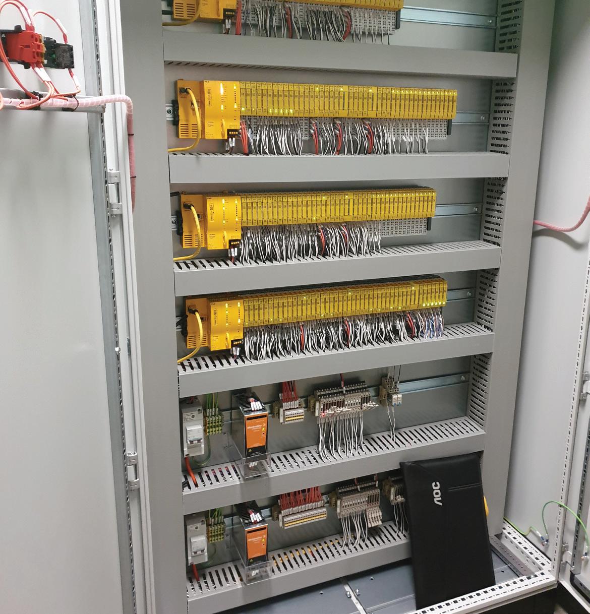
The solution monitors over 1200 sensors and interacts with the BMS to keep people in the building safe based on safety requirements specifications and a Cause-andEffÂect Matrix. It also monitors safe working conditions in multiple rooms and laboratories over seven levels of the building.

The integrated system allowed SPREE
to maximise resources and reduce cost by conducting integrated audits and assessments, helping them use their resources the best way possible. The new system also oÂffers a more efficient and productive day-to-day operational use. It oÂffers a user-friendly monitoring system that provides positive impacts on specific management system components such as improvement in quality, safety, risk and productivity. Today, after more than 18 months since project completion on 11 May 2021, SPREE’s staÂff and students feel more confident and safer when conducting groundbreaking research and experiments with the new Life Safety System.
“Pilz has been integral throughout the construction, commissioning, testing and troubleshooting of the new Life Safety System,” said Geoffrey Lim, Portfolio Delivery Manager/Project Manager Development, Estate Management, UNSW. “They have been present on a day-to-day basis and provided excellent service and responsiveness to date. Pilz was helpful in
working through user interfaces with the stakeholders and tailoring the HMI to user needs.
“I’m happy with the service and responsiveness of Pilz and the commitment to understanding the needs of the customer and committing to achieving the outcomes required.”
As safety experts, Pilz aim to help more businesses address their safety management concerns, from providing award-winning, certified products like PLC controllers, relays, drives and networks to consulting services like Plant Assessments, Risk Assessments, Safety Concepts, Safety Designs, Validations, and continuous development program of the stakeholders through certified Training Courses — all focused on the safety of man, machine and the environment.
Australia Industrial Automation LP www.pilz.com.au
A common challenge in the field of biological drug discovery and development is understanding the behaviour of biologics such as antibodies in serum or plasma to determine their stability and performance. Conventional bioassays, like surface plasmon resonance (SPR) or biolayer interferometry, have relied on enzyme or fluorescent labels to detect and monitor biomolecular interactions, but these methods tend to capture just one point in time and are fraught with non-specific binding issues that can skew results.
Malvern Panalytical’s Creoptix WAVEsystem is a label-free technology that is designed to deliver deep insight into previously undetectable interactions. Kinetic rate parameters and affinity constants (k a, kd, KD) along with binding specificity are derived in real time from even the most challenging sample types in a wide range of biological matrices with a resolution that’s not possible with other methods, the company says.
The power behind the Creoptix WAVEsystem is in the patented Grating-Coupled Interferometry (GCI) technology and non-clog WAVEchip microfluidics system. GCI measures the evanescent wave across the entire sensor surface and is not affected by temperature drifts or vibrations, allowing for sensitive measurement. Large molecules up to 1000 nm with high affinity and slow dissociation can be measured simply (even native proteins in complex matrices) without purification. The chip design also allows for determination of off-rates up to 10 s -1, meaning the kinetics of weakly binding fragments can be studied. The waveRAPID (Repeated Analyte Pulses of Increasing Duration) method can also be used to increase throughput compared to multi-cycle kinetics, particularly in screening applications, by generating a pulsating concentration profile.
The addition of two new wizards to the WAVEcontrol software aids to simplify and streamline workflows. They include ligand screening — a fast and flexible way to screen and characterise antibodies; and calibration-free concentration analysis (CFCA) — a quick and easy calibration-free approach to quantify active protein concentration.

ATA Scientific Pty Ltd www.atascientific.com.au
In February and September of each year, the World Health Organization (WHO) convenes with its network of advisors to decide on flu vaccine formulations for the Northern and Southern Hemispheres, respectively. To support ongoing vaccine reformulation programs, The Native Antigen Company develops season-specific antigens to support vaccine and diagnostic manufacturers.
The company has announced that haemagglutinin (HA) and neuraminidase (NA) antigens are now available to cover the 2023 Southern Hemisphere vaccine formulation. Available in multiple vial sizes and in bulk quantities, antigens can be used in a range of applications, including immunoassay development and as immunogens.
Expressed using Native Antigen’s VirtuE HEK293 expression system, the antigens are designed to exhibit proper glycosylation and folding. The latest haemagglutinin antigens feature C-terminal T4 foldon domains, stabilising them in their trimeric conformation to present more native-like conformational epitopes. BioNovus Life Sciences www.bionovuslifesciences.com.au

DAIHAN has two benchtop front-loading autoclave models in 25 and 40 L options. The products meet Australian Standards and can be ordered now for delivery to the laboratory.
The benchtop autoclave MaXterile BT40 has a capacity of 40 L, a water filling tank with a built-in separate water reservoir (5 L) and an intuitive user interface. With a temperature range of 110 to 135°C, it is suitable for medical, clinical, biotechnology and/or any laboratory environment with space limitations.
Safety features include errors on display as well as an audible alarm, overpressure release valve, automatic and manual door lock system, and overtemperature protector with thermostat. The product features sterilisation cycle type Class-B, as per EN13060-2 Standard. The exterior dimensions are 527 x 878 x 530 mm and it weighs 70 kg.
Pacific Laboratory Products www.pacificlab.com.au
www.LabOnline.com.au
The Thermo Scientific Vanquish Analytical Purification LC system is designed to combine the separation power of analytical LC with precise fractionation to generate high-purity products or for the isolation of contaminants. Moreover, with the introduction of the integrated Vanquish Fraction Collector, a wide application range is covered, which should give users precision in purification across their whole analytical workflow.

Biopharmaceutical laboratories working at the forefront of drug development require detailed characterisation of impurities. The user experience and separation performance of the Vanquish LC product line, with the addition of the Vanquish Fraction Collector, enable researchers to isolate and purify compounds right after their analytical separation. Biopharma labs can now fractionate with high recovery and low carryover, with additional automation features that will allow researchers to focus on success in their process results instead of handling the tool.
Features include: isolation of impurities or purification of active substances (antibodies, proteins, protein fragments, oligonucleotides, mRNA reagents, traditional medicines and natural products); wide application flexibility supporting a broad flow range from 0.05–10 mL/min and a wide variety of sample containers; compelling fraction purity performance using innovative valve technology for enhanced resolution, product recovery and minimised carryover; preserved sample integrity through fail-safe vessel rack recognition, precise fractionation movement and controlled sample storage; optimal system setup with Viper Fingertight fittings and automated delay volume determination; and confident biological sample processing through full system biocompatibility.

Thermo Fisher Scientific thermofisher.com

Engineers responsible for maintaining a sanitary environment in pharmaceutical, biotechnology and food/beverage plants will find that the FLT93C flow switch from Fluid Components International (FCI) provides liquid flow rate measurement for clean-in-place (CIP) system operational integrity.
The sanitary thermal flow switch, featuring stainless steel wetted materials and a 20 Ra finish, is available in either mechanical polish or electro-polish finishing. It supports both skid-mount and stationary CIP systems, the purpose of which is to ensure all the process piping and equipment is thoroughly cleaned per ASME BPE standards to avoid contamination.
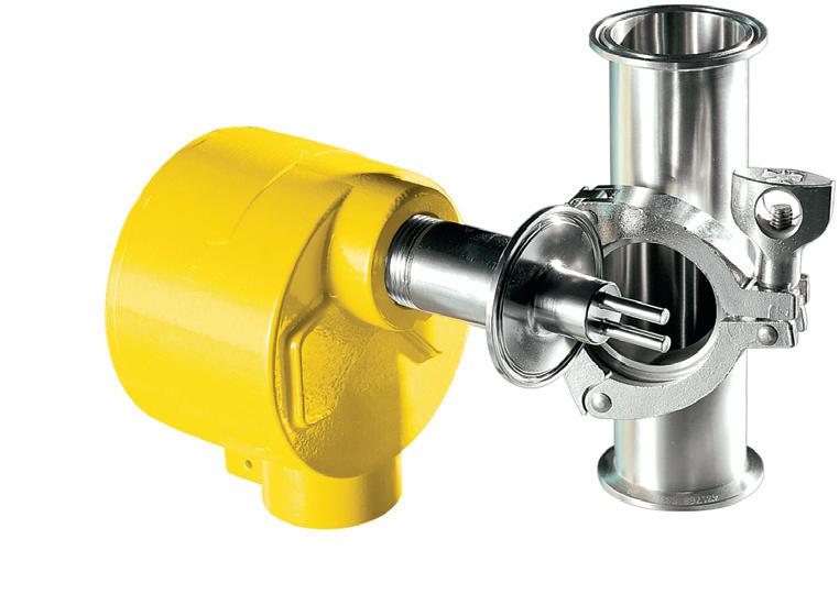
Monitoring the process cleaning solution’s minimum liquid flow rate with the thermal flow switch means the flow of liquid fluid is maintained during the entire cleaning process runtime. The switch operates over a wide flow range of 0.003 to 0.9 mps, and it also offers accuracy of ±0.5% reading or ±0.012 mps.
Relying on its temperature compensation technology, the flow switch is designed to offer setpoint accuracy for process temperatures that can vary up to 37.8°C. Suitable for 19.05 to 101.6 mm sanitary tubing process lines, the flow switch connects with a secure tri-clamp fitting for easy removal for inspection and servicing. It can easily be field-configured or factory-preset, providing flexibility and stability for all multiple process sensing and switching requirements.
Beyond its application in CIP systems, other pharmaceutical uses include compendial water systems (WFI, PW and HPW) and solution preparation systems (buffer solution). Special options are available for applications requiring more corrosion-resistant, wetted materials such as Hastelloy C and Class 1, Div 1 and 2 hazardous areas.
The switch is a dual-function instrument that indicates flow, temperature and/or level sensing in a single device. Dual 6A relay outputs are standard and are independently configurable to flow, level or temperature.
The switch is available with a choice of sensors including one that is suitable for process temperatures up to 176.67°C and one that is suitable for temperatures up to 260°C. Hazardous approvals available for the switch include ATEX and EAC/TRCU.
AMS Instrumentation & Calibration Pty Ltd www.ams-ic.com.au
Tacta is designed to make pipetting effortless and safe, while producing error-free results time after time. The product is comfortable to use due to the ergonomic handle and low pipetting and tip ejection forces.
The Sartorius Optilock system provides flexibility for volume adjustment and locking. Volume display is large, clear and facing the user. The volume is always shown with numbers, leaving the guesswork out. Tacta pipettes are easy to clean, with only few parts to disassemble, and fully autoclavable when sterilisation is needed. Single-channel pipettes cover a volume range of 0.1 to 10,000 µL and multichannel pipettes are available from 0.5 up to 300 µL volume.

Pathtech Pty Ltd www.pathtech.com.au

FlexAble from Proteintech is a novel antibody labelling kit for immunofluorescence (IF), western blot (WB) and flow cytometry (FC). The kits use an affinity linker to conjugate fluorochromes, enzymes and molecules to the user’s chosen antibody in any buffer condition.
The user can simplify their protocol by pre-labelling the primary antibodies with FlexAble kits, completely removing the need for secondary antibodies and multiple rounds of staining. Labelling is fast and easy using the provided two-step protocol and the antibodies are ready to use in just 10 min.
The kits are compatible with antibodies from any supplier regardless of concentration. There is also no requirement for buffer exchange as the kits are optimised to work with any buffer, including ones containing BSA and glycerol.
Currently the range includes kits for mouse IgG1 and rabbit IgG with four different colours: CoraLite 488, CoraLite Plus 550, CoraLite Plus 650 and CoraLite Plus 750.
United Bioresearch Products Pty Ltd www.unitedbioresearch.com.au
www.LabOnline.com.au
Targeted therapy is the foundation of precision medicine today and is driving research efforts particularly for cancer research. Antibody drug conjugates (ADCs) are biotherapeutic proteins gaining widespread use for delivery of chemotherapeutic drugs targeted to cancer cells. Unlike conventional methods, more potent drugs can be delivered using lower dosages while sparing healthy cells and minimising systemic toxicity. Critical to the success is maintaining the native structure of the monoclonal antibody within the conjugate since this underpins the targeting efficiency and stability. Consequently, the direct link between the structure and functionality of a biotherapeutic protein makes it essential to develop biopharmaceuticals that deliver the molecule with its target structure intact. By their own nature, proteins are prone to conformational change and aggregation in solution which can compromise efficacy and impact safety and thus must be understood and controlled. The development of an optimised formulation therefore relies on being able to sensitively detect changes in secondary and higher-order structure.
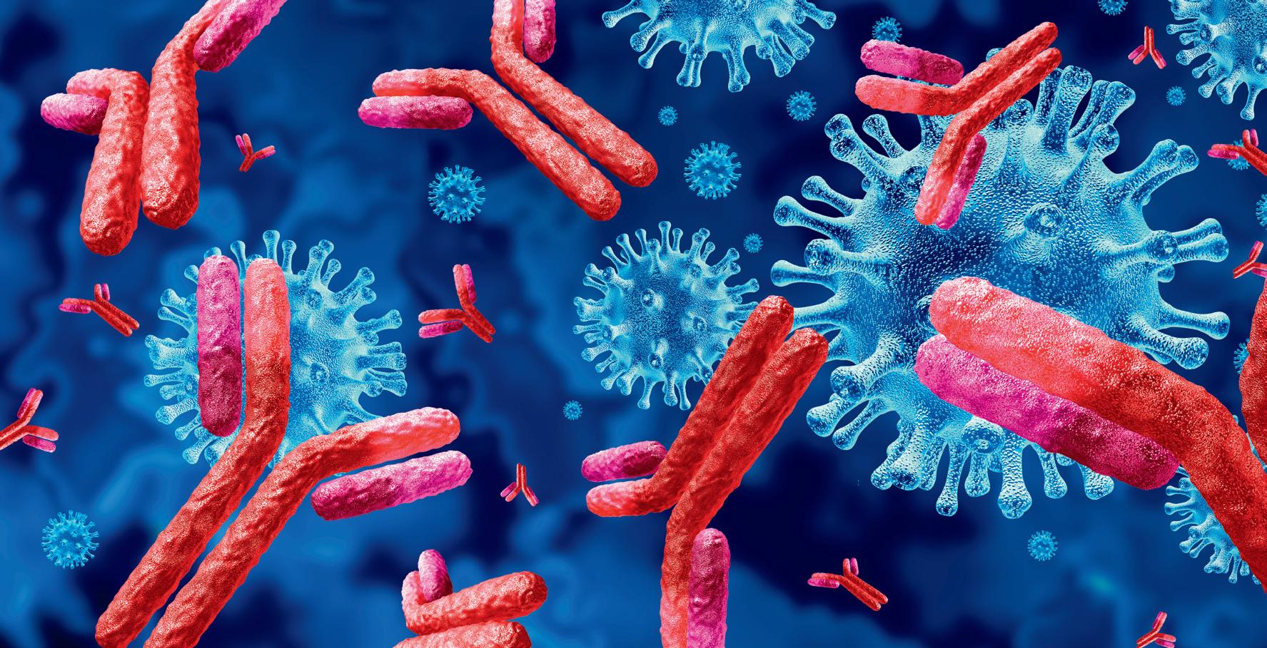
While circular dichroism (CD) and Fourier transform infrared (FTIR) spectroscopy are commonly utilised for characterising the secondary structure of proteins, they have recognised limitations. While CD works most effectively at lower concentrations, it is not compatible with many excipients and buffers. Ill-suited to automation and relatively slow, FTIR is manually intensive and lacks the sensitivity required to detect structural changes.
Microfluidic modulation spectroscopy (MMS) is a new technique that should make it easier to exploit the inherent advantages of IR spectroscopy for measurements of secondary structure. It uses a mid-IR light source to probe absorption within the Amide I band (1714 to 1590 cm-1) associated with C=O stretching in the polypeptide backbone. However, at the heart of an MMS system is a modulating microfluidic flow cell that continuously switches between the sample (protein solution) and reference (buffer) streams, used for automatic real-time background correction of the sample spectra. As a result, measurements should be less impacted by noise and less sensitive to background drift. MMS requires minimal sample preparation and it is automated for unmanned operation. It can be used for a wide range of biomolecules, from mAb-based biotherapeutics to robust measurements of ADCs, AAVs and mRNA.
The RedShiftBio Apollo is a second-generation flagship system featuring MMS technology. ATA Scientific Pty Ltd www.atascientific.com.au


Throughput is a measurement of the rate of productivity so, in terms of science, it could be how many experiments are conducted or results are obtained in a defined amount of time. Manual handling is one of the greatest limitations to throughput, as research is constrained by the size and efficiency of the labour force, and the quantity or quality of available equipment.
The manual manipulation of samples also increases the incidence of human error and limits the reproducibility of tasks, with the technique and skill of the laboratory worker having an impact.
The automation of workflows has been proven to offer significant advantages over manual procedures where high throughput and reproducible results are needed in short time frames. 1 Automation is more commonplace in industrial and clinical laboratories — where high productivity rates are paramount — but

adopting automation in academic laboratories could ease the translation from research to commercialisation, improve reproducibility and see researcher efficiency soar.
Performing key cell culture tasks manually can require many hours of repetitive and painstaking work, leaving scientists susceptible to fatigue and the risk of repetitive strain injuries. In addition, living cells require specific environmental parameters that need to be maintained throughout every stage of the cell culture workflow — cell growth, harvesting, seeding and analysis — so laboratory personnel need to work with the highest precision. Any mistakes that are made during the process can be costly to the reliability of results and, in the incidence of cell therapies and biopharmaceuticals, potentially hazardous to patients. Switching to automated workflows can help to carefully control and maintain environmental conditions for a high throughput of samples, to meet the strict standardisation requirements of cell culture.
The challenge of upscaling
Increasing the throughput of samples by automation is hugely beneficial for lab productivity, as well as for scaling up cell-based workflows for clinical or commercial applications. The transfer phase when switching from a manual to automated workflow is likely to be a learning curve that requires additional training until lab members are familiar with new equipment and supplies. Automating a stage in the process that does not directly influence the cell sample, such as pipetting medium or buffers, is relatively straightforward. However, when handling samples, the accuracy of each step needs to be validated to ensure that it is not exerting stressful forces on the living cells. It is also important that these steps are standardised, so that variations in pipetting speeds or pipette tips do not influence the viability of cells or the reproducibility of results. Once fully established, an automated workflow can increase throughput, improving the efficiency and productivity of the lab.
Automated systems can offer significant advantages for all key parts of the cell culture process — including growing cultures, plating cultures, dilutions and any other cell handling tasks — freeing up the lab technician. Complete automation would be ideal for routine workflows but, for other scenarios, stepwise automation with pipetting systems is a far more accessible route to establish a higher throughput. This can start anywhere from switching to electronic multichannel pipettes to investing in an automated plate filler.

different labware types for even faster pipetting workflows with fewer transcription errors. Furthermore, an assisted robotic platform can be used in conjunction with different electronic pipettes to combine the flexibility of handheld pipettes with the throughput of automation. Ideally, these robotic tools should be portable with a small footprint, allowing them to be seamlessly incorporated into any lab, as well as providing the option for sterile work in a laminar flow cabinet.
The need to invest in a fully automated set-up is frequently a topic of debate, despite having clear advantages for reproducibility and throughput. Academic researchers and small-scale research and development teams are often reluctant to invest in automation equipment due to concerns about the cost — with reports of full systems costing upwards of $1m — and the workspace that they require.2 The standardised procedures that come with complete automation also decrease process flexibility, which is often important as new research findings and protocols emerge. Therefore, the perfect balance between automated and manual workflows — considering the necessary throughput, flexibility, investment and reproducibility — will be determined by the requirements of the individual lab.
Multichannel pipettes can range from four to 384 channels, allowing the user to simultaneously pipette complete rows/columns — or even entire microplates — at the same time. Electronic pipettes also offer a variety of preset or customisable modes and settings, so that pipetting protocols and mixing routines can be selected and saved for repeated use in different workflow stages. For example, repeat dispense mode aspirates a large volume of liquid and dispenses it in small, defined amounts — reducing the need to move back and forth between the source and the plate — while custom programs allow users to define routines that suit their own requirements. Keeping automation to this simple yet effective standard of multichannel electronic pipettes offers a degree of flexibility in protocols and allows the pipette to be used for different stages in a workflow.
Electronic pipettes that offer automatic adjustable tip spacing provide a further degree of automation, allowing liquid to be transferred to
It is now widely agreed that automated pipetting is likely to become an industry standard for the accurate, reproducible, cost-effective and high-throughput development of cell-based products3, and electronic pipettes — together with robotic assistants — can help labs to adopt these practices sooner.

1. Holland, I., & Davies, J. A. (2020). Automation in the Life Science Research Laboratory. Frontiers in Bioengineering and Biotechnology, 8, 1326.
2. Wong, B. G., Mancuso, C. P., Kiriakov, S., Bashor, C. J., & Khalil, A. S. (2018). Precise, automated control of conditions for high-throughput growth of yeast and bacteria with eVOLVER. Nature Biotechnology, 36(7), 614-623.
3. Doulgkeroglou, M. N., Di Nubila, A., Niessing, B., König, N., Schmitt, R. H., Damen, J., ... & Zeugolis, D. I. (2020). Automation, monitoring, and standardization of cell product manufacturing. Frontiers in Bioengineering and Biotechnology, 8, 811.
This article was previously posted on the INTEGRA Biosciences website and has been republished here with permission.
INTEGRA Biosciences https://www.integra-biosciences.com/global/en
London Electronics has been manufacturing digital panel meters since 1992, useful for those who have a variable that needs measuring. The INTUITIVE range, which began production in 1998, has gone on to be one of the company’s most popular product ranges, with hundreds of thousands sold all over the world. The company is currently up to version 4, aptly named the INT4.

London Electronics panel meters are built to a high standard which conforms to ISO 9001:2015, and as such they can be expected to last for a considerable amount of time. The company recently quoted for an INT4 to replace an old original INTUITIVE which has been in service for over 20 years. The customer was impressed at the longevity of the product, indicating its quality.
AMS Instrumentation & Calibration Pty Ltd www.ams-ic.com.au
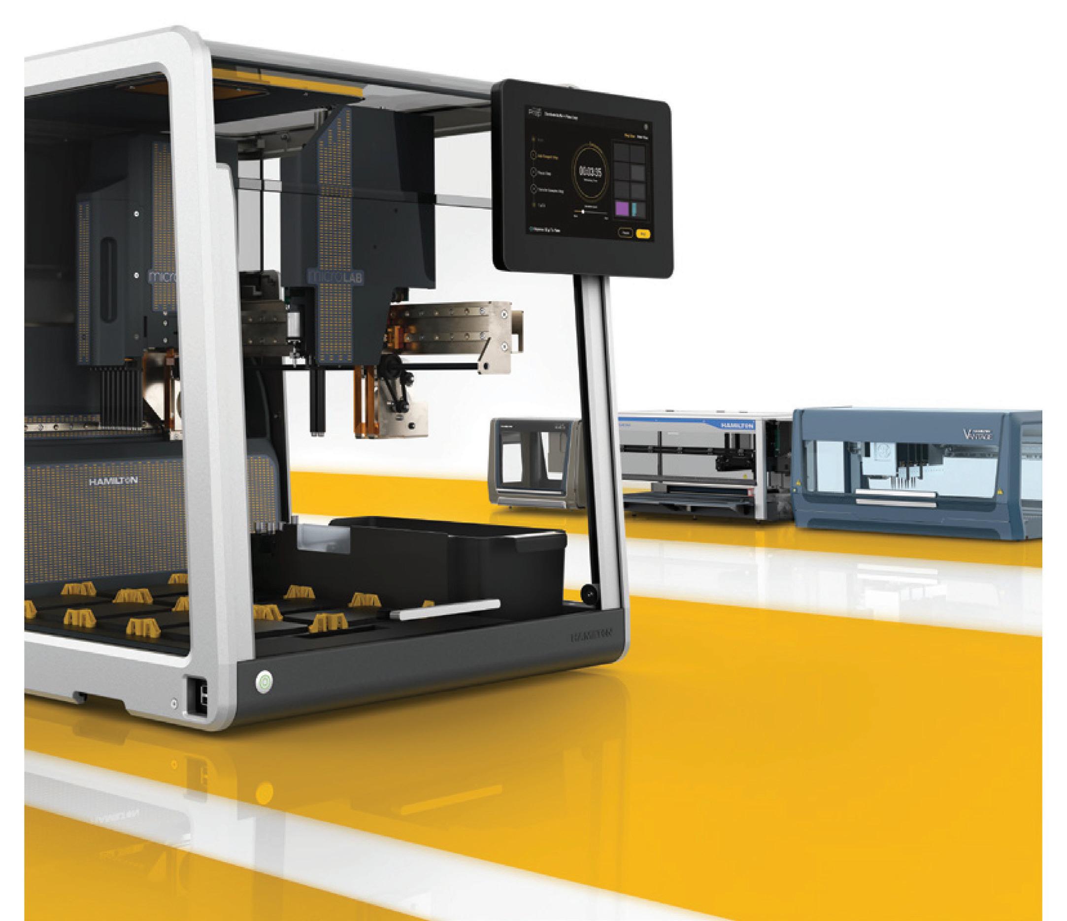
Hamilton Robotics’ Microlab Prep is a small liquid handler that features the same high-speed, powerful pipetting capabilities found in the company’s larger liquid handlers, including active pipette monitoring, air displacement pipetting, CO-RE technology and liquid level detection. These technologies are designed to provide labs with high-quality pipetting, no matter if the user is just getting started with automation or highly experienced. The product comes in a tiny footprint of only 534 x 610 x 610 mm.
The device was designed to be as simple to use as a smartphone, with clear walkthroughs that make automating basic lab tasks straightforward and quick. It automatically identifies labware on the deck using machine vision to save time and provide consistency between runs. It offers simple, touchscreen-based software that makes setting up liquid handling automation clear and understandable.
The product automates common time-consuming laboratory tasks, including replicating plates, performing a serial dilution, performing cherry-picking or hit-picking, adding reagent, normalisation, PCR preparation and LCMS preparation.

Bio-Strategy Pty Ltd www.bio-strategy.com

Laboratories performing environmental, food and biopharmaceutical testing can now increase productivity, reduce errors and minimise personnel time and solvent use with the Thermo Scientific EXTREVA ASE Accelerated Solvent Extractor — an all-in-one automated chromatography sample preparation system. The product can automatically extract and concentrate analytes of interest from solid and semi-solid samples — such as persistent organic pollutants (POPs), polycyclic aromatic hydrocarbons (PAHs) or pesticides — in a single instrument, eliminating manual sample transfer for a walk-away sample-to-vial workflow.
The system can also perform four sample extractions and concentrations in parallel, improving sample throughput and laboratory productivity. Manual intervention is reduced by a factor of three, in some cases, allowing users more time to focus on value-added tasks. Analytical labs extracting from solid or semi-solid matrices can pair the sample prep instrument with gas chromatography (GC), GCmass spectrometry (MS) or liquid chromatography (LC)-MS systems.
Traditional sample preparation for chromatography with solid or semi-solid samples is labour-intensive, prone to errors and can stifle lab productivity. Even with current automation solutions, the process can involve time-consuming manual navigation of multiple instruments. Streamlining workflows allows users to reduce manual sample preparation steps from hours to minutes, thereby increasing their productivity, minimising errors and reducing solvent use.
Thermo Fisher Scientific thermofisher.com
The Blazar Rodent Panel from Merck is claimed to detect a much broader range of adventitious viruses compared to traditional polymerase chain reaction methods, as it targets conserved regions within virus families.
The panel allows the biopharma industry to move away from using traditional animal experiments to detect rodent origin virus in the cell banks used in the production of the biologics. It also reduces the use of animals in accordance with the 4R principles — replacement, reduction, refinement and responsibility.
Merck Pty Ltd www.sigmaaldrich.com/AU/en
www.LabOnline.com.au
Apollo is RedShiftBio’s second-generation, fully automated flagship system designed to enable researchers to detect previously undetectable changes in biomolecule higher-order structure for analysis of formulations, stability, and lot-to-lot reproducibility. Measurement of these small changes allows the identification of the molecules, formulations, and conditions which are most likely to lead to stable drug products.
Apollo is powered by Microfluidic Modulation Spectroscopy (MMS) and provides ultra-sensitive, ultra-precise structural analysis of a wide range of biomolecules. When compared to CD or FTIR, MMS can detect structural change 20x faster and with 30x greater sensitivity*. MMS combines a high-power Quantum Cascade Laser with real-time buffer referencing. This provides the power to analyse both low- and high-concentration samples, in formulation buffer without excipient interference, and with the ability to detect small but critical structural changes. MMS delivers accurate and reproducible measurements from 0.1 mg/mL to >200 mg/mL under any conditions relevant to your study.
These unique features of MMS enable significant workflow improvements in different stages of biopharma development. In discovery, MMS can add more robust selection criteria for candidate screening by incorporating structural/stability monitoring earlier in the development process. In formulation, MMS has been shown to eliminate costly downstream failures by automatically analysing and comparing samples across a study to predictively identify optimal buffer formulations, stability profiles, and ideal storage conditions. Additionally, MMS tracks stability and structure — Critical Quality Attributes — across the entire manufacturing process, guaranteeing the safety, efficacy, and functionality of biotherapeutics.
Researchers at Northeastern University recently demonstrated the utility of MMS to characterise IgG light chains found in patients with amyloidosis as part of an effort to elucidate the aggregation mechanism and provide therapeutic insights. In this study, MMS was employed in direct comparison to CD to study the aggregation propensity of samples based on three different sources of IgGs (Light and variable chain IgGs from amyloid-disease positive, disease-prone, and disease negative samples). MMS interrogates the Amide I band of the IR spectrum to sensitively probe protein structure while modulating against the reference buffer for accurate background subtraction in aqueousbased samples. Compared to CD, MMS can much more accurately measure beta-sheet structures and distinguish between native beta-sheets (intramolecular) and aggregated beta-sheets (intermolecular), a distinction that is critical for monitoring amyloid formation progression. General qualitative trends were observed in the CD structural results, but due to poor sensitivity for beta-sheet content, no quantitative conclusions could be made.
MMS provided quantitative HOS characterisation and the ability to distinguish between native and aggregation-prone beta-sheet structures, which are critical for amyloidogenic protein studies.
MMS proved its value to formulation workflows in another recent example by directly measuring buffer-induced changes in protein structure and folding — a rapidly growing area of interest due to structure being a key quality attribute in biopharmaceutical development. In this instance, MMS characterised buffer-induced structural differences of lysozyme, a wellcharacterised alpha-helix-rich protein, in water and three common buffers: Phosphate Buffer (PB), Phosphate Buffered Saline (PBS), and Tris buffer. Absorption spectra in the Amide I region were automatically collected and processed to calculate higher order structure (HOS) percentages and the overall structural similarities between samples in all prepared conditions. The results showed the enzyme exhibited various degrees of structural change within these buffers, and that these changes could be quantified to inform buffer selection decisions to support ideal protein activity.
To request a demo, proof-of-concept, or a discussion on how MMS can enhance your research or product development goals, please contact ATA Scientific at enquiries@ atascientific.com.au or +61 2 9541 3500.

*Journal of Pharmaceutical Sciences, JAN 2020
ATA Scientific Pty Ltd www.atascientific.com.au

Meet Apollo, by RedShiftBio — your next-gen solution to the challenges of protein characterisation.
to assure the safety, efficacy and functionality of biotherapeutics
Data logging and conditions monitoring can be made easy and effortless with the help of Vaisala’s modern cloud solution. It allows storing of real-time and historical data and provides easy access to it from any location using various portable devices, eg, a mobile phone or a tablet.

Connect Vaisala temperature and humidity probes to the Jade Smart Cloud system and receive all the benefits of the cloud connectivity, including continuous storing of real-time measurement data, replacing manual data collection; access to real-time and historical data 24/7 from any location; the possibility to grant different access rights, eg, viewer; easy plug-and-play installation with wireless data loggers; and wireless radio communication from data loggers to access points.
No set-up or pairing is needed — devices appear directly on the user’s account — and there are no additional IT investments or extra maintenance costs. The system also provides accredited calibration when paired with Vaisala data loggers. This enables lab personnel to concentrate on their core activities.
Vaisala acknowledges that choosing the right solution for ensuring proper conditions monitoring might not be easy, and so has created a cheat sheet on what aspects to consider when choosing the right conditions monitoring system. The cheat sheet is available to download from the company’s website.

Vaisala Pty Ltd www.vaisala.com
The IKA TWISTER is a powerful single-position magnetic stirrer for mixing up to 5 L (max 3000 rpm, max 500 mPa·s). Connecting up to 30 units together, it becomes a multi-place stirrer, where each unit can be controlled independently or simultaneously. With high ingress protection rating (IP66) and a glass surface for corrosion resistance, the product helps users to effectively manage their bench space.
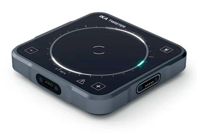
TWISTER introduces an innovative operating concept on the 360° LED glass display; all information is intuitively conveyed without any alphanumeric display. During operation, either the speed or the power required for stirring can be displayed (viscosity changes can be displayed as a percentage).
Mixing can be started by a button press or — thanks to integrated beaker recognition — automatically by placing or touching the beaker to the unit. Reverse function can be enabled to change the direction of rotation at definable time intervals.
Attaching the multi-position stirring TW.IX accessory, TWISTER is transformed into a 9-position magnetic stirrer for sample vessels with small volumes (8–60 mL). The product can further be transformed into a vortex or orbital shaker with the TW.VX shaker attachment.
The device is compact, innovative, versatile and robust.
Bio-Strategy Pty Ltd www.bio-strategy.com


The past few years have affirmed why science, technology and innovation are so important to the future of our global ecosystem. With COVID-19 expediting trends in mRNA technology platforms, vaccine manufacturing, research laboratories, cleanrooms and open data initiatives, the capacity for digital transformation in science is fast becoming an inevitable blueprint for a more sustainable future.
In 2021, the OECD outlined the importance of enabling collaborative, trans-disciplinary innovation in science and technology in order to build a more resilient and inclusive society. This, coupled with Australia’s Science Minister Ed Husic recently ordering a review of federally funded programs designed to improve diversity in STEM in a climate where women make up just 16% of people with STEM qualifications, is slowly paving the way for a collective effort driven by women in science.
Recently, I have been reflecting on the incredible contribution of Australian scientists to our knowledge base, as well as musing on my own role as a woman in STEM and advocate of life sciences. Since winning the design competition
for the University of Sydney’s Sydney Biomedical Accelerator (SBA) in August, I have been thinking about just how interdependent science, education, health and research truly are — and why my role at HDR as a Senior Laboratory Planner in the Architecture business is so rewarding.
In such a niche and specialised field, knowledgesharing needs to become more ingrained in our day-to-day so, in the spirit of igniting meaningful discussion, I thought I’d share some insights into my world — laboratory planning and design.
To put it simply, laboratories are among the most programmatically complex environments to plan, design and engineer. In my role I work across several typologies — life sciences, physical sciences, high containment and teaching labs just to name a few — and provide solutions to our clients’ often
complex challenges: “How can a facility be rightsized in a way that accounts for future growth and requirements?”, “How can a facility be retrofitted to allow for flexibility within a limited footprint?” and “How flexible should laboratory furniture and equipment systems be?”
A symbiotic relationship
Right-sizing and flexibility is at the heart of laboratory planning and design outcomes. These notions are not mutually exclusive, but it is not until all the pieces of the laboratory planning and design puzzle harmoniously fit together that a symbiotic relationship between the two can be fully realised.
At HDR, we use right-sizing and flexibility to extend the life cycle of every facility we design and provide a working, living and breathing entity that meets a laboratory’s operational needs and goals, both now and in the future.
6. Activity-based utilisation: This two-pronged approach involves the assignment of open benches and rezoning laboratory spaces to allow for cost-effective changes to the facility over time.
7. Furniture and equipment: Once the space strategy has been realised, it is important to consider whether fitout of furniture and equipment will involve traditional fixed systems, largely mobile systems or a hybrid system. This decision should be made using a benefit-cost ratio.
8. Laboratory utility and service systems: The laboratory furniture systems selected are only as good as the utilities that serve them. There are roughly three main options for service systems — traditional hard connections to fixed benches, suspended overhead carriers and highly flexible, quick-disconnect ceiling-mounted services.
Ultimately, right-sizing is about the avoidance of overbuilding or underbuilding lab space and facilities and, while it is primarily focused on optimising allocation of space, it also involves the right-sizing of utilities and services. As a lab planner, the key considerations here are a) the potential for too much space and ending up with underutilised labs, b) inadequate space to accommodate future growth or change and c) over- or under-designed mechanical, engineering and plumbing systems. In short, getting this balance right will optimise the allocation of space.
Similarly, flexibility applies at a number of scales — with the building scale being paramount — and must be integrated into modular planning strategies, laboratory furniture and equipment systems, and utility and service delivery systems. The end goals are to allow for easy expansion and contraction of research programs, staff and equipment over time; minimise the disruption and costs associated with programming changes over a building’s life cycle; and ensure minimal costs for additional renovations and physical changes. Together with right-sizing, flexibility will inform the long-term design outcomes of the laboratory.
Lab planning and design is a complex practice so, in the spirit of knowledge-sharing, here are some best-practice and emerging lab design strategies that can help organisations to right-size their facilities, account for flexibility and futureproof their business.

1. Programming and growth projections: Determining the size, scale, number of lab modules and personnel needs is critical to the programming and master planning phase, as is utilising data analytics and modelling tools to look at funding growth, staff growth and the facility’s implication.
2. Typology menu: Working with stakeholders to determine their bespoke requirements and developing a matrix which looks for commonalities between laboratories can assist in the creation of a menu of typologies that will help to populate and program space and/or design based on predictions of typology quantities.
3. Core and shared facilities: Core facilities can minimise the duplication of expensive equipment and instruments, as well as provide core resources for research programs that may change over time.
4. Modular planning: One size doesn’t fit all, so finding the right laboratory module and ascertaining the right concept layout is critical to creating a highly flexible research environment over the building life cycle.
5. Connected clusters: Once a module has been decided, it is important to consider how to arrange the spaces. While the approach to lab design in the last two decades has favoured the ‘open lab’ layout, we have recently taken this approach one step further by connecting clusters of smaller labs together to streamline connectivity and provide an opportunity to operate individual labs that may require isolation.
9. Pathways: Providing proper pathways in lab buildings and spaces can help organise the space efficiently in order to maximise productivity, workflow and flexibility. When integrated into a fluid, malleable design process, these strategies can help to navigate the complex nature of lab environments. There’s still so much unrealised potential to enable transformative and agile scientific research, experiments and measurement in laboratories. With more advocacy, strategic foresight and knowledge-sharing, we have the opportunity to enact lasting change and innovation in the industry — and hopefully broaden the pipeline of women in STEM.
*Amy Papas is an experienced architect and project leader, specialising in laboratory planning. With over 10 years’ experience in the industry, Amy’s passion for science and education has been realised in a number of significant projects at UNSW, including the Materials, Science & Engineering Building, Science & Engineering Building, and the Biological Sciences Stage 2 project. More recently, she has led the planning team on the Westmead Viral Vector Manufacturing Facility (VVMF) and the University of Sydney’s Biomedical Accelerator.
HDR www.hdrinc.com.au

The Azure Cielo real-time PCR system from Azure Biosystems is a qPCR system designed to provide high-quality data through high-performance optical technology, broad-spectrum detection capability, high specificity, precision, fast run times and reproducible qPCR data. Applications include quantitative and qualitative gene expression analysis, miRNA analysis, genetic mapping, genetic fingerprinting, NGS library quantification, pathogen quantification and six-channel multiplexing.

The Cielo is designed for multiplex experiments involving up to six different targets. With the ability to scan 16 wells simultaneously, an entire 96-well plate can be scanned for all six detection channels in 9 s. It uses fibre optics to deliver light to each individual well with precision, which reduces background excitation and the need for passive reference dyes.
With the option of three or six fluorescent channels, the Cielo 3 (with three detection channels) and the Cielo 6 (six detection channels) can detect up to six different fluorophores per well and provide flexibility for experimental design. The product’s high sensitivity and resolution are designed to save the user time, money and samples, and the device comes with a 12-month standard warranty.
qPCR is used to detect specific DNA sequences in bacterial, viral and parasitic pathogens. For accurate pathogen detection, assays need to be able to detect very low amounts of genetic material. This has been especially true with SARSCoV-2 where early detection and diagnosis are important. When tested, the company says the Cielo was able to detect as little as 0.625 copies of RNA per µL of sample, meaning it can be used for rapid detection of SARS-CoV-2.

SciTech Pty Ltd www.scitech.com.au
Perform spiral inoculations with the automated IUL Eddy Jet 2W Spiral Plater, a validated spiral plating system designed to deliver precise threefold dilutions on each plate; for spiral plating method validation, see ISO 7218/AOAC 977.27/FDA (BAM Ch.3). The innovative technology unifies diluting and plating, streamlining the process with no cross-contamination.
The Eddy Jet 2W uses patented, sterile, single-use Microsyringes that are discarded after each inoculation, eliminating cross-contamination, washing protocols and potential errors while delivering consistent results. The spiral plater’s cuttingedge stepper controller motor regulates the Microsyringe liquid dispensing and distribution pattern, based on user protocol.
Benefits to the microbiology laboratory may include a reduction in the number of serial dilutions and petri dishes, reduction in inoculation and colony counting procedure time, reduction in cost per test and use of broader petri dish diameters. With no requirement for bleach disinfection, users can proceed to incubation and colony counting.
The Eddy Jet 2W’s intuitive interface, 18 cm colour touch screen, icons layout and custom protocol guide users to set up in just a few minutes. Plate traceability can be achieved through barcode reader, keyboard and printer connectivity.
Agricultural, environmental, food, beverage, pharmaceutical and cosmetic microbiological laboratories are set to benefit most from the use of a spiral plater for bacterial determination. The technology is designed to accelerate efficiencies in throughput, accuracy and reproducibility, enabling users to focus on results.
Capella Science www.capellascience.com.au
Schott’s KL fibre-optic light sources with LED or halogen illumination are widely used in stereo microscopy for a broad range of applications. As cold light sources, they are useful in heat-critical situations, and with a halogen light option, natural colour reproduction is available across the full spectrum.

The KL series demonstrates its full strength in situations where uniform light quality is crucial. Carefully matched components deliver homogeneous illumination, while consistent colour temperature, controllable features and a variety of accessories are designed to enable their light sources to deliver correct results every time.
Coherent Scientific Pty Ltd www.coherent.com.au

A Melbourne-led team has for the first time shown that 800,000 brain cells living in a dish can perform goal-directed tasks — in this case, the simple tennis-like computer game ‘Pong’. Published in the journal Neuron, the team’s so-called ‘DishBrain’ system is evidence that even brain cells in a dish can exhibit inherent intelligence, modifying their behaviour over time.
Serving as lead author on the study was Dr Brett Kagan, Chief Scientific Officer of biotech startup Cortical Labs, which is dedicated to building a new generation of biological computer chips. He worked with collaborators from 10 other institutions on the project, including Monash University, RMIT University, University College London (UCL) and the Canadian Institute for Advanced Research.
“In the past, models of the brain have been developed according to how computer scientists think the brain might work,” Kagan said. “That is usually based on our current understanding of information technology, such as silicon computing.
“But in truth, we don’t really understand how the brain works.
“From worms to flies to humans, neurons are the starting block for generalised intelligence. So, the question was, can we interact with neurons in a way to harness that inherent intelligence?”
To perform their experiment, the research team took mouse cells from embryonic brains as well as some human brain cells derived from stem cells and grew them on top of microelectrode arrays that could both stimulate them and read their activity. While scientists have for some time been able to mount neurons on multi-electrode arrays and read their activity, this is the first time that cells have been stimulated in a structured and meaningful way.
Electrodes on the left or right of one array were fired to tell DishBrain which side the Pong ball was on, while distance from the paddle was indicated
by the frequency of signals. Feedback from the electrodes taught DishBrain how to return the ball, by making the cells act as if they themselves were the paddle.
“We’ve never before been able to see how the cells act in a virtual environment,” Kagan said. “We managed to build a closed-loop environment that can read what’s happening in the cells, stimulate them with meaningful information and then change the cells in an interactive way so they can actually alter each other.”
The researchers monitored the neurons’ activity and responses to this feedback using electric probes that recorded ‘spikes’ on a grid. The spikes got stronger the more a neuron moved its paddle and hit the ball. When neurons missed, their play style was critiqued by a software program created by Cortical Labs. This demonstrated that the neurons could adapt activity to a changing environment, in a goal-oriented way, in real time.
“The beautiful and pioneering aspect of this work rests on equipping the neurons with sensations — the feedback — and crucially the ability to act on their world,” said co-author Professor Karl Friston, a theoretical neuroscientist at UCL.
“Remarkably, the cultures learned how to make their world more predictable by acting upon it. This is remarkable because you cannot teach this kind of self-organisation, simply because — unlike a pet — these mini brains have no sense of reward and punishment.”
“An unpredictable stimulus was applied to the cells, and the system as a whole would reorganise its activity to better play the game and to minimise having a random response,” Kagan added. “You can also think that just playing the game, hitting the ball and getting predictable stimulation, is inherently creating more predictable environments.”
A microscopy image of neural cells where fluorescent markers show different types of cells. Green marks neurons and axons, purple marks neurons, red marks dendrites and blue marks all cells. Where multiple markers are present, colours are merged and typically appear as yellow or pink depending on the proportion of markers. Image credit: Cortical Labs.

The theory behind this learning is rooted in the free energy principle, developed by Friston, which states that the brain adapts to its environment by changing either its world view or its actions to better fit the world around it.
“We faced a challenge when we were working out how to instruct the cells to go down a certain path,” Kagan said. “We don’t have direct access to dopamine systems or anything else we could use to provide specific real-time incentives, so we had to go a level deeper to what Professor Friston works with: information entropy — a fundamental level of information about how the system might self-organise to interact with its environment at the physical level.
“The free energy principle proposes that cells at this level try to minimise the unpredictability in their environment.”
Kagan said one exciting finding was that DishBrain did not behave like silicon-based systems.
“When we presented structured information to disembodied neurons, we saw they changed their activity in a way that is very consistent with them actually behaving as a dynamic system,” he said.
“For example, the neurons’ ability to change and adapt their activity as a result of experience increases over time, consistent with what we see with the cells’ learning rate.”
By building a living model brain from basic structures in this way, scientists will be able to experiment using real brain function rather than flawed analogous models like a computer. Kagan and his team, for example, will next experiment to see what effect alcohol has when introduced to DishBrain.
“We’re trying to create a dose response curve with ethanol — basically get them ‘drunk’ and see if they play the game more poorly, just as when people drink,” Kagan said. This would potentially open the door for completely new ways of understanding what is happening with the brain, and could even be used to gain insights into debilitating conditions such as epilepsy and dementia.
“This new capacity to teach cell cultures to perform a task in which they exhibit sentience — by controlling the paddle to return the ball via sensing — opens up new discovery possibilities which will have far-reaching consequences for technology, health and society,” said Dr Adeel Razi, Director of Monash University’s Computational & Systems Neuroscience Laboratory.
“We know our brains have the evolutionary advantage of being tuned over hundreds of millions of years for survival. Now, it seems we have in our grasp where we can harness this incredibly powerful and cheap biological intelligence.”
The findings also raise the possibility of creating an alternative to animal testing when investigating how new drugs or gene therapies respond in these dynamic environments. According to Friston, “The translational potential of this work is truly exciting: it means we don’t have to worry about creating ‘digital twins’ to test therapeutic interventions. We now have, in principle, the ultimate biomimetic ‘sandbox’ in which to test the effects of drugs and genetic variants — a sandbox constituted by exactly the same computing (neuronal) elements found in your brain and mine.”
“This is the start of a new frontier in understanding intelligence,” Kagan said. “It touches on the fundamental aspects of not only what it means to be human but what it means to be alive and intelligent at all, to process information and be sentient in an ever-changing, dynamic world.”
Merck has launched its Milli-Q EQ 7008/16 ultrapure and pure water purification system, demonstrating its commitment to meeting the purified water needs of every scientist. The product will replace the existing Milli-Q Direct systems.
The system produces consistent ultrapure (Type 1) water quality direct from a tap water source and includes an option to obtain reverse osmosis (RO, Type 3) quality water. Final ultrapure water quality can be easily adapted to experimental requirements through a selection of final filters. Dedicated innovations make the system convenient and effortless to use.
The product features flexible set-up options, as the touchscreen display and dispenser may be wall mounted or placed up to 3 m from the system, saving valuable bench space. Its ‘Check & Dispense’ lights, located on the Q-POD dispenser arm, rapidly monitor and confirm the quality of all liquid dispensed. The redesigned total organic carbon (TOC) indicator measures TOC at the point of use, providing assurance that organic contamination is below 5 ppb.
Other design elements include: patented polishing cartridges for consistently high-quality ultrapure water; high-definition digital displays to simplify system operation; convenient handling with a range of easily accessible dispensing options; smaller system footprints and purification cartridges versus Merck’s previous-generation Milli-Q systems; hibernation mode to minimise water and energy consumption when the lab is closed for extended periods; and a space-saving design that can be easily integrated and neatly installed for a clutter-free workspace.
Merck Pty Ltd www.sigmaaldrich.com/AU/en

The C200 bomb calorimeter from PA Hilton consists of a well-stirred, highly insulated reactor with ignition wire, high-pressure valves, electronic digital thermometer and pressure regulator. The 300 mL reactor is manufactured from a corrosion-resistant stainless steel to allow experiments on a wide range of fuel.
The bomb calorimeter has been used in an engineering teaching and testing laboratory that specialises in energy and fuel research. Students or users can design experiments to measure the calorific value of liquid fuels and solid fuels. This would allow the students to compare the results from different fuels.
To suit the laboratory environment, the bomb calorimeter is equipped with components to facilitate ease of experiments such as during data collection and sample preparation. The digital thermometer provides data with two decimal places for high-resolution reading. It also includes a pellet press to assist preparation of powdered fuel to be fed into the calorimeter. The reactor is also equipped with a bursting disc for safety precautions and a bottle pressure regulator for safe discharge of gases.
The unit must be supplied with commercial oxygen capable of pressuring the reactor up to 25 bar.
Bestech Australia Pty Ltd www.bestech.com.au
The PatchScope Pro range of fully integrated electrophysiology rigs, from Scientifica, is specifically designed for patching with field stimulation and local perfusion in single cells, cultures or monolayers. The three models are suitable for neuroscience, cardiac and many other research areas. They can also be customisable to the user’s specific research needs, including fluorescence imaging.
PatchScope Pro 1000 is an integrated, compact and versatile package consisting of an inverted microscope and a PatchStar micromanipulator. This hands-free, electrophysiology system is specifically designed for patching in single cells or monolayers. The system is easy to set up and quick to master.
PatchScope Pro 2000 is a complete patch clamp rig, useful for pharmacological studies or other cultured cell stimulation. It consists of an inverted microscope, a PatchStar and a LBM-7 micromanipulator. It is an integrated electrophysiology system specifically designed for patching with field stimulation and local perfusion in single cells or monolayers.
PatchScope Pro 3000 consists of an inverted microscope and two PatchStar micromanipulators. The user-friendly design means that the system is simple to handle and work with.
One of the many advantages of the PatchScope Pro systems is that they incorporate the Scientifica PatchStar manipulators. Their stability and ultralow noise enable users to record even the tiniest signals and obtain data from long-term experiments. The automated technology is designed to provide experimental stability by enabling the user to smoothly switch between objectives and change the fluorescence illumination without having to touch the microscope.
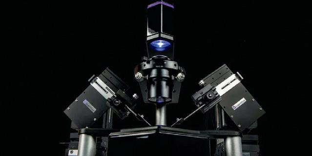
SciTech Pty Ltd www.scitech.com.au
www.LabOnline.com.au

The human gut microbiome has been found to affect metabolic health and nutrient absorption, and preliminary research also suggests that it contributes to the development of the immune response, food allergies and intolerances, obesity and a wide range of other conditions. This field is the focus of Ortho-Analytic, an integrative medicine laboratory based in Switzerland, that identifies the bacterial, fungi and parasite DNA found in stool samples using molecular genetic analyses, helping to build a detailed picture of a patient’s gut microbiome and aiding practitioners in formulating specific treatment plans.

The demand for gut microflora analysis has been growing rapidly over the last few years due to recent discoveries that bacteria in the digestive tract can have significant impacts on our general health. However, many smaller facilities are now struggling to keep up with the increased workflow. One solution that addresses this challenge is the automation of DNA extraction from stool samples, which represents an important step towards increasing lab throughput and reducing costs, and offers the potential to open up the medical benefits of microbiome profiling to greater segments of the general public. This approach was the one taken by Ortho-Analytic for its specialist service providing the detection and analysis of the bacterial DNA present in patients’ stool samples.
Ortho-Analytic extracts bacterial, fungi and parasite nucleic acid from stool specimens using the DreamPrep NAP workstation and the ZymoBIOMICS 96 MagBead DNA Kit from Zymo Research, minimising contact time with the samples and reducing the likelihood of introducing manual errors. The system also incorporates the Frida Reader module for built-in quantification and normalisation. Dr Philipp Lemal, Deputy Laboratory Manager at Ortho-Analytic, explained: “We tested several DNA extraction kits and found that the one from Zymo Research was the best for our application. Tecan had already automated this kit for use on its DreamPrep NAP equipment, so we could purchase the whole package and save ourselves a lot of time in the validation process.”
It took under two weeks for the system to be fully calibrated and adjusted in the lab, and it has proven to be very robust, allowing staff to perform other tasks while the machine runs in the background. The team has found the workstation itself to be very flexible, enabling employees to easily add or change scripts as necessary to fine-tune the extraction process. Lemal described how the move towards automation has had a positive impact on the lab’s productivity: “Before we had the DreamPrep NAP workstation, we did all of our extractions manually and could only process a maximum of 40 samples per day. The DreamPrep NAP
now automates most of our workflow, which has increased that more than fivefold to around 200 samples per day. The Frida Reader module also allows us to measure DNA concentration straight after we extract it, all on the same machine, and that’s very convenient.”
Speaking about the team’s current and upcoming research projects, Lemal said: “At the moment we are collaborating with several universities all over the world who are using our technical platform for their research. We also hope to be involved with a greater number of long-term studies in the near future that will help the research community to be able to clearly associate the microbiome with many conditions, including cancers and inflammatory bowel disease.”
To further improve the quality of microbial DNA extraction, Ortho-Analytic has collaborated with BÜHLMANN Laboratories to develop its own sample tube, the Calex NGS. The tube contains a special stabilisation buffer that kills bacteria, viruses and fungi, and hinders biological activity inside the tube to stabilise DNA prior to extraction. This ultimately produces a more realistic representation of microbiome composition for each sample.
Until recently, the lab was sending the samples to an external partner laboratory for DNA extraction and molecular genetic analysis. However, the team was concerned about sample stability during transport, due to the long transit times and inconsistent storage conditions. This limitation motivated staff to begin performing microbial DNA analysis themselves, with the aim of preserving their specimens and providing faster, more reliable results. These workflow adjustments promise to enhance the speed and quality of both DNA extraction and analysis, increasing the lab’s capacity to meet the demand for microbiome profiling well into the future.
“We are really happy that we can finally do all our DNA extraction and analysis in-house, and we hope that this will greatly benefit our clients and their patients going forward,” Lemal concluded. Tecan Australia www.tecan.com
March 8–10, Hobart
The 16th GeneMappers conference, GeneMappers2023, will be held at the Crown Plaza Hotel in Hobart’s CBD. Join Australia and New Zealand’s statistical genetics community to get up to speed on novel analysis methods, insights into human disease and more, all while catching up face to face.
The program and invited speakers reflect the landmark developments that the genetics field has been ignited by in the last few years:
• The promise of long-read sequencing in advancing understanding of unusual genetic variation.
• The continuing delivery of biobanks such as the UK Biobank and exiting promises ahead with novel biobanks and more data.
Exciting advances in our understanding of human diseases, in both common and single gene disorders.

• The development of novel analytical methods for the identification and understanding of these diseases. https://www.genemappers2023.org/
Lorne Proteomics 2023
February 2–5, Lorne https://www.lorneproteomics.org/
Lorne Proteins 2023 February 5–9, Lorne https://www.lorneproteins.org/
Lorne Cancer 2023 February 9–11, Lorne and online https://www.lornecancer.org/
Lorne Genome 2023 February 12–14, Lorne https://www.lornegenome.org/
Lorne Infection & Immunity 2023 February 15–17, Lorne https://www.lorneinfectionimmunity.org/
Quantum Australia 2023 February 21–23, Sydney and online https://quantum-australia.com/quantum-australia
Pathology Update 2023
February 24–26, Melbourne https://www.rcpa.edu.au/Events/Pathology-Update
Cutting Edge Symposium: Efficient use of plant biomass for different feedstocks
March 7–9, Canberra https://events.csiro.au/Events/2022/August/25/ Efficient-use-of-plant-biomass-for-different-feedstocks
SMP (Science Meets Parliament) Online March 7–9, online https://sta.eventsair.com/science-meetsparliament-2023/
Australasian Exploration Geoscience Conference
March 13–18, Brisbane https://2023.aegc.com.au/
SMP (Science Meets Parliament) On The Hill March 22, Canberra https://sta.eventsair.com/science-meetsparliament-2023/
Building the SABRE Biosecurity Alliance March 23, Canberra https://sta.eventsair.com/science-meetsparliament-2023/
TSANZSRS 2023 March 25–28, Christchurch https://www.tsanzsrsasm.com/
35th ISE Topical Meeting: Electrochemistry for energy, environment and health May 7–10, Gold Coast https://topical35.ise-online.org/
EcoSummit 2023 June 13–17, Gold Coast https://ecosummitcongress.com/
Cutting Edge Symposium: Locking Carbon in Minerals
June 19–23, Perth https://events.csiro.au/Events/2022/July/28/LockingCarbon-in-Minerals
The Australian Society for Microbiology Annual National Meeting July 3–6, Perth https://www.theasmmeeting.org.au/
20th International Conference on Biological Inorganic Chemistry July 16–21, Adelaide https://icbic2023.org/
31st International Symposium on Lepton Photon Interactions at High Energies July 17–21, Melbourne https://indico.cern.ch/event/1114856/
National Science Week 2023 August 12–20, Australia-wide https://www.scienceweek.net.au/
IUCr 2023
August 22–29, Melbourne http://iucr2023.org/
ASC 50th Annual Scientific Meeting November 3–5, Surfers Paradise https://www.cytology.com.au/annual-scientificbusiness-meeting
Acoustics 2023
December 4–8, Sydney https://acoustics23sydney.org/
Westwick-Farrow Media A.B.N. 22 152 305 336 www.wfmedia.com.au
Head Office Unit 7, 6-8 Byfield Street, (Locked Bag 2226) North Ryde BC NSW 1670, AUSTRALIA Ph: +61 2 9168 2500
Editor Lauren Davis LLS@wfmedia.com.au
Publishing Director/MD Geoff Hird
Art Director/Production Manager Julie Wright
Art/Production Linda Klobusiak, Marija Tutkovska Circulation Dianna Alberry circulation@wfmedia.com.au
Copy Control Mitchie Mullins copy@wfmedia.com.au
Advertising Sales Sales Manager: Kerrie Robinson Ph:0400 886 311 krobinson@wfmedia.com.au
Nikki Edwards Ph: 0431 107 407 nedwards@wfmedia.com.au
Tim Thompson Ph: 0421 623 958 tthompson@wfmedia.com.au
If you have any queries regarding our privacy policy please email privacy@wfmedia.com.au
Printed and bound by Dynamite Printing
Print Post Approved PP100008671
ISSN No. 2203-773X
All material published in this magazine is published in good faith and every care is taken to accurately relay information provided to us. Readers are advised by the publishers to ensure that all necessary safety devices and precautions are installed and safe working procedures adopted before the use of any equipment found or purchased through the information we provide. Further, all performance criteria was provided by the representative company concerned and any dispute should be referred to them. Information indicating that products are made in Australia or New Zealand is supplied by the source company. Westwick-Farrow Pty Ltd does not quantify the amount of local content or the accuracy of the statement made by the source.
to industry and business professionals


The magazine you are reading is just one of 11 published by Westwick-Farrow Media. To receive your free subscription (magazine and eNewsletter), visit the link below.


