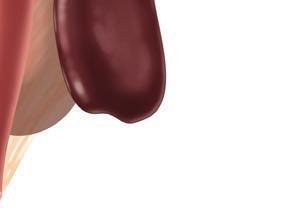
Anatomy of the


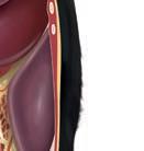






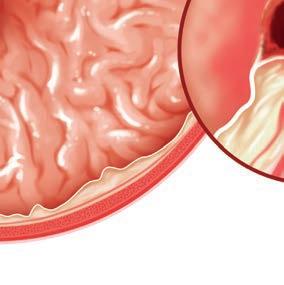
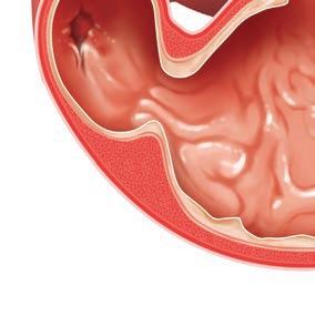
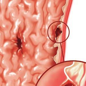
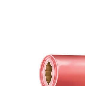
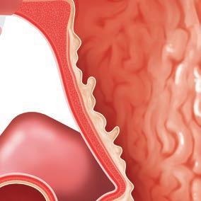
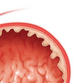
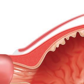
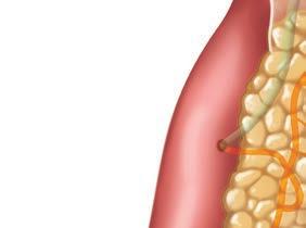
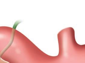
A gastroduodenal ulcer is a defect in the wall of the stomach or duodenum that extends through the muscularis mucosa into the deeper layers (submucosa) of the gastric or intestinal wall. Given the presence of vessels in these deeper tissues, life-threatening hemorrhage may result. With severe ulceration, perforation of the wall may occur, resulting in bacterial contamination of the abdominal cavity. The most common causes of ulceration are drugassociated injuries (NSAIDs), mast cell tumors, and primary gastric and duodenal tumors. Sled dogs may suffer from exercise-induced gastroduodenal ulcers. Gastrinomas, a tumor of the pancreas which produces the hormone gastrin, may result in ulcer formation, but mucosal hypertrophy and delayed gastric emptying are more commonly detected.
■ Vomiting.
■ Hematemesis, melena.


■ Abdominal pain.
■ Weight loss.
■ Abdominal ultrasound (a normal study does not rule out ulcers).
■ Endoscopy.
■ Biopsy to screen for underlying cancer, infection (Helicobacter), and inflammation.

■ Treatment of underlying causes when possible.
■ Sucralfate slurry to protect the gastric mucosa.

■ Proton pump inhibitors (omeprazole) to decrease the acidity of gastric fluid.
■ Low-fat, low-fiber, canned food in small frequent meals.
■ Systemic analgesics for pain management.
■ Surgical resection of ulcers that are refractory to medical management or have perforated.
■ Dependent on the underlying cause, severity of the clinical signs, and whether or not perforation has occurred.
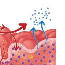

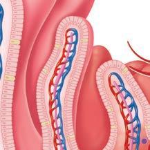
Chronic enteropathy (CE) is an umbrella term relating to a host of diseases characterized by gastrointestinal signs that persist for at least 3 weeks. Although inflammatory bowel disease (IBD) is a form of CE, the diagnosis of IBD requires a systematic therapeutic approach to rule out food-responsive, fiber-responsive, and antibiotic-responsive disease. By following this approach, many dogs and cats with CE can be successfully treated before undergoing invasive and costly endoscopic procedures.
■ Any combination of gastrointestinal clinical signs (e.g., vomiting, diarrhea, weight loss, changes in appetite) present for more than 3 weeks.

■ Exclusion of extragastrointestinal diseases:
■ Complete blood count, chemistry profile, urinalysis, and abdominal imaging.
■ Trypsin-like immunoreactivity.
■ Resting cortisol.
■ Total T4 in cats.
■ Geographically relevant infectious disease testing.
■ Fecal parasite testing.
■ Serum cobalamin and folate to screen for malabsorption.
■ Response to therapy (see below).
■ Endoscopy if there is no response to therapy.
Lymphatic vessel Vein Artery
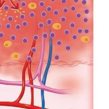
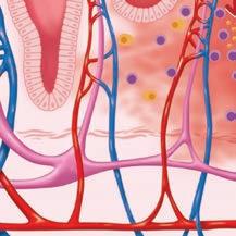
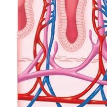
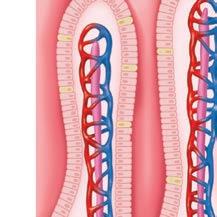
Decreased nutrient and electrolyte absorption
Water
■ Empirical deworming to rule out parasitic disease.
■ Diet trials: fed exclusively (no treats, flavored medications, or oral preventatives) for 2–3 weeks.
■ Easily digestible low-fat diet.
■ Fiber-rich diet if large bowel diarrhea.
■ Novel protein, limited ingredient, or hydrolyzed diets if there is no response to easily digestible diets.
■ Probiotic and antibiotic (tylosin, metronidazole) trials.
■ After the histopathologic diagnosis of lymphoplasmacytic ± eosinophilic enteritis and treating for parasitism and antibiotic- and food-responsive disease, corticosteroids for the treatment of idiopathic CE.
■ Variable and largely dependent on the underlying cause.
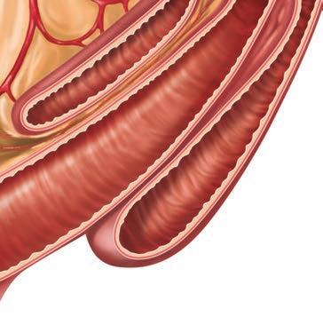
Gastrointestinal intussusceptions are an uncommon form of bowel obstruction that occur when a migrating peristaltic wave reaches a fixed or noncontractile segment. Usually, a proximal segment of the gastrointestinal tract (intussusceptum) telescopes into the lumen of the adjacent distal segment (intussuscipiens). The most common sites are the jejunum and the ileocolic junction. Intussusceptions occur most often in young animals (<1 year) but may be seen in older animals secondary to intestinal cancer. Most intussusceptions in dogs are idiopathic; however, any disturbance of intestinal motility (e.g., viral enteritis, cancer, foreign bodies, prior abdominal surgery) may lead to intussusception.
Intussusceptions can be chronic or intermittent (i.e., they reduce themselves spontaneously and then reform).
■ Vomiting.

■ Diarrhea.
■ Abdominal pain.
■ Anorexia.
■ The intussusception can often be palpated as an abdominal mass.
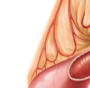
■ Abdominal radiographs: obstructive pattern if complete obstruction.
■ Abdominal ultrasound.
■ Surgery: manual reduction.
■ Enterectomy and anastomosis if the affected segment is not reducible, not viable, or associated with a mass lesion.

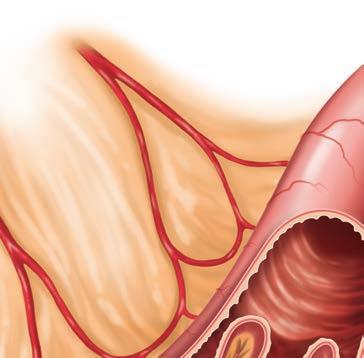
■ Percutaneous manual reduction may be successful, but the chance of recurrence is high.
■ Some intussusceptions spontaneously resolve with anesthesia.
■ Depends on the underlying cause. Good long-term prognosis in idiopathic cases.
■ Complications: recurrence, dehiscence of intestinal anastomosis, ileus, intestinal obstruction, peritonitis, short bowel syndrome.
■ Recurrence rate in dogs: 11%–27%. Enteroplication can be performed at the time of surgery to prevent recurrence.


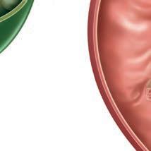
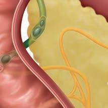
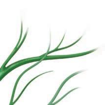
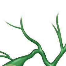
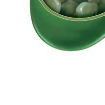
Stones within the gallbladder are termed choleliths, while stones in the biliary ducts are termed choledocholiths. Cholesterol, bilirubin, and mixed choleliths have been reported in dogs and cats. Many patients are asymptomatic; however, in some cases, the gallbladder or bile duct may become obstructed or rupture. Older female dogs are predisposed, particularly those of the Miniature Schnauzer and Miniature Poodle breeds.
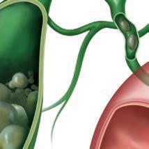
■ Patients may be asymptomatic to severely ill with signs of biliary obstruction or bile peritonitis.
■ Abdominal pain.
■ Vomiting.

■ Anorexia.
■ Jaundice.
■ Many stones are too small and are not mineralized enough to be seen on radiographs (14%–50% of choleliths can be seen).
■ Abdominal ultrasound to visualize stones and evaluate the gallbladder and biliary tree.
Choleliths and choledocholiths are often found incidentally. Pancreatitis, gastroenteritis, and gastrointestinal foreign bodies may present with similar clinical signs and should be considered before attributing the signs to a gallstone.

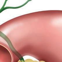
Hepatic ducts
Cystic duct
■ Most (53%–87%) stones are incidental findings and do not require intervention.
■ Supportive care with intravenous fluids, ursodeoxycholic acid, analgesics, and antimicrobials may be considered in clinical patients with partial biliary obstruction.

■ Surgery is indicated in cases with cholecystitis or biliary obstruction. Medical dissolution of stones in patients with severe acute obstruction is rarely successful.
■ Treatment of concurrent endocrine diseases.
■ Survival depends on the number and location of stones and the surgical procedure required.
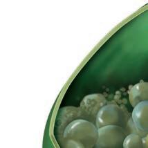
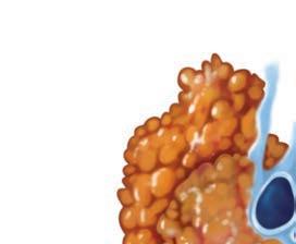
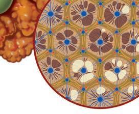
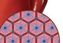
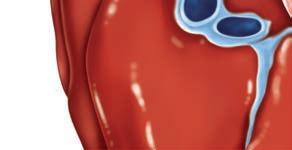
Cirrhosis is the end-stage consequence of liver inflammation secondary to chronic liver disease, drugs, or toxins. In cirrhosis, nonfunctional fibrous scar tissue replaces functional liver tissue, resulting in a small and irregular liver.

■ Jaundice.
■ Ascites.
■ Anorexia, vomiting, and/or diarrhea.
■ Signs of hepatic encephalopathy.
■ Biochemical parameters show evidence of liver failure (low BUN, albumin, cholesterol, and glucose; elevated total bilirubin, bile acids, and ammonia).
■ Small, nodular, irregular liver.
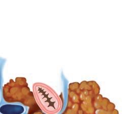
■ Multiple acquired portosystemic shunts may be seen on ultrasound or CT scan.
■ Although biopsy is necessary for a definitive diagnosis, the limitations of sampling error and poor prognosis result in diagnosis often being based on imaging and clinicopathologic parameters.
■ Treatment of the underlying chronic hepatopathy if possible.
■ Management of hepatic encephalopathy (see Sheet 52).
■ Management of ascites with diuretics (e.g., spironolactone).
Necrosis
Fat
■ Poor.
By the time clinical signs of cirrhosis are detected, less than 20% of functional liver tissue remains.

As a result, the prognosis is poor.
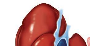
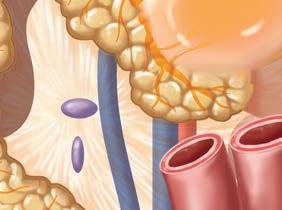
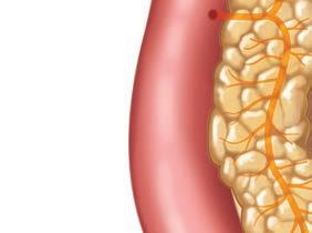
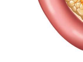
Pancreatic abscesses and pseudocysts are reported complications of acute and chronic pancreatitis. They occur infrequently in dogs and cats and are most often sterile. Pancreatic abscesses are a collection of pus, or white blood cells, whereas pancreatic pseudocysts contain a collection of enzyme-rich pancreatic fluid.
■ Small pseudocysts are clinically asymptomatic.
■ Mild, intermittent gastrointestinal signs.
■ Signs of acute pancreatitis (see Sheet 70).
■ Abdominal ultrasound: fluidfilled structure in association with the pancreas.
■ Fine needle aspiration to collect fluid from within the abscess or pseudocyst for cytological evaluation and culture.
■ Evaluation for acute or chronic pancreatitis (speciesspecific pancreatic lipase immunoreactivity).
■ Medical management of pancreatitis (see Sheet 70).
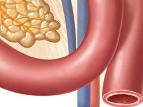
■ Sterile abscesses and pseudocysts can be drained using ultrasound guidance, but they often refill. Surgical intervention may be necessary for rapidly enlarging pseudocysts.
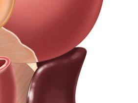

■ Surgical drainage and aggressive antibiotic therapy for infected pancreatic abscesses and pseudocysts.
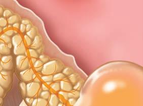
■ Generally good.
Pancreatic pseudocyst
■ Fair to grave in pets that require surgical drainage for infected pancreatic abscesses.
