What lies ahead for corneal endothelial disease treatment?
ALSO IN THIS ISSUE
Latest ESCRS Clinical Trends Survey Delegate response shows presbyopia IOL use steady, but obstacles remain.
Avoiding Pitfalls in IOL Power Calculations Navigate the best options for previous corneal refractive surgery patients.

Simulators Increase Access to Skills Training
ESCRS sends surgical simulators to help surgeons with limited access gain hands-on training.
https://congress.escrs.org/

BINKHORST MEDAL LECTURE
Jorge L Alió MD, PhD, FEBOphthal will speak on “Corneal Regeneration: The Future of Corneal Surgery.” Dr Alió is a professor and chairman of ophthalmology at the University Miguel Hernández de Elche in Alicante, Spain, and a leader in the emerging topic of corneal regeneration. He conducted the first worldwide clinical trial for the treatment of corneal dystrophies and particularly keratoconus with autologous mesenchymal stem cells, and he has published a series of important scientific papers on this innovative type of corneal surgery.
SMART AND @CTIVE MONDAY
A new addition to the ESCRS Congress, Smart and @ctive Monday will feature “brushups” on retinal surgery, glaucoma surgery, and oculoplastics, a medical writing workshop for young ophthalmologists, an ophthalmic anaesthesia symposium, a surgical video session, and practice management sessions. A “digital track” will offer a “Continents Going Digital” symposium, symposia on automating eye surgery and the digital operating room, and talks and panel discussions about artificial intelligence in ophthalmology.

HERITAGE LECTURE
Marie-José Tassignon, MD, PhD, FEBOS-CR will speak about “The Enigma of the Anterior Interface.” Dr Tassignon is the emeritus head and chief of the department of ophthalmology of the Antwerp University Hospital and University of Antwerp. The first female president of ESCRS (2004–2005), Dr Tassignon developed bagin-the-lens cataract surgery for the paediatric population and holds 10 patents (three earned since 2019). She has been published more than 370 times in peer-reviewed journals and is the author of 27 book chapters and two full textbooks in ophthalmology.
iNOVATION DAY
Building on the success of the inaugural iNovation® Day in 2022, ESCRS is hosting the second iNovation Day on Friday, 8 September. Sessions will focus on the most urgent clinical needs and barriers to success in anterior segment care and how new technologies may address those barriers and clinical needs within the next 5–10 years. Of special interest is a new feature, “The Innovators Den: EyeCare Pioneers,” which will highlight entrepreneurs who have personally developed unique ideas to address some of the biggest unmet needs of Congress attendees.
28 Pseudomonas Keratitis Outbreak
Allan R Slomovic MD
GLAUCOMA
30 Potential New Diagnostic Parameter for Glaucoma
Ingrida Januleviciene MD, PhD
RETINA
32 CRISPR for Retinitis Pigmentosa
Alessandra Recchia PhD
33 Potential Therapies and Biomarkers for Uveal Melanoma
Breandán K Kennedy PhD
Kayleigh Slater PhD
34 Progressive Polysulfate Pentosan Maculopathy
Nieraj Jain MD
35 What’s the Best Treatment for RVO?
Richard Gale BSc, MBChB, FRCP, FRCOphth, MEd, PhD
PAEDIATRIC
36 Low-Concentration Atropine for Preventing Myopia
Jason C Yam MB, Steven M Archer MD, David C Musch PhD, MPH, David A Bernsten OD, PhD
38 Pushing the Boundaries of Drug Delivery
Kris Morrill, Michael O’Rourke, Eyal Sheetrit, Patricia Zilliox, Thomas Reeves

2 EUROTIMES | MAY 2023 Acrylic IOLs 06 Inside ESCRS: Moving Simulators to Increase Access to Skills Training 08 ESCRS at a Glance: 27th ESCRS Winter Meeting Congress in Numbers 10 Ukraine Update: From Ukraine to New Zealand 12 Ensuring Safe and Timely Access to Advanced Cell and Gene Therapies COVER COMPANION 17 DMEK Sets the Bar High Mor Dickman MD, PhD 20 Latest ESCRS Clinical Trends Survey
MMA Nuijts MD, PhD
Kohnen MD, PhD, FEBO 22 Knowing Lens Tilt Theresa Höftberger PhD CORNEA 24 The Clinician or AI— Who Will Diagnose?
A Woodward MD, MSc
D Klyce PhD, FARVO 26 Cell Therapy for Corneal Endothelial Dysfunction Ula V Jurkunas MD Massimo Busin MD 14 Cover
PK: New
to
corneal endothelial disease. May 2023 | Vol 28 Issue 4 ESCRS CONTENT
Rudy
Thomas
Maria
Stephen
Beyond
approaches
treating
OPHTHALMOLOGY
OCULAR UPDATE
Publishers
Jemilah Senter
Mariska van der Veen
Mark Wheeler
Executive Editor
Stuart Hales
Editor-In-Chief
Sean Henahan
Senior Content Editor
Kelsey Ingram
Creative Director
Kelsy McCarthy
Graphic Designer
Jennifer Lacey
Circulation Manager
Mariska van der Veen
Contributing Editors
Cheryl Guttman Krader
Howard Larkin
Dermot McGrath
Roibeárd O’hÉineacháin
Contributors
Soosan Jacob
Leigh Spielberg
Colour and Print
W&G Baird Printers
Advertising Sales
Roo Khan
MCI UK
Tel: +44 203 530 0100 roo.khan@wearemci.com

EuroTimes® is registered with the European Union Intellectual Property Office and the US Patent and Trademark Office.


Published by the European Society of Cataract and Refractive Surgeons, Suite 7–9 The Hop Exchange, 24 Southwark Street, London, SE1 1TY, UK. No part of this publication may be reproduced without the permission of the executive editor. Letters to the editor and other unsolicited contributions are assumed intended for this publication and are subject to editorial review and acceptance.
ESCRS EuroTimes is not responsible for statements made by any contributor. These contributions are presented for review and comment and not as a statement on the standard of care. Although all advertising material is expected to conform to ethical medical standards, acceptance does not imply endorsement by ESCRS EuroTimes. ISSN 1393-8983
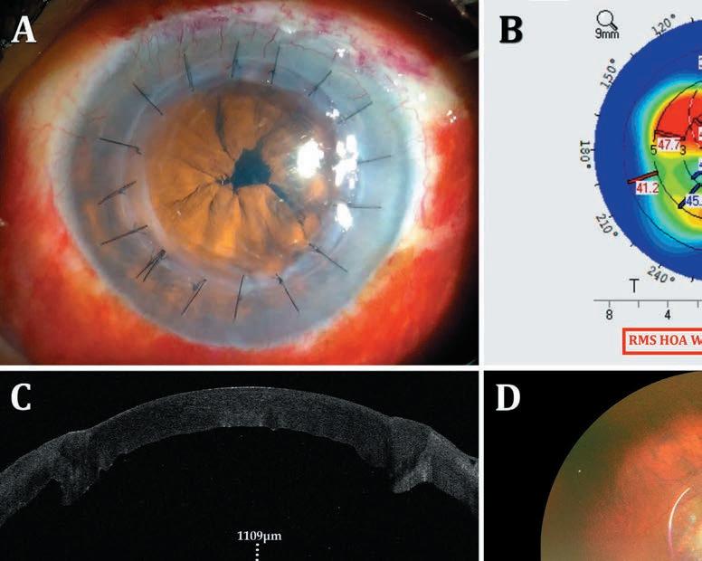
3 2023 MAY | EUROTIMES 22 12 40 Leadership and Business Innovation: Managing Public Ophthalmology 42 Everything You Always Wanted to Know About... Pinhole Optics 44 Industry News 45 JCRS Highlights 46 Index of Citations 48 Upcoming Events Learn more about EuroTimes or connect with ESCRS at ESCRS.org
ALSO IN THIS ISSUE
What’s Wrong with PK?
JOSÉ GÜELL MD, PHD
The Viennese ophthalmologist Eduard Konrad Zirm is credited with performing the first full-thickness penetrating keratoplasty in 1905—the first solid organ transplant of any kind. While the procedure undoubtedly has benefited many, many patients, it is not without drawbacks. Together with the inherent risk of an “open-sky” procedure, some persistent problems with traditional PK include relatively high rejection rates over the long term, suture-related complications, irregular astigmatism, long-term immune suppression, and poor tectonic stability.
PK is still performed—remaining relevant for a small number of indications, no doubt—but has been supplanted by an alphabet soup of lamellar keratoplasty procedures in many cases. Deep anterior lamellar keratoplasty (DALK), Descemet stripping automated endothelial keratoplasty (DSAEK), Descemet membrane endothelial keratoplasty (DMEK), and keratoprostheses (KPRO) are all widely performed.
Endothelial keratoplasty treatments continue to evolve. In this issue, we review some of the new approaches to treating corneal endothelial disease. Our cover article by Dr Soosan Jacob looks at the state of cell-based therapies, stem cell treatments, and bioengineered tissue, which could someday offer safer surgery with more efficient use of scarce cornea donor resources.
In a related article, Cheryl Guttman Krader reports on a debate between Dr Ula V Jurkunas and Dr Massimo Busin on when and if cellular therapy will become the treatment of choice for corneal endothelial disease.
Ensuring safe and timely access to advanced cell and gene therapies requires researchers and industry to navigate a complex maze of government agencies. Professor Mor Dickman
If we knew what we were doing,
would not be called research,
describes new initiatives in this area and discusses the important role independent registries should play in following these treatments once approved.
Disturbing cases of corneal blindness and death associated with contaminated eyedrops have received considerable media attention. Dr Allan R Slomovic tells EuroTimes about this outbreak and associated visual morbidity associated with Pseudomonas keratitis, reminding us of its fulminant nature, potential for a poor visual prognosis, and the need for rapid diagnosis and treatment.
There seem to be no escaping reports of the rise of artificial intelligence (AI) in the news. Will AI-based systems replace (or enhance) the experienced clinician in diagnosing keratoconus and infectious keratitis within the next ten years? Cornea specialists debated this question at a recent conference, reported here.
Finally, we want to congratulate the Union of Ukrainian Ophthalmic Surgeons for holding their annual conference in March, one year after the country was invaded by the Russian military. Slava Ukraini!
José Güell MD, PhD
EDITORIAL BOARD
INTERNATIONAL EDITORIAL BOARD
Noel Alpins (Australia)
Bekir Aslan (Turkey)
Roberto Bellucci (Italy)
Hiroko Bissen-Miyajima (Japan)
John Chang (China)
Béatrice Cochener-Lamard (France)
Oliver Findl (Austria)
Nino Hirnschall (Austria)
Soosan Jacob (India)
Vikentia Katsanevaki (Greece)
Daniel Kook (Germany)
Boris Malyugin (Russia)
Güell Medical Editor
Paul Rosen Medical Editor


Marguerite McDonald (US)
Cyres Mehta (India)
Sorcha Ní Dhubhghaill (Ireland)
Rudy Nuijts (The Netherlands)
Leigh Spielberg (The Netherlands)
Sathish Srinivasan (UK)
Robert Stegmann (South Africa)

Ulf Stenevi (Sweden)
Marie-José Tassignon (Belgium)
Manfred Tetz (Germany)
Carlo Enrico Traverso (Italy)
 Oliver Findl ESCRS President
Thomas Kohnen Chief Medical Editor
EDITORIAL
4
EUROTIMES | MAY 2023
José
it
would it? –Albert Einstein
Hydrophilic Acrylic IOLs
 GERD U AUFFARTH MD, PHD, FEBO AND BEN LAHOOD MD, PHD, MBCHB(DIST), PGDIPOPHTH(DIST), FRANZCO
Previously, this publication asked “IOL Calcification. Are hydrophilic IOLs more trouble than they are worth?”1 This question was addressed by Professor Gerd U Auffarth at the 40th meeting of the ESCRS in September 2022,2 as well as during a podcast with Doctor Ben LaHood in which they discussed the pros and cons of hydrophilic acrylic intraocular lenses (IOLs).3 Altogether the answer was a resounding “No!” According to both surgeons, hydrophilic acrylic IOLs play an important role in optimizing cataract surgery, for the patient and the surgeon.2, 3
Hydrophilic acrylic IOLs have been in use for more than 40 years. Their continued popularity (they account for approximately 29% of IOLs implanted worldwide) is due to characteristics provided by their higher water content and lower refractive index compared to other IOL materials. Usually 18–38% water compared to ≤ 5% for hydrophobic acrylic or silicone IOLs.4
Hydrophilic acrylic IOLs unfold within the eye effortlessly to assume their final position quickly.5 This is especially beneficial when implanting toric IOLs where the slower unfolding or self-adherence seen with some hydrophobic acrylic IOLs can be time-consuming and may lead to rotation if the surgeon is impatient.2, 23 If necessary, rotating or explanting a hydrophilic IOL is easier than with hydrophobic acrylic IOLs due to their greater flexibility. This gives surgeons more confidence to use advanced technology IOLs, as potential problems can be more safely managed. Although hydrophilic acrylic IOLs are highly flexible, with proper haptic design, they resist displacement or rotation as the capsular bag contracts.6 The hydrophilic material is also more resistant to forceps damage or fold marks, which Prof Auffarth noted makes these lenses a good choice when training residents.2
Further, the flexibility and compressibility of hydrophilic acrylic IOLs make them an excellent choice for microincision cataract surgery, minimizing surgically induced astigmatism and improving the predictability of post-surgical unaided visual function.7 As Dr LaHood notes: “If you can minimize the impact on refractive outcomes of factors we can’t currently predict—such as surgically induced astigmatism—then that’s one area where you can improve your overall predictability.”3
Hydrophilic acrylic IOLs are also highly biocompatible, a benefit for patients with uveitis or diabetes.8, 9 In these patients, implantation of hydrophilic acrylic IOLs results in “quieter” eyes and excellent visual outcomes.3
When compared to hydrophobic acrylic IOLs, hydrophilic acrylic IOL implants, with a higher ABBE number, show less light dispersion resulting in minimized chromatic aberration10 and glare.11 Unlike some of the most frequently implanted hydrophobic acrylic IOLs22, hydrophilic acrylic IOLs are significantly less likely to develop glistenings.12 These fluid-filled microvacuoles in hydrophobic acrylic IOLs can scatter light resulting in dysphotopsia, decreased contrast sensitivity, and other photic phenomena that interfere with vision.4, 13–15 In severe cases, they require explantation.13, 16
The impetus for the suggestion that hydrophilic acrylic IOLs be removed from the cataract surgeon’s armamentarium
originated in reports of calcification of hydrophilic acrylic IOLs in patients who underwent procedures using intracameral instillation of air or gas, such as Descemet membrane endothelial keratoplasty, pars plana vitrectomy, or Descemet stripping (automated) endothelial keratoplasty.17 Since surgeons cannot predict perfectly which patients may need keratoplasty or pars plana vitrectomy surgeries in the future, the suggestion was made that surgeons should just stop using hydrophilic acrylic IOLs. Both Professor Auffarth and Dr LaHood disagree with this suggestion and feel the issue of opacification has been blown out of proportion.
We do not yet have a perfect IOL. However, given the advantages of hydrophilic acrylic IOLs, perhaps a less drastic approach than banning them is available. As Dr LaHood says, “We are dealing with a risk of a risk of opacification” so adaptive techniques may be a suitable option for certain eyes at higher risk. Adaptive techniques have been developed that can safeguard IOLs against exposure to air/gas18–20 that may increase the risk of excessive calcium buildup. De Cock and colleagues propose that prior to air/ gas exposure, the anterior chamber should be irrigated with saline left in place for at least 8 minutes. The presence of the saline removes excess calcium ions from the IOL via passive diffusion.18 Ahad and colleagues also suggest minimizing the anterior chamber air fill from 1 hour to 10 minutes when performing endothelial transplant surgery. They identified the rebubbling of the endothelial graft as a major risk factor for opacification.19 Sise and colleagues demonstrated that a modified technique using a reduced volume air bubble (to reduce the total time of contact with the IOL) can significantly reduce IOL opacification even in cases when rebubbling was necessary.20
Further, researchers are developing IOL tests that can help identify a material’s susceptibility to calcification in the eye.21
It is inappropriate to recommend the removal of all hydrophilic IOLs from the surgeons’ armamentarium. It is up to the individual surgeon to determine the appropriate IOL to use for an individual patient, taking into consideration risks, benefits, and their patients’ overall ocular and health conditions. It is also incumbent upon companies manufacturing hydrophilic acrylic IOLs to extensively test their product before entering the marketplace.
For citation notes, see page 46.
5 2023 MAY | EUROTIMES LETTER TO THE EDITOR
Want to comment on an article in EuroTimes? Send your letter to seanh@eurotimes.org.
Moving Simulators to Increase Access to Skills Training
STUART HALES, EXECUTIVE EDITOR
ESCRS will send surgical simulators to countries where young doctors have limited access to hands-on training.
Performing a complex surgical procedure without posing any risk to a patient sounds like the stuff of virtual reality games, but simulators for cataract surgery have been in use for about 15 years and have been shown to significantly enhance surgical skills and reduce complications in early training.
The national ophthalmological societies of some of the larger European countries, such as the UK and France, operate simulators in different locations and allow aspiring ophthalmologists to use them at little or no cost during their training. In some countries, simulator training is mandatory for gaining access to surgical training in clinical settings.
The initial outlay to purchase a simulator to train young ophthalmologists can be quite substantial. Consequently, teaching hospitals in smaller countries and some countries in eastern Europe do not have simulators or are using older models.
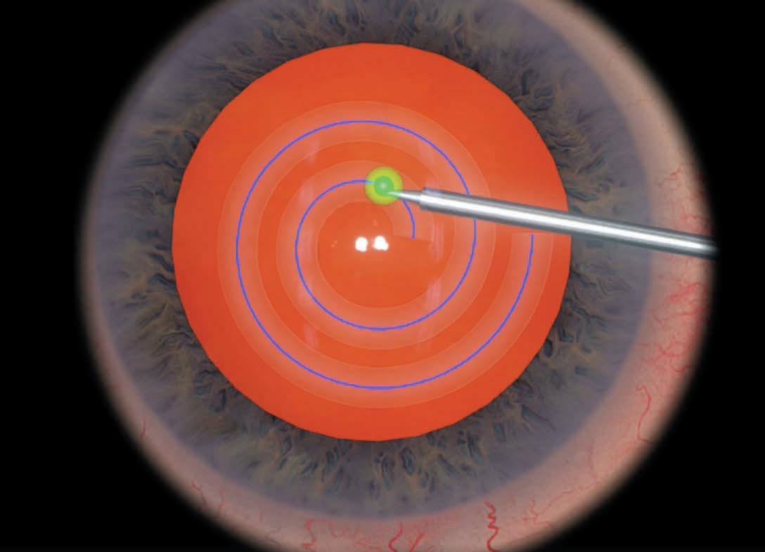
The ESCRS is helping to fill the void by purchasing Eyesi simulators from Haag-Streit and moving them from country to country. The simulators will be hosted by the national society, either in their office rooms or in a centrally located hospital.
“Through this project, ESCRS will intensify its role in providing hands-on skills training, which is of utmost importance to young surgeons,” says Dr Oliver Findl, president of the society. “This will allow them to undergo a larger part of the curriculum on the simulator, which is not possible at our annual Congress due to the restricted time frame.”
Travel logistics
The first simulator is being delivered from Haag-Streit on May 19 and will be shipped to Romania to begin the
training project. As currently envisioned, each simulator will stay in one location for at least four weeks and as many as eight, depending on country size and the number of interested trainees. Ideally, the simulators will travel to six or more countries per year, visiting each country every other year, based on need. The local society will set up the simulator and oversee access.
“We will start with countries that have dual membership with ESCRS, then invite other countries to offer this type of membership, and then move to countries with the most need,” Dr Findl says. “The trainees need to be or become ESCRS members, which is free for the first five years, and watch an instructional video before using the simulator.”
Training curriculum
A customised “ESCRS curriculum” comprising eight hours of instruction is being developed by Dr Artemis Matsou and Dr Alja Crnej. The training will be delivered in two four-hour sessions; during the first session, an instructor will be available for the first 15–20 minutes to help the trainee get oriented and answer possible questions.
“For practical reasons, and in order to provide this fantastic opportunity to as many trainees around Europe as possible, two four-hour sessions were deemed the optimal allocated time for each surgeon,” says Dr Matsou. “The emphasis is on basic cataract skills like anti-tremor tasks, bimanual coordination, and basic phacoemulsification steps, while some modules for more experienced surgeons will also be incorporated.”
Trainees will be able to view their performance scores and compare them to previous scores to see if they
improved their skills. They will also be able to download a record of completion for the various training modules to receive a certificate from the ESCRS verifying their progress.
More important than the certificate, however, is the self-assurance from practising surgical techniques in a secure, low-stress setting.
“The primary advantages of using a simulator, especially early on in surgical training, are that the surgeon has the opportunity to complete all the steps of a cataract procedure in a controlled environment and consolidate their theoretical knowledge of the surgical steps before operating on a real patient,” says Dr Matsou. “It is a great advantage to do all that in a risk-free environment, with permission to fail, which is one of the big sources of anxiety for many trainees at the start of their surgical career. It is a great tool to build confidence and be the safest version of yourself before operating on a real patient.”
6 INSIDE ESCRS EUROTIMES | MAY 2023
ESCRS CONTENT
Through this project, ESCRS will intensify its role in providing hands-on skills training, which is of utmost importance to young surgeons.
The ESCRS training programme for the moving simulators includes modules on anti-tremor techniques, bimanual surgery, and capsulorhexis. Images courtesy of Haag-Streit.


7
2023 MAY | EUROTIMES
27TH ESCRS WINTER MEETING CONGRESS IN NUMBERS
1,311
8 EUROTIMES | MAY 2023 ESCRS AT A GLANCE 2% 1% 86% 3% 8% 0%
69 COUNTRIES COUNTRIES REPRESENTED CONGRESS OVERVIEW
=25 PARTICIPANTS
4 MAIN SYMPOSIA 2 INSTRUCTIONAL COURSES 86 FREE PAPERS 24 WET LABS 21 CME CREDITS 191 E-POSTERS 13 DIDACTIC AND BASIC OPTICS COURSES 4 PORTUGUESE TRACK CORNEA DAY SESSIONS 1 PARALLEL ROOMS 92.5 TOP 10 COUNTRIES 443 105 79 75 60 51 45 35 31 30 PORTUGAL GERMANY UNITED KINGDOM UKRAINE SWITZERLAND BELGIUM NETHERLANDS SPAIN FRANCE ROMANIA
PARTICIPANTS 275 INDUSTRY MEMBERS 720 DOCTORS/HCPS 301 NURSES/TECHNICIANS/TRAINEES 15 ALLIED HEALTHCARE PROFESSIONALS ESCRS CONTENT





2023 MAY | EUROTIMES 9 ESCRS CONTENT
From Ukraine to New Zealand
Ukrainian ophthalmologists continue to function in the midst of the conflict their country continues to endure. The Russian aggression that began in February 2022 happened very close to the annual conference of the Union of Ukrainian Ophthalmic Surgeons. One year later, the organisation again held its meeting, led by chairman Volodymyr Melnyk MD, PhD.
Dr Melnyk said it was essential that the congress be held one year after the hostilities began. He noted such events inspire positive emotions, which are incredibly important for the Ukrainian people.
“I’m very proud of all our speakers and delegates, who found the courage to come to Kyiv from all Ukrainian regions and take part in our event in person. This year, we had more than 400 participants, and it is a great achievement, despite the war and all the complications,” he told EuroTimes
More than 50 Ukrainian speakers reviewed cataract, glaucoma, vitreoretinal, and corneal and refractive surgeries—including many young ophthalmologists.
The programme included online presentations by several ESCRS members, including Professor Thomas Kohnen, who spoke of surface photoablation, Professor Béatrice Cochener-Lamard on lenticular intrastromal surgery, Professor Filomena Ribeiro on the management of cataract after corneal refractive surgery, and Professor Rudy Nuijts discussing cataracts and corneal diseases. In addition, Professor Oliver Findl reviewed managing the unhappy patient, and Professor Dick Burkhard discussed innovations in cataract and MIGS surgery.
“Many Ukrainian ophthalmologists in eastern and southern parts of our country had to leave their cities, close their clinics, and stop working because of the active war,” Dr Melnyk reported. “But, thanks to our armed forces, we conserved and restored our practical activity in the biggest Ukrainian cities with a wide network of ophthalmic care.”
On to New Zealand
The ESCRS has an ongoing programme to support our Ukrainian colleagues that includes free registration to Ukrainian surgeons to attend our annual Congress (more than 400 attended in person in Milan), as well as grants for travel and accommodation. The Society has also arranged to provide observerships and some travel grants for these colleagues, one of which we recently finalised. Dr Iryna Ovchar will visit the Greenlane Clinical Centre in Auckland, New Zealand, hosted by Dr Richard Hart.
“This was very well done, and it is a great result,” said Tom Ogilvie-Graham, Managing Director of the ESCRS. “It will not only be a great experience for Iryna but also sends a signal to our Ukrainian colleagues—and the wider Ukrainian community—that they are not forgotten and, indeed, supported by a country as far away as New Zealand.”
In addition, as part of a long-term project sponsored by ESCRS at the St John Eye Hospital in Jerusalem, two Ukrainian junior doctors will undertake secondments in Jerusalem this June. There will also be four observerships in Oxford over the summer, co-sponsored with Oxford University and arranged by Professor Rob MacLaren.
The observership was made possible in part by donations from our members. As the war unfortunately enters its second year, the ESCRS will endeavour to continue providing targeted support for Ukrainian surgeons.

The Society has established a fund to accept financial donations that will exclusively support ophthalmology-related relief efforts arising from this conflict. We can accept donations to the fund from ESCRS members, industry partners, and fellow societies.
We can accept these donations through bank transfer. Simply log in at https://donate.escrs.org using your membership details to access information on how to donate, which is a straightforward process.
For industry partners or fellow societies, please email escrs@mci-group.com for information on how to make your donation.
10 EUROTIMES | MAY 2023 UKRAINE

11 2023 MONTH | EUROTIMES NOW OPEN! Applications for the Peter
Fellowship 2023 are The Fellowship of €60,000 will allow a trainee to work abroad at a centre of excellence for clinical experience or research in the field of cataract and refractive surgery, anywhere in the world, for 1 year. The application deadline is 2 May 2023. For more information: escrs.org/education/grants-awards/peter-barry-fellowship
Barry
Ensuring Safe and Timely Access to Advanced Cell and Gene Therapies
 DERMOT MCGRATH REPORTS
The development of advanced therapy medicinal products (ATMPs) for treating eye diseases has become a rapidly expanding field in recent years. Based on genes, tissues, or cells, ATMPs have the potential to revolutionize the treatment of a wide range of ocular conditions such as age-related macular degeneration, retinitis pigmentosa, Leber’s congenital amaurosis, Stargardt’s disease, optic nerve pathology, and limbal stem cell deficiency, among others.
With the number of approved ATMPs expected to increase, the legislative, regulatory, and access frameworks need to be sufficiently flexible and robust to ensure that patients across Europe have equitable access to these potentially life-changing treatments.
With this in mind, the European Alliance for Transformative Therapies (TRANSFORM) recently launched an MEP Charter in the European Parliament. The Charter presents seven main policy recommendations to promote safe and timely access to ATMPs while ensuring Europe remains attractive for investment and promoting healthcare systems’ sustainability.
The Charter launch included several roundtable discussions with TRANSFORM Alliance and MEP Interest Group members. Speaking at the panel discussion on new approaches to foster safe and timely patient access to ATMPs in Europe, Professor Mor Dickman of Maastricht University, Netherlands, and representative of the European Alliance for Vision Research and Ophthalmology (EU EYE), said it was a particularly exciting time to work as an ophthalmologist.
“We have some of the best scientists in the world working in Europe, and we have seen some very exciting developments in recent years in the field of ophthalmology,” he said. “We now have the first cell-based therapy with marketing approval to treat corneal blindness and the first gene therapy to treat inherited retinal disease with marketing approval. This is exciting for us as clinicians, but first and foremost, for patients who can experience life-changing therapies.”
Fragmented landscape
Prof Dickman said it was important for Europe to draw on the experience of such breakthrough treatments to improve the process of taking ATMPs from the lab to the bedside.
“This is a very fragmented landscape, and there is a clear need for harmonization, even though health is the prerogative of the European member states. What happens, for example, if an ATMP is approved, patients start benefiting from it, and then the company decides to abandon it?” he observed. “It has happened in the past and will undoubtedly occur again. One possible solution might be to apply the principle of hospital exemption (HE), which allows for ATMP use without marketing authorization under certain strictly controlled circumstances. The COVID-19 pandemic has really made clear how important it is that Europe becomes self-sufficient.”
Another key issue relates to data collection both within and across the continent’s borders, which Prof Dickman said is very complex due to the number of partners involved.
GLOBAL 12 EUROTIMES | MAY 2023
Non Contact Tono/Pachymeter
“What are the standard data sets, what are the outcome measures, and how do we collect them considering the issues of interoperability? How do we find funding for registries? And if one does receive funding, how do we ensure the sustainability of these registries once the funding is over? There is a need to impose data collection in a way that collects long-term data and captures patient-reported outcome measures (PROMs) in an independent platform in collaboration with the industry,” he said.
Lessons from COVID
The recent COVID-19 pandemic, Prof Dickman said, has also demonstrated the benefit of international collaboration in working towards a common goal.
“COVID showed how regulatory authorities throughout the world could come together and approve a vaccine in a historically short time. So, there should be room for ATMPs as well,” he said. “Someone earlier mentioned the FDA as our competitor, whereas I think they are our partner. We need to work together and harmonize our processes with the United States to make sure there is one pathway to the patient.”
Registries can play an important role in helping patients gain access to groundbreaking new treatments, Prof Dickman said, citing the Dutch RD5000 registry for patients with inherited retinal diseases (IRDs).
“Anticipating that more and more advanced therapies will become available soon, we’ve set up a registry where all these patients with IRDs in the Netherlands are registered,” he said. “It essentially means we are building an infrastruc ture in how we can reach these patients as and when a new treatment becomes available. I think if we had multinational registries or European registries with similar information, we could reach our target populations much more quickly.”
Another advantage is how they foster collaboration and enable benchmarking.
“One of the major benefits is they create networks where people learn from each other, share their experi ences, and improve,” he added. “For surgeons, it allows benchmarking so you can compare your performance to others and see that what you are doing adjusted to the pa tients that you are treating is delivering the same results.”
New Design Innovations that Incorporate Operator and Patient Comfort with Gentle Measurements
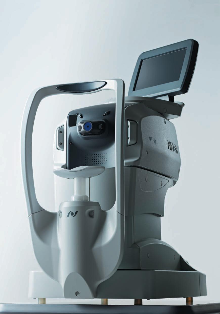
• Fully-automatic measurement
• Gentle voice guidance (available in 9 languages)
• Reliable tono/pachymeter
• Flexible and space-saving design
• A variety of options to meet your needs
Mor M Dickman MD, PhD is professor of ophthalmology at the University Eye Clinic, Maastricht UMC, Netherlands. m.dickman@maastrichtuniversity.nl
Prof Dickman’s work with the European Parliament is part of the greater efforts of EU EYE to increase awareness at the EU level about the research and policy needs in ophthalmology. This work is continuously supported by Dr Ioanna Psalti, who develops and finely tunes the communication and visibility strategy for EU EYE.

www.nidek.com ET 93 x 266mm European
Vision Research
Ophthalmology Help us to build a healthier Europe! Go to
or send us an email at info@eye.org to learn
our
Alliance for
and
https://www.eueye.org
about
work with the European Commission and the European Medicines Agency.
What lies ahead for corneal endothelial disease treatment?

EUROTIMES | MAY 2023 COVER ARTICLE 14
Currently, eye banks can meet only 1.5% of the global keratoplasty demand for donor corneas. Researchers around the world have taken this as a challenge to develop innovative approaches that could help make the most of the existing supply of donor corneas, even someday growing tissue on demand.
Though endothelial keratoplasty for Fuchs’ endothelial corneal dystrophy (FECD) and pseudophakic bullous keratopathy has become increasingly successful, the basic prerequisite is availability of good quality donor corneas. New keratoplasty techniques under investigation that minimise the need for a donor cornea include Descemet stripping only (DSO) and Descemet membrane transplantation (DMT).





DSO does away with the transplant altogether, waiting for the patient’s endothelial cells to migrate into place. With DSO, the central 4 mm of Descemet’s membrane is removed to allow central migration of healthy peripheral cells for central oedema clearance over the next three to four months. DMT transplants a decellularized DM following the central stripping.
DSO and DMT are indicated only in FECD with mild central oedema or central guttae and sufficient peripheral endothelial cell reserve (> 1400 cells/ mm2). Rough Descemet membrane edges and stromal scoring interfere with central endothelial migration and should be avoided. Rho-kinase (ROCK) inhibitors can augment cell migration.
Patients considered for these procedures need preoperative counselling
regarding the slow clearance of oedema and the possible need for EK in the eventuality of non-clearance by four months after surgery. Long-term steroids and rejection are avoided in DMT since the decellularized Descemet membrane is non-immunogenic. Non-optical-grade corneas can be used in DMT. The reliance on high-quality donor cornea is removed both for DMT and DSO procedures.
ROCK inhibitors impede apoptosis and promote endothelial cell proliferation, adhesion, and migration. They play a role in many of the new treatments for endothelial disease and are thought to enhance the results of endothelial keratoplasty, DSO, and DMT. They are also used in cell therapy cultivation methods.
Corneal endothelial cell therapy
Corneal tissue engineering aims to overcome donor tissue shortages and eliminate immune rejection. The ideal cell therapy would be non-toxic, immunologically compatible, genetically stable, free of transmissible diseases, and have long-term, stable functionality and survival. Important steps in cell therapy are tissue removal, isolation, differentiation, and cultivation in vitro, followed by implantation into the patient.

15 2023 MAY | EUROTIMES
DR SOOSAN JACOB REPORTS
Descemet’s stripping only: A: Descemetorhexis performed along inked 4-mm circle; B: Completed Descemetorhexis; C: Localized overlying corneal oedema postoperatively; D: Three months postoperative, Descemetorhexis edges seen on iris retroillumination. Corneal clearance seen (inset).
The challenges of this therapy include difficulty in obtaining a monolayer of cells with flat hexagonal morphology, programmed cell cycle arrest, avoiding morphological deviations, fibroblastic contamination, premature cellular senescence, and the need for a biocompatible carrier.
Human corneal endothelial cells (CEnC) possess limited proliferative ability within the eye since the cells are arrested in the quiescent G1 stage of the cell cycle. However, they may be induced to grow in vitro. Cell lines are obtained either from corneal endothelial stem cells or direct expansion. Descemet’s membrane is peeled from a corneal graft and digested using enzymes. The remaining cells are then cultured in vitro. Challenges include the risk of allogenic graft rejection and the limited regenerative capacity of donor cells.
Autologous adult stem cells under research include skin-derived precursors (SKP), mesenchymal stem cells (MSCs), peripheral blood monocyte cells (PBMCs), induced pluripotent stem cells (iPSCs), and even fibroblasts. MSCs are readily available in adipose tissue, bone marrow, and skeletal muscle. Ethical issues are few, and the risk of tumorigenesis is low. Disadvantages of stem cell therapy include variable differentiation capacity and differences in morphology.
Kinoshita’s landmark research
Following decades of research, Professor Shigeru Kinoshita and colleagues reported success in culturing corneal endothelial cells and implanting them in patients. This opens the door to taking a donor cornea, stripping out corneal endothelial cells, and growing it in the lab, thereby showing real potential for treating multiple patients from one donor cornea.
In the only published trial of endothelial cell therapy in humans, Prof Kinoshita and colleagues report that
11 bullous keratopathy patients received intracameral injections of in-vitro expanded CEnCs treated with a ROCK inhibitor, followed by prone positioning for 3 hours to achieve attachment to recipient corneas.1 After 24 weeks, corneal oedema regressed and CEnC density exceeded 500 cells per square millimetre (range 947 to 2833) in all eyes. Vision improved by two lines or more in 9 of the 11 patients, and the results were stable for up to 24 months.

Caveats include the inability to rule out the possibility of unattached CEnCs seeding systemically via the trabecular meshwork with subsequent tumorigenesis, a potential safety issue.
The techniques developed by Prof Kinoshita have been licenced to two companies—Aurion Biotech in the US and ActualEyes in Japan—with clinical trials underway. Proponents of this approach believe it may soon be possible to treat 50 patients or more from a single donor cornea.
An intact DM is essential for cell injection therapy. It is, therefore, preferable in early disease. Cell delivery systems such as superparamagnetic embedding and iron endocytosed CEnCs, are under investigation to increase adhesion. One novel approach, EO2002 (Emmecell), uses a magnetic cell delivery nanoparticle platform and a magnetic patch over the closed eyelid for a few hours after intracameral injection of CEnCs.
Tissue engineering and beyond
Tissue-engineered endothelial keratoplasty (TE-EK) is another approach under development. In this procedure, cells are cultured and differentiated onto supports, which are then implanted into the patient’s eye using standard endothelial keratoplasty techniques. The ideal support should be easily implantable, biocompatible, and biodegradable. Support materials tried thus far include ultrathin human stromal lamellae, anterior lens capsule, amniotic membrane, DM, and various biomimetic scaffolds of natural (collagen, gelatin) or synthetic (polyvinyl alcohol, polyethylene glycol) materials.
TE-EK is preferable in advanced disease when the recipient DM is scarred and needs removal. Large guttae are toxic to injected cells, and DM removal is indicated, again making TE-EK preferable.
16 COVER ARTICLE EUROTIMES | MAY 2023
Disadvantages of stem cell therapy include variable differentiation capacity and differences in morphology.
EndoArt (EyeYon Medical), a 50-micron artificial lamella positioned similarly to EK, deturgesces the cornea by acting as a fluid barrier, helping to avoid long-term steroids and rejection. There are also no concerns about damaging the graft or disease transmission. The device is approved in the US and Europe in patients with corneal oedema following graft rejection or those at risk of graft rejection.
Gene therapy offers the promise of a long-term cure. The cornea is ideal for gene therapy because of easy


access and immune privilege. Gene therapy’s previous use improved allograft survival by transducing the donor cornea before transplantation and inducing regression of host corneal neovascularization. It is also being tried in eye bank corneas to maintain endothelial cell density, thus allowing long-term corneal preservation and making donor corneas with low cell density viable for transplantation. Identified biomarkers may serve as potential therapeutic targets for treating FECD.
DMEK SETS THE BAR HIGH
MOR DICKMAN MD, PHD
There are many reasons why we try to avoid penetrating keratoplasty today. One is the notion PK is a very safe procedure with very little rejection, but this is not the case. Nearly half of the people undergoing PK will have an episode of rejection within 10 years of their transplant. Other issues involve suture-related complications— such as irregular astigmatism requiring contact lenses in a significant number of patients—and a risk to the integrity of the globe. Transplant dehiscence can occur even decades after surgery.

PK recipients also face the ongoing risk of infection, along with the need for lifelong immune suppression and those associated risks. Perhaps most important, especially in young patients with strong immune systems, the risk of rejection is higher. Those patients will likely need a second or a third transplant. We know the results of a second or third PK are worse than the primary transplant and, in some cases, might require systemic immune suppression. Indeed, in ECCTR, we have shown that repeated transplants are the second leading indication for corneal transplantation in Europe.
Fortunately, lamellar keratoplasty techniques have largely supplanted traditional PK in most of the western world. Performing PK when lamellar surgery is possible is a shame because patients could benefit from the new-
er techniques. In my institution, we have changed completely from PK to endothelial keratoplasty for diseases involving the corneal endothelium.
DMEK is our most common transplant technique, which accounts for 70% of transplants in the Netherlands and around 50% worldwide. In very difficult cases, such as when the endothelium needs replacing, we use ultrathin DSAEK. DALK is a wonderful solution for diseases involving the stroma.
We do DMEK for all standard cases, as outcomes are so good, its high bar is difficult to improve. DMEK has less than a 5% rejection over five years. Issues with DMEK include the shortage of donor tissue, but this is a problem for all transplant methods. Other issues include graft dislocation, seen in unplanned surgery in 10–20% of cases. Regarding whether DMEK patients require lifelong immune suppression, our randomised controlled clinical study, OPTIMISE, stops immune suppression after the second postoperative year. We think if surgeons carefully and continuously monitor the patients, they may not need lifelong immune suppression.
New techniques may allow more efficient use of donor tissue. If opting for a lamellar technique, the surgeon could use the endothelium to treat one or more patients and the stroma to treat another. DSO in combination
Gene therapy has been used in mouse models to treat early-onset FECD. Viral and non-viral vectors and techniques to introduce genes into the cornea are being researched, showing how gene editing and CRISPR technologies may offer breakthroughs in FECD.
For citation notes, see page 46.
with ROCK inhibitors offers a tissue-free alternative for select cases, but recovery is long and unpredictable. EndoArt, a synthetic implant that does not contain cells, is another interesting alternative for treating corneal oedema. While initial results are promising, understanding the longterm effects remains necessary.
There are some astonishing developments in this field from around the world. DALK, DSEK, and DMEK came from the Netherlands, regenerative therapies for corneal endothelium from Japan and Singapore, and regenerative therapies for corneal epithelium from Italy. However, no one has yet replicated the clinical grade Japanese process; Until now, only very young donor corneas have been used, the authorisation regime is extensive, and equivalent or superior results to DMEK are needed. Using single-cell RNA sequencing, we have demonstrated the changes that endothelial cells undergo in culture, and the overall yield is limited. Stem cell-derived corneal endothelial cell replacement is a promising alternative because young donors are not needed (and there is virtually no limit on cell numbers), but we must show safety and efficacy. Compared to the US and Japan, dedicated funding for ophthalmic research is limited in Europe. Imagine what would happen with more funding!
17 2023 MAY | EUROTIMES
Avoiding Pitfalls in IOL Power Calculations
IOL power calculations after previous corneal refractive surgery (CRS) may remain challenging for ophthalmologists despite the availability of advanced measurement technologies and new formulas, according to Professor Basak Bostanci.
“Today, with the help of many new formulas, novel devices, and artificial intelligence, the refractive results we obtain in post-CRS patients are satisfactory,” she said. “Having said that, I still recommend using as many variables and formulas as possible and comparing the predicted results. The final decision should be made on clinical judgment, depending on your expertise. But if you do not have special equipment or the clinical history data of the patient, then the Barrett True-K No-History formula may be a good fallback option.”
IOL power is usually calculated by measuring several biometric parameters—notably axial length (AL) and corneal refractive power—and estimating the effective lens position (ELP).
“This is not difficult in a standard virgin eye, but since we make many alterations to the cornea in refractive surgery, those can be sources of biometric error,” she added.
The main sources of biometric error after CRS are the miscalculation of the refractive index of the cornea, radius error, and formula error.
When calculating corneal power with keratometry and topography, the keratometric refractive index of the cornea serves as a constant value, but this is not the case after CRS.
“Because we change the ratio between the anterior and posterior surfaces of the cornea with CRS, we are actually changing this value,” Prof Bostanci explained. “So, after myopic ablation, we estimate the corneal power as higher. And after hyperopic ablation, the corneal power is estimated as lower. As the refractive correction increases, the error also increases.”
The radius error derives from the fact that keratometry and Placido-based topography devices measure the paracentral area of the cornea and then extrapolate the central refractive power from this measurement.
“This leads to an overestimation after myopic LASIK and a hyperopic surprise and an underestimation after hyperopic LASIK and a myopic surprise,” she said.
Third-generation IOL power formulas such as Hoffer Q, Holladay I, and SRK/T, Prof Bostanci explained, use corneal power to generate ELP, leading to its underestimation and a false low IOL calculation after myopic LASIK. This creates an even greater hyperopic surprise, whereas the opposite is true for hyperopic LASIK which results in a myopic surprise.
18 EUROTIMES | MAY 2023
DERMOT MCGRATH REPORTS
Cataract surgeons should know the best approaches in biometry to obtain emmetropic outcomes for post-refractive surgery patients.
CATARACT & REFRACTIVE
She recommended employing a formula such as Haigis L or Shammas to overcome this problem since they do not use corneal power to infer ELP.
“We can also perform a correction using the Aramberri Double K formula, which includes the pre-LASIK K value for calculating ELP and a post-LASIK K value to calculate the IOL power,” she said.
Surgeons need to keep in mind the other challenges with post-CRS eyes, Prof Bostanci said.

“These are eyes that are longer or shorter than the normal range, so they are prone to biometric errors. In addition, they may also have chronic ocular surface problems from dry eye or topographic irregularities caused by CRS, which need to be addressed before biometry,” she explained. “Furthermore, these patients were motivated to pay money and take risks to obtain spectacle-free vision with their original CRS surgery, so it is important to obtain an emmetropic solution, or they will not be satisfied.”
For eyes that previously underwent radial keratotomy (RK) and for which the pre-RK refraction is known, she suggested using the Barrett True-K history method with biometry data from the IOLMaster 500 or 700 (Carl Zeiss Meditec). If the pre-RK refraction is not known, she advised applying the no-history Barrett True-K formula, as it has shown superior to Potvin-Hill, SRK/T, Hoffer Q, and Holladay formulas, among others. Other alternatives that have shown non-inferiority to the no-history Barrett True include the Haigis L formula, newer formulas based on OCT or intraoperative aberrometry, and the ASCRS calculator average, she said.
After LASIK and PRK, both versions of the Barrett True-K (history and no history) are now the standard for IOL power prediction with vergence formulas for post-myopic and post-hyperopic eyes—although Haigis-L is also useful for myopic eyes. Using the Shammas formula may result in a slight hyperopic shift, while OCT-based ones may induce a slight myopic shift. Prof Bostanci added intraoperative aberrometry formulas or the ASCRS calculator also produce good results.
Although there is insufficient empirical data on IOL power prediction accuracy after SMILE, she concluded some studies showed good outcomes with both versions of the Barrett True-K and Masket formulas. Haigis-L showed a tendency for myopic shift, but Okulix ray tracing based on Pentacam HR and IOLMaster 500 biometry also showed potential.
Dr Bostanci gave this presentation as part of a recent ESCRS eConnect Webinar.
Basak Bostanci MD, FEBO is an Associate Professor of Ophthalmology at Bahcesehir University School of Medicine, Istanbul, and a cataract and refractive surgeon at World Eye Hospital, Istanbul, Turkey. drbbostanci@gmail.com
Learn
19 2023 MAY | EUROTIMES
To be the leading community and trusted source for science, education, and professional development in the fields of cataract and refractive surgery.
more at ESCRS.org
Latest ESCRS Clinical Trends Survey
Cost to the patient, concerns over contrast visual acuity, and night-time quality of vision are the main reasons cited for not performing more presbyopia IOL procedures, suggest the data from the latest ESCRS Clinical Trends Survey.

Professor Rudy MMA Nuijts, University Eye Clinic Maastricht, Netherlands, presented the latest survey results at an ESCRS IME symposium on the opening day of the ESCRS Winter Meeting in Vilamoura, Portugal.
More than 1,700 delegates responded to 146 survey questions during the 40th ESCRS Annual Congress Milan, the eighth year this survey was conducted.
Eleven percent of respondents were implanting presbyopia-correcting IOLs, the same percentage as the previous year. Similarly, trifocal IOLs accounted for the same percentage as the previous year, 50% of cases. Extended depth of focus IOLs saw a small increase to 33% from 28%.
Accommodating and bifocal IOLs were used in 2% and 4% of cases, respectively.
Regarding their biggest concerns about performing more presbyopia-correcting IOL procedures, members cited cost to patient, concerns over contrast visual acuity, and night-time quality of vision. Twenty percent also expressed concerns about inadequate unaided near-vision results.
Since 2016 there has been an 8% increase in cataract procedures using toric IOLs. The survey showed no clear consensus on the best approach in cases where the cylinder was 1.25 D or less, with many surgeons preferring on-axis incisions. However, toric IOLS were the clear favourite in cases of astigmatism of at least 1.75 D.
Cost remains the leading concern limiting the use of toric IOLs. If cost were not an issue, 37% of cataract patients with clinically significant astigmatism would receive a toric IOL, Prof Nuijts noted.
Since 2016, there has been an 0.08% increase of cataract procedures involving
If cost were not an issue, 37% of cataract patients with clinically significant astigmatism would receive a toric IOL.
20 EUROTIMES | MAY 2023
7% 7% 11% 9% 12% 8% 14% 9% 14% 8% 15% 11% 15% 11% Toric IOLs Presbyopia-correcting IOLs 2016 2017 2018 2019 2020 2021 2022
toric IOLs.
CATARACT & REFRACTIVE
What percentage of your CURRENT cataract procedures involve presbyopia-correcting or toric IOLs in qualified patients?
Presbyopia IOL use steady, but obstacles remain.
The survey comes on the heels of a JCRS review of data gleaned from the previous ESCRS surveys.1
“This current ESCRS survey reports on trends in our field over time and demonstrates nicely how our subspecialty continues to change,” said Professor Thomas Kohnen in an accompanying editorial. “Research in cataract and refractive surgery keeps moving us forward to evolve our practices for the benefit of our patients.”
Postoperative success with toric IOLs, he added, is dependent on placement accuracy and rotational stability: “The introduction of digital image guidance systems has improved the former; however, the latter varies across different toric IOL models, and ASCRS and ESCRS delegates are variably stringent with acceptable thresholds of postoperative rotational error,” he said. “Comparing ESCRS and ASCRS respondents, 16.0% and 9.4% are comfortable with ≥10 degrees, compared with >65.0% and 53.8%, respectively,
agreeing visual quality and acuity would be significantly affected by ≥5 degrees of rotational error.
“Surveys such as these are important tools to evaluate how real-world practice patterns align with recommended evidence-based standards of care and highlight areas where patient care can be enhanced through the adoption of new technologies and/or surgical approaches.”

For citation notes, see page 46.
The ninth ESCRS Clinical Trends Survey will be conducted in September 2023 at the ESCRS Annual Congress in Vienna.
What type of presbyopia-correcting IOL technology is used in the majority of your presbyopia correction patients?
21 2023 MAY | EUROTIMES
2020 Survey 2021 Survey 2022 Survey Extended depth of focus IOLs 32% Extended depth of focus IOLs 28% Extended depth of focus IOLs 33% Accom. IOLs 3% Other 4% 2% 2% 2% 2% 6% 4% Bifocal IOLs 8% Trifocal IOLs 53% Trifocal IOLs 51% Trifocal IOLs 50% Enhanced monofocal IOLs 11% Enhanced monofocal IOLs 8%
Rudy MMA Nuijts MD, PhD is professor of ophthalmology, vice-chairman, and director of the Cornea Clinic and the Center for Refractive Surgery at the University Eye Clinic Maastricht, Netherlands.
Thomas Kohnen MD, PhD, FEBO is professor and chair, Department of Ophthalmology, Goethe University, Frankfurt, Germany.
This current ESCRS survey reports on trends in our field over time and demonstrates nicely how our subspecialty continues to change.
Knowing Lens Tilt
Assessing physiological lens tilt can help improve post-surgical outcomes.

 DERMOT MCGRATH REPORTS
Preoperative crystalline lens tilt varies significantly between patients—and in a small but relevant number of patients, the tilt values exceed a threshold that may negatively impact the postoperative refractive outcome, according to Dr Theresa Höftberger.
“Our study showed the lens tilt of a certain percentage of patients exceeds the critical level of 7 degrees. It could be relevant to detect these cases and include the predicted postoperative tilt in the IOL power calculation,” she said. “Including the tilt prediction in the IOL power calculation would help to improve long-term post-surgical outcomes.”
Although modern imaging devices typically provide detailed information about phakic intraocular lens tilt, little data currently exists on the values that influence crystalline lens tilt. Misalignment of intraocular lenses, such as tilt, commonly causes visual deterioration and negatively affects optical performance after cataract surgery.
8.3%
Dr Höftberger said the physiological tilt of the phakic eye showed a normal distribution, with a phakic tilt above 7 degrees in 8.3% of all cases.
said. “This differs from a recent study by Li [et al.] that found a statistically significant difference of the crystalline lens among several age groups, especially with a lower physiological tilt in the age group from 18 to 40 years.”1
In the physiological tilt distribution of right and left eyes, the researchers found a mean tilt of 4.9 degrees with an inferior temporal tilting and a mirror symmetry of tilt. Other significant parameters affecting the tilt—such as axial length, decentration, Chang-Waring chord, and lens equator thickness—showed an area under the curve (AUC) value of 0.9. These parameters were then entered in a random forest plot machine learning algorithm, resulting in an out-of-bag error rate of 0.1, showing tilt can be predicted with good accuracy.
Dr Höftberger gave this presentation at the 27th ESCRS Winter Meeting in Vilamoura, Portugal.
For citation notes, see page 46.
“Previous studies already showed there is a significant correlation between the physiological tilt and the IOL tilt after cataract surgery. In 2017, we already reported that preoperatively measured lens tilt is useful for predicting the postoperative tilt,” Dr Höftberger explained. “The IOL tilt is a very important source of error in the IOL power calculation. Therefore, including preoperative measurements would help to obtain better outcomes and improve long-term post-surgical outcomes in patients receiving the IOL.”
Dr Höftberger’s study included data from 4,731 phakic eyes of 2,531 patients collected between June 2020 and October 2022 at the Kepler University Clinic in Linz, Austria. Patients scheduled for preoperative assessments were biometrically measured during the preliminary examination using the IOLMaster 700 (Zeiss) and the Casia2 (Tomey) swept source OCT devices. The IOLMaster 700 measured the axial length, decentration, central lens thickness, and the lens equator thickness, while the Casia2 obtained the IOL tilt and physiological tilt—defined as the angle between the optic axis of crystalline lens and the cornea’s vertex normal.
Discussing the results, Dr Höftberger said the physiological tilt of the phakic eye showed a normal distribution, with a phakic tilt above 7 degrees in 8.3% of all cases.
“We found no statistically significant difference between females and males, nor a significant correlation with age,” she
22 CATARACT & REFRACTIVE EUROTIMES | MAY 2023
Theresa Höftberger PhD is a clinician and researcher at the Kepler University Clinic, Linz, Austria. theresa.hoeftberger@kepleruniklinikum.at
We found no statistically significant difference between females and males.
2023 ESCRS Pioneer Award
True innovation comes from good research.
The ESCRS Pioneer Award is an initiative sponsored by the ESCRS to support and encourage independent clinical research in the field of cataract and refractive surgery.
The competition is open to young ophthalmologists up to the age of 40 with at least three years of ESCRS membership holding a full-time clinical/research post at an EU-based clinical or academic centre.
https://www.escrs.org/education/grants-awards/pioneer-research-awards/

23 2023 MONTH | EUROTIMES
The Clinician or AI— Who Will Diagnose?
Will artificial intelligence-based systems replace the experienced clinician in diagnosing keratoconus and infectious keratitis within the next 10 years? Cornea specialists debated this question at the American Academy of Ophthalmology annual meeting.
To support her conclusion that technology-based, clinician-support software will replace today’s clinicians alone, Dr Maria A Woodward described the shortcomings of today’s clinicians in delivering quality care and introduced opportunities through AI.
“A clinician is someone who triages, diagnoses, and manages disease and listens to, communicates with, and cares for the sick. But do we effectively diagnose and manage keratoconus and corneal ulcers? Are we being compassionate?” she asked.
Citing a study her group conducted, Dr Woodward said almost 50% of clinicians lacked confidence in their ability to differentiate organism types when diagnosing microbial keratitis.

“In contrast, AI-based algorithms could distinguish between organism categories and will only get better over the next decade.”
Similarly, she questioned whether clinicians effectively diagnose and manage keratoconus and suggested advantages for AI-based strategies.
“Do we know how frequently to see patients, which are at risk for progression, should get cross-linking, and should go on to transplantation? Now there is a lot of good evidence that AI algorithms can help distinguish between keratoconus subtypes and help us predict which patients should go on to get cross-linking or a transplant.”
These are the situations where Dr Woodward proposed AI could enhance clinician ability to provide compassionate quality care.
“How much time do we spend talking to our patients and interacting with them as human beings? There is plenty of data showing how we could be delivering better care, reducing our medical error rate, and reducing our burnout [with] compassion,” she concluded. “By leveraging technology for improving how we triage, diagnose, and manage disease, we can connect more personally with our patients. Technology is not the enemy—it is up to us to drive technology-aided care.”
Not the sole solution
Dr Stephen D Klyce assured his colleagues AI would not replace them in diagnosing keratoconus and microbial keratitis. Those tasks, he said, require more than machine-based analysis of corneal images because the original AI-based keratoconus screening program provided only an interpretation of topographic images, not a diagnosis.
“AI can identify the corneal topography has the characteristics associated with clinical keratoconus but is insufficient by itself to make a diagnosis,” Dr Klyce asserted.
Newer systems—including scanning devices that give information on anterior and posterior corneal surface thickness, corneal biomechanics, and epithelial thinning patterns— provide additional information to help determine whether a cornea has keratoconus or if a patient is at risk for ectasia after laser refractive surgery. Even if clinicians had the physical
24 EUROTIMES | MAY 2023 CORNEA
CHERYL GUTTMAN KRADER REPORTS
Debate seeks common ground on the value of a combined approach to corneal disease diagnosis.
space and financial resources to install all the devices used to acquire those measurements, the collective data still do not provide a clinical diagnosis.
But do we effectively diagnose and manage keratoconus and corneal ulcers? Are we being compassionate?
“You need to know the patient’s history,” Dr Klyce said, explaining that a patient who appears to have forme fruste keratoconus based on AI-based interpretation of images could instead have contact lens-related corneal warpage.
Regarding AI-based diagnosis of microbial keratitis, Dr Klyce noted some models are effective in classifying images, though they are not completely accurate.
Citing a study showing that an AI-based algorithm for diagnosing infectious keratitis based on clinical image classification had an 80% accuracy rate compared to just 49% for ophthalmologists, Dr Klyce focused on the algorithm’s error rate.
“What about the 20% of patients given the wrong therapy because of the algorithm’s mischaracterisation of the keratitis? The consequences could be disastrous.”

Describing other limitations of AI-based systems, Dr Klyce said they are only as good as the training data, whereas clinicians have access to many data sources beyond those available and used to train AI. In addition, AI algorithms need constant revision as technology advances. Even with expert consensus, there will be bias because independent groups have different ideas about the characteristics considered for an AI algorithm.
“AI will not replace the clinician but will continue to provide access to new tools to aid in clinical diagnosis,” Dr Klyce said in closing. “Clinicians and their patients will benefit from advances in the diagnostic capabilities afforded by the enormous strides in digital healthcare. The future will be AI and human intelligence working together.”
OCULUS Myopia Master ® For early myopia detection and management
The Myopia Master® combines all important parameters for myopia management in a myopia analysis software: axial length, refraction values and the central corneal radii.
New GRAS Module: All individually measured refractive components of the eye are automatically matched with the Gullstrand standard eye model.
 Maria A Woodward MD, MSc is Service Chief, Cornea, External Disease, and Refractive Surgery, University of Michigan, Ann Arbor, US. mariawoo@med.umich.edu
Stephen D Klyce PhD, FARVO is an adjunct professor of ophthalmology at the Icahn School of


Medicine at Mount Sinai, New York, US. sklyce@klyce.com
Myopia management –much easier and more reliable than ever!


myopia-master.com

25 2023 MAY | EUROTIMES
The availability of products and features may vary by country. OCULUS reserves the right to change product specifications and design. Eurotimes Myopia Master International 4er Kollage 93x266 e 4c 04.23 v2.indd 1 06.04.2023 18:41:45
Cell Therapy for Corneal Endothelial Dysfunction
Experts agree on the potential benefits of implanting ex vivo expanded endothelial cells, but how soon will it become a clinical strategy?
CHERYL GUTTMAN KRADER REPORTS
Significant progress has been made in corneal endothelial dysfunction management thanks to advances in selective endothelial keratoplasty (EK). However, two significant challenges remain—a shortage of donor tissue in many areas of the world and a need for strategies to increase long-term graft survival considering the risk of graft failure increases with time while patients can live for decades after EK.
Researchers are exploring cell therapy as an alternative approach to address these issues. Cornea specialists presented differing views on whether transplantation of ex vivo allogeneic corneal endothelial cells will become
the standard for managing endothelial cell loss during a special session at the American Academy of Ophthalmology annual meeting.
Dr Ula V Jurkunas explained why this technique will replace EK in the future.
“It improves tissue accessibility worldwide and might be cost-saving,” she said. “Compared to EK, it can also give better quality control and outcome predictability, be safer, and give control of cell product dose.”
Highlighting the need for increased tissue accessibility, Dr Jurkunas cited published data estimating that 12.7 million people are waiting for a corneal transplant worldwide.

CORNEA 26 EUROTIMES | MAY 2023
Fuchs’ dystrophy is the leading indication for keratoplasty, underscoring the need for constructs to treat endothelial cell dysfunction. Since the expansion of cells obtained from one donor cornea might allow for treating numerous recipients, there is potential for both improved access to treatment and cost savings.
“Currently, one donor cornea equals one transplant,” Dr Jurkunas observed. “With many passages of cultured cells, however, one donor cornea could potentially be used for treating 30 eyes or even 100.”
As another benefit of a cell therapy approach using ex vivo expanded allogeneic endothelial cells, Dr Jurkunas said sorting using cell markers could allow preselection of optimally functioning cell populations. Cultured expanded cells can also be subjected to additional pathogen screening, resulting in improved safety.
“A cell therapy product can be tested for mycoplasma and various viruses not tested for in donor corneas,” she explained. There’s also opportunity for dose control such that it might be possible to increase the cell dose to match the needs of the individual.
“With a cornea transplant, you are getting the tissue as is,” Dr Jurkunas said.

An opposing opinion
Dr Massimo Busin offered a different view, suggesting current evidence shows the cell therapy approach will not become the standard for managing endothelial cell loss. However, he held the door open to changing his view pending future research developments.
Dr Busin agreed a cell expansion approach could help address the paucity of donor tissue in some regions. He also said it might increase graft lifespan by protecting against accelerated endothelial cell loss and immunologic rejection, although implanted cell survival remains an unknown.
“We don’t know why some eyes lose more endothelial cells after graft surgery than other eyes or if a ‘super graft’ with a cell density of 3,000 to 4,000 cells/mm2 will last longer. It could be that with more cells implanted, there will be more loss. The problem is we do not know the mechanism that allows better survival of certain grafts with minimal endothelial cell loss and what causes a sharp decline and graft decompensation in others.”
Disadvantages he cited include the lack of standardisation, the high cost of developing the necessary techniques, and unanswered safety concerns.
“We don’t know if these cells will continue to replicate and populate the eye in ways that might be harmful,” he explained. “Are we really sure there is no risk in terms of mutagenicity with the possible development of tumours?”
Dr Jurkunas agreed there is a need to understand the mechanisms underlying endothelial cell loss post-keratoplasty and improve cell culture techniques. However, she was optimistic that continued research would address these issues.
A different approach to cell therapy
Rather than using allogeneic cells, Dr Busin proposed pursuing autologous endothelial cell expansion using peripheral corneal endothelial cells expected to retain proliferative potential.
“The idea is to get cells from patients before they undergo transplantation and then modify, replicate, and reimplant the autologous cells,” he explained. “This might solve the longterm problem of immunologic rejection and would be better than implanting more and more cells from a donor.”
Results obtained in early research exploring different culture conditions for ex vivo expansion of corneal endothelial cells as an autologous culture were disappointing, however, with endothelial to mesenchymal transition emerging as an obstacle.
“We are also assuming the transplanted cells will work perfectly if their morphology is similar to those we transplant with DMEK or DSAEK, but we don’t know if that is true. That is why I think that, at the moment, cell therapy is not the future,” Dr Busin concluded.
27 2023 MAY | EUROTIMES
Dr Ula V Jurkunas sees transplantation of ex vivo allogeneic corneal endothelial cells as a potential replacement for EK.
Ula V Jurkunas MD is an Associate Professor of Ophthalmology and Co-director of the Cornea Center of Excellence, Harvard Medical School, Boston, US. ula_jurkunas@meei.harvard.edu
Massimo Busin MD is a Professor of Ophthalmology at Università degli Studi di Ferrara, Ferrara, Italy. massimo.busin@unife.it
Pseudomonas Keratitis Outbreak
Artificial tear recall underscores management concerns.
CHERYL GUTTMAN KRADER REPORTS
The recent eyedrop-related outbreak of Pseudomonas keratitis in the US is a strong reminder to have a high index of suspicion in such cases.
Testing used bottles of the artificial tears found carbapenem-resistant P. aeruginosa with Verona integron-mediated metallo-β-lactamase and Guiana extended-spectrum-β-lactamase. The organism matched the outbreak strain and had never been isolated previously in the US.
According to the US Centers for Disease Control and Prevention, more than 50 patients were affected in multiple states. The Pseudomonas strain was identified in cornea scraping cultures in 11 patients. Five patients experienced vision loss, and one patient died.
This outbreak and associated visual morbidity place a spotlight on Pseudomonas keratitis and serve as a reminder of its fulminant nature, potential for a poor visual prognosis, and the need for rapid diagnosis and treatment, Dr Allan R Slomovic told EuroTimes.

A review of cases of bacterial keratitis seen at the University of Toronto over 16 years ending in December 2015 found that while Gram-negative organisms accounted for only 25% of bacterial isolates, P. aeruginosa represented 42% of the Gram-negative pathogens and 11% of total isolates.
“Suspicion for P. aeruginosa should be higher, however, in contact lens wearers—among whom it is the causative organism in 55% of microbial keratitis cases,” said Dr Slomovic, Professor of Ophthalmology at the university.
Clinical care
Patients with P. aeruginosa keratitis typically present soon after onset because of severe pain. However, no current pathognomic findings enable diagnosis based solely on clinical features. Therefore, corneal culture is needed to identify the causative organism.
If the lesion is large and/or vision-threatening, Dr Slomovic recommends corneal scraping for culture and sensitivity testing before starting empiric treatment. Because he tends to see vision-threatening or treatment-refractory cases of unilateral or bacterial keratitis at his tertiary care centre, he initiates empiric broad-spectrum antimicrobial therapy with compounded topical fortified vancomycin for Gram-positive coverage and fortified tobramycin for Gram-negative coverage. He instructs patients to use the drops hourly for 24 hours and return for a follow-up the next day.
It would be reasonable, however, to forego microbiology testing and start empiric therapy for suspected Pseudomonas keratitis with an intensive regimen of a commercially available fourth-generation fluoroquinolone in cases where there is a small lesion involving only the peripheral cornea and assuming the patient can be relied on to be compliant with treatment and follow-up recommendations.
“Susceptibility results in our study showed the P. aeruginosa isolates were highly susceptible to ciprofloxacin, gentamicin, tobramycin, ceftazidime, and piperacillin/tazobactam with sensitivity rates ranging from 91% to 96%, and there were no trends for increasing resistance over time,” Dr Slomovic said.
He added, “Ideally, clinicians should be familiar with local antibiograms. P. aeruginosa keratitis should respond quickly to appropriate antibiotic therapy with symptomatic improvement. Although the eye may look worse on the first post-treatment day because of the effect of pathogen-released lytic enzymes, patients typically report a marked improvement in their ocular pain. When this occurs, you know you are on the right track with your treatment.”
Caution with steroids
A topical steroid could be used as adjunctive treatment to control inflammation, thereby enhancing symptomatic relief and reducing the risk of permanent stromal scarring. Dr Slomovic does not recommend the treatment, however, before knowing the culture results due to the risk of worsening when a fungus, herpes virus, Nocardia, or Acanthamoeba cause keratitis.
“In addition, I avoid a steroid if there is corneal thinning because it can result in progressive thinning that may go on to perforation,” Dr Slomovic said.
Patients who are not responding to initial treatment should be referred to a cornea specialist, as they may have a
CORNEA 28 EUROTIMES | MAY 2023
polymicrobial infection. Dr Slomovic has used corneal cross-linking successfully to treat a few patients whose keratitis was not responding to initial therapy.


An ongoing updated review of microbial keratitis at the University of Toronto will give insight into whether P. aeruginosa resistance to commonly used antibiotics is increasing. Consistent with other reports, the previous study documented the concerning prevalence of infections caused by multidrug-resistant Staphylococcus aureus and coagulase-negative Staphylococci
“These findings underscore the need for new topical antibiotics,” Dr Slomovic said. “Unfortunately, this area holds little interest for pharmaceutical companies because of a poor expected return on what is a costly investment.”
A lesson learned from the COVID-19 pandemic suggests government collaboration could address the gap in therapy, said Dr Slomovic.
“Safe, effective COVID-19 vaccines were quickly developed, tested, and brought to market with government funding. Perhaps this type of financial support could also take down the barrier to opening the antibiotic development pipeline,” he said.
The outbreak initially led to the recall of two artificial tear products—EzriCare and Delsam Pharma—which then expanded to include Delsam Pharma’s Artificial Eye Ointment. For the latest information, please use the QR code on this page.
29 2023 MAY | EUROTIMES
Systematic Review Award 2023–2024 Evidence-based medicine supports quality science For more information, visit https://www.escrs.org/education/grants-awards/ systematic-review-awards/
Allan R Slomovic MD is professor of ophthalmology at the University of Toronto, Canada. allan.slomovic@utoronto.ca
Potential New Diagnostic Parameter for Glaucoma

Reduced ICP a risk factor for visual field loss in normal tension glaucoma.
 ROIBEÁRD O’HÉINEACHÁIN REPORTS

Anew study adds to the growing body of evidence that low intracranial pressure (ICP) may be an underlying pathological mechanism in normal tension glaucoma (NTG) development.
Data showed that in 80 patients with previously diagnosed NTG, low ICP was significantly associated with the lowest averaged pattern deviation scores in the nasal visual field zone and higher translaminar pressure difference—i.e., intraocular pressure minus ICP—correlated with lower mean deviation.1
The study involved 80 early-stage NTG patients with a mean age of 59 years, all of whom had IOP below 21 mmHg, along with characteristic optic nerve head changes, optic nerve changes, and nerve fibre layer loss. The team assessed patients with Goldmann tonometry, visual field perimetry, and a non-invasive ICP via a novel transcranial Doppler ultrasound device (Vittamed UAB, Lithuania).
The research may offer clues as to why glaucoma patients with very low IOP continue to progress while some ocular hypertension patients don’t develop glaucoma, study co-author Professor Ingrida Januleviciene told EuroTimes. Rather than being a result of intraocular ocular hypertension alone, it may be an imbalance between the IOP and intracranial pressure translaminar pressure gradient that causes damage to the optic nerve and the resulting visual field loss.
“Although lowering IOP helps to decelerate or stabilise the disease, the vast majority of patients still show signs of glaucomatous damage despite an IOP within normal range,” she explained. “Growing evidence suggests that intracranial pressure and translaminar pressure difference may play an important role in the pathophysiology of glaucoma, especially in normal-tension glaucoma patients.”
Intraocular and intracranial pressures are both interrelated and relatively independent systems, she said. As the optic nerve is surrounded by cerebrospinal fluid in the subarachnoid space, higher intraocular pressure may result in posterior displacement of the optic disk, while higher intracranial pressure results from the disk’s anterior displacement. Optic disc displacement may, in turn, lead to the injury of ganglion cell axons that transverse the lamina cribrosa leading to optic nerve structural changes as glaucomatous optic neuropathy.
Investigation of the role of reduced intracranial pressure in the pathogenesis of glaucoma has been limited due to the invasive nature of the standard means of measuring it, which include measuring pressure in the cerebrospinal fluid by lumbar puncture or implanting the pressure sensor into the brain ventricle. Such approaches carry the risk of intracranial haem-
orrhage or infection. Moreover, cerebrospinal fluid pressures in the spinal canal and cranial cavity differ in upright posture due to biophysical characteristics, she said.
“Several approaches have been proposed to estimate intracranial pressure non-invasively, including transcranial Doppler ultrasonography, tympanic membrane displacement, ophthalmodynamometry, measurement of optic nerve sheath diameter, and two-depth transcranial Doppler technology. But these techniques are still under investigation,” Prof Januleviciene added.
New diagnostic instrument
The non-invasive transcranial pressure sensor used in the study is an invention of Professor Arminas Ragauskas PhD from Kaunas University of Technology in Lithuania. The device simultaneously measures blood flow velocities in the intracranial and extracranial segments of the ophthalmic artery. It has a head frame with an ultrasound transducer and applies external pressure through a small inflatable ring cuff placed over the closed eye. Automatically, pressure increases gradually from 0 to 20–28 mmHg by 2 or 4 mmHg pressure steps in a 10-minute procedure.
Because of its non-invasive nature, the new device offers the prospect of the routine measurement of ICP and the translaminar pressure difference as a clinical parameter in assessing glaucoma patients on a more routine basis. In addition, it may play a role in deciding whether to treat patients with ocular hypertension with IOP-lowering therapies. Some research indicates ICP is elevated in OHT patients without glaucoma, possibly counterbalancing the elevated intraocular pressure and thereby preventing glaucoma development or slowing its progression.
“It would be difficult to explain the pathogenesis of glaucoma by a single measurement,” Prof Januleviciene added. “If it would be possible to evaluate changes in intraocular and intracranial pressures over time, that would definitely have an additive value in the evaluation of the development and progression of glaucoma. Non-invasive evaluation of intracranial pressure and translaminar cribrosa pressure difference could serve as an important biomarker, and offer innovative glaucoma diagnosis, monitoring, and treatment modalities.”
For citation notes, see page 46.
30 GLAUCOMA EUROTIMES | MAY 2023
Ingrida Januleviciene MD, PhD is Head of the Outpatient Department at the Eye Clinic of the Lithuanian University of Health Sciences Hospital, Kaunas, Lithuania. ingrida.januleviciene@kaunoklinikos.lt
2023 ESCRS
Clinical Research Awards
Real-world studies with improved patient outcomes.

The ESCRS Clinical Research Awards (the “Awards”) is an initiative sponsored by the ESCRS to support and encourage independent clinical research in the field of cataract and refractive surgery.
The competition is open to all clinicians and researchers with at least three years of ESCRS membership holding a full-time clinical/ research post at an EU-based clinical or academic centre.
https://www.escrs.org/education/grants-awards/clinical-research-awards/
CRISPR for Retinitis Pigmentosa
ROIBEÁRD O’HÉINEACHÁIN REPORTS
CRISPR-Cas9 gene-editing technology is showing promise as a potential treatment for retinitis pigmentosa, reports Professor Alessandra Recchia.

Prof Recchia’s team demonstrated in both in vitro and animal models that it is possible to use CRISPR-Cas9 technology to silence the most common pathogenic rhodopsin variant (p.Pro347Ser) in European autosomal dominant retinitis pigmentosa patients without affecting the expression of the wild-type rhodopsin gene.1
The researchers transduced HeLa cells with wild-type or p.Pro347Ser rhodopsin genes using lentiviral vectors. Once they obtained verifiably pure HeLa clones, they transfected them with SpCas9 or its high-fidelity variant (VQRHF1-SpCas9) and the appropriate allele-specific RNA guides, gRNA1 and gRNA5, which selectively target the p.Pro347Ser variant of the rhodopsin gene.
Allele-specific analysis showed nearly all insertions and deletions generated in Pro347Ser RHO HeLa by SpCas9_gRNA1 or VQRHF1-SpCas9_gRNA5 occurred in the variant transgene. Moreover, the p.Pro347Ser protein reduced to 40%, but the expression of the wild-type gene remained unchanged.
The second part of the study involved transgenic mice carrying the human p.Pro347Ser variant RHO allele as well as wild-type murine RHO alleles. At postnatal day seven, the transgenic mice received a single subretinal injection per eye of two AAV2/8 vectors carrying either SpCas9 or VQRHF1-SpCas9 in combination with either gRNA1 or gRNA5, with a scramble gRNA used as a control.
The researchers found a 30% to 60% reduction in the expression of the Pro347Ser variant rhodopsin gene in the eyes treated with gRNA1 or gRNA5 properly combined with SpCas9 or
VQRHF1-SpCas9. In addition, electroretinography performed at 40 postnatal days demonstrated significantly improved responses in treated eyes compared to the controls. On the other hand, there was only a partial amelioration of the retinal outer nuclear layer, and the reduction of the p.Pro347Ser rhodopsin variant did not prevent photoreceptor death.
“This is probably because we need to improve the delivery efficiency in the retina either by combining the Cas9 with the guide RNA in a single AAV vector or by other means of gene delivery, as lipid nanoparticles already employed in clinical trials,” Prof Recchia said.
CRISPR stands for clustered regularly interspaced short palindromic repeats, a nucleotide sequence which in bacteria serves as a repository of genetic information from viruses which previously infected them. The CRISPR-associated protein 9 (Cas9) is an enzyme that takes an RNA copy of the viral genes contained in the spaces between the palindromic repeats and uses them as a guide to recognize and cleave specific strands of viral DNA complementary to the CRISPR sequence. In CRISPR-Cas9 gene editing, a sequence from the target gene serves as the guiding RNA, allowing the removal of existing genes and/or new ones added in vivo.
Prof Recchia presented her research at the Retina 2022 Congress in Dublin, Ireland.
For citation notes, see page 46.
RETINA 32 EUROTIMES | MAY 2023
Alessandra Recchia PhD is an Associate Professor of molecular biology, Centre for Regenerative Medicine, Department of Life Sciences, University of Modena and Reggio Emilia, Modena, Italy. alessandra.recchia@unimore.it
Gene-editing technology successfully silences RP mutation but leaves healthy genes intact.
Potential Therapies and Biomarkers for Uveal Melanoma
Research highlights new approaches to challenging disease.
CLARE QUIGLEY REPORTS
Apreclinical study highlights the potential utility of a candidate drug and biomarker in treating uveal melanoma. The international study of Irish and Spanish patients revealed the potential diagnostic utility of CysLT 1 and ATP51F1B as biomarkers of uveal melanoma and the therapeutic potential of 1,4-dihydroxy quininib with ATP5F1B as a companion diagnostic to treat metastatic uveal melanoma.
“This research builds on our previous work evaluating compounds that can interfere with CysLT receptors in UM cells in the laboratory,” said Dr Kayleigh Slater, first author on the study, published in the January 2023 issue of Frontiers in Medicine
“We developed a laboratory model of primary uveal melanoma from patient samples and a preclinical model of metastatic uveal melanoma. Then, we used biochemical and pharmacological tests to gather a range of data,” she explained. “This preclinical data shows us firstly that higher CysLT receptor levels in primary uveal melanoma tumours indicate poor prognosis. Secondly, our candidate drug affects the molecular hallmarks of the disease that enable the cancer to grow and spread in uveal melanoma models. Thirdly, we identified a biomarker that appears to predict which patients will not develop metastatic disease.”
8%
There are limited treatment options once the disease spreads to the liver, with as few as 8% of patients surviving beyond two years.
usually to the liver, in one out of every two patients. There are limited treatment options once the disease spreads to the liver, with as few as 8% of patients surviving beyond two years.
Led by Professor Breandán Kennedy, this team used patient samples and experimental models to see if levels of these molecules, CysLT receptors, are linked to patient survival and what effect the candidate drug (1,4-dihydroxy quininib) has on them. CysLTs are involved in inflammation and known for playing a role in allergic diseases such as asthma. More recently, they have been implicated in many diseases, including central nervous system diseases and cancer.
“These are positive findings using an Irish and a Spanish cohort of patient samples in a disease that is the primary cause of eye cancer in Ireland,” Prof Kennedy said. “We have shown this candidate drug can act on the tumour and that the biomarkers could be valuable prognostic tools for clinicians to assess which patients are unlikely to develop metastatic disease.”
“Unfortunately, effective treatment for advanced stage (metastatic) uveal melanoma remains elusive in many cases,” said Mr Noel Horgan, consultant ophthalmologist, Royal Victoria Eye and Ear Hospital, Dublin. “Research continues to advance our understanding of this type of melanoma.”
Uveal melanoma is a rare cancer with an annual incidence of five per million in the United States, rising to as high as nine per million in Ireland. It is the most common intraocular malignancy, and death from metastatic disease occurs in up to half of patients affected. For uveal melanoma, as for cutaneous melanoma, the cancer arises from melanocytes. While fair complexion is an important risk factor in melanoma, an association with exposure to UV radiation for uveal melanoma is limited to iris melanoma and less well described in more posterior ciliary body and choroidal melanoma.
Current standard treatment is radiotherapy or enucleation. Despite this treatment, the melanoma will metastasize,
For more information, visit https://www.frontiersin.org/articles/ 10.3389/fmed.2022.1036322/full
Breandán N Kennedy PhD is a Professor at the UCD Conway Institute and the UCD School of Biomolecular and Biomedical Science, University College Dublin, Ireland. brendan.kennedy@ucd.ie
33 2023 MAY | EUROTIMES
Kayleigh Slater PhD was a post-doctoral scientist in the UCD Conway Institute and the UCD School of Biomolecular and Biomedical Science, University College Dublin, Ireland.
DR
Unfortunately, effective treatment for advanced stage (metastatic) uveal melanoma remains elusive in many cases.
Progressive Polysulfate Pentosan Maculopathy
Five years ago, clinical researchers first reported a novel form of vision-threatening maculopathy associated with pentosan polysulfate (Elmiron, Janssen), a drug used to treat interstitial cystitis.
The 2018 retrospective case study identified a new form of maculopathy that is now known as PPS maculopathy.1 These early cases showed no evidence of neovascularization and were not consistent with inherited dystrophies.
The first patients, mostly women, presented symptoms including difficulty reading and prolonged dark adaptation. Fundus examination revealed subtle paracentral hyperpigmentation at the retinal pigment epithelium (RPE) with a surrounding array of hypopigmented deposits. Some eyes presented with paracentral RPE atrophy. Retinal imaging showed RPE abnormality, mostly contained in the posterior pole.
“Up until 2018, many affected individuals were misdiagnosed with something else, AMD or macular pattern dystrophy,” Dr Nieraj Jain told EuroTimes. “When you just look at the ocular fundus with our normal examination techniques, this condition is quite subtle and could potentially resemble one of these other conditions.”
A subsequent article in JAMA Ophthalmology retrospectively evaluated the course of the disease in 11 patients.2 None of the patients improved upon cessation of the drug. Rather, the maculopathy continued to evolve over periods as long as 10 years. The research team observed functional and structural changes as well as an association between treatment duration and disease severity.
patients showed marked declines in ETDRS letter scores. Letter scores for four eyes declined by 15 or more, all of which were associated with complete RPE and outer retinal atrophy (cRORA). Statistically significant changes were seen in the median microperimetry average and percent reduced thresholds.
“There is now so much evidence of a direct link between this drug and maculopathy that I believe we have met the bar for a causal relationship,” he said.
Dr Jain noted that when he tells his patients to stop taking the drug, “most of them do surprisingly well” in terms of cystitis. There is no treatment for this retinal toxicity. He counsels patients to develop healthy lifestyle behaviours, including not smoking, eating a healthy diet, and avoiding UV exposure by wearing sunglasses.
The latest report—a prospective cohort study of 12 patients—followed patients for two years after initial presentation.3 All 12 patients showed a progressive loss in both subjective and objective visual function measures, said Dr Jain, senior author of this study and several earlier studies on the topic.
Most of the 12 patients seen at the Emory Eye Center in Atlanta, Georgia, US, were female, with a median age at enrolment of 58 years. Participants had been off pentosan for a median of 0.6 years. Over the two-year study period,

Pentosan, a semisynthetic sulfated polysaccharide, has been on the market in the US since 1996 and is still the only oral treatment available for cystitis. The drug gained the CE mark in 2017 in Europe. Pentosan has not been recalled despite numerous studies linking long-term use to vision-threatening effects, though its label now reflects its macular toxicity.
For citation notes, see page 47.
RETINA 34 EUROTIMES | MAY 2023
Nieraj Jain MD is an associate professor of ophthalmology, Vitreoretinal Surgery and Diseases (Retina) service, Emory University, Atlanta, Georgia, US.
SEAN HENAHAN REPORTS
Research shows “causal relationship” between interstitial cystitis drug and vision-threatening maculopathy.
Up until 2018, many affected individuals were misdiagnosed with something else, AMD or macular pattern dystrophy.
What’s the Best Treatment for RVO?
Real-world outcomes diverge from clinical trial results.
DR GEARÓID TUOHY REPORTS
Several options exist for treating patients with macular oedema secondary to branch retinal vein occlusion (BRVO). A recent British study assessed the real-world outcomes for the three principal treatments: anti-VEGF (ranibizumab or aflibercept), a steroid (intravitreal dexamethasone), or macular laser.1 Professor Richard Gale used a UK electronic medical record (EMR) group to evaluate the effectiveness and treatment burden of these approaches for BRVO. The research showed the advantages and disadvantages of these approaches need assessment outside of the controlled environment of formal clinical trials.
While randomised controlled trials (RCTs) are the gold standard for generating efficacy and safety evidence under restricted experimental settings, such trials have challenges in the real world. The differences between RCT outcomes versus RWE (Real World Evidence) require clinicians and patients to evaluate as much information as possible, Prof Gale noted.
Secondary macular oedema is the most common cause of visual loss in BRVO, and 5–15% of these patients develop macular oedema within 12 months of diagnosis. The three principal treatment options for macular oedema secondary to BRVO are intravitreal injections of anti-vascular endothelial growth factor (anti-VEGF), steroid implants, and macular laser. The 2015 Royal College of Ophthalmology guidelines state, “although any of these drugs may be used as first line for this condition, anti-VEGF is preferred in eyes with a previous history of glaucoma and younger patients who are phakic. Ozurdex (steroid implant dexamethasone) may be a better choice in patients with recent cardiovascular events and those who do not favour monthly injections.”
The recent UK real-world study used the UK EMR group’s data from up to 27 sites recording ophthalmology treatments between 1 February 2002 and 3 September 2017. Patients were divided into three cohorts according to the treatment received, and researchers collated the data from 5,661 treatment-naive patients with a single mode of treatment and no history of cataract surgery. The patients’ mean age was 72.1 years, and 47.4% were male.
It is the largest study to report on a cohort of UK patients with BRVO, providing detailed information on outcomes and treatments of more than 5,188 patients, with 246 patients followed up for three years. The summary results showed a mean baseline visual acuity of 57.1/53.1/62.3 letters in the anti-VEGF/dexamethasone/ macular laser groups, respectively. This VA then changed to 66.72 (+9.6)/57.6 (+4.5)/63.2 (+0.9) letters at 12 months. Adequate numbers allowed analysis at 18 months for all groups (66.6 [+9.5]/56.1 [+3.0]/60.8 [-1.5] letters), and anti-VEGF at 36 months (68.0 [+10.9] letters). The mean
number of treatments were 5.1/1.5/1.2 letters at 12 months, 5.9/1.7/1.2 at 18 months for all three groups and 10.3 at 36 months for anti-VEGF (following switching).
The mean baseline visual acuity was higher for patients treated with macular laser and lower for those treated with steroid. The clinician or patient choice of treatment may be influenced by the perceived severity of the occlusion, with less severe being macular laser, Prof Gale said.
“Relative gains in VA for patients treated with anti-VEGF were lower than in key clinical trials: 11.5 at 12 months compared with 18.4 in the 0.5 mg ranibizumab cohort in the BRAVO trial,” he commented in the context of RCTs versus RWE.2
Referencing a previous report3, Prof Gale added, “[p] atients in the anti-VEGF cohort had a mean of 5.1 injections in the first 12 months. This partly reflects that many more patients in this study dropped out, but the figure is still only 6.0 when patients who dropped out before 12 months are excluded. This compares to 8.5 over 12 months in BRAVO and 9.0 in the first 48 weeks of the VIBRANT study.4 Mean baseline VAs in these studies was comparable at 56 ETDRS letters for BRAVO, and 59 for the treatment group in VIBRANT, compared with 57.1 in this cohort.” Similarly, gains in VA with the steroid cohort at 12 months (+4.5) appeared to fall short of historical trial outcomes, compared to formal clinical trial study comparison (+6) GENEVA trial.5
“This is the largest study yet in the UK for patients with this condition, and the outcomes should help inform clinician and patient choice and the planning of services,” he said. “It shows that patients treated with the highest baseline VA have the best outcomes, presenting a strong argument for the prompt initiation of treatment.”
For citation notes, see page 47.
Richard Gale BSc, MBChB, FRCP, FRCOphth, MEd, PhD is Consultant Medical Ophthalmologist and the Clinical Director at York Teaching Hospital NHS Foundation Trust, York, UK. richard.gale@york.ac.uk
35 2023 MAY | EUROTIMES
The patients’ mean age was 72.1 years, and 47.4% were male.
Low-Concentration Atropine for Preventing Myopia
In a test of low-concentration atropine eyedrops in non-myopic children aged four through nine years at high risk of developing myopia, a 0.05% solution instilled nightly significantly reduced both the incidence of myopia and fast myopic shift compared with placebo after two years but a 0.01% solution did not.1
Published in JAMA, the study results suggest that in addition to slowing progression in already myopic children, adding atropine eyedrops to preventive measures such as increasing outdoor time might delay or even prevent myopia in high-risk children. The drops might reduce the final degree of myopia, and the risk of myopia-related pathologies.

Further research is needed to better understand the longterm effects, the authors wrote. “We plan to follow up for six years to look into the longer-term effect of 0.05% atropine compared with the placebo group,” Dr Jason C Yam, the study’s lead author, told EuroTimes in an email.
Other questions requiring additional studies include how early atropine treatment should begin, how long it should continue, and whether it is effective outside the Asian populations in which it has been most studied, according to an editorial on the study in JAMA Ophthalmology 2
Assessing the treatment’s effect on very high myopia will also require stratifying outcomes by myopia degree, according to the editorial’s co-author Dr Steven M Archer. “If the effect is to keep -3.00 D patients from becoming -5.00 D, that’s nice.
But to fulfil the promise of reducing myopia-related pathology, it must be shown to reduce the incidence of high myopia, not just keep a patient at -12.00 D who otherwise would have been -15.00 D,” he wrote in an email.
Promising results
The study results are based on 353 non-myopic children aged four through nine years who were at risk of developing myopia and completed two years of treatment. All started with a cycloplegic spherical equivalent between +1.00 D and 0.00 D and astigmatism less than -1.00 D, and at least one parent with at least -3.00 D myopia. Highly myopic parents increase myopia risk by about 12-fold, while low hyperopic reserve at a young age—for example, +0.50 D at age four years—is another significant risk factor, Dr Yam noted.
Of 116 patients receiving 0.05% atropine, 33 (28.4%) developed myopia during the study, defined as -0.50 D or more in either eye. By comparison, 56 of 122 patients (45.9%) receiving 0.01% atropine and 61 of 115 (53.0%) receiving placebo developed myopia.
36 EUROTIMES | MAY 2023
HOWARD LARKIN REPORTS
A 0.05% eyedrop solution reduced incidence and fast myopic shift in young children.
PAEDIATRIC OPHTHALMOLOGY
Given the numerous questions yet to be addressed that we and others have identified, further study is warranted.
Similarly, only 25% of the 0.05% group underwent a fast myopic shift—a change of -1.00 D or more—compared with 45.1% and 53.9% in the 0.01% atropine and placebo groups, respectively. The differences between the 0.5% group’s outcomes and those for the other two groups were statistically significant, but those between the 0.01% and placebo groups were not.
Photophobia was the most common adverse event, but its rates of occurrence were not statistically different among the groups.

Clinical implications
It is too soon to recommend changes in treatment protocols, Dr David C Musch, co-author of the JAMA Ophthalmology editorial, told EuroTimes. “Given the numerous questions yet to be addressed that we and others have identified, further study is warranted before a preferred practice guideline recommends atropine treatment to prevent myopia onset or progression.”

However, low-concentration atropine drops could be used off-label. “For those high-risk children, they could be offered this option for myopia prevention,” Dr Yam said.
But identifying high-risk children before they develop myopia creates challenges, Dr David A Berntsen, who co-authored a separate JAMA editorial3 commenting on the study, said in an email: “Pre-myopic children often don’t receive eye examinations unless they fail a routine vision screening, so we need to change how we manage children at risk of myopia to identify and educate those who are most likely to benefit from this new treatment strategy.”
Dr Berntsen also stressed the need for additional studies to determine if delaying onset will result in lower amounts of myopia later in life. “For now, knowing that certain concentrations of low-dose atropine can at least delay onset is promising.”
For citation notes, see page 47.
Jason C Yam MB BS (HK), MPH (HK), FRCSEd, FCOphth HK, FHKAM (Ophthalmology) is an associate professor in the Department of Ophthalmology and Visual Sciences of the Chinese University of Hong Kong, Kowloon, Hong Kong. yamcheuksing@cuhk.edu.hk
Steven M Archer MD holds the Ida Lucy Iacobucci Collegiate Professorship in Ophthalmology and Visual Sciences and is professor, ophthalmology and visual sciences, at the University of Michigan Kellogg Eye Center, Ann Arbor, US. sarcher@med.umich.edu
David C Musch PhD, MPH is professor in the departments of ophthalmology and visual sciences and epidemiology at the University of Michigan Kellogg Eye Center, Ann Arbor, US. dmusch@med.umich.edu
David A Berntsen OD, PhD is associate professor, Golden-Golden Professor of Optometry, and chair, Department of Clinical Sciences, at the University of Houston College of Optometry, Houston, Texas, US. dberntsen@uh.edu


CTF/TCT optic designed to:


PRESBYOPIA & ASTIGMATISM CORRECTION REINVENTED


37 2023 MAY | EUROTIMES
1) Broader Toric meridian designed to be more tolerant of misalignment. White paper: Evaluation of a new toric IOL optic by means of intraoperative wavefront aberrometry (ORA system): the effect of IOL misalignment on cylinder reduction. By Erik L. Mertens, MD Medipolis Eye Center, Antwerp, Belgium 2) The misalignment tolerance and the use of segments instead of concentric rings reduces photic phenomena, helping patients to adapt more naturally to their new vision. 3) The central zone of 1.4 mm in diameter is larger than most available mIOLs and allows a wider tolerance so that the visual axis passes through the wider central segment avoiding visual disturbances. 4) In cases of tilt or misalignment, the patient can still benefit from good near and far vision, as the segmented zones allow a balanced far/near light distribution in a steady optical platform. OPHTEC | Cataract Surgery
REDUCE GLARE & HALOS1
TOLERATE THE KAPPA ANGLE2
TOLERATE DECENTRATION 3
TOLERATE MISALIGNMENT 4
Pushing the Boundaries of Drug Delivery
Four companies show eyedrops are not the only means of therapeutic efficacy.
DERMOT MCGRATH REPORTS
Ocular drug delivery systems continue to push the boundaries in a drive to improve therapeutic efficacy, enhance patient comfort and compliance, and reduce side effects in treating a wide range of ocular conditions.
Along with new diagnostic technology, drug delivery has the highest number of new companies with products in development, said Kris Morrill, Founder and President at Medevise Consulting, who chaired a recent Oracles of Eye Innovation webinar on the subject.

“The global ophthalmic drug delivery system’s market size was valued at $12.1 billion in 2021 and is projected to expand at a compound annual growth rate of over 8% from 2022 to 2030,” she said. “We have identified more than 50 companies in the US and EU that have raised over $1.6 billion since their inception.”
Although most of the companies in this space are focused on eyedrops (despite the known drawbacks of this approach), other firms are exploring the potential of technologies such as contact lenses, punctal plugs, intracameral implants, prodrugs, iontophoresis, nanocarriers, and hydrogels, among others, to deliver drugs safely and effectively into the eye.
“The hot areas therapeutically are sustained release for glaucoma, wet AMD, and diabetic retinopathy, as well as topical administration for retinal diseases and gene delivery,” said Morrill before introducing four companies hoping to take their pipeline products to market soon.
Nanotechnology approach
A nanoparticle punctal plug (Eximore Ltd) offers several advantages over eyedrops for drug delivery into the eye.
“The challenges for eyedrops include non-compliance and overuse, pulsatile drug concentration, deviation from the therapeutic window, around 5% bioavailability, and known side effects from preservatives,” said Eyal Sheetrit, CEO of Israeli biotech firm Eximore Ltd.
Eximore’s approach uses patented technology to mould the active drug into the nano-layered plug, which is then implanted into the tear duct.
“Natural tears carry the active drug to the eye for effective and better-targeted treatment. It is a fast, easy, and minimally invasive procedure that can be done in the physician’s office,” he said. “There is a high drug retention rate and long-lasting effect. The plug is non-degradable so we can replace it every three to six months.”
The plug’s other potential uses include glaucoma, dry eye, myopia, and ocular allergies. Combination therapies are another possibility, as the plug can carry more than one drug at the same time.
Eximore has completed phase I trials in glaucoma patients and anticipates starting phase II trials for glaucoma in the US early next year and in Asia for dry eye.
Gene therapy approach
Harnessing the capability of the eye’s ciliary body to generate its own therapeutic proteins is the novel approach to drug delivery proposed by French biotech company Eyevensys.
“By introducing proprietary DNA plasmids encoding therapeutic proteins directly into the ciliary muscle via a patented electro-transfection system, our approach offers the possibility of a wide range of targeted, long-lasting treatments that are safe, convenient, and effective,” explained Patricia Zilliox, President and CEO of Eyevensys.
Although the platform’s safety and feasibility have been demonstrated in late-stage non-infectious uveitis, Zilliox said target indications for the technology include wet AMD, geographic atrophy, retinitis pigmentosa, diabetic macular oedema, retinal vein occlusion, and glaucoma.
38 EUROTIMES | MAY 2023
OCULAR
UPDATE
Biologics approach
Sustained release products delivered into the eye in a preformed implant or via an in situ depot forming implant photo cross-linked during injection is the novel approach Re-Vana Therapeutics developed to deliver larger drug volumes for extended therapeutic benefit.
EyeLief® and EyeLief-SD® (Super Dense) are biodegradable implants injected into the eye through a narrow-gauge needle,
designed to achieve significant high loading of small molecule and biologic drugs, explained Michael O’Rourke, CEO of Re-Vana. The SD model provides higher release rates and drug loading, thus broadening the wide range of biologics and small molecule therapeutics capable of more than six months of sustained release. Another product OcuLief® is composed of gel made from polymeric materials that, upon exposure to short UV light, results in a photo cross-linked implant in the eye.
“We have overcome the main barriers to delivering biologics in the eye: tolerability, high drug loading, and sustained biological activity, so the protein is not denaturing over time,” O’Rourke said.

Prodrugs approach
Ripple Therapeutics believes it has overcome the limitations of polymer-based drug delivery systems, using prodrug implants that deliver surface-mediated drug release over time into the eye.
“It is a platform technology, but we focus on ophthalmology because we think there are multiple markets where the current standards of care are hampered by challenges such as refractory patients, limited duration of effect, patient frustration with invasive repeat dosing, compliance challenges, and costs,” said Thomas Reeves, President and CEO of Ripple Therapeutics.

The company’s first product—IBE-814 IVT—is a patented intravitreal dexamethasone prodrug implant targeting diabetic macular oedema (DME) and retinal vein occlusion (RVO) with a six- to nine-month duration. A phase 2 trial is underway, with six-month data expected in the second quarter of this year. Its RTC-1119 is an intracameral latanoprost prodrug implant targeting open-angle glaucoma and ocular hypertension with a six- to twelve-month duration.
“Our preclinical data is compelling. This is a fully degradable implant once the drug releases with the possibility for retreatment without issues of stacking, which has plagued polymer-based products,” Reeves said.
Kris Morrill: kris@medevise-consulting.com
Michael O’Rourke: mor@revanatx.com
Eyal Sheetrit: eyal@eximore.co.il
Patricia Zilliox: patricia.zilliox@eyevensys.com
Thomas Reeves: treeves@rippletherapeutics.com
39 2023 MAY | EUROTIMES
The hot areas therapeutically are sustained release for glaucoma, wet AMD, and diabetic retinopathy, as well as topical administration for retinal diseases and gene delivery.
MANAGING PUBLIC OPHTHALMOLOGY
Team building and business skills help make a successful department.
BY HOWARD LARKIN
Keeping up with rapidly advancing patient needs, technology, and evolving public-private medical service delivery partnerships can be a challenge for public ophthalmology departments. Cultivating team building and business skills is essential for success, said presenters in the Leadership and Business Innovation Programme at the 40th Congress of the ESCRS in Milan.
Public and private systems share many goals and needs, said Dr Sorcha Ní Dhubhghaill. Both aim for high clinical quality, efficiency, and patient service. And both require high individual and team performance to achieve these.
Yet surgeons are an independent lot and notoriously difficult to manage, and the surgeon elevated to department head in a public system is not always the best manager. They are more likely a high-performing and ambitious clinician, scientist, or teacher. Now, suddenly they need leadership, team building, and change management skills.
Building a sustainable team
Dr Ní Dhubhghaill reviewed four steps for building an effective team and creating a trusting environment that facilitates teamwork, honest communication, and change based on the pioneering work in group dynamics of psychologist Dr Bruce Tuckman, first published in 1965.
“We are not unique in getting people to work together,” she said.
First comes transitioning individuals to a team, which requires establishing norms and behaviours and determining individual roles and responsibilities. Dr Ní Dhubhghaill advised picking a discrete goal of high importance, such as building a retinal unit. Then work with team members to specify the process and define roles. Creating open communication channels to identify and
balance competing goals, such as promoting efficiency versus teaching and building trust by being fair, are essential building blocks for future functioning.
The second step is storming, which begins when team members realise the task is more difficult than they imagined. Ensuring to address all issues identified in the forming stage and clarifying or modifying the ground rules, roles, and responsibilities, as needed, are critical.
“This is the stage where everyone complains,” Dr Ní Dhubhghaill explained.
Transparency and fairness in compensation are essential, as are honestly navigating conflicts and listening. Perceived unfairness in compensation or perks erodes team cohesion and degrades individual performance.
“If you are providing incentives, you also have to provide information and a pathway so other colleagues can get there, too,” she said.
Third is norming, which occurs when members accept the team, their roles, and the individual members. The team can then focus on detailed planning and developing goal-completing criteria. Building on positive norms and changing unhealthy norms are important, as is encouraging continued team spirit.
“You have to be a bit of a cheerleader at this stage,” Dr Ní Dhubhghaill noted.
Last is performing, when team members see the results they have been working for and recognise they are worth the effort, Dr Ní Dhubhghaill said. At this stage, the team can better understand its strengths and weaknesses and focus on improving quality and taking the greatest advantage of each member’s skills to improve outcomes. Setting deadlines for every task is critical to ensure they are accomplished.
Business training needed
While the relationship between public and private ophthalmic services is shifting toward more services performed under contract by private clinics in much of Europe, public hospitals still provide about half of cataract services and greater shares of specialised glaucoma, vitreoretinal and other secondary and tertiary care, said Dr Paul Rosen.

Barriers to effectively managing performance in the public sphere include a lack of incentive payments for increased production and effi-

40 EUROTIMES | MAY 2023 LEADERSHIP AND BUSINESS INNOVATION
ciency and a lack of understanding of procedure and process costs, Dr Rosen said. And while shortening patient waiting lists is a key driver of public health system performance, patient satisfaction is much further down the list of priorities.

“What you should be offering in the private and public service is the same sort of patient service.”
ESCRS Academies
Committee representatives of ESCRS organise and present sessions at meetings organised by our national and sister societies. These sessions are typically delivered by a group of speakers on a current topic selected by ESCRS in person or virtually.

These sessions provide useful education as well as collaboration between societies promoting and sharing benefits across both memberships.
skills, consensus building, policy development, defining key performance indicators, and working with non-medical managers.
“We should be teaching business and financial skills in medical schools or during residency,” he emphasised. “The idea is clinical managers should be masters of their own and their departments’ destinies.”
escrs.org/education/academies/
PINHOLE OPTICS
DR SOOSAN JACOB MS, FRCS, DNB
Ophthalmologists have used pinholes for more than 100 years to aid in the diagnosis and treatment of visual problems, such as pinhole occluders for testing visual potential and pinhole glasses. The small aperture allows light from a single source and direction to fall upon the retina while at the same time also removing scattering, thereby allowing an individual with refractive error to see clearly. This results in an extended depth of focus as well as clearer vision by reduction of both lower and higher order aberrations.
Pinhole IOLs
The IC8 intraocular lens (Acufocus) is a 5-micron thick annular ring composed of polyvinylidene fluoride and carbon nanoparticles embedded in a single-piece hydrophobic acrylic IOL. The inner and outer diameters are 1.36 and 3.23 mm respectively. It is implanted in the non-dominant eye to provide increased depth of focus and 3.0 D functional range of presbyopic correction. It can also correct astigmatism up to 1.5 D and in some cases, even up to 2.5 D; combine with mild residual refractive error of -0.75 D to increase presbyopic correction further; and be used in patients with previous radial keratotomy or LASIK where other presbyopic IOLs may result in sub-optimal correction.
The XtraFocus Pinhole intraocular lens implant (Morcher) was first introduced in 2014 by Trindade (et al.) and received the CE mark in 2016. This foldable hydrophobic acrylic 14.0 mm diameter sulcus IOL with 6.0 mm optic is opaque and black with a 1.3 mm central clear aperture without any refractive power. It is used mainly for treating irregular astigmatism secondary to penetrating keratoplasty, radial keratotomy, and keratoconus.
Its pinhole effect neutralizes the effect of corneal aberrations. The black pigment of this sulcus-placed IOL allows infrared rays to pass through, enabling fundus examination and providing view of the retro-IOL
structures, such as the in-the-bag IOL placed behind it. Implanted through a small incision, the XtraFocus may even be implanted in the bag, together with primary IOL implantation, the concave-convex design preventing primary IOL touch. Previous use includes treating irregular astigmatism in complex eyes.
Both the IC8 and XtraFocus IOLs may produce photic phenomena in patients with large mesopic pupil size greater than 6.0 mm, where light may enter around the external diameter of the opaque annulus or the optic.
Pinhole Pupilloplasty (PPP)
First described by Professor Amar Agarwal, pinhole pupilloplasty (PPP) reduces pupil size surgically and can be done in pseudophakic eyes or in combination with cataract surgery and IOL implantation. Surgeons preoperatively test a range of pupil sizes to determine the ideal pupil size for the patient, which then becomes the surgical aim. They centre the PPP on the first Purkinje reflex marked preoperatively on the slit lamp. Though possible to complete using the surgeon’s choice of pupilloplasty technique, it is commonly done using the single-pass four-throw pupilloplasty.
Advantages of PPP are the ability to size and centre the pupillary aperture more accurately as well as cost efficiency. The pinhole pupil can be cantered under the first Purkinje reflex even in patients with large chord mu values, unlike pinhole IOLs which centre automatically in the bag or sulcus. Fundus visualisation is possible since the dilator muscles still act to enlarge the parts of the pupil that are not tied down. In addition, newer wide-angle retinal imaging systems can
capture fundus images even through undilated small pupils. However, it is not possible in phakic eyes and in eyes where iris tissue is missing over large areas, such as in post-traumatic or post-surgical iris loss.
Corneal inlays
The Kamra intracorneal inlay (CorneaGen) has been implanted in more than 20,000 eyes, although the manufacturer discontinued the product in the US in February 2022. It is used for presbyopia correction in the non-dominant eye in both phakic and pseudophakic patients. It is a 3.8 mm diameter, 6 microns thick opaque corneal inlay with microperforations and a 1.6 mm central opening—implanted in a femtosecond laser-dissected pocket within the corneal stroma. Haze and refractive shift were seen in a small percentage of patients, though combining with laser vision correction to obtain a slight myopic shift helped achieve better results.
Miotic drops
As another option, miotic drops induce a pinhole effect through the parasympathetic pathway. Recently approved by the US FDA for treating presbyopia, topical 1.25% pilocarpine (Vuity, Allergan) can be used uniocularly for monovision or in both eyes. They may cause headaches and dim vision at night. Long-term safety and efficacy are not known, but long-term miotic use has previously been associated with chronic inflammation and retinal detachment.
Other investigated miotic agents include brimonidine, phentolamine, carbachol, aceclidine, AGN-241622, CSF-1, and drug combinations. The need for
42 EUROTIMES | MAY 2023
EVERYTHING YOU ALWAYS WANTED TO KNOW ABOUT...
The advantages of PPP are the ability to size and centre the pupillary aperture more accurately as well as cost efficiency.
daily dosing, issues with tolerance, and inability to address refractive error and cataract are disadvantages.
Pinhole aperture optics
Mostly used monocularly, pinhole optics find use in certain situations, such as bilateral radial keratotomy or bilateral corneal grafts with high irregular astigmatism, where it may be used in both eyes. Despite the possibility that small aperture optics could lead to difficulty in low light conditions, there have been contrasting reports. Trindade (et al.) had only one of 21 patients complain of reduced acuity. Perceived brightness through small apertures showed 1.25 to 1.5 times more than expected from the aperture size because of the Stiles-Crawford effect, binocular effect, and other unknown effects.
In monocular surgeries, binocularity also helps increase perceived brightness. Binocularity has shown to provide better contrast sensitivity than multifocal IOLs—and though some drop shows at certain lighting conditions or spatial frequencies, in unilateral surgeries, there is very little change in binocular contrast sensitivity. Additionally, making the pupil too small may result in image degradation from diffraction, so surgeons should avoid extremely small pinholes, especially when attempting to treat presbyopia.
Diagnostic tests before surgery include uncorrected and best spectacle-corrected visual acuity followed by pinhole testing on the spectacle refraction, visual acuity with rigid contact lens, topography, and aberrometry. Other tests, such as dominance and macular
OCT, may be decided based on specific patient characteristics and needs.
Pinhole optics should be avoided in patients with scars in the cornea. Some of the devices and procedures can however be tried in patients with iris defects and those unhappy with multifocal IOLs. The pinhole can get rid of the symptoms in patients with night vision disturbances, such as halos from multifocal IOLs, while still providing sharp vision and an extended depth of focus.
43 2023 MAY | EUROTIMES
Dr Soosan Jacob is Director and Chief of Dr Agarwal’s Refractive and Cornea Foundation at Dr Agarwal’s Eye Hospital, Chennai, India, and can be reached at dr_soosanj@hotmail.com.
A: Pinhole pupilloplasty done in a patient with post-penetrating keratoplasty irregular astigmatism; B: Irregular astigmatism seen on topography; C: Anterior segment OCT showing a pupil size of 1.1 mm; D: Wide-angle fundus photograph taken through the small pupil.
Ophtec New Look
Ophtec is celebrating 40 years in the ophthalmic industry with a new logo and website.
Marketing manager Remko Bos explains the reasons behind the rebranding: “Ophtec always had a full focus on making unique products. Our people are highly dedicated to support ophthalmologists to do their best job possible. Our branding was somehow underexposed. This year is a great moment for us to honour our heritage, freshen up our brand, and connect to a new generation of eye doctors.”
Already this year, the company has launched the ArtiPlus, an iris-fixated presbyopia correction phakic IOL, in South Korea. Clinical studies are underway in Europe. The company is also introducing a new preloaded injector system for its Precizon cataract lenses and expects to launch a new lens this summer.
www.ophtec.com/blog/newbrand
New User-Friendly SLT Entering Clinical Practice in Germany
Researchers at the University Eye Clinic in Bochum, Germany, are among the first to use a new automated, non-contact selective laser trabeculoplasty (SLT) device from BELKIN Vision called the Direct SLT Eagle™. Unlike conventional SLT, using the device does not require gonioscopy or direct contact with the eye. Instead, the new laser administers 120 laser shots to the trabecular meshwork through the limbus using a proprietary algorithm and eye-tracking technology. In research conducted to date, 70% of patients who received DSLT as a first-line treatment did not require eyedrops one year after treatment, the company says. The Eagle device received its CE mark in May 2022 and is being introduced to other key markets in Europe later this year.
www.belkin-vision.com
www.youtube.com/watch?v=Rmt5rFUxCJI&t=13s
Norlase Receives EU MDR Certification
The ophthalmic laser manufacturer Norlase has received EU MDR certification for its products, which include LEAF, a highly compact laser photocoagulator that mounts directly on the slit lamp; LION, the first fully integrated green laser; and ECHO, an ultra-portable pattern scanning laser. The EU MDR replaces the previous EU Medical Device Directive (MDD) and is designed to strengthen protection against risks posed by medical devices.
www.norlase.com
Tracey Upgrades Software
Tracey Technologies has released a software upgrade called iTrace Prime for their iTrace Ray Tracing aberrometer and corneal topographer. This new software version (7.0) has a “Prime Dashboard” that adds two new indices to the device’s proprietary Dysfunctional Lens Index (DLI™): the Corneal Performance Index (CPI™) and the Quality of Vision Index (QVI™). The upgrade also includes Tear Film Analysis, which uses a proprietary algorithm to generate a Tear Film Index (TFI™) based on changes in the shape and sharpness of Placido rings after a patient blinks their eye.
www.traceytechnolgies.com
First Geographic Atrophy Treatment Approved
The US FDA has approved pegcetacoplan (SYFOVRE™, Apellis Pharmaceuticals) for treating geographic atrophy (GA) secondary to age-related. It is the first treatment available for this indication and is administered by intravitreal injection once every 25 to 60 days. The active ingredient, pegcetacoplan, binds to and inhibits complement protein C3, which plays a central role in the complement system. The approval of SYFOVRE is based on the results the Phase 3 OAKS and DERBY studies, which involved a total of more than 1,200 patients.
https://apellis.com
44 INDUSTRY NEWS EUROTIMES | MAY 2023
NEWS IN BRIEF
BARRETT TRUE-K FORMULAS BEST FOR KERATOCONUS

For eyes with keratoconus, the Barrett True-K intraocular lens calculation formulas have a higher prediction accuracy than new-generation formulas, but similar to the Kane keratoconus formula, according to a multicentre retrospective case series. The chart review of 87 eyes of 57 consecutive subjects with keratoconus who underwent cataract surgery between August 2012 and April 2021 covered preoperative evaluations such as corrected distance visual acuity, manifest refraction, corneal tomography, and biometry using either partial coherence interferometry-based or swept-source OCT. The researchers found the prediction error was not significantly different from zero for SRK/T, Barrett True-K, and Kane keratoconus formulas. In addition, using direct or predicted posterior cornea measurements did not improve the Barrett formula’s prediction accuracy.

M M S Vandevenne et al., “Accuracy of intraocular lens calculations in eyes with keratoconus”, 49(3): 229–233.
ICL SAFE FOR HIGH MYOPES
Implantation of the implantable collamer lens (ICL) in patients with high myopia does not appear to increase the long-term risk of rhegmatogenous retinal detachment (RRD), according to a retrospective cohort study. After a follow-up of at least 10 years, 7 of 409 eyes (1.71%) of 252 patients with a mean preoperative spherical equivalent (SEQ) of -12.6 D developed RRD—a median 66 months after surgery. The RRD rate was not significantly different than 221 non-operated patients with a mean SEQ of -10.5 D, among whom 5 of 400 eyes (1.25%) developed the complication after a median follow-up of 81 months.
L Arrevola-Velasco et al., “Ten-year prevalence of rhegmatogenous retinal detachment in myopic eyes after posterior chamber phakic implantable collamer lens”, 49(3): 272–277.
CARE REQUIRED FOR RP PATIENTS IN CATARACT PROCEDURES

In most patients with retinitis pigmentosa, cataract surgery will result in visual improvements, although it requires special consideration to avoid complications and disease worsening, according to a systematic review of the peer-reviewed literature. Authors note posterior subcapsular cataract among RP patients is a common complication that occurs more frequently and earlier than in the general population. Cataract extraction in such patients is challenging due to the higher risk of postoperative complications, including posterior capsular opacification, aggressive anterior capsular contraction, and intraoperative macular phototoxicity. Strategies for reducing the complication rate include using mechanical iris retractors, large capsulorhexis, capsular tension ring, careful hydrodissection, chopping technique, careful epithelial cell removal, and proper IOL selection.
H Khojasteh et al., “Cataract surgery in patients with retinitis pigmentosa: systematic review”, 49(3): 312–320.
Research Education Innovation
45 JCRS HIGHLIGHTS 2023 MAY | EUROTIMES
ESCRS’s vision is to educate and help our peers excel in our field. Together, we are driving the field of ophthalmology forward.
INDEX OF CITATIONS
Debunking Hydrophilic Acrylic IOLs, Page 5
1. O’hÉineáhain R. “IOL Calcification: Are hydrophilic IOLs more trouble than they’re worth?” EuroTimes (Suppl). 2022 (July): 27(6); 1–3. https://www.escrs.org/eurotimes/iol-calcifications, Accessed 13 January 2023.
2. Auffarth GU. “Should hydrophilic acrylic IOLs be abandoned? NO”. Presented at 40th Annual Meeting of the European Society of Cataract and Refractive Surgeons: September 2022; Milan, Italy. Available at ESCRS On Demand, https:// escrs.conference2web.com/#!resources/should-hyrdophillicacryclic-iols-be-abandoned-no. Accessed 16 January 2023.
3. Auffarth GU, LaHood B. “IOL Materials – What We Know Today.” Peer2Peer: The Podcast. Recorded 9th September 2022. Available for streaming in March 2023. https://rayner.com/ peer2peer/#podcasts
4. Werner L. “Glistenings and surface light scattering in intraocular lenses”. J Cataract Refract Surg. 2010; 36: 1398–1420.
5. Hood B. “Toric IOLs for beginners: Advice on selecting and calculating toric IOL sphere and cylinder power”. Ophthalmology Times Europe. 2021: 17(09). https://europe.ophthalmologytimes.com/view/toric-iols-for-beginners-advice-on-selectingand-calculating-toric-iol-sphere-and-cylinder-power. Accessed 13 January 2023.
6. Haripriya A, Gk S, Mani I, Chang DF. “Comparison of surgical repositioning rates and outcomes for hydrophilic vs hydrophobic single-piece acrylic toric IOLs”. J Cataract Refract Surg. 2021; 47: 178–183.
7. Tjia KF. “Is there an optimal incision size for routine cataract surgery? A personal review of incision size in relation to IOL injection and phacoemulsification”. Cataract Refract Surg Today Europe . Nov 2009. https://crstodayeurope.com/ articles/2009-nov/crsteuro1109_13-php/ . Accessed 13 January 2023.
8. Abela-Formanek C, Amon M, Schauersberger J, Kruger A, Nepp J, Schild G. “Results of hydrophilic acrylic, hydrophobic acrylic, and silicone intraocular lenses in uveitic eyes with cataract: Comparison to a control group”. J Cataract Refract Surg. 2002; 28: 1141–1152.
9. Abela-Formanek C, Amon M, Kahraman G, Schauersberger J, Dunavoelgyi R. “Biocompatibility of hydrophilic acrylic, hydrophobic acrylic, and silicone intraocular lenses in eyes with uveitis having cataract surgery: Long-term follow-up”. J Cataract Refract Surg. 2011; 37: 104–12.
10. Eppig T, Rawer A, Hoffmann P, Langenbucher A, Schröder S. “On the chromatic dispersion of hydrophobic and hydrophilic intraocular lenses”. Optom Vis Sci. 2020; 97: 305–313.
11. Tetz M, Jorgensen MR. “New hydrophobic IOL materials and understanding the science of glistenings”. Current Eye Research. 2015; 40: 969–981.
12. Chang A, Kugelberg M. “Glistenings 9 years after phacoemulsification in hydrophobic and hydrophilic acrylic intraocular lenses”. J Cataract Refract Surg. 2015 Jun; 41(6): 1199–204.
13. Matsushima H, Nagata M, Katsuki Y, Ota I, Miyake K, Beiko GHH, Grzybowski A. “Decreased visual acuity resulting from glistening and sub-surface nano-glistening
formation in intraocular lenses: A retrospective analysis of 5 cases”. Saudi J Ophthalmol. 2015; 29: 259–63.
14. Labuz G, et al. “Glistening formation and light scattering in six hydrophobic-acrylic intraocular lenses”. Am J Ophthalmol. 196, 112–120.
15. Dhaliwal DK, Mamalis N, Olson RJ, Crandall AS, Zimmerman P, Alldredge OC, Durcan FJ, Omar O. “Visual significance of glistenings seen in the AcrySof intraocular lens”. J Cataract Refract Surg. 1996; 22: 452–7.
16. van der Mooren M, Steinert R, Tyson F, Langeslag M, Piers PA. “Explanted multifocal intraocular lenses”. J Cataract Refract Surg. 2015; 41: 873–877.
17. Grzybowski A, Zemaitiene R, Markeviciute A, Tuuminen R. “Should We Abandon Hydrophilic Intraocular Lenses?” Am J Ophthalmol. 2022; 237: 139–145.
18. De Cock R, Lacey JC, Ghatora BK, Foot PJS, Barton SJ. “Intraocular lens calcification after DSEK: a mechanism and preventive technique”. Cornea. 2016; 35: e28–30.
19. Ahad MA, Darcy K, Cook SD, Tole DM. “Intraocular lens opacification after Descemet stripping automated endothelial keratoplasty”. Cornea. 2014; 33: 1307–11.
20. Sise A, Mekhail J. “Modified DMEK technique for eyes with hydrophilic intraocular lenses”. Can J Ophthalmol. 2022 Mar 7: S0008-4182(22)00051-5. doi: 10.1016/j.jcjo.2022.02.008.
Epub ahead of print.
21. Britz L, Schickhardt SK, Yildirim TM, Auffarth GU, Lieberwirth I, Khoramnia R. “Development of a standardized in vitro model to reproduce hydrophilic acrylic intraocular lens calcification”. Sci Rep. 2022 May 10; 12: 7685.
22. Yildirim TM, et al. “Quantitative evaluation of microvacuole formation in five intraocular lens models made of different hydrophobic materials”. PLOS One. 2021. https://journals. plos.org/plosone/article?id=10.1371/journal.pone.0250860
23. Luo C, Wang H, Chen X, Xu J, Yin H, Yao K. “Recent Advances of Intraocular Lens Materials and Surface Modification in Cataract Surgery”. Front Bioeng Biotechnol. 2022 Jun 8. https://doi.org/10.3389/fbioe.2022.913383
Beyond PK, Page 14
1. S Kinoshita et al. N Engl J Med, 2018; 378: 995–1003.
Latest ESCRS Clinical Trends Survey, Page 20
1. J Cataract Refract Surg, 49(2): p 115–116, February 2023.
Knowing Lens Tilt, Page 22
1. JCRS, 2021 Oct 1; 47(10): 1290–1295.
Potential New Diagnostic Parameter for Glaucoma, Page 30
1. A Stoskuviene et al., Diagnostics 2023; 13: 174.
CRISPR for Retinitis Pigmentosa, Page 32
1. Patrizi et al., Am J Hum Genet. 2021; 108(2): 295–308.
46 EUROTIMES | MAY 2023
Progressive Polysulfate Pentosan Maculopathy, Page 34
1. Ophthalmology. 2018; 125(11): 1793–1802.
2. JAMA Ophthalmol. 2020; 138(8): 894–900.
3. JAMA Ophthalmol. 2023; 141(3): 260–266.
What’s the Best Treatment for RVO? Page 35
1. Gale et al., BJO, 2021; Apr; 105(4): 549–554.
2. Brown et al., Ophthalmology, 2011; 118: 1594–602.
3. Gale et al., BJO, 2021 Apr; 105(4): 549–554.
4. Clark et al., Ophthalmology, 2016; 123: 330–336.
5. Haller et al., Ophthalmology, 2011; 118: 2453–2460.

Low-Concentration Atropine for Preventing Myopia, Page 36
1. Yam JC et al., JAMA. 2023; 329(6): 472–481. doi:10.1001/ jama.2022.24162.
2–3. Musch DC et al., JAMA Ophthalmol. Published online February 14, 2023. https://jamanetwork.com/journals/jamaophthalmology/article-abstract/2801373
47 2023 MONTH | EUROTIMES
JOIN the leading community and trusted source for SCIENCE, EDUCATION & PROFESSIONAL DEVELOPMENT in the fields of cataract and refractive surgery. learn more about membership at escrs.org
Apply
for the
John Henahan Writing Prize
What is the potential role of artificial intelligence (AI) in ophthalmology, and what are the negative implications and caveats?
Young ophthalmologists are invited to submit their answer to that question in an 800-word essay for the John Henahan Writing Prize. The author of the winning essay will receive a €500 bursary and a specially commissioned trophy, awarded during the 2023 ESCRS Congress in Vienna, Austria. The winning essay will be published in EuroTimes
The competition is open to ESCRS members (including the free membership available to trainees) age 40 or younger on 1 January 2023. Please submit your application form and essay no later than 1 June to seanh@eurotimes.org.
For more information, visit https://www.escrs.org/eurotimes/john-henahan-writing-prize/

Upcoming Events
May 5–8 ASCRS

San Diego, US
May 5
May 22–25

Royal College of Ophthalmologists
Birmingham, UK

June 15–17
European Society of Ophthalmology (SOE)
Prague, Czech Republic

June 28–July 1
World Glaucoma Congress
Rome, Italy

Sep 8–12
41st Congress of the ESCRS
Vienna, Austria
May 22
June 15
June 28
49
2023 MAY | EUROTIMES
POWERFUL PREDICTABLE PROVEN


INTERVENE EARLIER WITH ISTENT INJECT® W DELAYING GLAUCOMA DISEASE PROGRESSION1-8
81% OF PATIENTS BELOW 15MMHG AFTER 5 YEARS FOLLOW-UP2
97% OF 778 PATIENTS IN A METAANALYSIS OF STANDALONE ISTENT INJECT EYES DID NOT REQUIRE SECONDARY INCISIONAL SURGERY DURING FOLLOW-UP6

† Data on file.
40+ PUBLICATIONS DEMONSTRATE ISTENT TECHNOLOGIES PROTECT AGAINST VISUAL FIELD LOSS †

1. Berdahl, J., Voskanyan, L., Myers, J. S., Katz, L. J., & Samuelson, T. W. (2020). iStent inject trabecular micro-bypass stents with topical prostaglandin as standalone treatment for open-angle glaucoma: 4-year outcomes. Clinical & Experimental Ophthalmology, 48(6), 767-774. 2. Hengerer, Fritz H., Gerd U. Auffarth, and Ina Conrad-Hengerer. “iStent inject Trabecular Micro-Bypass with or Without Cataract Surgery Yields Sustained 5-Year Glaucoma Control.” Advances in Therapy (2022): 1-15. 3. Ferguson, Tanner J., et al. “iStent trabecular micro-bypass stent implantation with phacoemulsification in patients with open-angle glaucoma: 6-year outcomes.” Clinical Ophthalmology (Auckland, NZ) 14 (2020): 1859. 4. Ziaei, Hadi, and Leon Au. “Manchester iStent study: long-term 7-year outcomes.” Eye 35.8 (2021): 2277- 2282. 5. Salimi, Ali, Harrison Watt, and Paul Harasymowycz. “Long-term outcomes of two first-generation trabecular microbypass stents (iStent) with phacoemulsification in primary open-angle glaucoma: eight-year results.” Eye and Vision 8.1 (2021): 1-12. *Consistent cohort. 6. Healey, Paul R., et al. "Standalone iStent trabecular micro-bypass glaucoma surgery: A systematic review and meta-analysis." Journal of Glaucoma 30.7 (2021): 606-620. 7. Samuelson TW, on behalf of the iStent inject Pivotal Trial Study Team. Three-Year Effectiveness and Safety of 2nd-Generation Trabecular MicroBypass (iStent inject). Paper at the Annual Meeting of the American Academy of Ophthalmology (AAO). Virtual Meeting: November 13-15 2020. 8. Samuelson, Thomas W., et al. "Prospective, randomized, controlled pivotal trial of an ab interno implanted trabecular micro-bypass in primary open-angle glaucoma and cataract: two-year results." Ophthalmology 126.6 (2019): 811-821. iStent inject® W IMPORTANT SAFETY INFORMATION
INDICATION FOR USE: The iStent inject ® W, is intended to reduce intraocular pressure safely and effectively in patients diagnosed with primary open-angle glaucoma, pseudo-exfoliative glaucoma or pigmentary glaucoma. The iStent inject ® W, can deliver two (2) stents on a single pass, through a single incision. The implant is designed to stent open a passage through the trabecular meshwork to allow for an increase in the facility of outflow and a subsequent reduction in intraocular pressure. The device is safe and effective when implanted in combination with cataract surgery in those subjects who require intraocular pressure reduction and/or would benefit from glaucoma medication reduction. The device may also be implanted in patients who continue to have elevated intraocular pressure despite prior treatment with glaucoma medications and conventional glaucoma surgery. CONTRAINDICATIONS: The iStent inject ® W System is contraindicated under the following circumstances or conditions:
• In eyes with primary angle closure glaucoma, or secondary angle-closure glaucoma, including neovascular glaucoma, because the device would not be expected to work in such situations.
• In patients with retrobulbar tumor, thyroid eye disease, Sturge-Weber Syndrome or any other type of condition that may cause elevated episcleral venous pressure. WARNINGS/PRECAUTIONS:
• For prescription use only. • This device has not been studied in patients with uveitic glaucoma.
• Do not use the device if the Tyvek® lid has been opened or the packaging appears damaged. In such cases, the sterility of the device may be compromised. • Due to the sharpness of certain injector components (i.e. the insertion sleeve and trocar), care should be exercised to grasp the injector body. Dispose of device in a sharps container. • iStent inject ® W is MR-Conditional; see MRI Information below. • Physician training is required prior to use of the iStent inject ® W System. • Do not re-use the stent(s) or injector, as this may result in infection and/or intraocular inflammation, as well as occurrence of potential postoperative adverse events as shown below under “Potential Complications.” • There are no known compatibility issues with the iStent inject ® W and other intraoperative devices. (e.g., viscoelastics) or glaucoma medications. • Unused product & packaging may be disposed of in accordance with facility procedures. Implanted medical devices and contaminated products must be disposed of as medical waste. • The surgeon should monitor the patient postoperatively for proper maintenance of intraocular pressure. If intraocular pressure is not adequately maintained after surgery, the surgeon should consider an appropriate treatment regimen to reduce intraocular pressure. • Patients should be informed that placement of the stents, without concomitant cataract surgery in phakic patients, can enhance the formation or progression of cataract. ADVERSE EVENTS: Please refer to Directions For Use for additional adverse event information. CAUTION: Please reference the Directions For Use labelling for a complete list of contraindications, warnings and adverse events. © 2023 Glaukos Corporation. Glaukos, iStent inject ® and iStent inject ® W are registered trademarks of Glaukos Corporation PM-EU-0235


 Oliver Findl ESCRS President
Thomas Kohnen Chief Medical Editor
 GERD U AUFFARTH MD, PHD, FEBO AND BEN LAHOOD MD, PHD, MBCHB(DIST), PGDIPOPHTH(DIST), FRANZCO


 DERMOT MCGRATH REPORTS


 DERMOT MCGRATH REPORTS


 Maria A Woodward MD, MSc is Service Chief, Cornea, External Disease, and Refractive Surgery, University of Michigan, Ann Arbor, US. mariawoo@med.umich.edu
Stephen D Klyce PhD, FARVO is an adjunct professor of ophthalmology at the Icahn School of


