EL INSTITUTO OFTALMOLÓGICO FERNÁNDEZ-VEGA, MEJOR CENTRO PRIVADO DE OFTALMOLOGÍA DE ESPAÑA (MERCO 2022) REVISTA GRATUITA Nº43 31 Carlos Fitz-James Stuart y Martínez de Irujo: “el mayor reto de La Casa de Alba es mantener al máximo nivel su patrimonio para que la sociedad sea partícipe y pueda disfrutar de todo el legado” 38 3er cuatrimestre 2022 Los doctores Luis Fernández-Vega Cueto-Felgueroso y Belén Alfonso Bartolozzi, entre los mejores oftalmólogos jóvenes de Europa
www.fio.fernandez-vega.com


Colabora con nuestra Fundación de Investigación Oftalmológica y promueve la ciencia que ilumina
al código donativo: 10000
RECONOCIMIENTO Y GRATITUD
No han sido pocas las distinciones que la práctica oftalmológica de la familia Fernández-Vega ha recibido a lo largo ya de tantas décadas y cinco generaciones de ejercicio de la misma.
Las hay de la naturaleza más diversa y tienen su origen en entidades a lo largo de casi toda la geografía española. Pero, si algo tienen en común es, desde luego, la generosidad de quienes la conceden, y que siempre supongan un estímulo para avanzar, para ir más allá en nuestra profesión y poder atender cada vez mejor a nuestros miles de pacientes. Por supuesto unimos a ello un profundo sentimiento de gratitud de
cuantos formamos parte de un equipo que es el verdadero destinatario, y que se esfuerza todos los días por ser acreedor a ellas.
Viene esto a colación, porque iniciamos el último trimestre del año con cuatro galardones de características bien distintas, que creo que añaden luz sobre una gran parte de lo que hemos realizado, lo que somos ahora y aquello que esperamos ser en un futuro próximo.
El último recibido supone el reconocimiento como el centro oftalmológico privado con mejor reputación de España según el estudio llevado a cabo entre profesionales y gestores sanita-
rios por MERCO – a través de más de seis mil encuestas -y que goza de un merecido prestigio dentro del sector.
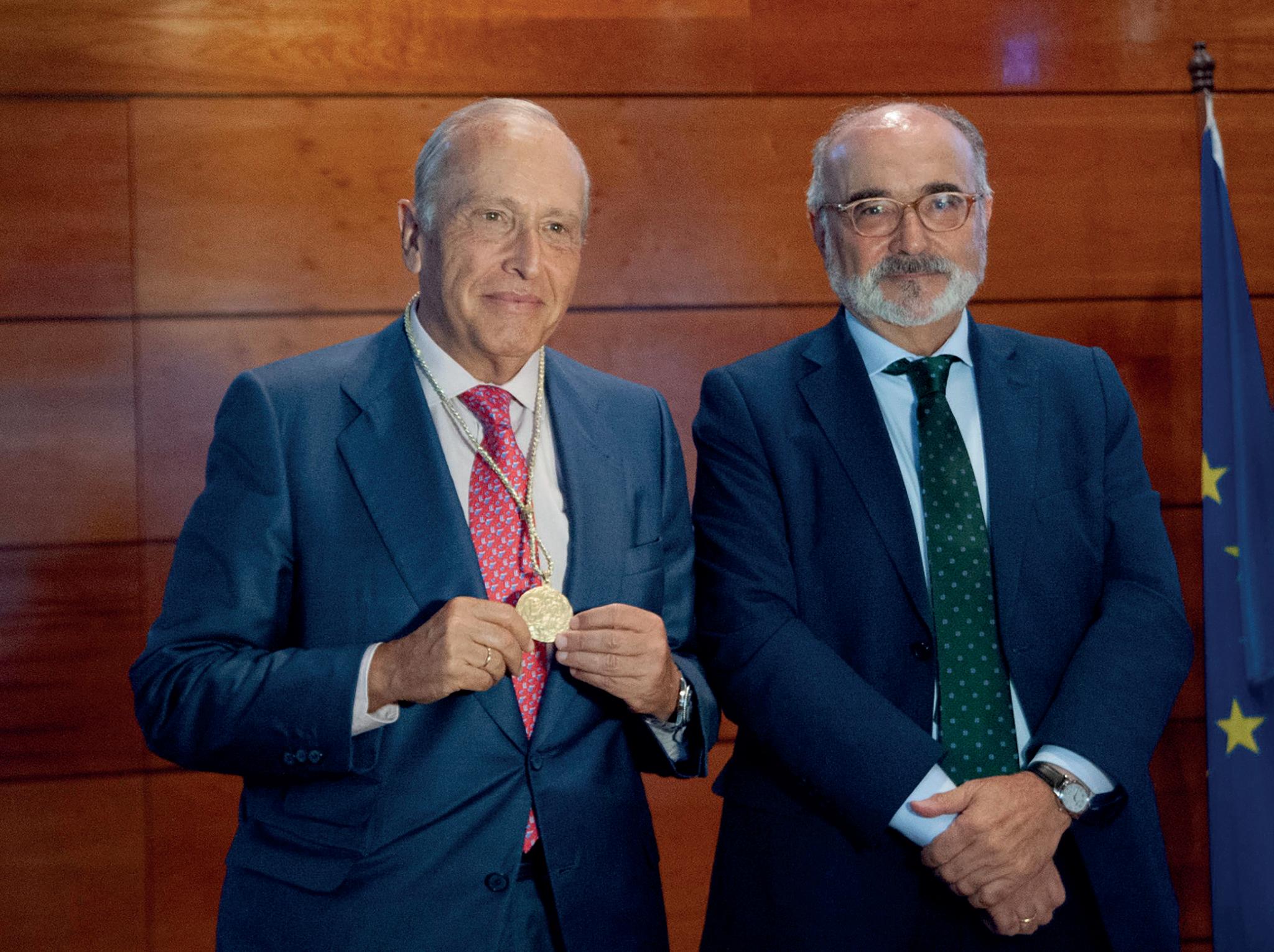
Los “Cursos de La Granda” – entidad académica de gran tradición y enorme prestigio – tuvo a bien concederme su Medalla en atención a lo que consideró una singular trayectoria en el apartado médico, desde luego, pero también en el universitario y ciudadano, por un continuado compromiso – en este último de los campos – entre los que destaca la presidencia de la Fundación Princesa de Asturias.
Las Cámaras de Comercio de Asturias, por su parte, han elegido a nuestro Instituto como
PYME del año en el Principado en atención, tanto por lo que supone su aportación directa al PIB de Oviedo y el conjunto de la región, como por la imagen de excelencia que proyecta hacia el exterior y que se traduce en beneficios económicos generales de indudable importancia.
Por otra parte, en el plano científico, durante el Staar Experts Meeting celebrado recientemente en Milán, el Dr. Luis Fernández-Vega Cueto-Felgueroso y la Dra. Belén Alfonso Bartolozzi fueron distinguidos entre los mejores Jóvenes Oftalmólogos Europeos. En esa misma reunión también tuvieron menciones especiales el Dr.
José Alfonso y el Dr. Carlos Lisa.
Todo ello pone de manifiesto que nuestra continuada apuesta por la formación de excelencia de nuestro cuadro médico es una magnífica realidad y una sólida esperanza del mejor futuro. Hecho que refuerza, también, los premios obtenidos por el Dr. Ignacio Rodriguez Uña en el congreso de la Sociedad Española de Oftalmología celebrado en Pamplona, en los apartados de fotografía científica y comunicación en panel.
Son reconocimientos que, como otros anteriores, nos obligan y responsabilizan en nuestro desempeño y que agradecemos profundamente.
EDITORIAL
3
ASTURIAS Avda. Dres. Fernández‑Vega, 34 33012 OVIEDO T. 985 240 141 MADRID Príncipe de Vergara, 131 28002 MADRID T. 91 577 99 59 email: instituto@fernandez-vega.com www.fernandez-vega.com Dirección y coordinación, redacción de contenidos y gestión editorial: Atlántica Empresas Publicidad: SGANMEDIOS 678 684 457 Diseño: Eteria, Marketing y Comunicación D.L. AS 5668 2007 REVISTA DEL INSTITUTO OFTALMOLÓGICO FERNÁNDEZ-VEGA 4
SUMARIO
EN PORTADA
El Instituto Oftalmológico Fernández-Vega, de nuevo elegido centro privado de Oftalmología con mejor reputación de España

SOMOS NOTICIA
Los cursos de La Granda entregan su medalla a Luis Fernández-Vega Sanz
CONSULTORIO
Dudas de los pacientes sobre glaucoma y miopía
A FONDO
Álvaro Fernández-Vega González: “La mayoría de los desprendiemientos de retina pueden ser tratados con éxito por un cirujano de la especialidad”
INVESTIGACIÓN
La erupción cutánea por viruela del mono puede afectar a los ojos en el 20% de los casos
ACTIVIDADES SOLIDARIAS
Visita de la Fundación Fernández-Vega a la Asociación Nora
ACTIVIDAD CONGRESUAL
Los doctores Luis Fernández-Vega Cueto-Felgueroso y Belén Alfonso Bartolozzi, entre los mejores oftalmólogos jóvenes de Europa
NOS CUIDAMOS
¿Quién inventó la operación de cataratas?
OTRAS ESPECIALIDADES
Hábitos saludables y control de los niveles en sangre, clave para prevenir las enfermedades cardiovasculares
NOS VEN CON BUENOS OJOS
Carlos Fitz-James Stuart y Martínez de Irujo: “El mayor reto de la Casa de Alba es mantener al máximo nivel su patrimonio para que la sociedad sea partícipe y pueda disfrutar de todo el legado”
RECOMENDAMOS
Oviedo es música
CRUCE DE MIRADAS Ana de Armas
/
8
14/
16/
/
26
/
28
/
31
/
35
37/
38/
39/
40/
6/ 5
l Instituto Oftalmológico Fernández-Vega ha sido reconocido como el mejor centro privado de Oftalmología de España por segundo año consecutivo.
La apuesta por el compromiso con los pacientes, la investigación, la formación y la alta calidad asistencial le han valido el primer puesto de la clasificación de
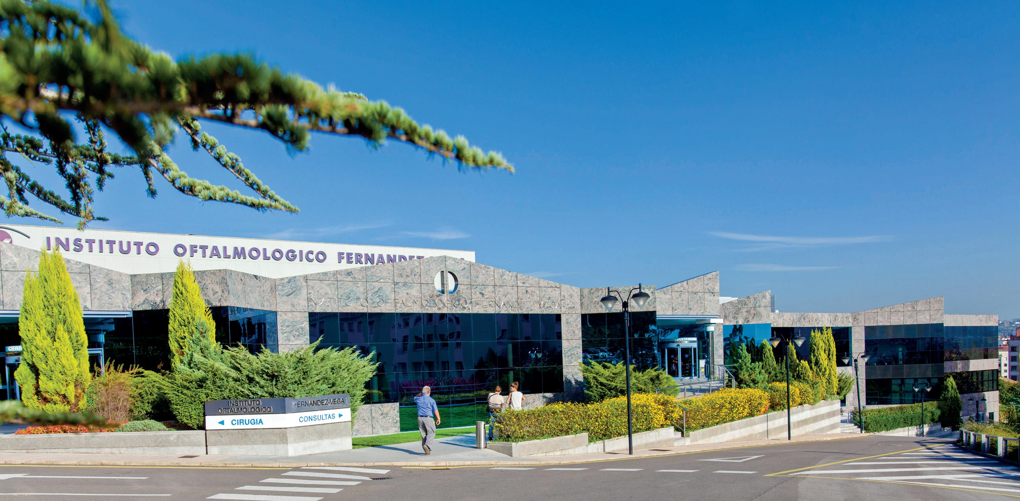
EL INSTITUTO OFTALMOLÓGICO FERNÁNDEZ-VEGA, DE NUEVO ELEGIDO CENTRO PRIVADO DE OFTALMOLOGÍA CON MEJOR REPUTACIÓN DE ESPAÑA E EN PORTADA Gracias a su apuesta por la investigación, la formación y la alta calidad asistencial, el Instituto Fernández-Vega se sitúa en el primer puesto de la clasificación de los centros oftalmológicos privados de España del Monitor de Reputación Sanitaria (MRS) que elabora MERCO 6
8.ª EDICIÓN DEL MONITOR DE REPUTACIÓN SANITARIA
los mejores centros oftalmológicos privados de España en el Monitor de Reputación Sanitaria (MRS) que anualmente elabora MERCO.
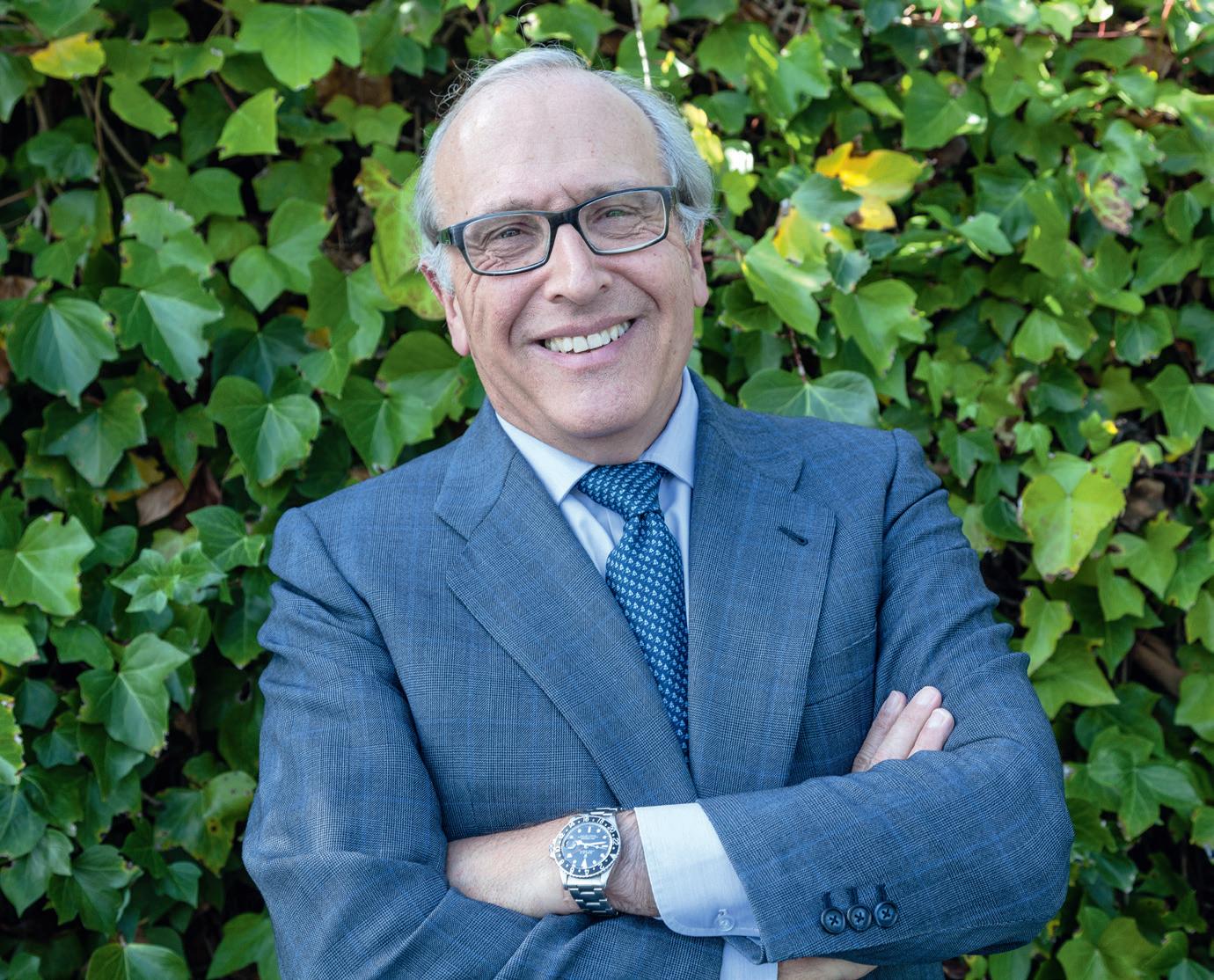
“Este reconocimiento nos motiva a continuar invirtiendo en la excelencia y apostando por ofrecer a los pacientes un servicio oftalmológico de máxima calidad”, comenta el Prof. Luis Fernández-Vega Sanz, director médico del Instituto Oftalmológico Fernández-Vega “Sin duda, es un orgullo para todos nosotros habernos consolidado como un referente nacional e internacional” añade el profesor.
El ranking general está integrado tanto por los servicios de Oftalmología de los hospitales públicos, como por los centros privados dedicados a esta especialidad. En
concreto, el I.O. Fernández-Vega se sitúa en el top 3, tras los hospitales públicos.
El Instituto Oftalmológico Fernández-Vega se distingue no solo por su calidad asistencial, sino también por su firme apuesta por la investigación y la docencia de la mano de la Fundación de Investigación Oftalmológica (FIO) y el Instituto Universitario Fernández-Vega. Además, cuenta con la Fundación Fernández-Vega, que lleva a cabo una importante labor social proporcionando cuidados oftalmológicos a colectivos desfavorecidos dentro y fuera de España.
Para llevar a cabo el Monitor de Reputación Sanitaria 2022, el más prestigioso ranking independiente de este tipo, se han realizado más de 6.526 encuestas a
médicos, enfermeras, asociaciones de pacientes, periodistas sanitarios, gerentes de hospitales, gestores de enfermería, miembros de la administración pública sanitaria, responsables de farmacia hospitalaria y directivos de empresas farmacéuticas.
El MRS evalúa más de 3.010 indicadores objetivos de 2.912 servicios clínicos que aportan una perspectiva de la calidad de la atención de cada hospital, observando datos de actividad tales como la oferta asistencial, la experiencia del paciente y las técnicas aplicadas o los resultados obtenidos.
7 EN PORTADA
LOS CURSOS DE LA GRANDA ENTREGAN SU MEDALLA A LUIS FERNÁNDEZ-VEGA SANZ
han reunido durante el mes de agosto a una buena parte, sino a lo mejor, de la intelectualidad española, quienes en un ambiente sosegado han compartido y divulgado sus conocimientos con un espíritu que mucho se asemeja al perseguido en su día por el llamado Grupo de Oviedo de la Universidad asturiana o, más cerca en el tiempo, al preconizado por Ortega y su escuela.
Por estos cursos de La Granda, presididos hoy por Benigno Pendás, que lo es a su vez de la Real Academia de Ciencias Morales y Políticas, pasan historiadores, investigadores, artistas, escritores, políticos… de tal manera que casi ninguna disciplina les resulta ajena.
a Granda es un paraje idílico en el centro de Asturias. Arcelor Mittal posee allí una antigua residencia que ha servido a lo largo del tiempo para innumerables encuentros empresariales, pero también como sede de unas jornadas que, durante ya 44 años,
En esta ocasión, como es tradicional, han hecho entrega de su Medalla a la persona que a su juicio representa el conocimiento, compromiso, valores y trayectoria profesional que son seña de identidad de la Fundación que promueve esta iniciativa, y que ha recaído en el Profesor Luis
El Profesor Luis Fernández-Vega y Benigno Pendás, presidente de los Cursos de La Granda
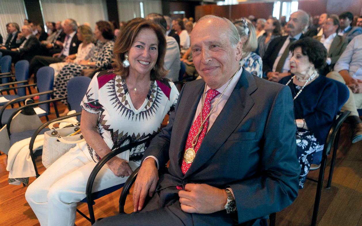
El profesor Luis Fernández-Vega Sanz y su esposa, Doña Victoria Cueto-Felgueroso Botas, durante el acto.
SOMOS NOTICIA
L
8
Foto propiedad de LNE.
Fernández-Vega, presidente del Instituto Oftalmológico que lleva el apellido de su familia.
Fue Raimundo Abando, destacado empresario asturiano, el encargado de resaltar las circunstancias humanas y profesionales del homenajeado, con quien le unen lazos de amistad desde sus años jóvenes, y que subrayó su compromiso ciudadano al tiempo que enfatizó la importancia que, bajo la perspectiva económica, tiene la citada
entidad médica, ya que -por ejemplo -supone el 5% del PIB de la capital asturiana.
Luis Fernández-Vega no solo se mostró honrado y agradecido por la distinción recibida – que dedicó a su mujer Victoria Cueto-Felgueroso – sino también muy orgulloso y responsabilizado por pasar a formar parte de un grupo que reúne en el apartado de la medicina y la investigación a personalidades como Severo
Ochoa, Santiago Grisolia, Segovia Arana o Grande Covián entre otros.
Fernández-Vega achacó a la tradición familiar su vocación, y a su empeño en aunar -junto a otros miembros de éstala actividad clínica con la investigadora y docente, que el Instituto Oftalmológico Fernández-Vega sea hoy una referencia que atiende a más de cien mil pacientes todos los años y cuente, entre sus más de doscientos profesionales,
con especialistas capaces de atender cualquier patología oftalmológica.
No faltaron en el emotivo acto de entrega referencias a su presidencia de la Fundación Princesa de Asturias en un periodo lleno de satisfacciones, pero también signado por la terrible pandemia que obligó a innovar y modificar varias de las actividades que tradicionalmente se llevan a cabo en torno a la entrega de los Premios que todos los octubres tiene
lugar en la capital asturiana.
Luis Fernández-Vega quiso finalizar sus palabras con un renovado compromiso cívico con la sociedad y un reforzado estímulo para continuar al frente del Instituto Oftalmológico que, precisamente junto a la Fundación Princesa de Asturias, y aunque sea en diferentes niveles, resultan, en encuestas sobre las entidades e instituciones del Principado, las más valoradas.
9
SOMOS NOTICIA
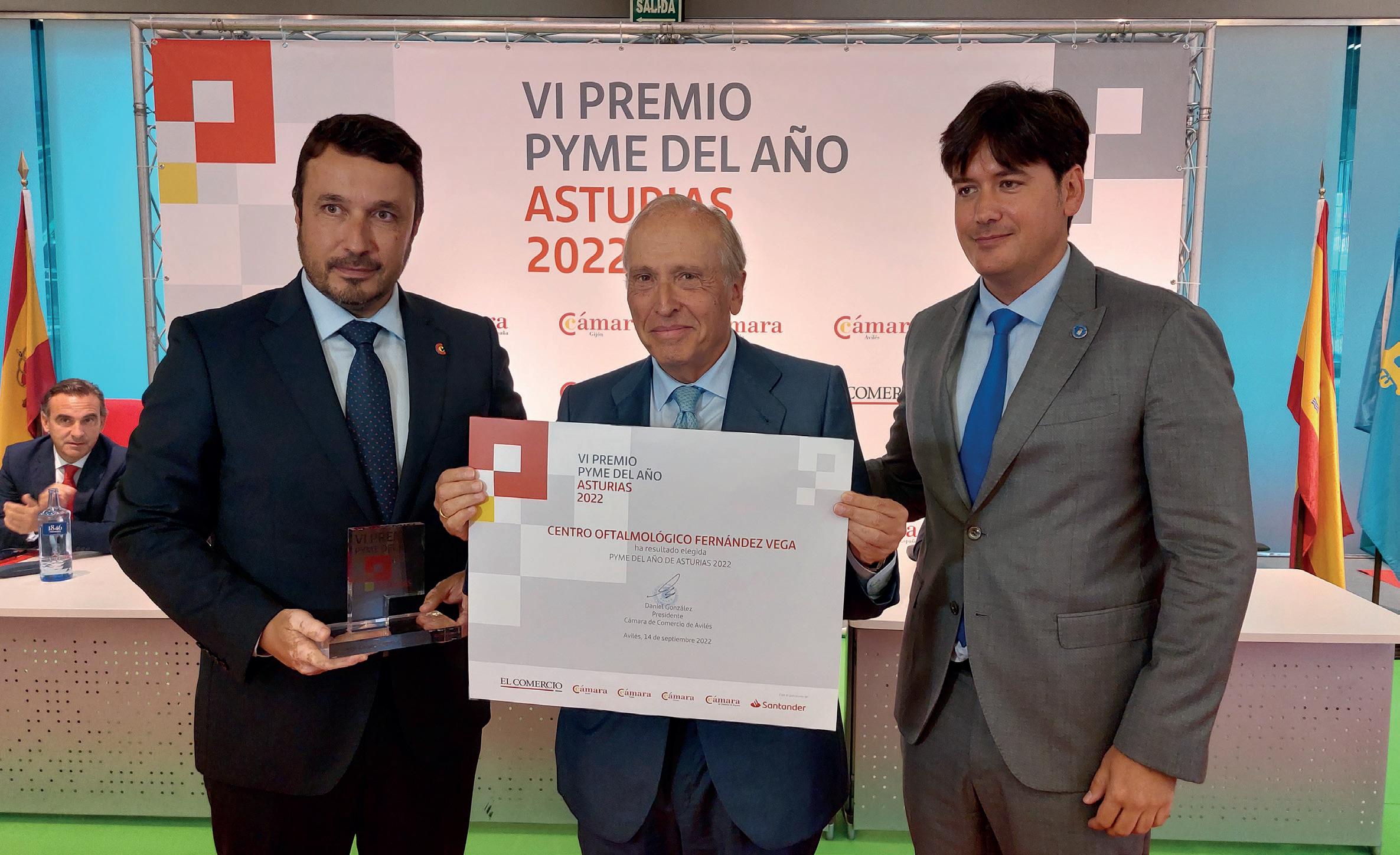
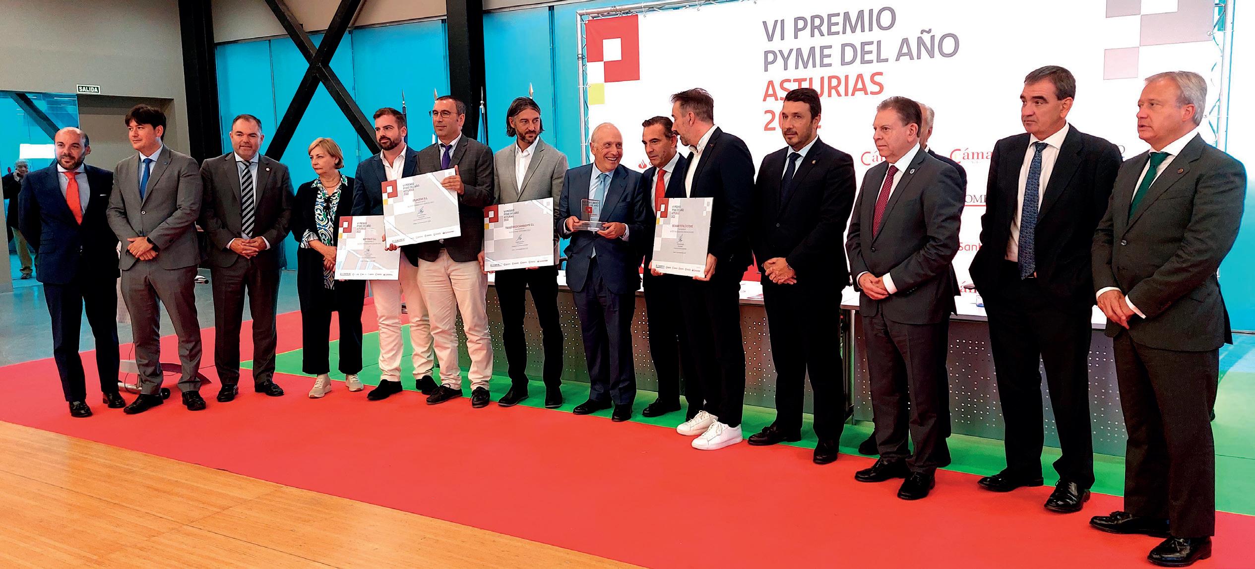
EL INSTITUTO, PYME DEL AÑO EN ASTURIAS
El premio, otorgado por las Cámaras de Comercio asturianas, valoró de forma muy especial su contribución a la economía y la mejor imagen del Principado.
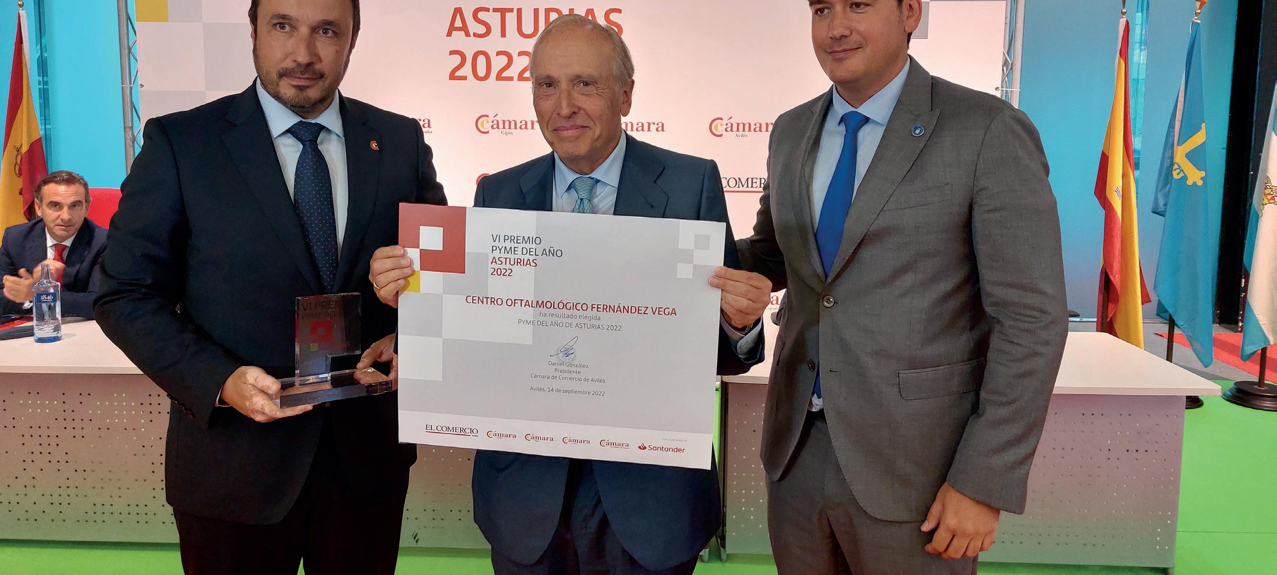
En el acto de entrega, que tuvo lugar en Avilés, con presencia de la vida económica, política y ciudadana de la región se distinguieron también otras empresas con accésits por su buen desempeño en aspectos como la innovación, la digitalización o la proyección exterior, mientras que al Ins-
tituto Oftalmológico Fernández-Vega le correspondió el premio a la mejor PYME de Asturias 2022. Se trata de una iniciativa auspiciada por Cámara España y el Grupo Vocento y, por ello, el Instituto será quien representará a Asturias en el ámbito nacional de la misma, que tendrá lugar en el
primer trimestre del próximo año.
Fue Carlos Paniceres, presidente de la Cámara de Oviedo, el encargado de glosar la trayectoria de una familia – con cinco generaciones ya de oftalmólogos – y el impacto en la economía de una entidad médica que atiende todos los años a más de cien
10 SOMOS NOTICIA
Daniel González, presidente de la Cámara de Comercio de Avilés; el Profesor Luis Fernández-Vega Sanz, director médico del IOFV, y Borja Sánchez, consejero de Ciencia, Innovación y Universidad del Principado de Asturias.
Los galardonados, al finalizar el acto.
mil pacientes y lleva a cabo cerca de 10.000 cirugías, y contribuye, además, a proyectar una imagen de excelencia que repercute en muy distintos sectores. No olvidó Paniceres una referencia al compromiso profesional y cívico del profesor Luis Fernández-Vega, de quien dijo ser una persona con
la que “siempre se puede contar”.
“Si mi vocación médica fue temprana, la empresarial puede decirse que fue más tardía y sobrevenida”, manifestó tras recoger la distinción Luis Fernández-Vega, ya que ésta derivó de la necesidad de “hacer empresa” en el paso de una
clínica tradicional prestigiosa a un Instituto de referencia en el sector, capaz de atender toda clase de patología oftalmológica con profesionales de excepción y la mejor tecnología. De ahí que se mostrara más acostumbrado a recibir distinciones en su calidad de médico que de empresario.
SOMOS NOTICIA
Enfatizó su compromiso, y el de su familia, con Asturias, pero no dejó de señalar la importancia de que ésta disponga de las condiciones necesarias para poder competir en igualdad con las comunidades más avanzadas de España.
11
El IOFV será quien represente a Asturias en el ámbito nacional de la iniciativa, que tendrá lugar en el primer trimestre del próximo año
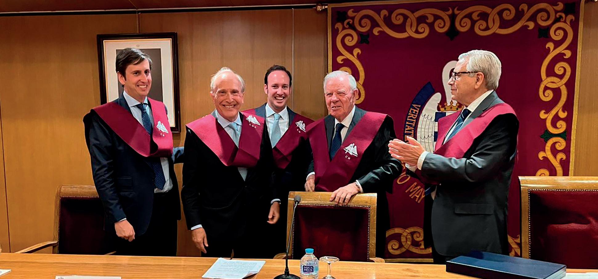
EL PROFESOR LUIS FERNÁNDEZ-VEGA SANZ, NOMBRADO ‘COLEGIAL MAYOR DILECTO’ POR EL FORO SAN PABLO
El Consejo de Colegiales Mayores del Foro Mayor San Pablo ha otorgado el reconocimiento de “Colegial Mayor Dilecto” al Profesor Luis Fernández-Vega Sanz, director médico del Instituto Oftalmológico Fernández-Vega. Durante el acto de entrega se reconoció su singular aportación a la sociedad española, su significativa vinculación con los valores del espíritu paulino y su excepcional trayectoria como referente internacional de la oftalmología.
Tras unas palabras de bienvenida del presidente del Foro Mayor, Andrés Contreras, el Profesor fue presentado por los antiguos colegiales José Ramón Álvarez Rendueles, antiguo gobernador del Banco de España y expresidente de la Fundación Princesa de
Asturias, y Javier López-Galiacho, antiguo director del Mayor. Ambos destacaron los méritos académicos y profesionales del director médico del Instituto, así como los valores que representan el espíritu paulino. Los Dres. Luis y Andrés Fernández-Vega Cueto-Felgueroso, oftalmólogos de la quinta generación de la familia, hijos del Profesor y colegiados del Mayor durante sus 6 años de carrera, asistieron al acto, durante el que se le impuso la beca colegial y se le entregó una placa conmemorativa.
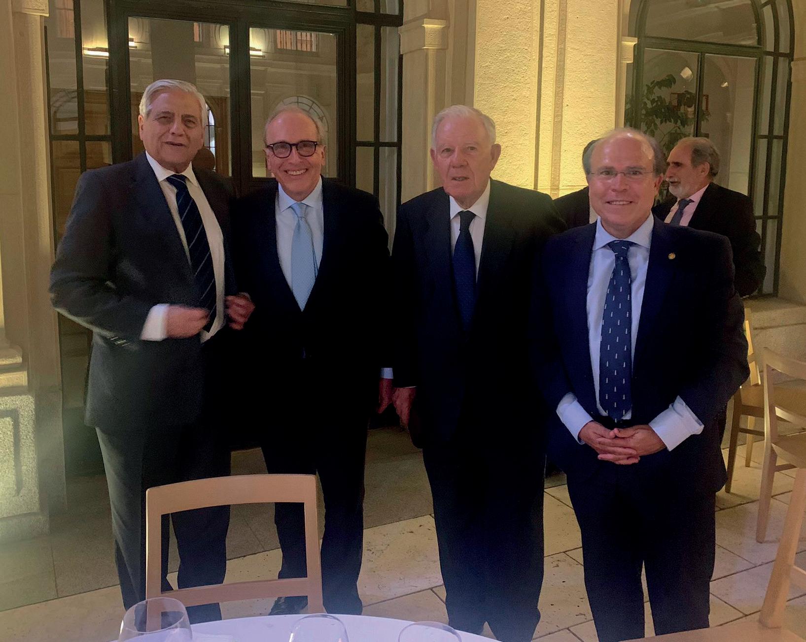
Al evento asistieron también los
miembros de la junta directiva del Foro, Pascual Cervera, Juan Carlos Fouz, Alfonso Pérez, Pablo Fernández-Canedo, Aitor Errasti y David Rojo -promotor de la iniciativa de concesión del premio-. Además, participaron Carlos Gregorio Hernández y José Manuel Varela -director y director adjunto del Colegio Mayor-, numerosos antiguos colegiales y destacados miembros del equipo médico del Instituto Oftalmológico Fernández-Vega, así como varios amigos y familiares de los asistentes.
José Ramón Álvarez Rendueles y Javier López-Galiacho destacaron los méritos académicos y profesionales del Profesor.
12 SOMOS NOTICIA
El Prof. Luis Fernández-Vega Sanz, acompañado de sus hijos y con José Ramón Álvarez Rendueles y Andrés Contreras Salido.
El Consejo de Colegiales Mayores del Foro Mayor San Pablo reconoció su singular aportación a la sociedad española.
Por la izquierda, Andrés Contreras, Prof. Luis Fernández-Vega Sanz, José Ramón Álvarez Rendueles y Javier López-Galiacho.
SOMOS NOTICIA
DUDAS DE LOS PACIENTES SOBRE GLAUCOMA Y MIOPÍA
Buenas tardes, tengo 18 años y 6 dioptrías de miopía. Mis padres no tienen glaucoma, pero he leído que yo tengo más riesgo por ser miope. ¿Es así?
Cabe destacar que la miopía constituye un factor de riesgo significativo de desarrollar glaucoma, especialmente cuando se trata de miopía alta o magna. Es cierto que el riesgo está presente en cualquier miope, pero dicho riesgo aumenta conforme aumenta el grado de miopía. Por ello, aunque a priori no deberías preocuparte, sí que recomendamos la realización de revisiones oftalmológicas periódicas completas a todos los jóvenes miopes para el diagnóstico y tratamiento precoz del glaucoma u otras patologías a las que los miopes tienen mayor predisposición.
Los menores de 20 años pueden desarrollar glaucoma de dos tipos: congénito y juvenil. El juvenil tiene una incidencia de uno por cada 300.000 menores de 20 años, aunque lo típico es que aparezca entre los 5 y los 18 años y habitualmente se diagnostica en jóvenes miopes.
Hola, soy una mujer de 32 años y me estoy planteando ser madre. Tengo glaucoma diagnosticado desde hace unos años, ¿mi hijo podría tenerlo también?
Lo primero y más importante sería hacer una revisión para ver el estado actual de su glaucoma, pues hay fármacos que utilizamos para disminuir la tensión ocular que no deben aplicarse estando embarazada. Sin embargo, tenemos otras opciones que están avaladas por la literatura científica y que son alternativas seguras, como diferentes gotas hipotensoras o incluso algunos tipos de láser si fuera necesario, en función del grado de glaucoma. Por otro lado, el glaucoma sí que tiene un componente hereditario y el glaucoma congénito se cree que afecta a uno de cada 30.000 recién nacidos por lo que sería conveniente que observara a su bebé buscando alguno de los síntomas típicos: fotofobia, epifora, blefarospasmo u ojos más opacos y más grandes de lo normal. De todos modos, el 80-90% de los casos de glaucoma congénito son esporádicos y aproximadamente el 10% son los que presentan una herencia autosómica recesiva con penetrancia incompleta, lo cual indica que haría falta la presencia del gen en ambos progenitores (portadores), para que se desarrollara la enfermedad.
CONSULTORIO
Dr. Andrés Fernández-Vega Cueto-Felgueroso
14
Buenos días Doctor, soy una joven a la que le encanta el pádel y el otro día, jugando, una bola me golpeó un ojo. ¿Puedo, en un futuro, tener problemas por ello?
Tras un traumatismo ocular es muy importante realizar una exploración oftalmológica minuciosa y completa incluyendo la gonioscopia, así como estar atentos a diferentes signos y síntomas que se puedan desarrollar. Cuando el ojo sufre un traumatismo puede haber erosiones corneales, un aumento agudo de la presión intraocular debido a un potencial daño en las estructuras angulares, una recesión del ángulo que pueda dar problemas tensionales con el paso de los años (por ello recomendamos un seguimiento a largo plazo de estos ojos), un hipema (sangre en la cámara anterior del ojo), inflamación intraocular o uveítis, que puede hacer que se desarrolle un glaucoma inflamatorio, formación de una catarata o incluso un desprendimiento de retina. Los traumas oculares son una causa muy frecuente de hipertensión ocular, la cual, si no se detecta pronto, puede llevar a una atrofia progresiva del nervio óptico con el paso del tiempo.
Tengo 35 años, tres dioptrías de miopía y uso gafas habitualmente. Después de hacer deporte veo borroso, ¿es normal o es necesario que vaya a revisión oftalmológica?
Sin duda, te recomendamos que acudas al oftalmólogo para realizar una revisión. Lo que describes es un signo típico del síndrome de dispersión pigmentaria, más común en varones jóvenes miopes. No significa esto que vayas a tener dicha dispersión, pero sin duda creemos que sería conveniente descartarlo. Este síndrome se produce cuando el iris, por una mala posición, roza contra la parte anterior del cristalino, liberándose pigmento en el interior de la cámara anterior y pudiéndose depositar en alguno de los tejidos que tenemos en la parte delantera del ojo.
Una de las zonas en las que el citado pigmento se puede depositar es el ángulo iridocorneal, que es por donde se produce el “desagüe” natural del humor acuoso del ojo. Cuando se obstruye esa zona de filtración, no sale bien el líquido, produciéndose lentamente una subida de la presión intraocular. Este sería el mecanismo que, mantenido, podría producir glaucoma.
CONSULTORIO
“LA MAYORÍA DE LOS DESPRENDIMIENTOS DE RETINA PUEDEN SER TRATADOS CON ÉXITO POR UN CIRUJANO DE LA ESPECIALIDAD”
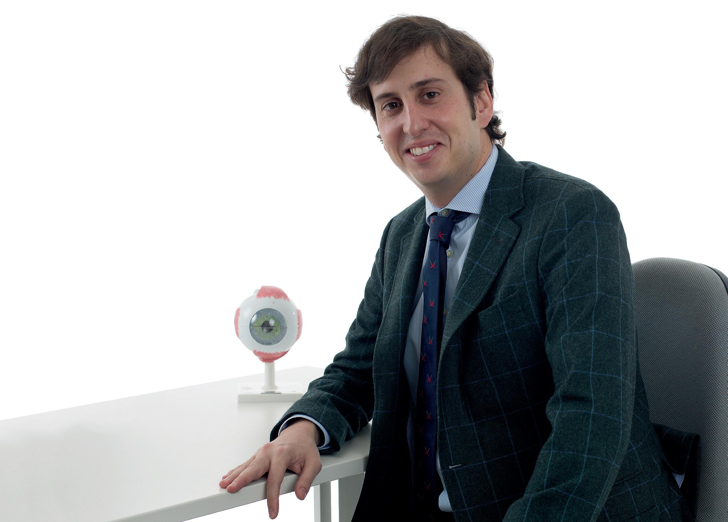
El desprendimiento de retina es una patología relativamente frecuente en pacientes con alta miopía y por ello es importante la prevención. El doctor Álvaro Fernández-Vega González, oftalmólogo de la Unidad de Retina y Vítreo del Instituto Oftalmológico Fernández-Vega, explica en esta entrevista en qué consiste esta patología.
¿Qué es la retina?
Es un tejido constituido por neuronas que forman una capa que tapiza por dentro el interior del globo ocular, como si fuera el papel pintado de una habitación. El sistema óptico del ojo está planeado para que las imágenes que miramos sean enfocadas en la retina (igual que el objetivo de una cámara de fotos enfoca las imágenes en la película o sensor), que se encarga de recoger dichas imágenes y
transmitirlas al cerebro, donde serán interpretadas.
¿Qué es un desprendimiento de la retina?
Es la separación de la retina neurosensorial de la pared posterior donde debería estar adherida. Para que la retina sea capaz de captar las imágenes tiene que estar pegada a esta, que le permite alimentarse y funcionar correctamente, de forma que, si se separa, la retina empieza a degenerar y las células que
la forman mueren, produciendo una pérdida de visión que puede ser grave.
¿Cómo se produce un desprendimiento?
Lo más frecuente es que un desprendimiento de retina se produzca tras un desgarro/agujero en la misma. En este proceso tiene un papel fundamental el gel que rellena el ojo por dentro denominado vítreo. Cuando somos jóvenes, el vítreo está adherido a toda la retina a lo largo de
su superficie, pero a medida que envejecemos el vítreo se va despegando de la retina provocando un desprendimiento del mismo. Este suceso es un proceso normal de envejecimiento del vítreo que en la mayoría de las personas no va a causar ningún problema. Sin embargo, en un pequeño porcentaje, el vítreo al separarse puede tirar de la retina y romperla en un punto, generando un desgarro retiniano. Si esto sucede, la parte del vítreo que es más líquida puede colarse detrás de la retina a través de ese desgarro y despegar a esta de su pared posterior, dando lugar a un desprendimiento.
¿Cuáles son sus síntomas?
El más característico de que el vítreo se
está desprendiendo de la retina es la aparición repentina de “moscas volantes”, que son multitud de manchas negras con diferentes formas (moscas, puntos, telarañas), que aparecen en el campo visual. A esto se puede asociar la visión de una nube que se mueve y destellos o relámpagos de luz. Estos síntomas indican que el vítreo se está separando de la retina y el paciente debe saber identificarlos para acudir urgentemente a revisar la retina, ya que este es el momento donde se pueden producir los desgarros retinianos.
¿Se
puede prevenir?
En muchas ocasiones, si el paciente acude a urgencias y se descubre un desgarro antes de que se haya produ-
16 A FONDO
Dr. Álvaro Fernández-Vega González
cido el desprendimiento de retina, se puede impedir su desarrollo mediante la fotocoagulación con láser de dicho desgarro. Cuando además del desgarro está presente un desprendimiento de retina, lo que el paciente observaría es una cortina o mancha negra en una zona de su campo visual periférico que poco a poco avanza hacia el centro hasta quitar completamente la visión. En esta situación, donde ya no solo tenemos un desgarro, sino que está presente un desprendimiento de retina, el pronóstico visual empeora y el tratamiento es quirúrgico.
¿En qué consiste la fotocoagulación con láser?
En hacer una serie de pequeñas quemaduras alrededor de la rotura retiniana, que en poco tiempo van a generar una cicatriz adherente en torno al desgarro que permita su sellado y evite el paso de fluido debajo de la retina. Actuaría, por tanto, como una soldadura alrededor de la rotura retiniana, evitando la progresión de un desgarro a desprendimiento de retina. Algunos pacientes que tienen lesiones predisponentes al desprendimiento de la retina (como zo-
nas de ésta adelgazadas), pueden ser tratados con láser alrededor de estas lesiones, pero esto no evita un desprendimiento de retina al cien por cien, por lo que la mejor prevención es que los pacientes conozcan los síntomas de la separación del vítreo y acudan cuanto antes al especialista.
¿Se puede tratar un desprendimiento de retina?
Hoy día la mayoría pueden ser tratados con éxito, gracias a la alta tecnología y las sofisticadas técnicas que emplean los cirujanos de retina. El principal objetivo de la cirugía es volver a posicionar la retina en su lugar original para que recupere su función. Sin embargo, dependiendo del tipo y de su tiempo de evolución puede ser que, aunque la cirugía sea un éxito, quede un déficit visual permanente, ya que las células de la retina que han muerto no son recuperables. Por eso es fundamental acudir cuanto antes al especialista si el paciente tiene síntomas.
¿Cómo se trataría el desprendimiento de retina?
Existen tres diferentes técnicas: la vitrectomía, el cerclaje y la retino-
pexia neumática. Dependiendo de las características del desprendimiento y del paciente estará más indicada emplear una técnica u otra.
¿En qué consisten?
Cada una de estas técnicas tiene ventajas e inconvenientes. La vitrectomía, que es la técnica más utilizada hoy, consiste en entrar dentro del ojo a
través de microincisiones, quitar el vítreo, drenar el fluido de detrás de la retina para que ésta vuelva a su posición original, hacer láser alrededor de las roturas y poner una burbuja de gas al final de la cirugía. El objetivo de la burbuja de gas es presionar y bloquear el desgarro, evitando que la retina se vuelva a desprender durante el tiempo que se
forma la cicatriz del láser. El cerclaje es una banda de silicona que se pone alrededor del ojo, suturada, con el objetivo de acercar la pared del globo ocular a la retina y facilitar que la retina vuelva a su posición. Por último, la retinopexia neumática es una técnica menos agresiva que permite tratar algunos desprendimientos sin realizar una cirugía, con tan solo la inyección de una burbuja de gas y posicionamiento. Con el tratamiento adecuado, la mayoría de los desprendimientos de retina pueden solucionarse, la clave es seleccionar en cada paciente la técnica más adecuada.
17 A FONDO
“La fotocoagulación con láser de los desgarros puede prevenir su desarrollo “
“LOS ÚLTIMOS AVANCES EN CIRUGÍA PERMITEN CORREGIR LA PRESBICIA DE FORMA SEGURA, RÁPIDA Y DEFINITIVA”
La presbicia es un proceso inevitable que se produce como consecuencia del envejecimiento natural del ojo1. Aparece a partir de los 40 años2 y se caracteriza por la pérdida gradual para enfocar objetos de cerca, lo que popularmente se conoce como vista cansada. Afortunadamente, hoy en día existen soluciones eficaces, rápidas y seguras que permiten corregir esta afección de forma definitiva y prescindir del uso de gafas a todas las distancias.
El Profesor Luis Fernández-Vega Sanz, director médico del Instituto Oftalmológico Fernández-Vega, explica los principales tratamientos para la presbicia.
¿Qué es la presbicia? ¿qué síntomas presenta?
Es un defecto refractivo asociado a la edad que provoca visión borrosa y dificultad para ver de cerca. Con el tiempo, el cristalino, la lente natural del ojo, pierde su flexibilidad y capacidad para enfocar la luz, sobre todo en distancias cortas1 Este fenómeno es conocido popularmente como “vista cansada”1
Entre sus síntomas más frecuentes están la pérdida gradual de la capa-
cidad para enfocar los objetos en visión cercana, necesidad de alejar el libro o el teléfono móvil para poder ver mejor, sensación de que las letras de un texto “bailan” o están borrosas, dolor de cabeza al fijar la vista en la lectura durante mucho tiempo y fatiga ocular.
¿Se puede prevenir la presbicia?
Se trata de un proceso degenerativo que se produce como consecuencia inevitable del envejecimiento del ojo. Se desarrolla generalmente a partir de
los 40 años2 y, con la edad, todas las personas desarrollarán presbicia en cierta medida3
¿Qué relación existe entre la catarata y la presbicia?
Ambas afecciones afectan el cristalino del ojo y están asociadas al envejecimiento, por lo que su incidencia irá en aumento debido a la mayor longevidad de la población. Aunque son condiciones diferentes, tienen algunos síntomas en común, como la visión borrosa. Hoy día es posible poner
A FONDO
Profesor Luis Fernández-Vega Sanz
18
solución a ambas en un mismo procedimiento quirúrgico, ya que la cirugía implanto-refractiva permite reemplazar el cristalino afectado y reestablecer la función visual.
¿Qué opciones existen para corregirla?
Existen diversos tratamientos para mejorar los síntomas de la presbicia. El primero es la corrección óptica mediante el uso de gafas o lentes de contacto1
Otra opción es la corrección quirúrgica, ya que hay diferentes técnicas como la cirugía láser, la cirugía de reemplazo del cristalino por lentes intraoculares o los implantes corneales1 y todas ellas permiten corregir la
presbicia de forma segura, rápida y definitiva.
¿Cómo se determina cuál es la opción terapéutica más recomendable para cada paciente?
El oftalmólogo analizará cada caso de forma personalizada, realizando un estudio detallado a nivel anatómico y funcional y teniendo en cuenta las preferencias y el estilo de vida del paciente para ofrecer la alternativa que mejor se adapte a su situación y a sus necesidades.
¿En qué consiste la cirugía para corregir la presbicia? ¿Qué ventajas ofrece?
La cirugía implanto-refractiva, que
es la técnica quirúrgica más habitual, consiste en la sustitución del cristalino por una lente intraocular, restaurando totalmente la función visual. Esta nueva lente permitirá corregir determinadas afecciones o problemas que tuviese el paciente antes de la intervención, en función del tipo de lente intraocular que se utilice. Al sustituir el cristalino, esta operación evita el desarrollo de cataratas en el futuro.
La cirugía es, por tanto, una de las soluciones más recomendadas en la mayoría de los casos para aquellos pacientes que desean prescindir del uso de gafas, ya que ofrece resultados de larga duración, especialmente con las lentes intraoculares, que son para toda la vida. Además, permite corregir de forma simultánea otros problemas refractivos o la catarata, de forma definitiva.
¿Qué tipo de lentes intraoculares existen para corregir la presbicia? ¿cuáles son los últimos avances?
Las lentes intraoculares correctoras de la presbicia cuentan con avanzadas tecnologías y materiales que permiten ofrecer una calidad de imagen de alta resolución a todas las distancias: lejos,
intermedia y cerca,y se pueden subdividir en multifocales (las trifocales son las más habituales) y lentes de visión extendida.
Las lentes multifocales (o difractivas) corrigen la mayoría de los defectos refractivos ofreciendo un rendimiento alto de la visión y una corrección óptica estable a lo largo del tiempo. Cada vez existen materiales y plataformas más avanzados que ofrecen una gran claridad a todas las distancias.
Hoy existe una nueva clase de lente intraocular de visión extendida que ofrece la posibilidad de prescindir del uso de gafas para, prácticamente, todas las
actividades diarias. Estas lentes proporcionan un rango de visión completo, permitiendo recuperar la visión de lejos, intermedia y cerca funcional, y un nivel de molestias visuales muy bajo.
Referencias:
1. Sociedad Española de Oftalmología (SEO). Presbicia - Oftalmoseo. (2022). Retrieved 27 July 2022, from https://www.oftalmoseo. com/patologias-frecuentes-2/ presbicia/
2. Consejo General de Ópticos y Optometristas. Libro Blanco de la Salud Visual en España 2019 https://www.cgcoo.es/ libro-blanco-salud-visual-enespana-2019
3. National Eye Institute. Presbicia (2022). Retrieved 27 July 2022, from https://www. nei.nih.gov/espanol/aprendasobre-la-salud-ocular/ enfermedades-y-afeccionesde-los-ojos/presbicia
A FONDO
“La catarata y la presbicia tienen algunos síntomas en común, como la visión borrosa. Hoy en día es posible poner solución a ambas en un mismo procedimiento quirúrgico”
19
LA OFTALMOLOGÍA PEDIÁTRICA PREVIENE Y TRATA LAS DOLENCIAS OCULARES EN LA INFANCIA
El avance de las técnicas diagnósticas y terapéuticas en oftalmología, tanto en procedimientos clínicos como quirúrgicos, ha propiciado la creación de subespecialidades médicas que tienen como objetivo tratar las enfermedades oculares específicas más complejas en algunas partes del ojo o en cierto tipo de pacientes. El Instituto Oftalmológico Fernández-Vega, en su afán por mantener una atención excelente, cuenta con una unidad especializada en Oftalmología Pediátrica, dirigida por el Dr. Javier Fernández-Vega Sanz y con la Dra. Lucía Fernández-Vega Sanz y la Dra. Belén Sánchez Cañal en el cuadro de oftalmólogos especialistas.
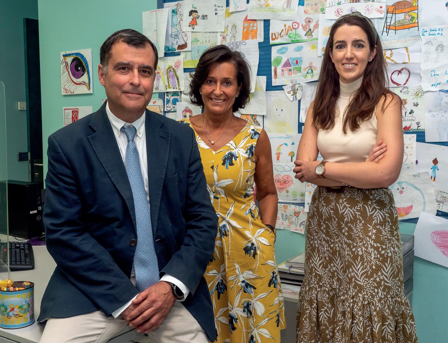
La oftalmología pediátrica trata las afecciones oculares desde el nacimiento del bebé hasta la entrada en la etapa adulta. Es muy importante que los trastornos oculares en niños sean detectados a tiempo con el fin de ser tratados lo antes posible y evitar así problemas visuales, garantizando una buena visión durante el desarrollo y resto de la vida. El éxito de los trata-
mientos depende, en gran medida, del diagnóstico precoz de las enfermedades oculares.
Se aconseja la primera revisión en niños sin un problema visual previo en torno a los tres años de edad. Bien es cierto que, después del nacimiento, el pediatra realiza un examen completo de la morfología ocular para detectar enfermedades oculares congénitas
(cataratas, glaucoma, estrabismo) y es recomendable permanecer atentos a cualquier cambio en el comportamiento del niño (se empieza a caer frecuentemente, alteración en la posición del ojo, se acerca mucho a los objetos para mirarlos, presencia de mancha blanca en el ojo), momento en el que debemos consultar sin demora a un Oftalmólogo Pediátrico.
20 A FONDO
El seguimiento y control desde la infancia y las medidas de prevención adecuadas, la mejor estrategia para evitar problemas
Por la izquierda, el Dr. Javier Fernández-Vega Sanz, la Dra. Lucía Fernández-Vega Sanz y la Dra. Belén Sánchez Cañal.
Retinopatía del prematuro y retinoblastoma, las dolencias más graves en bebés
Dos de los problemas más graves que pueden aparecer en la etapa postnatal y antes de los 2/3 años de edad son la retinopatía del prematuro y el retinoblastoma. La primera afección se da en niños prematuros y nacidos de bajo peso. En ellos es necesario examinar el fondo del ojo para evitar la aparición de lesiones retinianas, lo que debe hacerse aproximadamente al mes del nacimiento del bebé. Un examen tardío podría llevar a la aparición de un desprendimiento de retina que conduzca a la ceguera del bebé.
Por otro lado, el reti-
noblastoma es el tumor maligno intraocular más frecuente en niños menores de tres años, y supone una amenaza para la visión y la vida del niño. Si bien la tasa de supervivencia ronda el 95%, es determinante la detección temprana. El síntoma más común es la presencia de una mancha blanca en la pupila (leucocoria). La sospecha clínica del retinoblastoma por un oftalmólogo pediátrico implica la necesidad de traslado inmediato a un centro de referencia nacional. Aunque la mayoría son espontáneos (60%), existe un 40% con genética positiva los cuales suelen ser más agre-
sivos y afectar a los dos ojos.
Otra patología ocular con gran repercusión en la visión es la catarata congénita. Estas son opacidades del cristalino presentes desde el nacimiento. En muchas ocasiones no se conoce la causa, pero a veces puede ir asociada a enfermedades infecciosas o a alteraciones sistémicas.
La presencia de esta opacidad no permite ver bien al niño y es por eso que es necesario realizar cirugía precoz que, junto con una posterior rehabilitación, logrará una buena agudeza visual en el ojo en cuestión.
Tras
nacimiento,
el glaucoma
Estrabismo infantil, diferente al de los adultos
El estrabismo es la subespecialidad oftalmológica que hace referencia a la motilidad ocular, es decir, cuando los dos ojos no se alinean en la misma dirección cuando miran a un objeto lejano o cercano. Esto puede ocurrir por diferentes razones y en distintas etapas de la vida. Cuando sucede en edad adulta su principal sintomato-
logía será la diplopía o visión doble de los objetos. Si esta aparece de manera aguda, lo indicado en la mayoría de las ocasiones es realizar una prueba de imagen y posterior examen neurológico urgente. Si la visión doble permanece en el tiempo, y no se resuelve, es posible que sea necesario realizar tratamientos intervencionistas como inyecciones
con toxina botulínica o cirugía.
En la etapa infantil un estrabismo se comporta de manera diferente al de los adultos ya que ellos no manifiestan visión doble. Si detectamos un estrabismo en un niño debemos llevarlo al oftalmólogo para realizar un examen completo de la visión, ya que en la mayoría de ocasiones la causa
del mismo es un defecto refractivo no corregido, como la hipermetropía o la miopía.
Es crucial ante la presencia de un estrabismo en un niño detectar la existencia o no de ambliopía u ojo vago, ya que los tratamientos actuales únicamente son útiles hasta los 8/9 años de edad, y si no se realizan a
tiempo en esta etapa es posible que el niño no desarrolle una visión al 100% por uno de sus ojos. Finalmente, si ese estrabismo no se corrige con gafas y presenta una magnitud significativa, muy probablemente necesitará intervención quirúrgica y será el oftalmólogo pediátrico el que decida cuál será el mejor momento para realizarla.
21
el
el pediatra realiza un examen completo para detectar enfermedades oculares congénitas como las cataratas o
A FONDO
EL FUTURO DE LA OFTALMOLOGÍA PASA POR LA APUESTA EN INVESTIGACIÓN S
En el siglo XXI han tenido lugar grandes avances en la investigación sanitaria y, en concreto, en el sector de la oftalmología. Esto se debe a múltiples factores, entre los que destaca el desarrollo de patologías relacionadas con el aumento del uso sin descanso de la visión de cerca (estudiando, leyendo, utilizando pantallas, etc.) y con el consiguiente descenso de actividades que permitan mirar a lo lejos.
e estima que el 52% de la población mundial en 2050 será miope. De hecho, ya es la principal causa de discapacidad visual moderada y grave en países desarrollados. Por ello, en los últimos años, la investigación se ha centrado en avanzar en la cirugía refractiva para corregir o minimi-
zar los defectos de refracción, entre los que se encuentran, además de la miopía, la hipermetropía, el astigmatismo o la presbicia.
En cuanto a las patologías más prevalentes relacionadas con el progresivo envejecimiento de la población, principal factor de riesgo para múltiples alteraciones oculares, la más extendida es la catarata, primera causa de ceguera reversible a nivel mundial. Su cirugía es la más frecuente en los países desarrollados: cada año se reali-
zan en el mundo en torno a 30 millones de intervenciones y en España, más de un millón.
En los últimos años, uno de los principales avances en la técnica quirúrgica en cataratas ha sido el láser de femtosegundos, aprobado en 2010 para su uso en esta cirugía, que ha conseguido aumentar la precisión y seguridad de la intervención. Otra innovación fundamental en este campo son las lentes intraoculares, que en los últimos años han alcanzado
A FONDO
22
Dr. Luis Fernández-Vega Cueto-Felgueroso
un nivel de calidad y prestaciones excepcional, permitiendo aportar buenas agudezas visuales para cualquier distancia.
Con el fin de avanzar en el conocimiento de las bases de las enfermedades que causan ceguera y alteraciones de la visión y así lograr nuevas medidas de prevención,
diagnóstico y tratamiento médico-quirúrgico, cada vez es mayor el esfuerzo realizado en investigación traslacional, es decir, aquella que va del laboratorio a la cabecera del paciente. En este sentido trabajamos en el Instituto Oftalmológico Fernández-Vega (IOFV), con amplia experiencia adquirida en
A FONDO
El láser de femtosegundos ha conseguido aumentar la precisión y seguridad de la intervención de cataratas.
nuestros 135 años de historia y cinco generaciones. Desde 2009, contamos con la Fundación de Investigación Oftalmológica (FIO), que dispone de cuatro grupos de investigación diferenciados: superficie ocular, genética del glaucoma, neurobiología de la retina e investigación clínica.
Las patologías de la superficie ocular afectan cada vez más a la población de los países desarrollados. Muchas de estas enfermedades no tienen tratamiento curativo en el momento actual, por ello desde la Fundación de Investigación Oftalmológica contamos con un grupo que tiene líneas de investigación abiertas en ojo seco, (envejecimiento y enfermedades neurodegenerativas, que no son de superficie), queratocono, medicina regenerativa de la córnea y terapias avanzadas (terapia celular, ingeniería tisular).
Disponemos también de un grupo de investigación del
glaucoma, segunda causa de ceguera en el mundo. Nuestros estudios en materia de genética ocular se centran en varias líneas diferenciadas para la detección precoz de esta patología mediante la búsqueda de biomarcadores proteicos y el estudio de los genes de riesgo asociados.
En cuanto a la neurobiología de la retina, estamos investigando las células dañadas (células ganglionares y otras) para evitar su muerte y lograr una supervivencia más larga mediante el empleo de factores que protegen la parte de la célula que le da energía (las mitocondrias).
Asimismo, nos centramos en la degeneración macular asociada a la edad (DMAE), una patología ocular para la que actualmente no existe cura. Esta afecta a la retina y en concreto a la mácula, que es donde se localiza la visión central, aquella que permite enfocar los objetos y diferenciar los detalles más pequeños. En
los últimos años las vías de investigación para esta patología han avanzado exponencialmente, sobre todo en los ensayos con terapias génicas. En nuestro caso, estamos estudiando la implicación del zinc y las metalotioneínas (las proteínas más importantes en la regulación de los niveles de zinc) en el desarrollo de esta enfermedad.
El cuarto grupo de investigación, relacionado con la investigación clínica, es fundamental para mejorar el abordaje de muchas patologías visuales, en muchos casos infradiagnosticadas. Esto se debe a que permite que las investigaciones básicas se trasladen a la consulta en beneficio del pa-
ciente, propiciando la multidisciplinariedad. En la FIO nos centramos en el estudio del segmento anterior y posterior del ojo. Un equipo multidisciplinar del IOFV, compuesto por oftalmólogos, optometristas, enfermeros, farmacéuticos y personal auxiliar, entre otros, se dedica a estudiar las patologías y tratamientos que afectan a la córnea, el cristalino, la retina y el glaucoma. Estas investigaciones son posibles gracias a la gran afluencia de pacientes que pasan por el Instituto al año, más de 110.000, lo que se traduce en más de 220.000 ojos que pueden ser analizados para poder avanzar en la investigación de las patologías oculares.
Ahora, en nuestro horizonte se encuentra el reto de seguir a la vanguardia de la investigación en la próxima década. Ante futuribles pandemias y nuevas enfermedades, tendremos que reforzar nuestras
líneas de trabajo y seguir consolidando la formación de equipos multidisciplinares. Porque algunas de estas patologías afectan también a los ojos, como por ejemplo la COVID-19, que hemos estudiado debido a que en algunos casos ha provocado secuelas oculares similares a las de los pacientes con neuropatía diabética y enfermedad de ojo seco, o el reciente brote de viruela del mono, que puede afectar a los ojos en el 20 % de los casos y llegar a producir queratitis, es decir, inflamación de la córnea.
No hay duda de que la oftalmología sigue presentándonos desafíos, tanto en el diagnóstico como en el tratamiento de muchas patologías visuales de difícil abordaje. Desde el Instituto Oftalmológico Fernández-Vega seguiremos trabajando para continuar mejorando la calidad de vida de nuestros pacientes.
A FONDO
La investigación clínica es fundamental para mejorar el abordaje de muchas patologías visuales, en muchos casos infradiagnosticadas
24
Nuestros estudios en materia de genética ocular se centran en varias líneas diferenciadas para la detección precoz del glaucoma, la búsqueda de biomarcadores proteicos y el estudio de los genes de riesgo asociados.
25
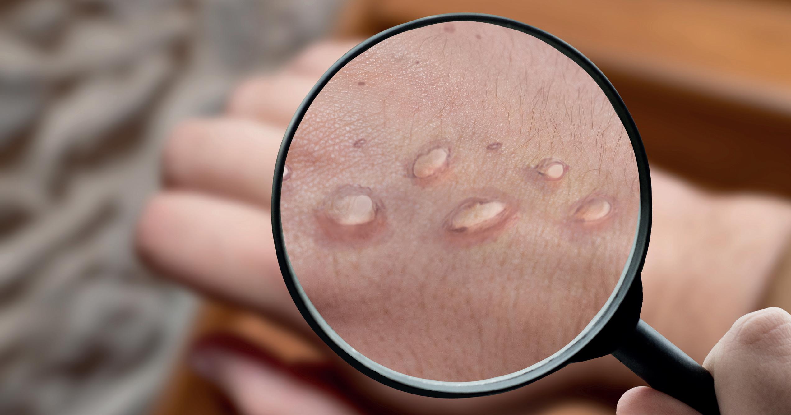
LA ERUPCIÓN CUTÁNEA POR VIRUELA DEL MONO PUEDE AFECTAR A LOS OJOS EN EL 20% DE LOS CASOS
Según recoge un estudio de The Lancet, las complicaciones oculares por viruela del mono pueden producir queratitis, es decir, inflamación de la córnea. Esta dolencia puede generar diferentes complicaciones, como infecciones virales recurrentes, inflamación crónica, reducción de la visión e, incluso, ceguera.
Referencias:
[1] Organización Mundial de la Salud. Viruela símica: https://www.who.int/es/ news-room/fact-sheets/ detail/monkeypox
[2] The Lancet. Clinical features and management of human monkeypox: a retrospective observational study in the UK: https:// www.thelancet.com/ journals/laninf/article/ PIIS1473-3099(22)002286/fulltext
La viruela del mono puede generar graves consecuencias para la salud ocular. Según la Organización Mundial de la Salud (OMS)[1], la erupción cutánea que se da con esta enfermedad puede propagarse en el 20% de los casos a la conjuntiva, la membrana que recubre la parte blanca del ojo. Además, tal y como recoge un estudio de The Lancet[2], estas complicaciones pueden producir inflamación de la córnea, llamada queratitis.
“Esto se debe a que la córnea forma junto a la conjuntiva la superficie ocular, es decir, la primera capa de defensa del ojo. Por esta relación, y al ser ambas la primera envoltura del globo ocular, muchas veces se afectan de forma conjunta, como es el caso de la viruela del mono”, explica el Dr. Luis Fernández-Vega Cueto-Felgueroso, de la unidad de córnea y cristalino del Instituto Oftalmológico Fernández-Vega. “La queratitis -aña-
Los síntomas más comunes, además de la alteración de la agudeza visual, son el dolor agudo y la irritación, que pueden llegar a producir dificultad
INVESTIGACIÓN
a la hora de parpadear.
26
de- es una inflamación que puede derivar en molestias oculares y también en pérdidas importantes de la agudeza visual, de hecho, puede acabar en ceguera”.

Además de las infecciones por virus, como el de la viruela símica, la queratitis puede producirse por bacterias, hongos y protozoos, sequedad ocular, irritaciones por agentes físicos y químicos (incluidos las radiaciones ultravioletas y el uso de lentes de contacto) y procesos alérgicos. Casi siempre estas queratitis son
superficiales, pues afectan a las capas más externas de la córnea.
Entre los síntomas más comunes, además de la alteración de la agudeza visual, están el dolor agudo y la irritación, que pueden llegar a producir dificultad a la hora de parpadear. “También suelen ser habituales los ojos rojos, el picor ocular, el lagrimeo excesivo y, en muchas ocasiones, la fotofobia, es decir, el exceso de sensibilización a la luz”, indica la Dra. Pilar Quiroga, oftalmóloga del Instituto Oftalmológico Fernández-Vega.
La queratoplastia, un tratamiento eficaz Con menos frecuencia puede haber afectación de capas profundas y entonces las consecuencias son más graves, llegando a ocasionar opacidades corneales permanentes. Para tratarlas, es preciso un trasplante corneal denominado queratoplastia. Este trasplante puede ser lamelar, es decir, solo se trasplanta la parte afectada de la córnea (epitelio, estroma o endotelio). “Hace unos años, la córnea solo se podía sustituir por otra completa, trasplantando todas sus capas. Ahora, los avances han permitido que podamos sustituir solo las capas dañadas, lo que reduce drásticamente el índice de complicaciones como rechazos, fallos del injerto o glaucoma, así como el tiempo de recuperación visual. Además, si con el paso de los años ocurre una recidiva de la patología o cualquier otro problema, se puede sustituir ese botón corneal por otro nuevo conservando siempre el endotelio del paciente”, señala el Dr. Fernández-Vega.
El diagnóstico precoz, clave para evitar complicaciones
Si no se trata o el diagnóstico se retrasa, la infección puede generar diferentes complicaciones, tales como infecciones virales recurrentes, inflamación crónica, reducción de la visión e, incluso, ceguera.
Por ello, para detectar esta afección a tiempo, es imprescindible acudir al oftalmólogo y someterse a un examen ocular con el que se detectará la extensión y el efecto que ha causado sobre las estructuras del ojo. Además, se puede pedir un análisis de las lágrimas con el fin de detectar el carácter infeccioso del problema.
INVESTIGACIÓN
Para detectar esta afección a tiempo hay que someterse a un examen ocular para medir la extensión y el efecto que ha causado.
27
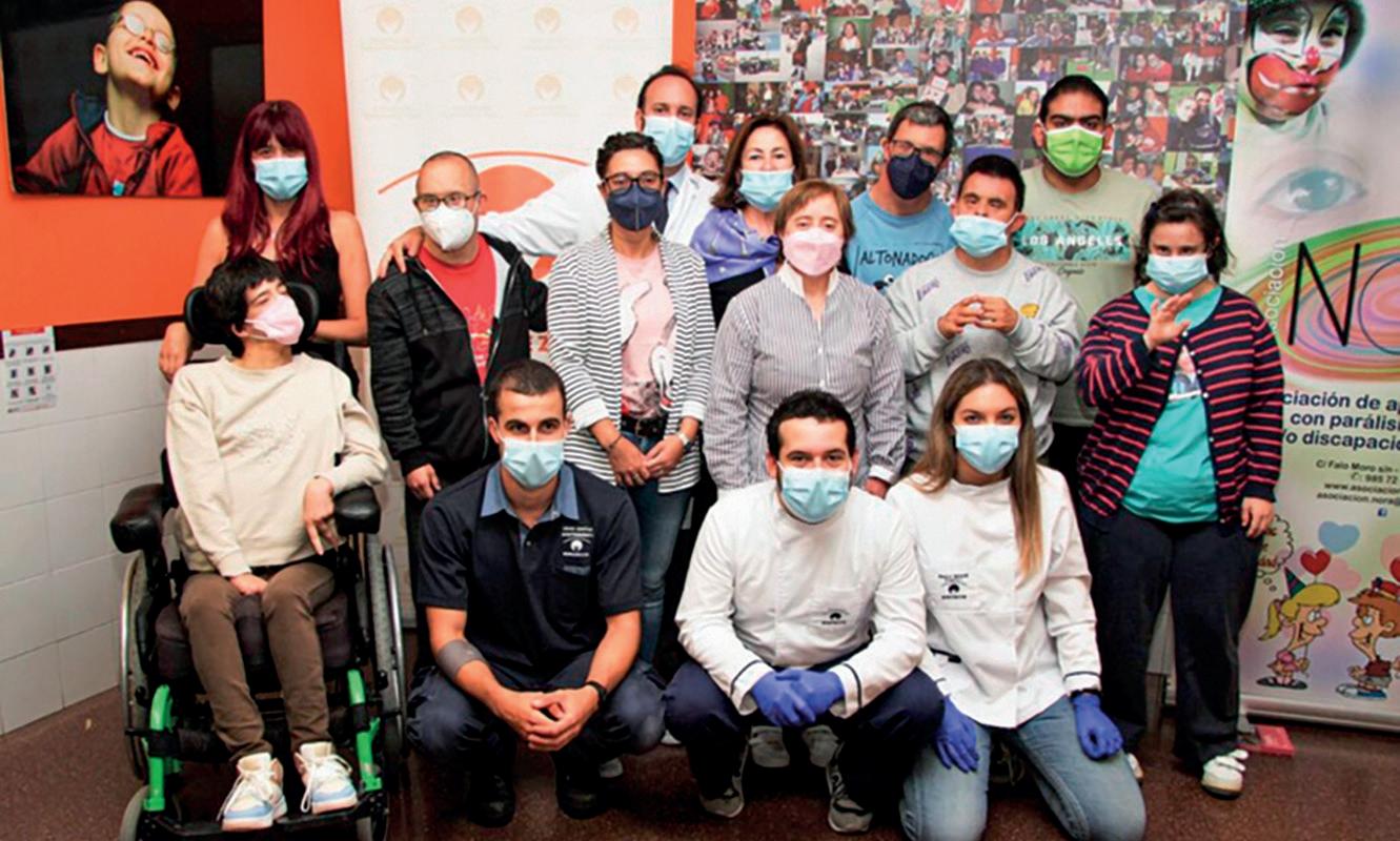
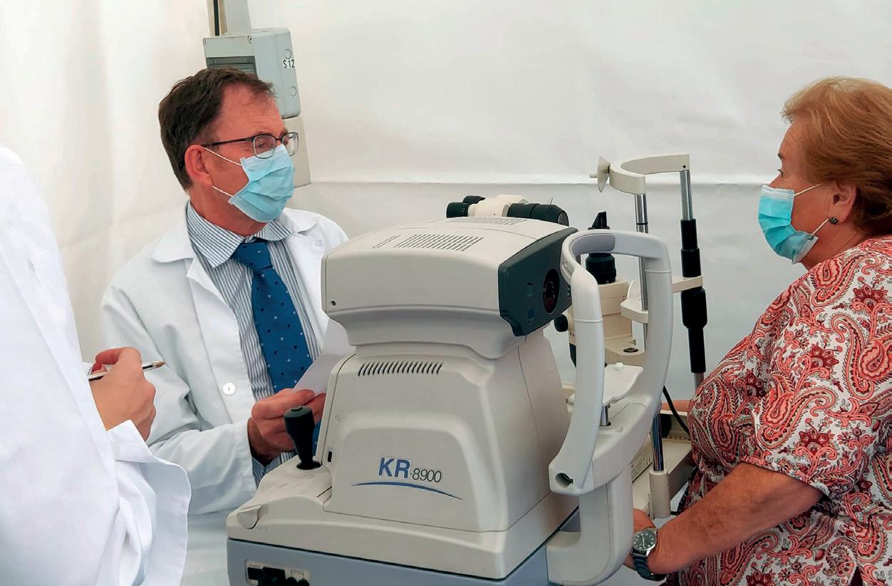
VISITA DE LA FUNDACIÓN FERNÁNDEZ-VEGA A LA ASOCIACIÓN NORA
Coincidiendo con las celebraciones del 30 aniversario de la Asociación Nora, un grupo de profesionales de la Fundación Fernández-Vega visitó la entidad para atender a más de veinte usuarios de todas las edades y llevar a cabo revisiones oftalmológicas.
Concretamente, a la visita asistieron Dª Victoria Cueto-Felgueroso Botas, directora de la Fundación Fernández-Vega; el Dr. Andrés Fernández-Vega Cueto-Felgueroso, oftalmólogo de la unidad de glaucoma del Instituto Oftalmológico
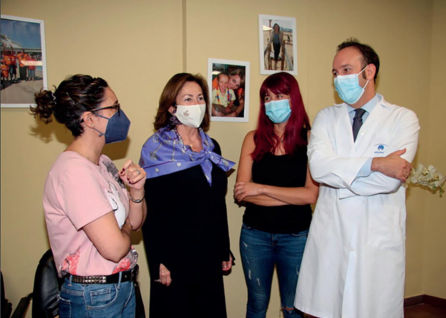
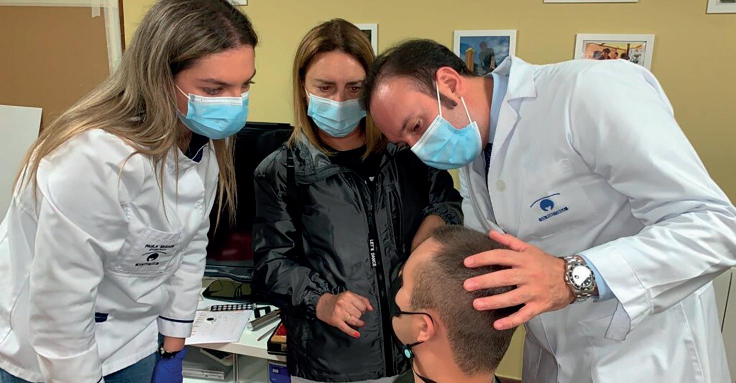
Fernández-Vega; los ópticos-optometristas Paula Segade y Álvaro Cuéllar y Diego Costas, de mantenimiento. Tatiana Llorente, presidenta de la Asociación Nora, agradeció la iniciativa de la Fundación, que lleva realizándose varios años. “Estas revisiones oftalmológicas completas son muy importantes para los pacientes pues pueden mejorar su calidad de vida realizando las tareas del día a día con
mayor facilidad”, señaló el Dr. Andrés Fernández-Vega Cueto-Felgueroso.
Por su parte, Victoria Cueto-Felgueroso Botas indicó que “como directora de la Fundación Fernández-Vega, venir con los médicos del Instituto para ayudar a las personas que lo necesiten es uno de los mayores reconocimientos personales que puede tener una persona, ayudar a los demás es lo mejor”.
ACTIVIDADES SOLIDARIAS
28
El Dr. Andrés Fernández-Vega Cueto-Felgueroso; Doña Victoria Cueto-Felgueroso Botas, directora de la Fundación Fernández-Vega, y el resto de profesionales del Instituto fueron recibidos por Tatiana Llorente, directora de la asociación Nora.
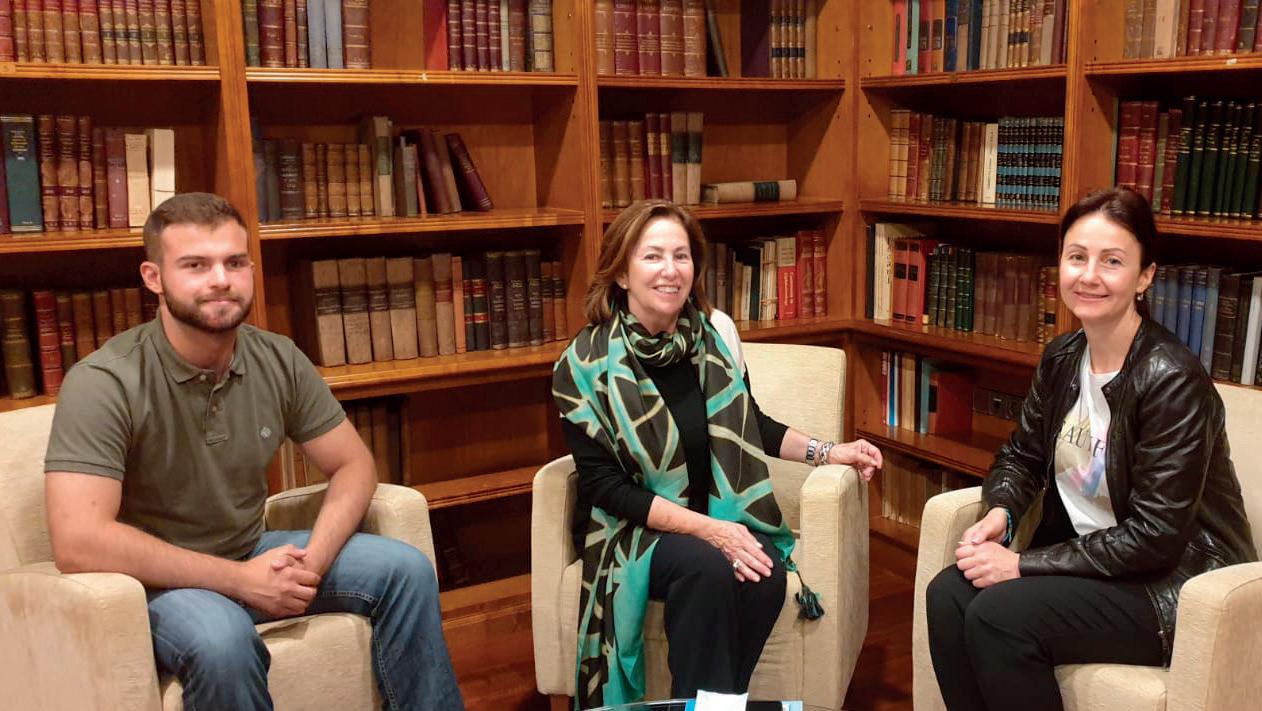
LA FUNDACIÓN FERNÁNDEZ-VEGA, CON UCRANIA
ACTIVIDADES SOLIDARIAS
¿Cómo es el día a día de una familia en guerra?
Depende de muchos factores. Las hay que se tuvieron que ir y solo ven el conflicto por la televisión, mientras que otras viven en bunkers y salen solo a veces. Algunas personas sufren escasez de comida porque, aunque tratan de asegurarse el abastecimiento, hay zonas muy castigadas por la guerra en las que resulta muy difícil acceder a los alimentos. Otras tienen familiares
que han tenido que alistarse y sufren también por la incertidumbre que ello supone.
La mejor situación es, por lo general, la de la gente que huyó. Están a salvo y eso marca la diferencia. En las zonas de conflicto hay familias que no pudieron marchar o no pensaron que éste fuera a ser tan largo.
Yo, desde los primeros días de la guerra, estuve en la zona más peligrosa, en la frontera con Rusia. Mi familia es-
taba cerca, en una casa de pueblo, y cuando los soldados rusos entraron en Ucrania, mi padre y yo decidimos ir al frente. El resto de la familia salió del país.
¿Cómo conoció la Fundación Fernández-Vega?
A través de familiares. Cuando conocieron mi problema, alguien que está aquí, en España, se reunió con unos amigos para tratar de ayudarme.
Encontraron esta clínica en Oviedo y conectaron con una asociación de ayuda al pueblo ucraniano.
Les mostraron las recomendaciones de distintas clínicas oftalmológicas y, dado que ésta era la mejor, no dudamos en venir. En
Tiene 21 años recién cumplidos y es ucraniano. Tras terminar el colegio, comenzó sus estudios de técnico informático en Ucrania. En 2014, coincidiendo con las primeras tensiones con Rusia, prometió proteger a su patria fuera como fuese e ingresó en el ejército, tras formarse en defensa y ayuda médica. La situación por la que atraviesa su país le aconseja la mayor discreción sobre sus datos personales en aras a su seguridad, pero también a la de sus personas más próximas.
Ucrania no había esperanza para mi problema.
¿Cómo ha sido su experiencia en la clínica?
Me encantó el trato y el desempeño de los profesionales, así como la rapidez con la que me atendieron. Son muy buenos y la tecnología que utilizan, muy avanzada. En Ucrania no vi nada igual. Estoy muy contento por el trato recibido.
¿Qué necesidades tiene más urgentes la sociedad ucraniana?
Necesitamos ayuda militar, especialmente armamento. Rusia está mejor dotada y eso hace que muera mucha gente. También medicamentos para la población civil y los heridos de guerra, así como alimentos, tecnología y recursos para reconstruir el país cuando esto termine, que esperemos que sea pronto.
“La mejor situación es, por lo general, la de la gente que huyó. Están a salvo y eso marca la diferencia”
“Los doctores del IOFV son muy buenos y la tecnología que utilizan, muy avanzada. En Ucrania no vi nada igual”
29
Doña Victoria Cueto-Felgueroso Botas, directora de la Fundación Fernández-Vega, acogió la visita.
ACTIVIDADES SOLIDARIAS
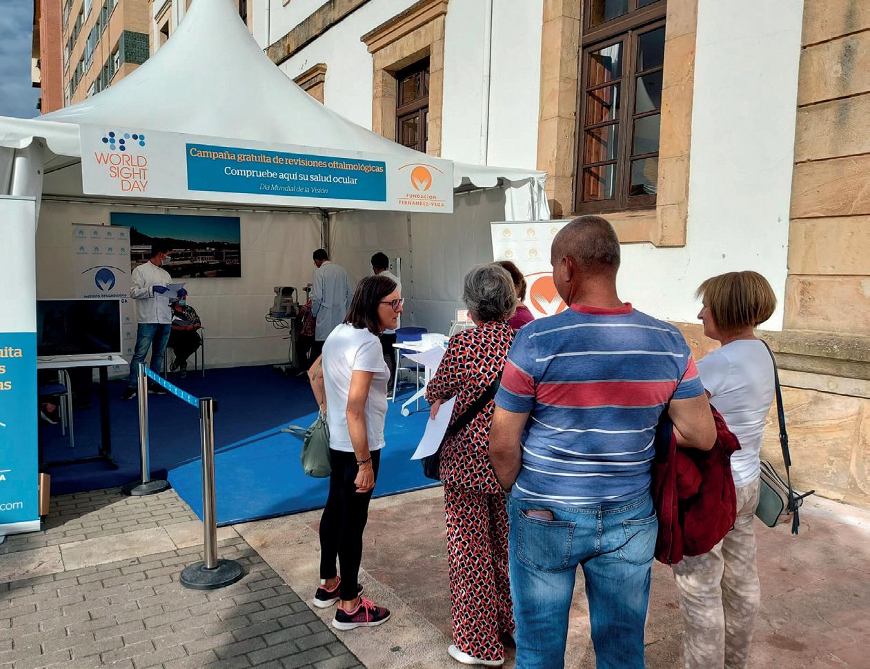
CELEBRAMOS EL DÍA MUNDIAL DE LA VISIÓN EN CANGAS DE ONÍS
Con motivo de la celebración del Día Mundial de la Visión, la Fundación Fernández-Vega ha recuperado su tradicional carpa solidaria para atender a los pacientes de forma gratuita. Este año, el Dr. Andrés Fernández-Vega Cueto-Felgueroso y el Dr. Tomás Parra Rodríguez se trasladaron a Cangas de Onís, la localidad asturiana en la que se ubicó este año.
Las revisiones, que se realizaron durante toda la jornada, permitieron valorar la salud visual de docenas de vecinos de la localidad. De esta manera, la Fundación Fernández-Vega se sumó al movimiento promovido por la ONU, enfocado a la concienciación ciudadana. Las revisiones oftalmológicas periódicas resultan cruciales para detectar a tiempo posibles enfermedades oculares, especialmente las asintomáticas.
En la primera infancia, la Unidad de Oftalmología Pediátrica del Instituto Oftalmológico Fernández-Vega, dirigida por el Dr. Javier Fernández-Vega Sanz y con la Dra. Lucía Fernández-Vega Sanz y la Dra. Belén
Sánchez Cañal, aconseja realizar revisiones periódicas. La primera, al nacer, para descartar patologías congénitas. Si no se aprecian molestias ni problemas de visión, también es recomendable una segunda
consulta a los tres años. De esta forma se pueden prevenir algunos problemas como el ojo vago, el estrabismo o posibles defectos de refracción como la miopía, el astigmatismo o la hipermetropía.
En la etapa adolescente es posible que desarrollen queratocono, una deformidad de la córnea.
Ya en la edad adulta, la catarata es el problema más frecuente y puede, incluso, provocar la ceguera. Antes de este problema, los pacientes suelen presentar presbicia, entre los 45 y los 55 años. Se trata de una dolencia causada por la pérdida de elasticidad del cristalino, lo que conlleva que el paciente tenga dificultad para enfocar a corta distancia.
En personas mayores de 50 años, la Degeneración Macular Asociada a la Edad o DMAE es la enfermedad más frecuente y provoca la pérdida visual irreversible.
En todos estos casos, las revisiones oftalmológicas periódicas son imprescindibles para diagnosticar las diferentes dolencias a tiempo y frenar su evolución.
30
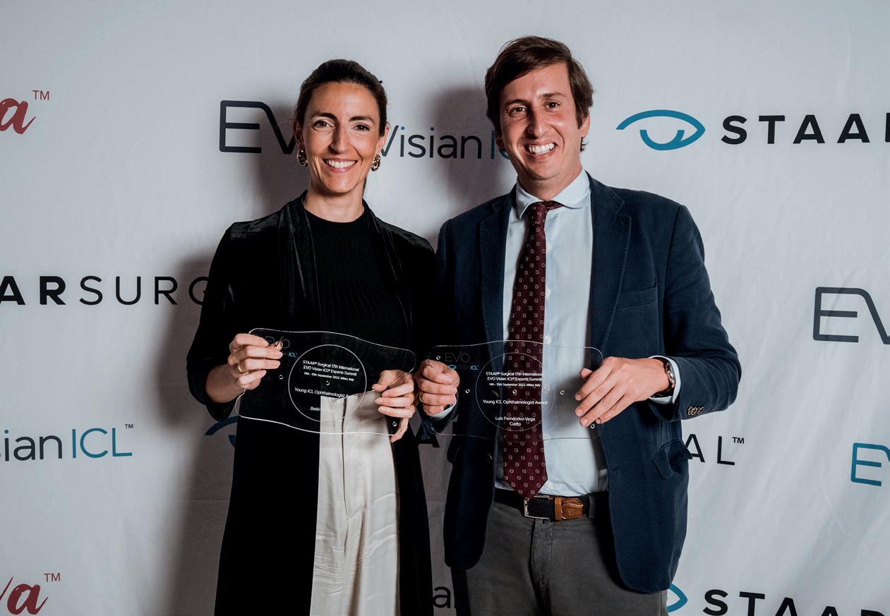
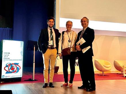
LOS DOCTORES LUIS
FERNÁNDEZ-VEGA CUETO-FELGUEROSO Y BELÉN
ALFONSO BARTOLOZZI, ENTRE LOS MEJORES OFTALMÓLOGOS JÓVENES DE EUROPA
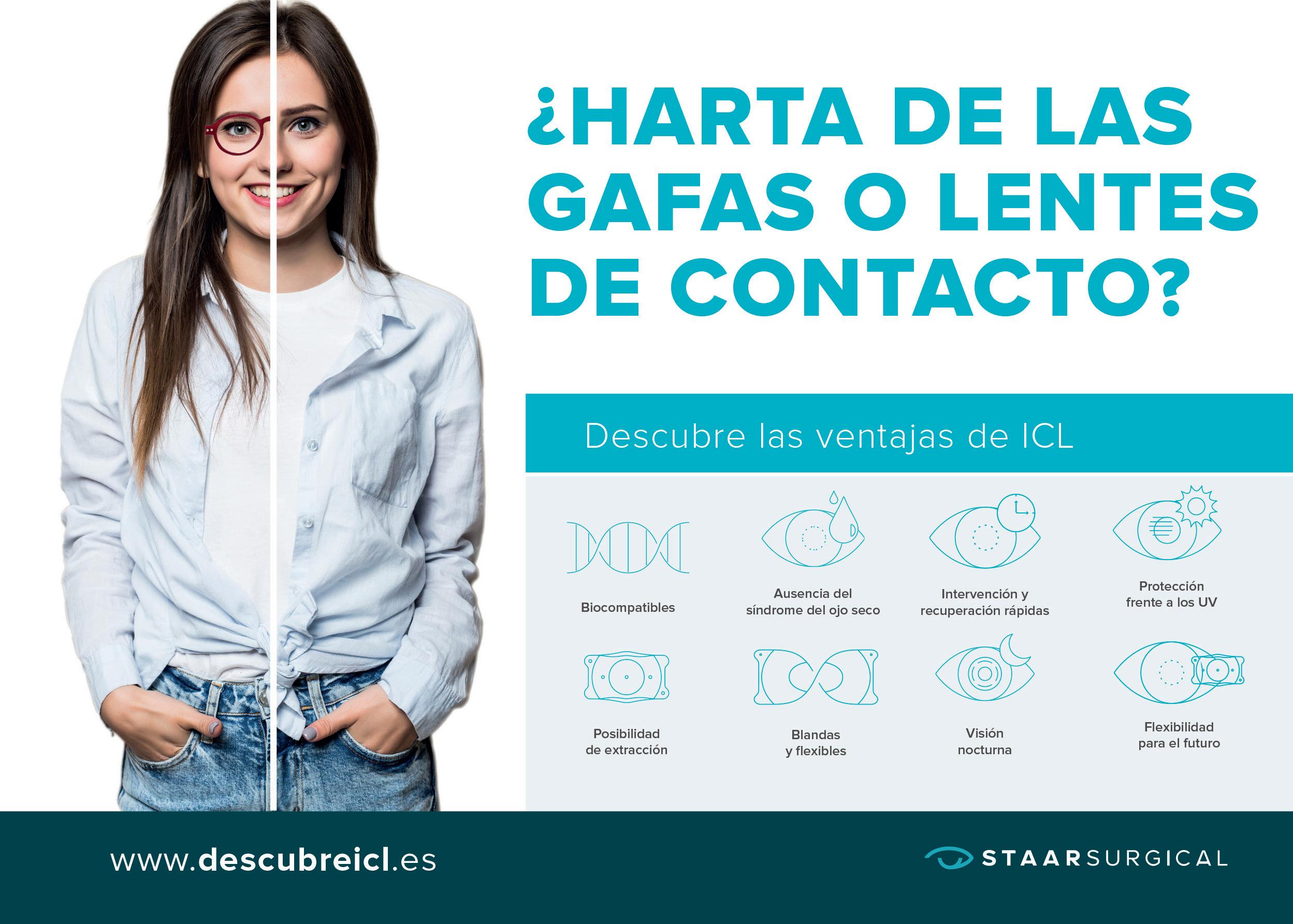
sí se ha decidido el pasado septiembre en Milán, durante el encuentro anual de la Sociedad Europea de Cirugía Refractiva (ESCRS), que reunió a más de cuatro mil oftalmólogos que cuentan, entre sus especialidades, con la de implantación de lentes ICL – intraoculares – para combatir miopías, hipermetropías y astigmatismos.
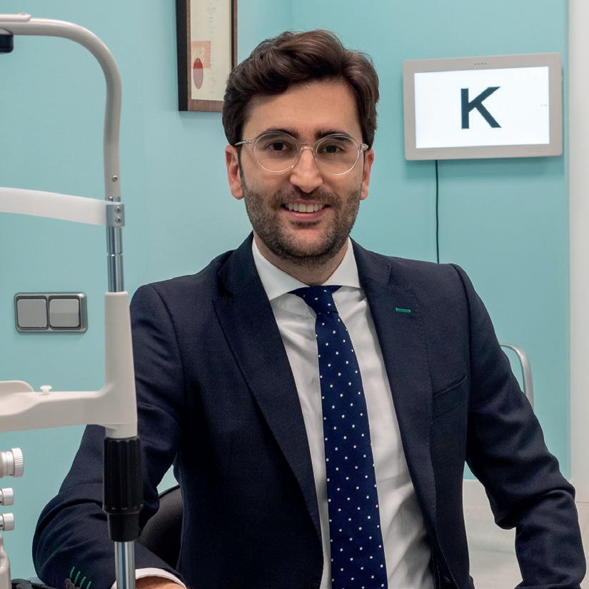
Fue en esta convención donde se reconoció a los doctores Luis Fernández-Vega Cueto Felgueroso y Belén Alfonso Bartolazzi
Aentre los mejores jóvenes oftalmólogos europeos, dada su brillante trayectoria y su ya más que acreditada experiencia en la aplicación de una tecnología que ha venido a aportar mayor seguridad, fiabilidad y comodidad al paciente.
Junto a ellos, también fue distinguido otro profesional del Instituto, el Dr. Carlos Lisa, por haber implantado ya más de 1.000 lentes de las características citadas, así como el Dr. José Alfonso, por su muy valiosa contribución a las publicaciones científicas que difunden y profundizan en las ventajas de un dispositivo y una tecnología en la que es uno de los mayores expertos europeos.
ACTIVIDAD CONGRESUAL
31
Los Dres. Luis Fernández-Vega Cueto-Felgueroso y Belén Alfonso Bartolozzi, en el evento.
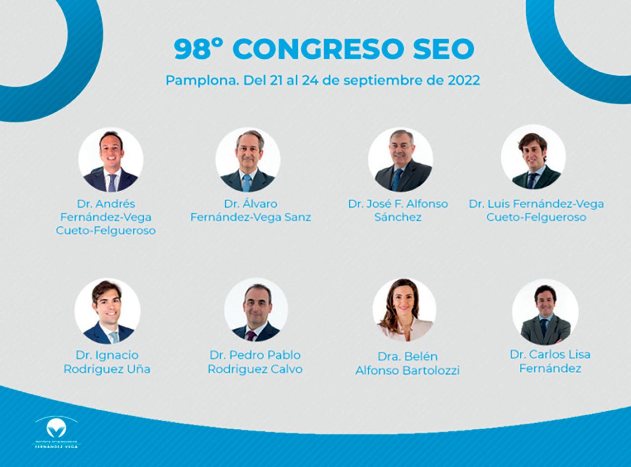
98º CONGRESO DE LA SOCIEDAD ESPAÑOLA DE OFTALMOLOGÍA
Comunicaciones libres
En la primera jornada, los Dres. Belén Alfonso Bartolozzi, Luis Fernández-Vega Cueto-Felgueroso y Pedro Pablo Rodríguez Calvo participaron como ponentes para hablar sobre “DALK + LIO” y “Lensectomía alta miopía – Seguridad quirúrgica”, respectivamente.
Las comunicaciones libres de la segunda jornada contaron con la participación del Dr. José F. Alfonso Sánchez, que disertó sobre la lente “Vivity” y del Dr. Ignacio Rodríguez Uña, que lo hizo sobre “Evaluación Cierre Angular OCT.
Cursos de actualización
La Sociedad Española de Oftalmología (SEO) celebró en Pamplona, a finales de septiembre, la 98ª edición de su congreso, recuperando la presencialidad tras dos años en los que el encuentro tuvo carácter virtual por la situación sanitaria. En esta importante cita se reunieron los expertos de todas las subespecialidades del sector de la Oftalmología, entre los que se encontraba una amplia representación de doctores del Instituto Fernández-Vega, de las unidades de córnea, cristalino, retina y glaucoma. El Profesor Luis Fernández-Vega Sanz intervino en el marco para la subespecialidad de Cataratas y Refractiva para hablar sobre los “Motivos de lensectomía precoz”, con el Dr. José F. Alfonso Sánchez como moderador. Sobre retina, el Dr. Álvaro Fernández-Vega Sanz intervino como moderador y ponente con una exposición sobre “Desprendimiento de Retina”. Además, el simposio sobre “Asquelio” contó como panelista con el Dr. Luis FernándezVega Cueto-Felgueroso, quien también participó en el Debate Intergeneracional de Cirugía en Segmento Anterior del simposio de Alcon.
Por lo que se refiere a los cursos de actualización, el Dr. José F. Alfonso Sánchez fue el director del curso “ENDO-K”, en el que la Dra. Belén Alfonso Bartolozzi y los Dres. Luis Fernández-Vega Cueto-Felgueroso y Carlos Lisa Fernández intervinieron como ponentes. En este curso avanzado, dirigido a oftalmólogos expertos en córnea, los doctores expusieron los últimos avances de la prótesis corneal llamada endo-queratoprótesis, que se implanta utilizando técnicas lamelares, evitando la perforación del globo ocular en pacientes que necesitan un trasplante de córnea. El concepto y diseño inicial de la Endo-queratoprótesis ha sido desarrollado en el Instituto Universitario Fernández-Vega, que ha trabajado conjuntamente con la empresa AJL, encargada de la optimización y la fabricación del dispositivo.
En el curso sobre glaucoma, los doctores Pedro Pablo Rodríguez Calvo e Ignacio Rodríguez Uña ejercieron de directores y el Dr. Andrés Fernández-Vega Cueto-Felgueroso intervino como ponente.
ACTIVIDAD CONGRESUAL
32
El Dr Ignacio Rodríguez Uña fue distinguido en este congreso, en los apartados de fotografía científica y comunicación en panel
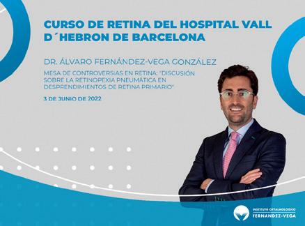
CURSO DE RETINA DEL HOSPITAL VALL D’HEBRÓN
3 junio
El Dr. Álvaro Fernández-Vega González, de la Unidad de Retina y Vítreo del Instituto Oftalmológico FernándezVega, ha participado en el curso de retina del Hospital Vall D’Hebron de
Barcelona. Concretamente, el Doctor ha intervenido en la mesa de controversias en retina: «Discusión sobre la Retinopexia Pneumática en desprendimientos de retina primario».
CONGRESO DE LA SOCIEDAD EUROPEA DE OJO SECO
10-11 junio
El Profesor Jesús Merayo Lloves, director del Instituto Universitario FernándezVega, participó como ponente y moderador en este Congreso, organizado por la Sociedad Europea de Ojo Seco (EuDES) y celebrado en París. Durante el
encuentro, el Dr. Merayo Lloves aportó las ponencias “Focus the problem of severe dry eye”, “Regeneration of the ocular surface with plasma enriched in growth factors” y “A new esthesiometer for measuring corneal sensitivity”.
25º CONGRESO DE LA SOCIEDAD ESPAÑOLA DE RETINA Y VÍTREO (SERV)
17-18 de junio
El 25º Congreso de la Sociedad Española de Retina y Vítreo (SERV), retransmitido en directo pero con carácter presencial, contó con la participación de los Dres. Álvaro Fernández-Vega Sanz y Álvaro Fernández-Vega González.
Durante dos días, numerosos expertos hablaron sobre los últimos avances tecnológicos tanto para el diagnóstico como para el abordaje de las principales enfermedades de la retina, coroides y vítreo.
Concretamente, el Dr. Álvaro Fernández-Vega Sanz ejerció como moderador en la mesa de casos clínicos mientras que el Dr. Álvaro Fernández-Vega González defendió la comunicación oral “Resultados de la técnica de manguito de silicona para la sutura a esclera de lentes intraoculares luxadas” y participó en la mesa de retina quirúrgica para hablar de “Pneumorretinopexia: aspectos clave para el éxito”.
EUROPEAN CORNEA CONFERENCE
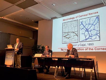
16-19 junio
El encuentro, celebrado en Amberes (Bélgica), contó con la participación del Dr. Jesús Merayo Lloves, director del Instituto Universitario FernándezVega. Concretamente, revisó temas de inervación corneal en esta reunión que
agrupa a destacados líderes de opinión en enfermedades del segmento anterior de Europa. Además, aportó la ponencia sobre «Measurement of Ocular sensitivity with a New Esthesiometer».
ACTIVIDAD CONGRESUAL
33
4TH EUROPEAN MEETING OF YOUNG OPHTHALMOLOGIST
9-10 julio
El Dr. Luis Fernández-Vega CuetoFelgueroso participó en este evento, celebrado en Bruselas (Bélgica) y que agrupa a los más destacados jóvenes oftalmólogos de Europa. En su
intervención expuso el tema «Selección de pacientes y preoperatorio para las distintas técnicas de cirugía refractiva corneal» en la sección de Cirugía Refractiva y de Catarata.
22º CONGRESO DE EURETINA
1-4 septiembre
El congreso, celebrado en Hamburgo y retransmitido en directo, reunió a numerosos expertos que hablaron sobre los últimos avances en retina. El Dr. Álvaro Fernández-Vega Sanz realizó la ponencia “Combined severe trauma involving anterior and posterior segments”, mientras que el Dr. Álvaro Fernández-
Vega González participó con dos vídeos quirúrgicos: “The silicone bolster technique: A new surgical approach for repositioning dislocated intraocular lenses” y “Management of subretinal gas detachment after pneumatic retinopexy: External degassing technique”.
PREVENCIÓN Y CLAVES DEL GLAUCOMA
13 octubre
ONCE, el Instituto Oftalmológico Fernández-Vega y la Asociación Nacional de Informadores de la Salud (ANIS) han celebrado la jornada Prevención y Claves del Glaucoma con motivo del Día Mundial de la Visión. El objetivo principal del
evento es informar y debatir acerca de los últimos avances en el tratamiento de esta patología, primera causa de ceguera irreversible en el mundo. En el encuentro participaron el Dr. Pedro Pablo Rodríguez Calvo y la Dra. Montserrat García Díaz.
CONGRESO ANUAL LIVE-MED AAP
18 y 19 octubre
Este año, Live-Med incluye como parte de la agenda del Congreso una ponencia de oftalmología bajo el título “Un recorrido por la DMAE y otras enfermedades retinianas”, a cargo del Dr. Álvaro Fernández-Vega González, de la Unidad de Retina y Vítreo del Instituto Oftalmológico Fernández-Vega. Durante la ponencia, se pudo participar a través de los casos clínicos presentados y el doctor recibió pre-
guntas por medio de la app de Live-Med. El Programa de Actualización en Atención Primaria está dirigido a profesionales de la Medicina Familiar y Comunitaria como parte de su formación médica continuada, quienes, al cumplir con los requisitos y recibir la notificación de disponibilidad, podrán descargar el diploma acreditativo desde su cuenta en la página oficial de Live-Med.
ACTIVIDAD CONGRESUAL 34
¿QUIÉN INVENTÓ LA OPERACIÓN DE CATARATAS?
Descubre la evolución de la oftalmología y la historia de las cataratas, todas las innovaciones y descubrimientos científicos que han permitido que el tratamiento de esta enfermedad sea algo sencillo y rápido.
¿Sabías que la oftalmología fue la primera rama especializada de la medicina? Ya desde la Antigüedad, el cuidado de los ojos fue un tema de inquietud e investigación por parte de los eruditos. Dentro de todas las enfermedades oculares que ya se conocían, destacan las cataratas, una enfermedad tan común que hoy en día es la principal causa de ceguera en el mundo y que, ya se trataba en la antigüedad. Por eso, a continuación, vamos a hacer un recorrido por la evolución de la oftalmología y, en concreto, por la historia de la cirugía de cataratas.
Antes de seguir, recordemos que las cataratas son una enfermedad que genera una opacidad
en el cristalino, ese tejido transparente en la parte interna del ojo que permite la visión de cerca y de lejos. A lo largo de los años la eficacia de este tejido va disminuyendo, lo que obliga al uso de lentes oculares para facilitar la visión. Junto con esto, es posible que también se pierda transparencia, apareciendo así las cataratas y limitando la visión incluso con gafas.
Como apuntábamos, las cataratas son un problema tan común, que desde muy pronto atrajeron el interés de médicos y científicos. En la actualidad, en muchos países la operación de cataratas es un procedimiento muy común con escasos riesgos, pero no siempre ha sido así.
Un poco de historia: desde los egipcios a la actualidad
Los egipcios, famosos por sus vastos conocimientos astronómicos y científicos, también fueron grandes médicos. De hecho, el tratado de medicina más antiguo que se conoce data del Antiguo Egipto y se remonta hasta el 1550 a.C. Se trata del llamado Papiro de Ebers, que recoge en 877 apartados diversas enfermedades de la vista y sus prescripciones.
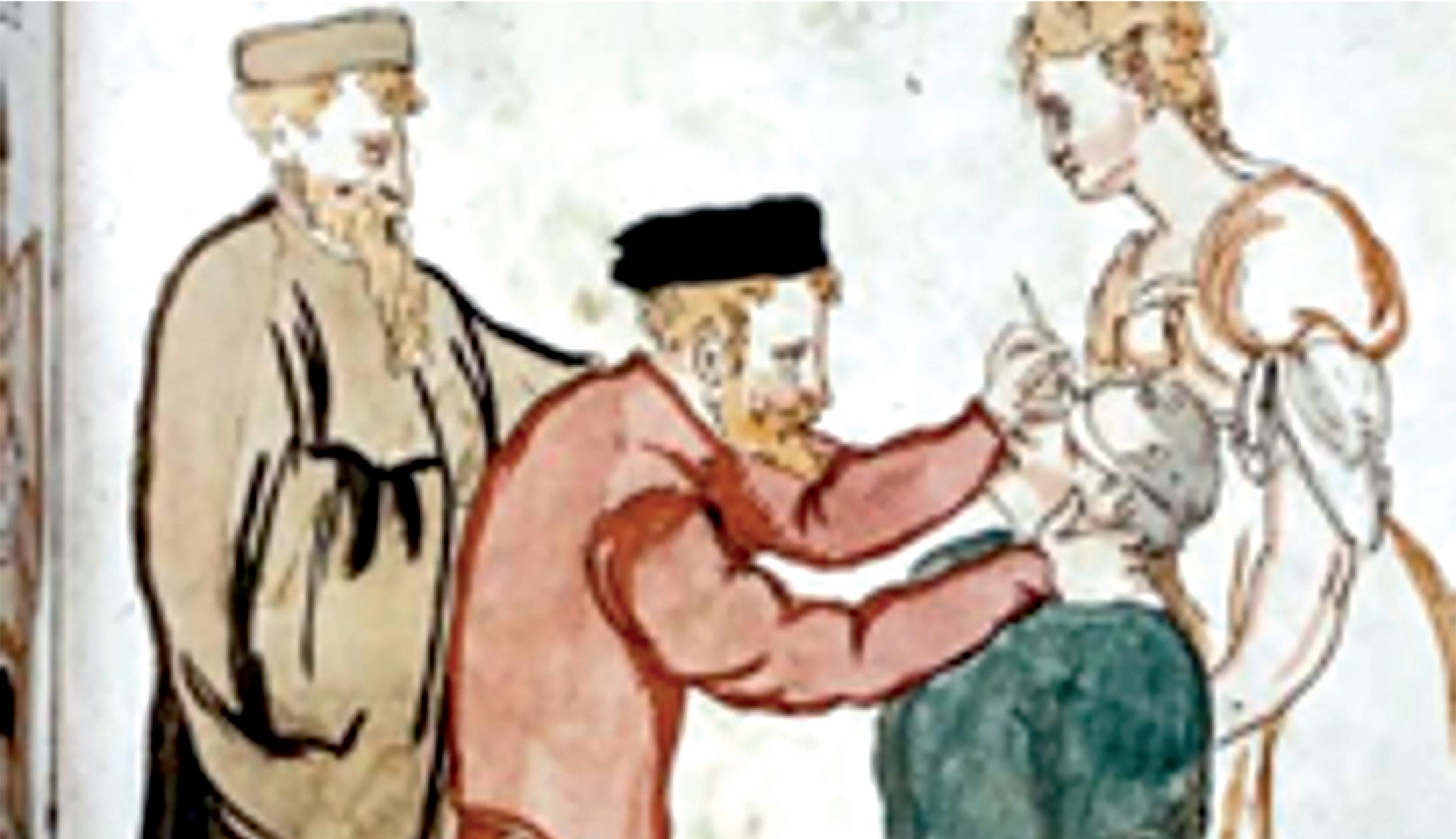
De este periodo data la primera intervención quirúrgica para combatir las cataratas, la llamada técnica del reclinamiento o del abatimiento. Este procedimiento
NOS CUIDAMOS
35
consiste en incidir en la pared del ojo con una aguja empujando el cristalino para abatirlo hacia el interior del ojo. Evidentemente, el índice de fracaso de esta técnica era bastante alto, era muy fácil que generara complicaciones y el postoperatorio era complejo.
Aun así, a falta de otra técnica mejor y como la alternativa era la ceguera, esta intervención fue muy extendida por todo el mundo y, de hecho, se sigue utilizando en algunas zonas remotas de África y Asia. Por ejemplo, tenemos testimonios de cirugías de este tipo llevadas a cabo con éxito durante la Grecia clásica y el Imperio romano. Así mismo, aparece una alusión a este procedimiento en el Susruta Samhita, un tratado de los siglos III-IV donde se recogen los conocimientos de la medicina ayurveda tradicional india.
Durante la Edad Media destaca la
invención de las gafas a partir de las bases teóricas de Alhacén (s. XI). Este erudito y astrónomo árabe desarrolló los preceptos de Séneca, que utilizaba una bola de cristal llena de agua para facilitar la lectura, y propuso el uso de las llamadas “piedras de lectura”: cristales de cuarzo pulido que funcionaban como lupa. Algunos siglos después, en el s. XIII, estos cristales se engarzaron en una montura, tomando la forma de primitivas lentes. Por esta época, en China, estos mismos cristales de cuarzo se ahumaban para dar lugar a las primeras gafas de sol de la historia.
En cuanto a las cataratas, de este periodo data la que probablemente sea una de las intervenciones oftalmológicas más famosas de la historia: el médico judeo-catalán Abnarrabí, un cirujano, oftalmólogo y astrólogo cabalista del siglo XV, logró operar de cataratas con total éxito al rey
Juan II de Aragón, el padre de Fernando el Católico.
No fue hasta la Ilustración, en 1748, cuando se produjo la siguiente gran innovación en el tratamiento de las cataratas. Jacques Daviel, oftalmólogo francés, desarrolló la técnica extracapsular que partía del método tradicional utilizado hasta entonces, pero realizando una pequeña incisión en la parte inferior de la córnea para extraer mediante presión los restos del cristalino. A continuación, se vendaba el ojo para su recuperación. Este proceso era mucho más seguro y eficiente, aunque también tenía complicaciones y efectos secundarios.
A partir de entonces, la operación de cataratas se fue perfeccionando, pasando de la técnica intracapsular a la extracapsular, hasta que Harold Ridley implantó la primera lente intraocular (LIO) después de la Segunda Guerra
Mundial. Se dio cuenta de que los pilotos ingleses tenían trocitos de astillas de plástico acrílico, procedentes de las ventanas de los aviones y que los toleraban perfectamente sin producir infección ni inflamación. Así se le ocurrió implantar la primera LIO el 29 de noviembre de 1949, en el Hospital St Thomas de Londres. Esto permitió afinar mucho la cirugía de cataratas y, sobre todo, facilitar el postoperatorio.
Charles D. Kelman: facoemulsificación para el tratamiento de las cataratas
A la técnica de Ridley se le sumó otro gran avance por parte de Charles D. Kelman, cuando se introdujo la facoemulsificación. Mediante ondas ultrasónicas se ablanda la lente del ojo para poder romperla antes de su retirada, haciendo que el tamaño de las incisiones se pueda reducir de entre 6 y 7mm a solo 3,5mm. Además, esta reducción de la incisión permite que no haga falta dar puntos de sutura.
Desde entonces se han ido perfeccionando las técnicas y los instrumentos y, hoy en día, con el láser de Femtosegundo, la operación de cataratas es una intervención rápida, con un riesgo mínimo de complicaciones o efectos secundarios y un postoperatorio sencillo.
html?ref=https%3A%2F%2Fwww.google.com%2F
• https://www.visionquestmedical.com/la-historia-de-la-cirugiade-cataratas
• https://www.doctorgimenez.com/noticias/historia-de-laoftalmologia/
• https://www.vistaoftalmologos.es/un-paseo-por-la-historiade-la-oftalmologia/#:~:text=La%20oftalmolog%C3%ADa%20 fue%20la%20primera,usados%20en%20momias%20y%20 estatuas.
• https://www.maspormas.com/especiales/los-lentes-unahistoria-de-salud-visual/
NOS CUIDAMOS
Fuentes: • https://oftalmologovigo.com/la-historia-de-lasgafas/#:~:text=Las%20gafas%20se%20inventaron%20en%20 la%20%C3%A9poca%20medieval.&text=Fue%20en%20 dichos%20monasterios%20donde,un%20escrito%2C%20 aumentaba%20las%20letras • https://es.wikipedia.org/wiki/Cresques_ Aviatar#:~:text=%E2%80%8B-,Operaci%C3%B3n%20de%20 cataratas%20de%20Juan%20II%20de%20Arag%C3%B3n,de%20 Rivesaltes%2C%20y%20sus%20anteriores • https://www.vista-laser.com/operacion-cataratas-historia/ • https://www.larioja.com/culturas/201406/10/ cirugia-catarata-largo-historia-20140610003933-v.
36
HÁBITOS SALUDABLES Y CONTROL DE LOS NIVELES EN SANGRE, CLAVE PARA PREVENIR LAS ENFERMEDADES CARDIOVASCULARES
de fuertes dolores de cabeza de forma súbita, sin causa aparente, vértigos, pérdida de equilibrio o caídas repentinas inexplicables.
Las enfermedades cardiovasculares, aquellas que afectan al corazón o al sistema circulatorio, son la primera causa de mortalidad en todo el mundo con 17 millones de fallecidos al año (1 de cada 3 muertes), por encima del cáncer. En España, en 2020, murieron 119.853 personas por causa cardiovascular, el 24,3% de los fallecimientos totales. La mayoría de estas enfermedades se deben a la acumulación de colesterol en las paredes de los vasos sanguíneos. Controlar los factores de riesgo y reconocer los síntomas de un fallo cardiovascular resultan clave para prevenir este tipo de dolencias.
Los expertos reconocen que hasta el 80-90% de los infartos podrían prevenirse actuando sobre los factores de riesgo. El principal síntoma de los infartos es la presión o dolor torácico, de más de 20 minutos
de duración, con sensación de malestar general, náuseas y sudoración profusa, que se asocia a dificultad respiratoria, normalmente con irradiación de dolor a mandíbula, espalda o brazos. Las mujeres pueden presentar los mismos síntomas de infarto que los hombres, pero ellas tienden a minimizarlos (al pensar erróneamente en que los infartos no son frecuentes en la mujeres). En otros casos presentan solo cansancio y fatiga, así como mareo, palpitaciones o síncopes.
En el caso del ictus, los síntomas son diferentes. Los pacientes que lo sufren suelen notar debilidad o falta de sensibilidad súbita de la cara, brazo, o pierna en un lado del cuerpo. También pérdida repentina de la visión, especialmente en un ojo, incluso pérdida del habla o entender.
Estos síntomas pueden ir acompañados
Si se sospecha de un infarto o un ictus, se debe pedir ayuda médica de forma urgente (llamar al 112); el tiempo es vital y cuanto antes se confirme el diagnóstico antes se podrá realizar el tratamiento de elección.
También es cierto que no todos los dolores precordiales son de origen coronario (angor/ infarto) sino que también pueden deberse a pericarditis, patología digestiva (gastritis/úlcera duodenal), disección de aneurisma de aorta, enfermedades osteoarticulares, costocondritis. Es importante consultar de forma urgente si el dolor es agudo, dura más de 20 minutos, se asocia a malestar general y sobre todo,
Las
si ya hemos sufrido alguna enfermedad cardiovascular previamente o si somos conocedores de padecer algún factor de riesgo como hipertensión arterial, tabaquismo, colesterol o antecedentes familiares de cardiopatía isquémica precoz.
Tras sufrir un ictus o un infarto, es aconsejable practicar ejercicio físico de forma progresiva, comenzando con ejercicio de intensidad moderada), ya que es fundamental para recuperar la calidad de vida y para ayudar a prevenir un segundo evento. Los programas de rehabilitación cardiaca han demostrado mejora de la calidad de vida y aumento de la supervivencia de hasta un 27%.
Cómo prevenir las enfermedades cardiovasculares
1. Seguir dieta variada y equilibrada, disminuir la ingesta de azúcares (control de obesidad y diabetes) y sal (control de hipertensión arterial).
2. Practicar ejercicio físico de forma regular, evitando el sedentarismo.
3. Control de peso, así como mantener buenos hábitos de sueño (mínimo 7 horas al día) y controlar el estrés.
4. Dejar de fumar
5. Controlar los factores de riesgo (colesterol, tensión arterial y glucosa en sangre)
6. Realizar reconocimientos médicos, al menos una vez al año
OTRAS ESPECIALIDADES
Dra. Rosario Cortina Rodríguez, FEA Cardiología Hospital Universitario de Cabueñes. Cardióloga en Hospital Covadonga, Gijón y en el Centro Médico de Asturias, Oviedo.
Las mujeres tienden a minimizar los síntomas de infarto y algunas solo presentan cansancio y fatiga, mareo, palpitaciones o síncopes
enfermedades cardiovasculares son la primera causa de mortalidad en todo el mundo con 17 millones de fallecidos al año (1 de cada 3 muertes)
37
FITZ-JAMES STUART Y MARTÍNEZ DE
SEA PARTÍCIPE Y PUEDA DISFRUTAR DE TODO EL LEGADO”
¿Qué valores rigen la Casa de Alba?
Desde su ya larga historia la Casa de Alba tiene como valores básicos la defensa de España, la lealtad al Rey, y para poder mantener esos valores, la conservación de su valioso y variado patrimonio.
¿Entraña dificultad preservar y divulgar el patrimonio histórico de la casa ducal?
Carlos Fitz-James Stuart y Martínez de Irujo es el mayor de los seis hijos de Cayetana Fitz-James Stuart, la duquesa de Alba. Ostenta el título de Duque de Huéscar y, desde el fallecimiento de su madre, en 2015, ha heredado el de Duque de Alba. De carácter serio pero afable, es poco dado a aparecer en el papel couché. Desde hace algunos años, asume la responsabilidad de conservar el patrimonio histórico y artístico de la Casa de Alba.
No es tarea fácil al tratarse de un patrimonio muy variado y complejo, pero para conseguirlo estoy asesorado por un equipo de profesionales de primer nivel.
¿En qué otras iniciativas participa la Casa de Alba para fomentar la cultura?
Una de las decisiones importantes en este ámbito ha sido abrir a la ciudadanía las tradicionales residencias de la Casa para que sus colecciones puedan ser disfrutadas por todos.
Igualmente, la cesión de cuadros, esculturas y otros objetos artísticos para exposiciones que organizan museos nacionales e internacionales.
¿Qué retos tiene la Casa de Alba en el horizonte?
Mantener al máximo nivel y si es posible aumentar el patrimonio de la Casa con el objetivo de que la sociedad sea partícipe y pueda disfrutar de todo el legado.
¿Con qué causas sociales tiene especial sensibilidad?
Con los mayores y todos los necesitados, pero sobre todo con los mayores.
Dª Cayetana Fitz-James Stuart, su madre, sigue siendo admirada por su carácter sociable, su vitalidad y buen hacer frente a la Casa de Alba. ¿Qué aspectos destacaría de su figura?
Su gran responsabilidad social en todos los sentidos. Su gran sensibilidad e inquietud social para con los enfermos y personas necesitadas
¿Por qué elige el Instituto Oftalmológico Fernández-Vega?
Por su excepcional nivel profesional, enorme prestigio internacional y por mi gran amistad con el Dr. Luis Fernández –Vega, gran amigo.
38 NOS VEN CON BUENOS OJOS
CARLOS
IRUJO: “EL MAYOR RETO DE LA CASA DE ALBA ES MANTENER AL MÁXIMO NIVEL SU PATRIMONIO PARA QUE LA SOCIEDAD
“De mi madre destaco su gran sensibilidad e inquietud social para con los enfermos y personas necesitadas”
“Una de las decisiones más importantes para fomentar la cultura ha sido abrir a la ciudadanía las tradicionales residencias de la Casa de Alba”
Carlos Fitz-James Stuart y Martínez de Irujo.
Con una tradición casi milenaria, que tiene sus más hondas raíces en los maestros y coros catedralicios, cuenta, año tras año, con una programación excepcional.
OVIEDO ES MÚSICA
Muy pocas cosas se improvisan con éxito y casi ninguna es fruto de la casualidad. La actividad musical en la capital asturiana es una buena prueba de ello. Y no solo porque se celebre este este año el 75 aniversario de su temporada de ópera, solo superada en este aspecto por las de Barcelona y Bilbao, sino porque a ello suma un ciclo de conciertos y una temporada de zarzuela que constituyen una rara excepción en una ciudad de tamaño medio, tanto por lo exhaustiva de su programación como por su calidad.
Para llegar a este punto ha sido necesaria la vocación y el trabajo de generaciones. No han existido atajos. No se ha dispuesto de presupuestos casi ilimitados – baste recordar dónde se encuentran hoy las fastuosas programaciones de algunas ciudades al hilo de la burbuja financiera y especulativa de finales de los pasados 90 – pero sí de magníficos profesionales
que han sabido dar continuidad a un trabajo que hunde sus raíces en las prácticas musicales de su catedral, y de su red conventual, traducidas en una educación musical ciudadana que es seña de identidad para quienes – con las lógicas salvedades – no dudan en calificar a Oviedo como la “Salzburgo española”.
Una temporada de ópera por la que ha pasado lo mejor del “bel canto” y que siempre en su programación va un poco más allá de lo tradicional y trillado, sin caer por ello en estridencias experimentales.
Que puede reunir a Verdi con Britten en una misma temporada y “atreverse” a programar la tetralogía del anillo wagneriano, al que, con ocasión del 200 aniversario del nacimiento del compositor alemán, solo se ha atrevido también El Teatro Real de Madrid.
Si el Teatro Campoamor acoge la ópera y la zarzuela, el Auditorio Príncipe Felipe lo hace con
un ciclo de conciertos y unas jornadas de piano en las que participan, sin excepción, las mejores formaciones musicales y solistas del mundo. Con un presupuesto en el que las estrecheces es el denominador común, solo el buen hacer y la seriedad con que se acomete esta actividad, hace posible que, por ejemplo, esta temporada recalen en la ciudad orquesta como la barroca de Friburgo o solistas de la categoría de Gregory Sokolov o Anne-Sophie Mutter, y que directores como Barenboim, Mheta, Mutti, Gardiner o Guergiev - por citar solo algunos -resulten de lo más familiar a los miles de ciudadanos que abarrotan los espacios citados todos los años.
Y todo ello sin olvidar la excepcional contribución que supone contar con dos orquestas sinfónicas como Oviedo Filarmonía y la de que es titular el Gobierno de Asturias, que prestan el imprescindible soporte a una programación que nunca defrauda.
39 PROGRAMACIÓN ÓPERA DE OVIEDO 2022-2023 13-19 noviembre Don Giovanni 13-17 diciembre Hamlet 29 enero-4 febrero Ernani Más información: www.operaoviedo.com RECOMENDAMOS
DE ARMAS ANA
i la calidad del film deja opiniones divididas, todo el mundo parece estar de acuerdo en que Blonde no es una biografía de Marilyn Monroe, ni siquiera de Norma Jeane Baker, su alter ego en la vida real. Desde luego, no se trata de un relato riguroso en el que los hechos narrados
Sse corresponden de manera fidedigna con lo ocurrido, con lo documentado. Y, sin embargo, nadie duda tampoco de que lo que vemos en la pantalla tiene pie y medio anclado en la realidad. Lo que hace la película (y antes la novela de Joyce Carol Oates) es someter los hechos a un juego, consistente en mezclar sobre el mismo plano ambos lados del espejo, hasta el punto de no saber cuánto es vivido por Norma Jeane, cuánto soñado por Marilyn, cuánto

CRUCE DE MIRADAS
40
fabulado por el espectador a partir de lo que conocemos, o creemos conocer, de la infortunada actriz. Guste o frustre, la forma de contar se ajusta como un guante (uno de esos que llegan hasta el codo) al perfil escurridizo del personaje, partida en dos por exigencias del guion, enfrentada a una realidad inclemente, obligada a huir hacia delante una y otra vez, intentando alcanzarse a sí misma, a no olvidar quién era, o aquella que le contaron que era.
Si Blonde no es una biografía, ¿Ana de Armas es Marilyn? No alabaremos las proezas del maquillaje y la caracteriza-
ción, que en no pocos momentos de la película nos hace dudar sobre qué es original y qué imitación. En cuanto aceptas que las cosas no van por ahí, que la recreación es importante, pero no un fin en sí misma, dejas de fijarte en el parecido. Y ese es, seguramente, el gran mérito de la actriz hispano-cubana: hacernos creer que Norma Jeane/Marilyn era así, tal y como ella la interpreta. ¿Eran estos sus gestos, era así como la recordábamos? Da igual. Los primeros planos que soporta Ana de Armas muestran más de una vez un maquillaje cuarteado, evi-
dencian los rasgos de su suplantadora. Entonces el icono se derrumba, la cámara nos asoma al ser humano más allá de la apariencia, a la fractura íntima de una mujer que podría ser muchas otras a lo largo de la historia. El desconcierto, la fuerza que intenta abrirse bajo tanta fragilidad, la voluntad de seguir siendo o de empezar a ser...
Cuando en la próxima ceremonia de los Oscar Ana de Armas suba a por su estatuilla, no le quepa duda: no será un premio a la mejor imitación, sino a la mejor actriz.
Pachi Poncela
PETICIÓN DE ENVÍO DE REVISTA
Si desea recibir la revista Vega en su domicilio sin coste alguno, por favor cumplimente los datos relativos a la dirección postal que se solicitan a continuación y hágalos llegar a la Avda. Dres. Fernández-Vega, 34 33012 Oviedo.
Con el objetivo de contribuir al medio ambiente, si prefiere recibir la revista en versión digital, solo es necesario que rellene además de nombre y apellidos el email.
También puede ser solicitada a través del e-mail revistavega@fernandez-vega.com
Los datos personales facilitados serán tratados conforme a la Ley Ogánica de Protección de Datos de Carácter Personal, con la máxima confidencialidad, y para utilización exclusiva del Instituto Oftalmológico Fernández-Vega. Le recordamos su derecho de acceso, modificación y cancelación de los mismos de acuerdo con la normativa vigente.
NOMBRE Y APELLIDOS DIRECCIÓN Nº PISO LETRA C.P. LOCALIDAD PROVINCIA EMAIL TELÉFONO
41
 Pachi Poncela
Ana de Armas goes to get a statuette, be in no doubt: it will not be an award to the best impersonation, but to the best actress.
When in the next Oscar’s Awards
we know, or think we known about the unfortunate actress. Whether we like it or not, the way of telling it fits like a glove (one of those that reach the elbow) to the slippery profile of the celebrity, cut in two by screen demands, faced with a merciless reality, required to scape forwards once and again, trying to reach herself, to remember who she was, or who she was told that she was.
If Blonde is not a biography, is Ana de Armas Marilyn? We will not praise the great achievements of make-up and characterisation, which often during the film makes us doubt what is the original and what is fake. As soon as you accept that that is not the question, that recreation is important but not an end in itself, you stop looking at the resemblance. And that is probably the great feat of the Hispanic-Cuban actress: making us believe that Norma Jeane/Marilyn was like that, like she interprets her. Were these her expressions, was this how we remembered her? It does not matter. The first close-ups of Ana de Armas show more than once cracked make-up, showing the features of her imitator. Then the icon collapses, the camera shows us the human being beyond appearances, the intimate fracture of a woman who could be many others throughout history. The bewilderment, the strength that tries to come out from under so much fragility, the drive to remain the same or to start being…
the
of Personal Data, with the
We remind you
your right to
Vega
home free of e,charg please fill in the
your mailing address and send them to Avda. Dres.
the environment, if you prefer to receive the digital
it is only necessary to fill in your e,nam surname and email
It
also be requested via
revistavega@fernandez-vega.com
ADDRESS Nº PISO LETRA POSTAL
PROVINCIA EMAIL TELEPHONE Nº
REQUEST TO SEND THE JOURNAL The personal data provided will be treated under the Spanish Organic Law on
Protection
utmost confidentiality, and for the sole use of the Instituto Oftalmológico Fernández-Vega.
of
access, modify and cancel these at any time in compliance with the regulations in force. If you desire to receive the journal
at
following data pertaining to
Fernández-Vega, 34 33012 Oviedo. Aiming to contribute to
version of the journal,
address.
can
email at
NAME AND SURNAME
CODE CITY
41
hile the opinions on the film are divided, everyone agrees that Blonde is not Marilyn Monroe’s biography, not even that of her alter ego in real life, Norma Jeane Baker. Indeed, it is not a rigorous account in which the facts told correspond accurately with what happened, with what has been documented. However, no one doubts either that what we see on the screen is at least half way anchored in reality. What the film does (and before that the novel by Joyce Carol Oates) is to submit facts to a game, consisting on mixing on the same shot both sides of the mirror, to the point of not knowing how much was Norma Jeane’s life, how much Marilyn’s and how much was fabricated by the audience from what
WDE ARMAS ANA
OF GLANCES
CROSSING
40
And all without forgetting the exceptional contribution implied by having two symphonic orchestras like the Oviedo Filarmonía and that of the Gobierno de Asturias, which provide essential support to a programme that never disappoints.
The Teatro Campoamor welcomes opera and zarzuela; the Auditorio Príncipe Felipe does it with a cycle of concerts and piano sessions with the participation, without exception, of the best group performers and soloists of the world. With a very tight budget, only good work and responsibility with which this activity is undertaken make possible that this season the city will see orchestras such as the baroque of Friburg or renowned soloists such as Gregory Sokolov or AnneSophie Mutter, and directors like Barenboim, Mheta, Mutti, Gardiner o Gergiev, just to name a few, are so well-known to the thousands of citizens that fill up the aforementioned spaces every year.
To reach this point it has been -neces sary the vocation and work of -gene rations. There have been no shortcuts. There have not been almost -unlimi ted budgets – suffice it to remember where the -sump tuous programmes of some cities -cu rrently are with -re gard to the -econo mic and speculative bubble at the end of the 90s – but there have been great professionals that have known how to provide continuity to a work with deep roots in the musical performances of its cathedral, and its convent network, translated into a citizen musical education that is hallmark to those who – with obvious exceptions – do not hesitate in -descri bing Oviedo as the “Spanish Salzburg”.
An opera season that has seen the best of “bel canto”, and with a programme that always goes beyond the traditional and cliché, without falling because of this into experimental stridencies. It can unite Verdi and Britten in one same season and “dare” to programme the tetralogy of the Wagnerian ring, with which only the Teatro Real de Madrid has dared on the occasion of the anniversary of the birth of the German composer.
OVIEDO IS MUSIC
Further information: www.operaoviedo.com
29 January-4 February Ernani
13-17 December Hamlet
13-19 November Don Giovanni
OVIEDO 2022-2023
OPERA PROGRAMME IN
With an almost thousand-year-old tradition, with deep roots in cathedral masters and choirs, each year has an exceptional programme. Things are rarely successfully improvised and almost none are by chance. The musical activity in the Asturian capital proves this point. An not just because this will be the 75th anniversary of its opera season, only overcome in this aspect by those in Barcelona and Bilbao, but because it also has a cycle of concerts and a season of zarzuela that constitute a rare exception in a middle-sized city, both due to its exhaustive programme and its quality.
39
WE
RECOMMEND
For its exceptional professional level, great international standing and my great friendship with Dr Luis Fernández-Vega.
Why do you -choo se the Instituto Oftalmológico Fernández-Vega?
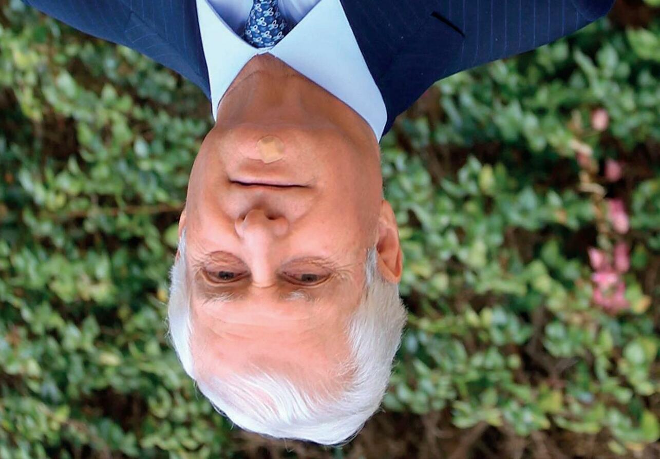
Her great social commitment in every respect. Her great social sensitivity and concern for the ill and those in need.
With the elderly and all those in need, but mainly with the elderly.
To with social -cau ses are you especially sensitive?
To maintain at the highest level the heritage of the House and add to it if possible, aiming for society to participate in and benefit from the full legacy.
Ms Cayetana Fitz-James Stuart, your mother, is still admired for her sociable nature, her vitality and good work heading the Casa de Alba. What would you -highli ght with regards to her?
One of the main decisions in this regard was to open to the public the traditional residences of the House so that everyone can enjoy their collections.
Is the Casa de Alba involved in any other initiatives to promote culture?
What are the next challenges for the Casa de Alba?
It is not an easy task as it is such a varied and complex heritage, but I have a team of first-rate professionals that act as consultants.
Similarly, the -ces sion of paintings, sculptures and other artistic -ob jects to expositions organised by -natio nal and international museums.
Is it difficult to preserve and divulge the historical -heri tage of the ducal house?
During its long history, the Casa de Alba has as its basic values the defence of Spain, loyalty to the King and, to uphold those values, the preservation of its valued and varied legacy.
What are the values of the Casa de Alba?
Serious but good-natured, he is not keen to appear in couché paper. A few years ago he undertook the responsibility for preserving the historical and artistic heritage of the Casa de Alba.
Carlos Fitz-James Stuart y Martínez de Irujo is the eldest of the six children of Cayetana Fitz-James Stuart, the Duchess of Alba. He has the title of Duke of Huéscar, and since his mother passed in 2015 he has inherited that of Duke of Alba.
38 THEY LOOK KINDLY
US
UPON
CARLOS FITZ-JAMES STUART Y MARTÍNEZ DE IRUJO: THE GREATEST CHALLENGE OF THE CASA DE ALBA IS TO HOLD ITS HERITAGE TO THE HIGHEST STANDARDS SO THAT SOCIETY PARTICIPATES IN AND BENEFITS FROM THE FULL LEGACY”.
I
social sensitivity
“One of the main decisions in this regard was to open to the public the traditional residences of the Casa de Alba”
“From my mother
highlight her great
and concern for the ill and those in need”
Carlos Fitz-James Stuart y Martínez de Irujo.
Undergo medical exams at least once a year.
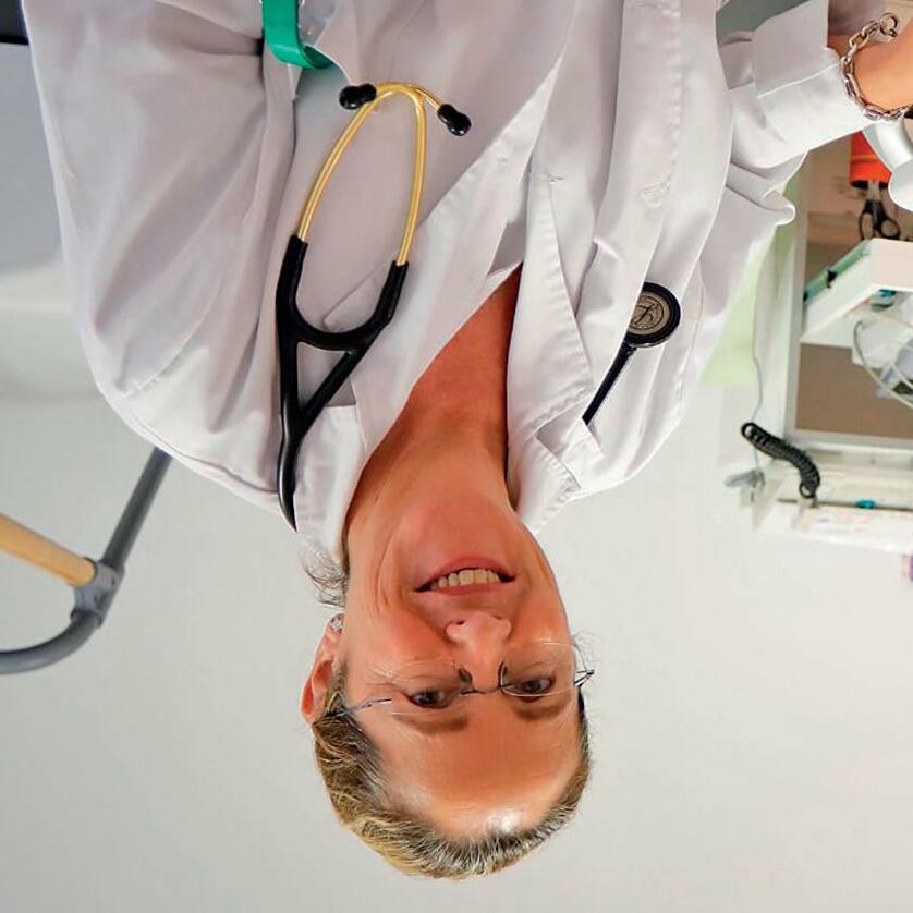
Control risk factors (cholesterol, blood pressure and glucose in blood)
Stop smoking.
Wight control, as well as good sleeping habits (at least 7 hours a day) and stress control.
Undertake regular physical exercise, avoiding a sedentary lifestyle.
Follow a varied and balanced diet, reduce intake of sugar (monitoring obesity and diabetes) and salt (monitoring high blood pressure).
Cardiovascular diseases are the leading cause of death worldwide with 17 million deceased every year (1 out of 3 deaths in the planet) How to Prevent Cardiovascular Diseases
Following a stroke or heart attack it is paramount for the recovery of quality of life, as well as to prevent a second event, to practise physical exercise (progressively, starting with moderate intensity exercise). Programmes of cardiac rehabilitation have shown an improvement in quality of life and up to 27% increase in survival.
If one suspects a heart attack or a stroke one must find medical help urgently (call 112); time is vital and the sooner the -diagno sis is confirmed the sooner the -appro priate treatment will be initiated.
Indeed, it is true that not every precordial pain is of coronary origin (chest angina/heart attack); this can also be due to -pe ricarditis, digestive pathology -(gastri tis/duodenal ulcer), aortic aneurysm dissection, -osteoar ticular diseases, costochondritis. It is important to visit the doctor urgently if the pain is acute and if it lasts more than 20 minutes, with general malaise and, especially if one has a previous cardiovascular disease or if one is known to have a risk factor such as high blood pressure, smoking, -choleste rol or family history or early ischemic cardiopathy.
in one eye, even loss of speech or understanding. These symptoms may be accompanied by sudden severe headaches, without apparent reason, dizziness, and loss of balance or sudden, unexplained falls.
OTHER SPECIALTIES
In the case of a stroke the symptoms are different. Patients who suffer from them usually experiment weakness or sudden numbness on the face, arm or leg on one side of the body. Also sudden loss of vision, especially
Cardiovascular diseases, those affecting the heart or the circulatory system, are the leading cause of death worldwide with 17 million deceased every year (1 out of 3 deaths in the planet), above cancer. In Spain, during 2020, 119,853 individuals died due to cardiovascular problems, 24.3% of all deaths. Most of these deaths are due to cholesterol build up on the walls of the blood vessels. Monitoring risk factors and recognising the symptoms of cardiovascular failure is key to prevent these type of diseases.
Experts recognise that up to 80-90% of heart attacks could be prevented by managing risk factors. The main symptom of heart attacks is pressure or chest pain, over 20 minutes long, with general malaise, nausea and profuse sweating, associated to shortness of breath, generally with pain irradiation to the jaw, back or arms. Women may present the same heart attack symptoms as men, but they tend to minimise these (wrongly thinking that heart attacks are uncommon among women). Other cases only report tiredness and fatigue, as well as dizziness, palpitations and syncope.
Women tend to minimise heart attack symptoms and some only present tiredness, dizziness, palpitations or syncope.
HEALTHY HABITS AND MONITORING BLOOD LEVELS, KEY TO PREVENTING CARDIOVASCULAR DISEASES
Dra. Rosario Cortina Rodríguez, Cardiology Specialist at Hospital Universitario de es.ñCabue Cardiology Specialist at Hospital Covadonga, nóGij and at Centro dicoéM de Asturias, Oviedo.
37
1.
2.
3.
4.
5.
6.
• https://www.maspormas.com/especiales/los-lentes-unahistoria-de-salud-visual/
https://www.vistaoftalmologos.es/un-paseo-por-la-historiade-la-oftalmologia/#:~:text=La%20oftalmolog%C3%ADa%20
Sources: fue%20la%20primera,usados%20en%20momias%20y%20 estatuas.
It wasn’t until the Enlightenment period, in 1748, when the next innovation occurred in the treatment of cataracts. Jacques Daviel, French ophthalmologist developed the extracapsular technique that took as a starting point the traditional method used to date but performing an incision in the inferior part of the cornea to extract through pressure the remainder of the crystalline. Subsequently, the eye was dresses to recover. This process was much safer and efficient despite also having complications and side effects. • https://es.wikipedia.org/wiki/Cresques_ Aviatar#:~:text=%E2%80%8B-,Operaci%C3%B3n%20de%20 cataratas%20de%20Juan%20II%20de%20Arag%C3%B3n,de%20 Rivesaltes%2C%20y%20sus%20anteriores • https://www.vista-laser.com/operacion-cataratas-historia/ • https://www.larioja.com/culturas/201406/10/ cirugia-catarata-largo-historia-20140610003933-v. html?ref=https%3A%2F%2Fwww.google.com%2F • https://www.visionquestmedical.com/la-historia-de-la-cirugiade-cataratas
Ever since, cataract surgery has been -ho ned, going from the intracapsular -techni que to the -extracap sular, till the point when Harold Ridley implanted the -intrao cular lens (IOL) after the Second World War. He realised that British pilots had bits of acrylic plastic splinters from the aeroplane windows and that they -endu red these perfectly without infection or inflammation. This is how he thought of implanting the first IOL the 29th November 1949 at St Thomas Hospital in London. This made possible to polish cataract surgery and, especially, facilitated the post-op. • https://www.doctorgimenez.com/noticias/historia-de-laoftalmologia/ •
Charles D Kelman: phacoemulsification for cataract treatment
However, in the absence of a better technique and as the alternative was blindness, this intervention was well spread throughout the world and, in fact, is still used in remote areas of Africa and Asia. For example, we have testimonials of this type of surgeries successfully performed during classical Greece and the Roman Empire. Likewise, there is an allusion to this procedure in Susruta Samhita, a treaty from the 3rd – 4th century that collects the knowledge of the Ayurveda medicine, traditional in India.
with a needle, thus pushing the crystalline to dislodge it into the interior of the eye. Obviously, the failure rate was very high with this technique and it very easily caused complications; it also had a complex post-op.
WE TAKE CARE OF OURSELVES
During the Middles Ages we can highlight the invention of glasses from the Alhace’s Theory (11th century). This Arab scholar and astronomer developed Seneca’s precepts, using a crystal ball full of water to enable reading and suggested the use of the so-called “reading stones”: polished quartz lenses that worked as a magnifying glass. Some centuries later, in the 13th century, these lenses were mounted into a frame, taking shape as primitive lenses. Around this time, in China, this same quartz glasses were filled with smoke to create the first sunglasses in history.
As for cataracts, this period has potentially the most famous eye intervention in history: the JudaeoCatalan physician Abnarrabí, surgeon, ophthalmologist and cabalist astrologist of the 15th century, performed a fully successful cataract surgery to King John II of Aragon, father of Ferdinand the Catholic. • https://oftalmologovigo.com/la-historia-de-lasgafas/#:~:text=Las%20gafas%20se%20inventaron%20en%20 la%20%C3%A9poca%20medieval.&text=Fue%20en%20 dichos%20monasterios%20donde,un%20escrito%2C%20 aumentaba%20las%20letras
Charles D Kelman added another great advance to that of Ridley; he introduced phacoemulsification. Ultrasound waves soften the lens of the eye to break it before being removed, this means that the size of the incisions can be reduced from 6 –7 mm to only 3.5 mm. Furthermore, this smaller incision means no suture is needed. 36
Ever since, the techniques and instruments have been improved, and currently, with Phemtosecond laser, cataract surgery is a fast procedure, with a minimal risk of complications or side effects and an easy post-op.
From this period is the first surgical intervention to fight cataracts, what as called couching technique. This procedure consists on impacting the wall of the eye
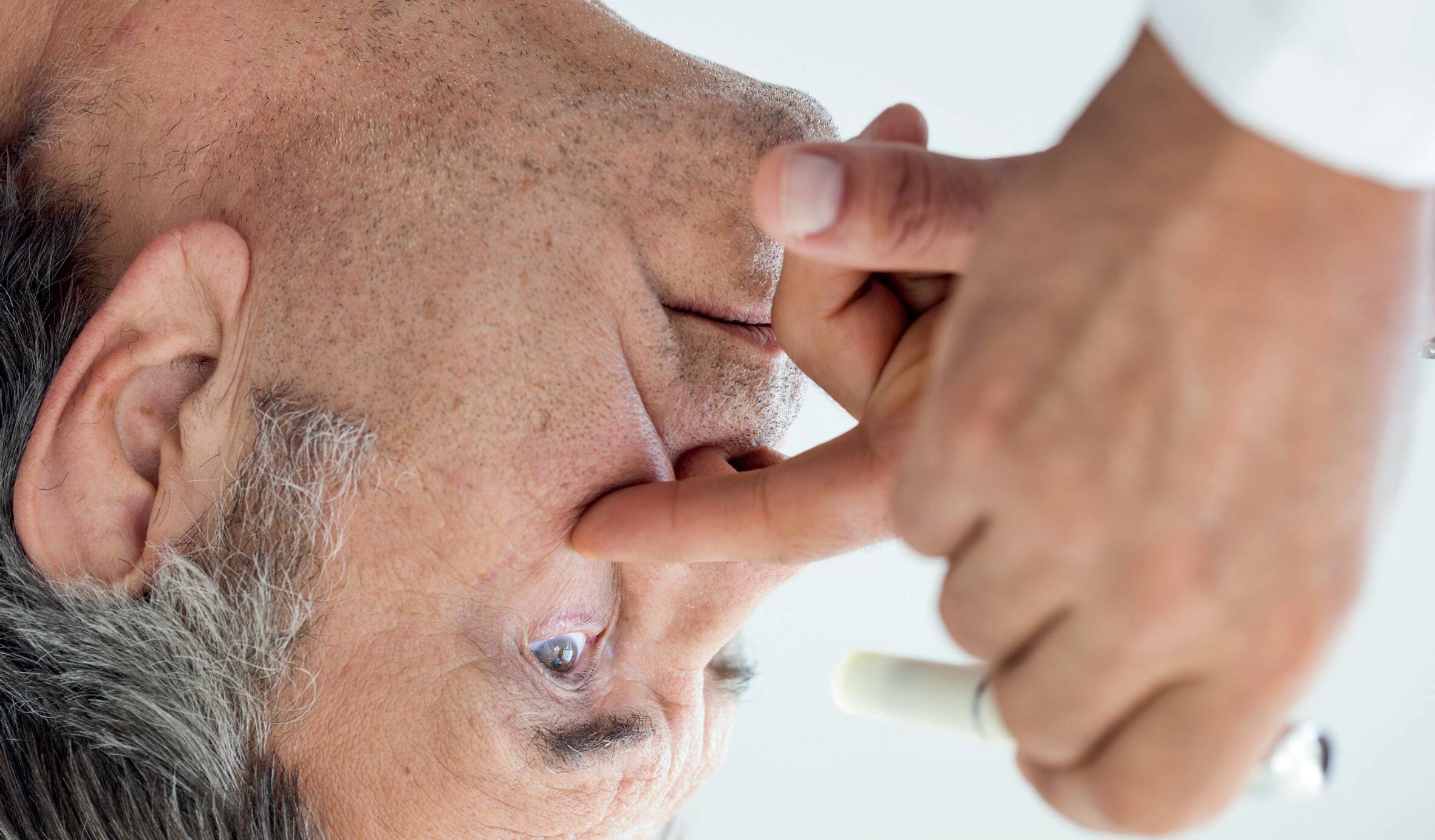
The Egyptians, famous for their extensive astronomic and scientific knowledge, were also great physicians. In fact, the oldest medical treaty dates from Ancient Egypt and goes as far back as 1550 B.C. It is called Ebers Papyrus and gathers in 877 sections different eye diseases and their prescriptions.
A little bit of history: from the Egyptians to the present
As we mentioned, cataracts is such a common problems that from very early on they attracted the attention of -phy sicians and -scien tists. Even though currently cataract surgery is a very common procedure with little risks in many countries, it hasn’t always been like this.
Did you know that ophthalmology was the first specialised branch in medicine? Even in ancient times, scholars were already interested in understanding and researching eye care. Among all the eye diseases already known and treated at the time, one might highlight cataracts, so common today that it is the first cause for blindness across the world. Therefore, we are subsequently going to go over the evolution of ophthalmology and, specifically, over the history of cataract surgery.
Before we continue, we remind you that cataracts is a disease that causes opacity in the crystalline, the transparent tissue inside the eye that enables near and far vision. The efficiency of this tissue diminishes over time bringing about the need for ocular lenses to facilitate vision. Furthermore, it is likely that the patient looses transparency, which is when cataracts appear hindering vision even with glasses.
Discover the evolution of ophthalmology and the history of cataracts, all the innovations and scientific discoveries that have enabled an easy and fast treatment of this disease.
WHO INVENTED CATARACT SURGERY?
WE TAKE CARE OF OURSELVES
35
The Updating Programme on Primary Care is devoted to Family and -Communi ty Medicine professionals as part as their continuous medical training; they will be able to download the certificate from their account on the Live-Med official webpage after meeting all the requirements and receiving the availability notification.
This year, Live-Med includes as part of its agenda an ophthalmology lecture -un der the title “A Tour Through DMAE and Other Diseases of the Retina” (Un -reco rrido por la DMAE y otras enfermedades retinianas), by Dr Álvaro Fernández-Vega González, from the Retina and Vitreous Department at Instituto Oftalmológico Fernández-Vega. During the lecture, it was possible to participate through the clinical cases presented and the physician answered questions through the Live-Med app.
th18 and th19 october
LIVE-MED AAP ANNUAL MEETING
ONCE, the Instituto Oftalmológico -Fer nández-Vega and the Asociación Nacional de Informadores de la Salud (ANIS) -cele brated the meeting Prevention and Key Aspects of Glaucoma in the framework of the World Sight Day. The main aim is to inform and debate the latest advances in the treatment of this pathology, first cause of irreversible blindness in the world.
Dr Pedro Pablo Rodríguez Calvo and Dr Montserrat García Díaz participated in the event.
th13 October
PREVENTION AND KEY ASPECTS OF GLAUCOMA
Detachment After Pneumatic Retinopexy: External Degassing Technique”.
“Combined Severe Trauma Involving Anterior and Posterior Segments”, while Dr Álvaro Fernández-Vega González will participate with two surgical videos: “The Silicone Bolster Technique: A New Surgical Approach for Repositioning Dislocated Intraocular Lenses” and “Management Of Subretinal Gas
The meeting, which takes place in Hamburg and is broadcasted live, gathers numerous experts that will talk about the latest advances in retina. Dr Álvaro Fernández-Vega Sanz will give the lecture
1-4 September
ND22 EURETINA MEETING
Dr Luis Fernández-Vega CuetoFelgueroso participated in this event that took place in Brussels (Belgium), gathering the most important young ophthalmologists in Europe. His intervention addressed the Selection of patients and pre-operative assessment for the different cornea refractive surgery techniques in the section of Refractive Surgery and Cataract.
9-10 July
TH4 EUROPEAN MEETING OF YOUNG OPHTALMOLOGIST
CONGRESS ACTIVITIES 34
Dr Jesús Merayo Lloves, director of the Instituto Universitario Fernández-Vega attended the meeting, which took place in Antwerp (Belgium). Specifically, he has revised issues of corneal innervation at this meeting that gathers renowned European opinion leaders in anterior segment.He also contributed the lecture «Measurement of Ocular Sensitivity With a New Esthesiometer».
16-19 June
EUROPEAN CORNEA CONFERENCE
For two days, numerous experts talked about the latest technology advances for the diagnosis and the management of the main diseases of the cornea, choroids and vitreous.
Drs Álvaro Fernández-Vega Sanz and -Ál varo Fernández-Vega González attended the 25thMeeting of the Spanish Society of Retina and Vitreous (Sociedad Española de Retina y Vítreo - SERV), broadcasted live but in a face-to-face manner.
Specifically, Dr Álvaro Fernández-Vega Sanz participated as moderator at the table of clinical cases while Dr Álvaro -Fernán dez-Vega González defended the oral communication “Results of the technique of silicone haptics to suture to the sclera dislocated intraocular lenses” (Resultados de la técnica de manguito de silicona para la sutura a esclera de lentes intraoculares luxadas) and participated in the table on surgical retina to talk about “Pneumatic -reti nopexy: key aspects for success” -(Pneumo rretinopexia: aspectos clave para el éxito)
17-18 June
TH25 MEETING OF THE SPANISH SOCIETY OF RETINA AND VITREOUS (SERV)
Professor Jesús Merayo Lloves, director of the Instituto Universitario FernándezVega, participates as lecturer and moderator at this Meeting organised by the European Dry Eye Society (EuDES) that took place in Paris. During the meeting, Dr Merayo Lloves provided the lectures “Focus the Problem of Severe Dry Eye”, “Regeneration of the ocular surface with plasma enriched in growth factors” and “A New Esthesiometer For Measuring Corneal Sensitivity”.
10-11 June
METING OF THE EUROPEAN DRY EYE SOCIETY
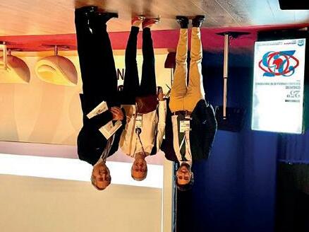
Discussion on Pneumatic Retinopexy in primary retinal detachment (Discusión sobre la Retinopexia Pneumática en desprendimientos de retina primario).
Dr Álvaro Fernández-Vega González, from the Unit of Retina and Vitreous at Instituto Oftalmológico Fernández-Vega, has participated in the course on retina at Hospital Vall D’Hebron in Barcelona. In the course, the physician has taken part in the table on controversies in retina:
3rd June
RETINA COURSE AT HOSPITAL VALL D’HEBRÓN
CONGRESS ACTIVITIES
33
On the course on glaucoma, doctors Pedro Pablo Rodríguez Calvo and Ignacio Rodríguez Uña acted as directors and Dr Andrés Fernández-Vega Cueto-Felgueroso took part as a lecturer.
As for the updating courses, Dr José F. Alfonso Sánchez directed the course “ENDO-K”, where Dr Belén Alfonso Bartolozzi and Drs Luis Fernández-Vega Cueto-Felgueroso and Carlos Lisa Fernández acted as lecturers. In this advanced course, aimed to ophthalmologists expert in cornea, the physicians presented the latest advances of the corneal prosthesis called Endokeratoprosthesis, implanted using lamellar techniques, avoiding the perforation of the eye in patients who need a corneal transplant. The concept and initial design of the Endokeratoprosthesis has been developed at the Instituto Universitario Fernández-Vega, which has worked alongside AJL, responsible for the optimisation and manufacturing of the device.
Updating Courses
Furthermore, Dr Luis Fernández-Vega Cueto-Felgueroso was a panellist at the symposium on “Asquelio”, he also participated in the Intergenerational Debate on Anterior Segment Surgery at the Alcon symposium.
The 98th meeting of the Spanish Society of Ophthalmology (SEO in Spanish) took place in Pamplona at the end of September, once again in person after two years in which the meeting was online due to the health situation. This important meeting gathered experts in all the subspecialties of ophthalmology, among which were a wide selection of physicians from the Instituto Fernández-Vega, from the departments of cornea, crystalline, retina and glaucoma. Professor Luis Fernández-Vega Sanz took part within the framework of the subspecialty of Cataracts and Refractive to talk about “Motives for early lensectomy” (Motivos de lensectomía precoz), with Dr José F. Alfonso Sánchez as moderator. On retina, Dr Álvaro Fernández-Vega Sanz intervened as moderator and lecturer with an exposition on “Retinal Detachment” (Desprendimiento de Retina).
on scientific photography and panel communication
Dr Ignacio Rodríguez Uña was recognised in this meeting, in the sessions
Dr José F. Alfonso Sánchez participated in the free communications of the second day with the dissertation on the “Vivity” lens and Dr Ignacio Rodríguez Uña spoke about “OCT Angle Closure Assessment” (Evaluación Cierre Angular OCT).
During the first day, Drs Belén Alfonso Bartolozzi, Luis Fernández-Vega Cueto-Felgueroso and Pedro Pablo Rodríguez Calvo participated as lecturers on “DALK + IOL” and “Lensectomy high myopia –Surgical safety” (Lensectomía alta miopía – Seguridad quirúrgica), respectively.
Free Communications
TH98 MEETING OF THE SOCIEDAD ESPAÑOLA DE OFTALMOLOGÍA
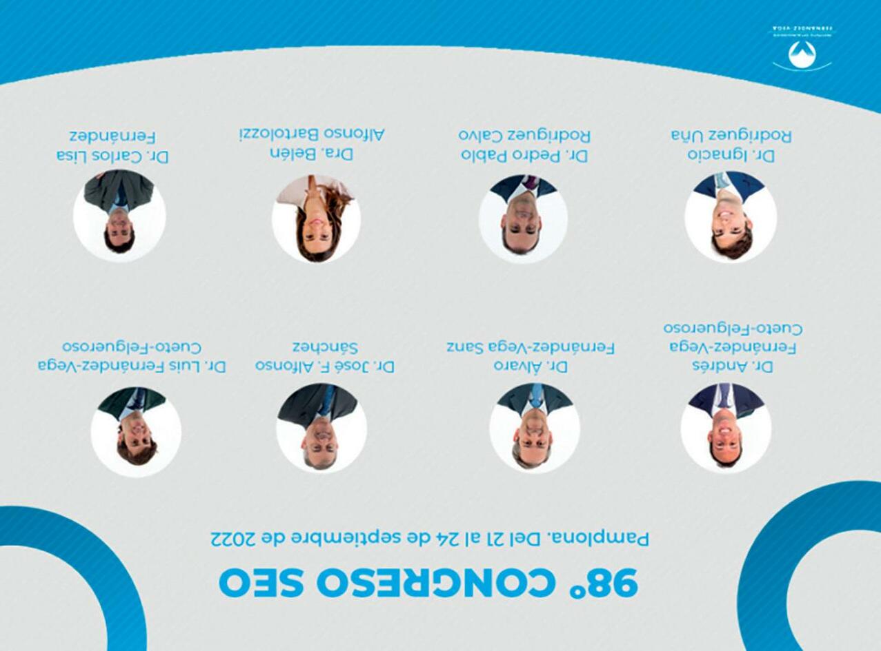
CONGRESS ACTIVITIES
32
Furthermore, another professional from the Institute was recognised alongside them, Dr Carlos Lisa, for having implanted over 1000 of the aforementioned lenses, as well as Dr José Alfonso, for his valued contribution to the scientific publications that spread and delve into the advantages of a device and technology in which he is one of the foremost experts in Europe.
It was during this meeting where Drs Luis Fernández-Vega Cueto Felgueroso and Belén Alfonso Bartolozzi were recognised as being among the best young ophthalmologists in Europe given their brilliant trajectory and their more than proven experience in the implementation of a technology that provides more safety, reliability and comfort to the patient.
hus has been decided last September in Milan, during the annual meeting of the European Society of Cataract and Refractive Surgeons (ESCRS) that gathered over four thousand ophthalmologists with, among others, the specialty of – intraocular- ICL lens implantation to address myopia, hyperopia and astigmatism.
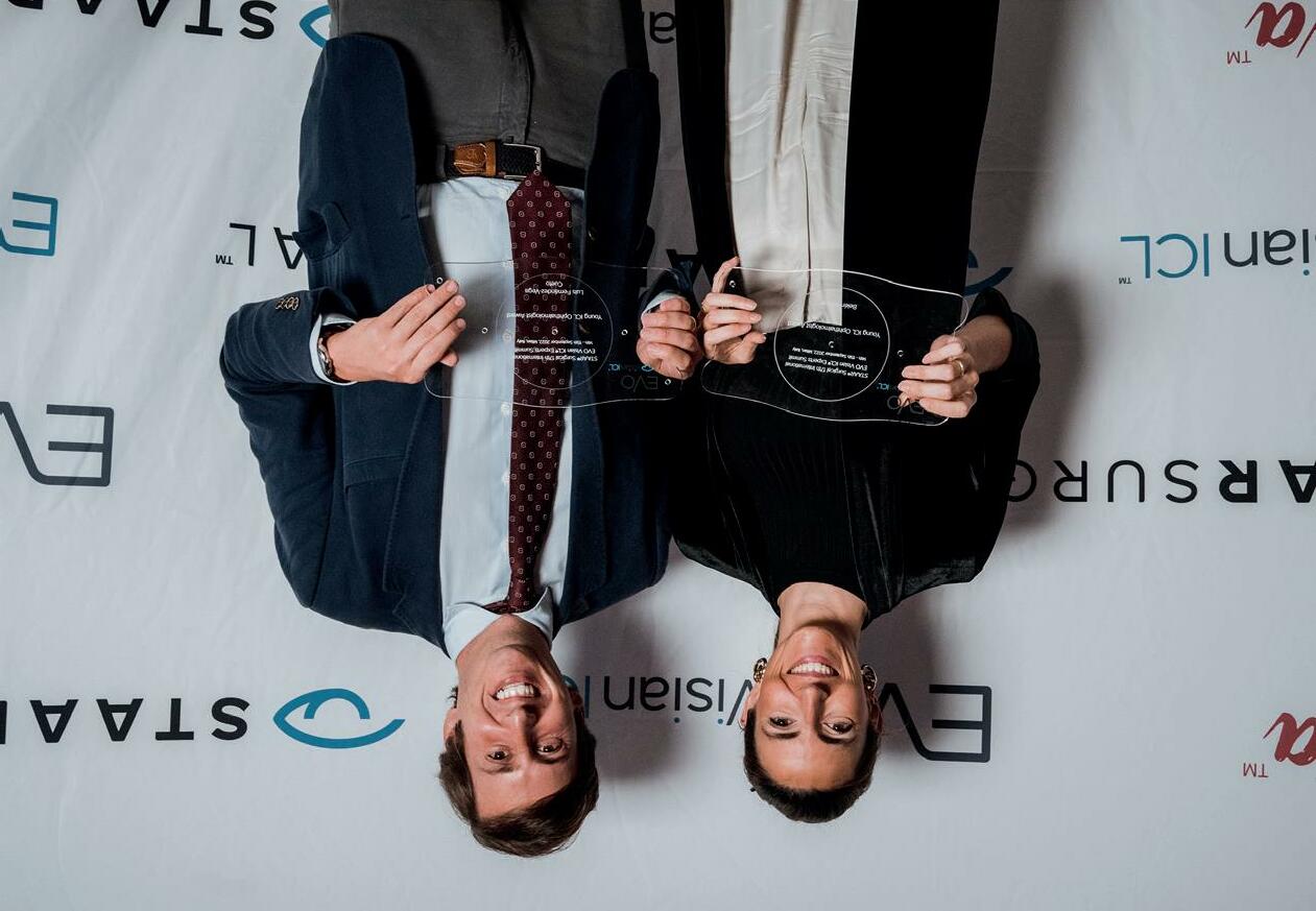
CONGRESS ACTIVITIES
LUIS FERNÁNDEZ-VEGA CUETO-FELGUEROSO AND BELÉN ALFONSO BARTOLOZZI, AMONG THE
YOUNG OPHTHALMOLOGISTS
PHYSICIANS
BEST
IN EUROPE
T
Drs. Luis Fernández-Vega
31
Cueto-Felgueroso and Belén Alfonso Bartolozzi, during the act.
Regular eye checkups are paramount in all of these cases to diagnose the different ailments on time and stop its progress.
In early childhood, the Department of Paediatric Ophthalmology at Instituto Oftalmológico Fernández-Vega, headed by Dr Javier Fernández-Vega Sanz and with Dr Lucía FernándezVega Sanz and Dr Belén Sánchez Cañal, recommends undergoing regular check-ups. The first visit as a newborn to rule out any congenital diseases. If there is no discomfort or visual problems it is also advisable to undergo a second check-up at the age of three. Several problems such as lazy eye, strabismus or potential refractive defects like myopia, astigmatism or hyperopia can be prevented.
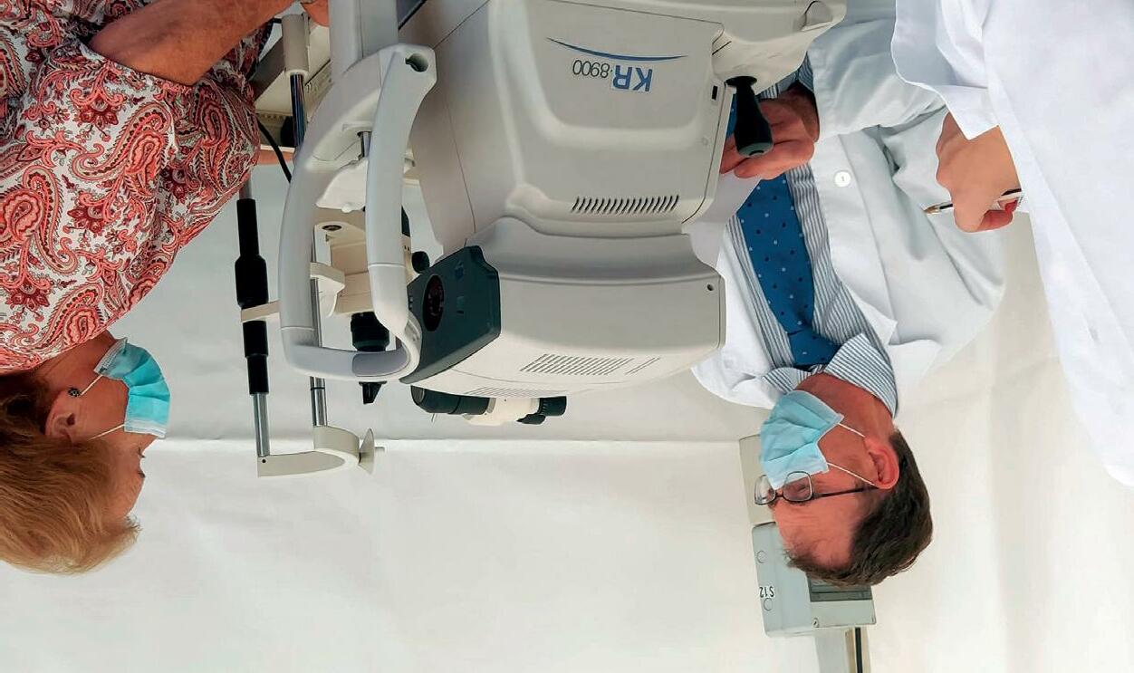
During adulthood, cataracts are the most common problem and can even cause blindness. Before this, patients usually present presbyopia, between the ages of 45 and 55. It is a disease caused by the loss of elasticity of the crystalline, which means patients have problems to focus at a near distance. In individuals over 50, Age-Related Macular Degeneration or AMD is the most common disease and causes irreversible visual loss. To treat this illness, the IOFV has the Department if Retina and Vitreous, headed by Dr Álvaro FernándezVega Sanz. 30
The check-ups performed during the day allowed to assess the visual health of dozens of neighbours of this town. Thus, the Fundación Fernández-Vega joined the movement promoted by the UN focused on raising citizen awareness. Regular eye checkups are paramount to detect on time potential eye diseases, especially the asymptomatic ones.
n the occasion of the World Sight Day, the Fundación Fernández-Vega recovered its solidarity tent to see patients free of charge. This year, Dr Andrés Fernández-Vega Cueto-Felgueroso and Dr Tomás Parra Rodríguez visited Cangas de Onís, the Asturian area where the tent was set this year.
During the teenage years it is possible to develop keratoconus, a deformity of the cornea. The Instituto Fernández-Vega has a specialised team headed by Dr José F. Alfonso, head of the Cornea and -Crystalli ne Department, and Luis Fernández-Vega Cueto-Felgueroso, specialist in this -pa thology. O WE CELEBRATES WORLD SIGHT DAY AT CANGAS DE ONÍS
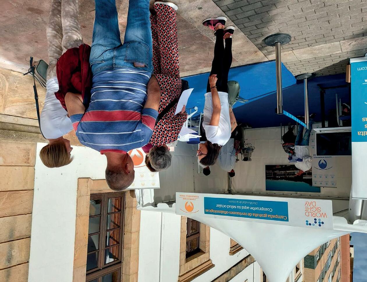
SOLIDARITY ACTIVITIES
We need military help, especially weaponry. Russia is better equipped and that means lots of people die. We also need medicines for the civilian population and war casualties, as well as food, technology and resources to build up the country when this is over, which we hope will be soon.
What are the most urgent needs of the Ukranian society?
I loved the treatment and work performed by the professionals, as well as how promptly they saw me. They are very good and use state-of-the-art technology. I didn’t see anything similar in Ukraine. I am very happy with the treatment received.
What was your experience like at the practice?
When they heard of my problem, someone who’s here in Spain met with some friends to try to help me. They found this practice in Oviedo and reached to an association that supports the Ukrainian people. They gave them the information for the different ophthalmology practices and, as this as the best one, we did not hesitate to come. There was no hope for my problem in Ukraine.
hosted the visit.
After finishing school he started his studies as a computer technician in Ukraine. In 2014, at the same time as the first tensions with Russia arose, he promised to protect his country no matter what and joined the army after training in defence and medical help. The situation his country is going through recommends being as discreet as possible about his personal details, not only for his own safety but also for that of his loved one’s.
He has just turned 21 and is from Ukaine.
SOLIDARITY ACTIVITIES
How did you get to know the Fundación Fernández-Vega?
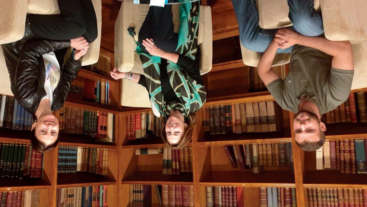
Generally speaking, the best situation is that of those who escaped. They are safe and that makes all the difference. There are families in conflict zones who could not leave or who didn’t think this would last for so long.
Ever since the beginning of the war I’ve been in the most dangerous area, in the border with Russia. My family was close, in a town house, and when the Russian soldiers entered Ukraine, my father and I decided to go to the front. The rest of the family left the country.
It depends on many factors. There are those who had to flee and only see conflict through TV, and those who live in bunkers and rarely go out. Some individuals suffer from food shortage because even though they try to secure supply, there are severely war-torn areas were it is very difficult to access foodstuff. Others have family members who have had to enlist and suffer due to the uncertainty this entails.
What is the everyday life like of a family during a war?
THE FUNDACIÓN FERNÁNDEZ-VEGA, WITH UKRAINE
Generally speaking, the best situation is that of those who escaped. They are safe and that makes all the difference
“The physicians at the IOFV are very good and use state-of-theart technology. I didn’t see anything similar in Ukraine”
29
Mrs Victoria
Cueto-Felgueroso Botas, director of Fundación Fernández-Vega,
28
For her part, -Victo ria -Cueto-Felgue roso Botas stated that “as director of Fundación -Fernán dez-Vega, being able to come with the physicians of the Institute to help people in need is one of the main personal -recogni tions one may have, helping others is the best”.
Segade and Álvaro Cuéllar and Diego Costas, from -main tenance, took part in this visit. Tatiana Llorente, -presi dent of Asociación Nora thanked the initiative of the Foundation, -some thing that has been taking place for several years. -“The se exhaustive eye check-ups are very important for the patients as they can improve their -quali ty of life by -perfor ming daily task in a much easier way” said Dr Andrés Fernández-Vega Cueto-Felgueroso. I
n the framework of the celebrations of the 30th anniversary of Asociación Nora, a team of professionals of the Fundación Fernández-Vega visited the entity to attend over 20 users of all ages and perform eye check-ups.
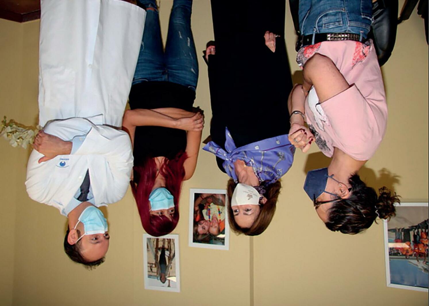

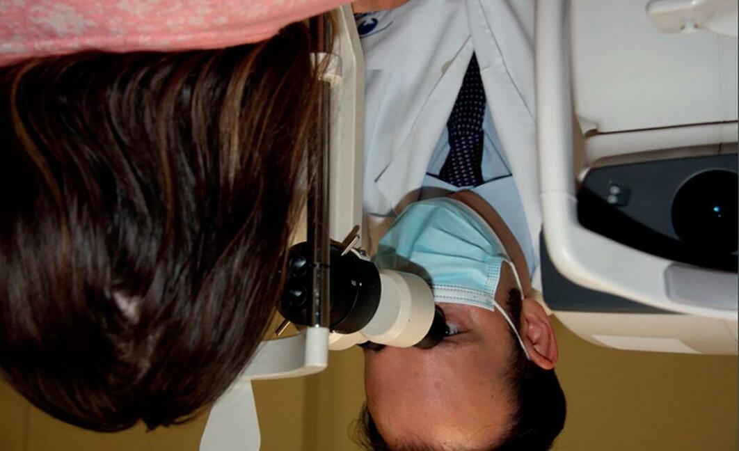
FERNÁNDEZ-VEGA VISITS ASOCIACIÓN NORA
FUNDACIÓN
ACTIVITIES
SOLIDARITY
Specifically, Mrs Victoria -Cueto-Felgue roso Botas, director of the Fundación Fernández-Vega; Dr Andrés -Fernán dez-Vega -Cue to-Felgueroso, -oph thalmologist of the Unit of Glaucoma at Instituto -Oftalmoló gico -Fernández-Ve ga; -opticians-op tometrists Paula
Tatiana Llorente, director of Association Nora, received Doña Victoria Cueto-Felgueroso Botas, director of Fundación Fernández-Vega, and the rest of professionals from the Institute.
Therefore, to detect this disease on time, it is paramount to visit the ophthalmologist and undergo an eye examination that will determine the extent and effect caused on the structures of the eye. Furthermore, the tears can be analysed to detect the infectious nature of the problem.
If it is not treated or if the diagnosis is delayed, the infection can cause different complications, such as recurrent viral infections, chronic inflammation, visual loss and even blindness.
Early diagnostic, key to avoid complications
Less frequently, the deeper layers can be affected and then the consequences are more severe, even causing permanent corneal opacification. The treatment for this is a corneal transplant known as keratoplasty. This transplant can be lamellar, i.e., only the affected part of the cornea is transplanted (epithelium, stroma or endothelium). “Several years ago, the cornea could only be substituted by another whole one, transplanting all its layers. Currently, advances have enabled us to substitute only the damaged layers, which drastically decrease the complication rate such as rejection, graft failure or glaucoma, as well as the visual recovery time. Furthermore, if over time there is a relapse of the pathology or any other problem, this corneal button can be substituted by a new one preserving the patient’s endothelium,” says Dr Fernández-Vega.
Keratoplasty, an efficient treatment
Among the most common symptoms, as well as the alteration of the visual acuity, are acute pain and irritation, which can cause difficulty when blinking. “It is also common to have red eyes, eye itchiness, excessive tearing and, often, photophobia, i.e., a severe sensitivity to light,” states Dr Pilar Quiroga, ophthalmologist at Instituto Oftalmológico Fernández-Vega.
Generally, these keratitis are superficial, as they affect the outer layers of the cornea.
As well as virus infections, such as monkeypox, keratitis can be caused due to bacteria, fungus and protozoa, dry eye, irritation caused by physical and chemical agents (including ultraviolet radiation and the use of contact lenses) and allergenic processes.
He adds, “keratitis is an inflammation that can lead to eye discomfort as well as to severe visual acuity loss, in fact, it can cause blindness”.
and crystalline at Instituto Oftalmológico Fernández-Vega.
INVESTIGATION
To detect this disease on time one has to undergo an eye examination to determine the extent and effect caused
27
“This is because the cornea and the conjunctiva constitute the ocular surface, i.e. the first defence layer of the eye. Due to this, and given that they are both the first cover of the eyeball, they are often affected together, as in the case of monkeypox,” explains Dr Luis FernándezVega CuetoFelgueroso, from the unit of cornea
onkeypox can cause severe consequences for eye health. According to the World Health Organisation (WHO)[1], skin rash caused by this disease can spread in 20% of the cases to the conjunctiva, the membrane that covers the white part of the eye. Furthermore, as presented in a study in the Lancet [2], these complications can cause inflammation of the cornea, called keratitis.
[2] The Lancet. Clinical features and management of human monkeypox: a retrospective observational study in the UK: https:// www.thelancet.com/ journals/laninf/article/ PIIS1473-3099(22)002286/fulltext
[1] World Health Organisation. Monkeypox: https://www.who.int/es/ news-room/fact-sheets/ detail/monkeypox
References:
MAccording to The Lancet, ocular complications due to monkeypox can cause keratitis, i.e. inflammation of the cornea. This ailment can cause different complications, such as recurring viral infections, chronic inflammation, vision reduction and even blindness.
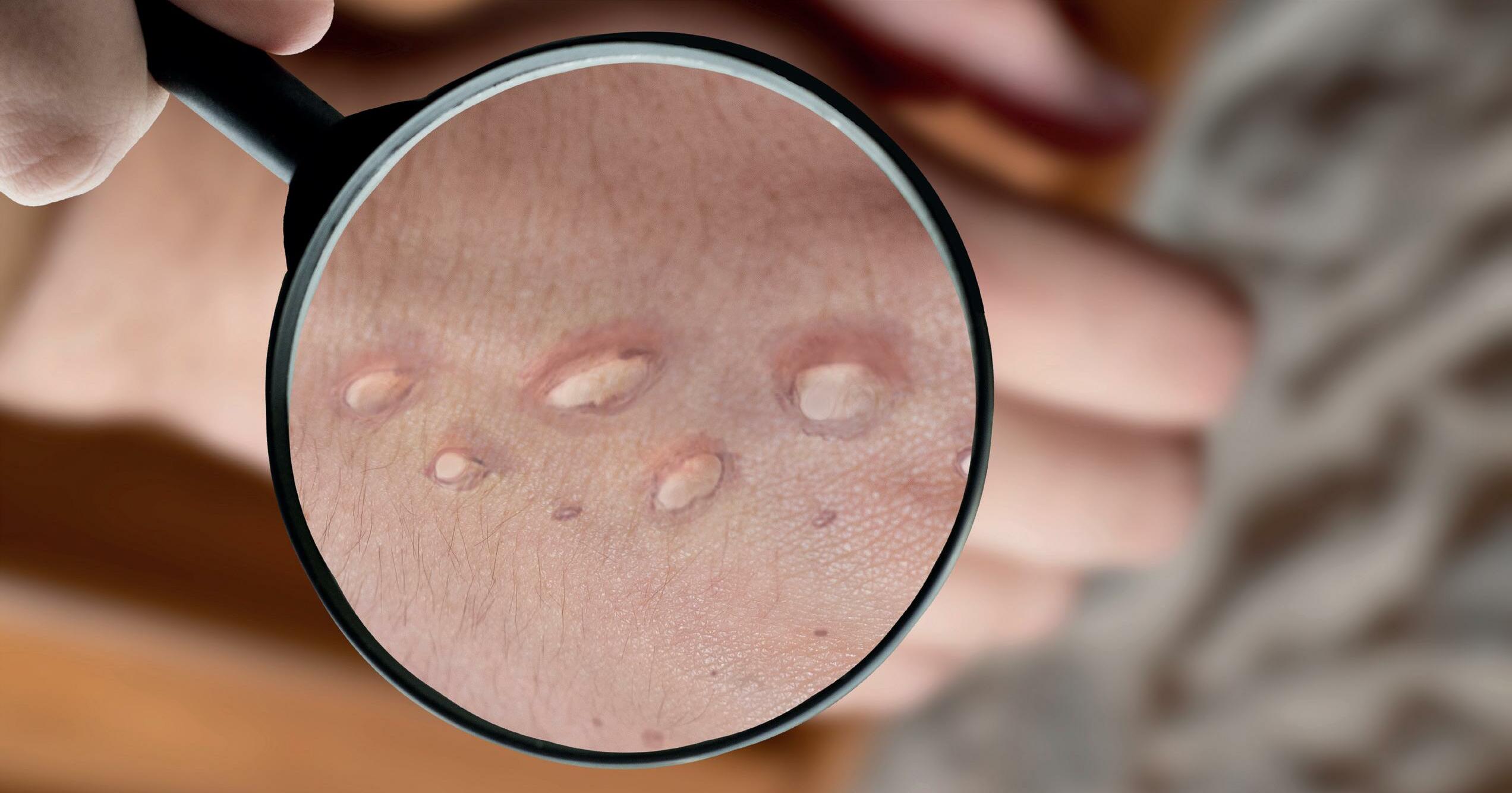
INVESTIGATION
IN 20% OF THE CASES, SKIN RASH DUE TO MONKEYPOX CAN AFFECT THE EYES The most common symptoms, as well as the alteration of the visual acuity, are acute pain and irritation, which can cause difficulty when blinking
26
25
Fernández-Vega we will continue to work to improve the quality of life of our patients.
At Instituto Oftalmológico
Undoubtedly ophthalmology still presents us challenges, both in the diagnosis and the treatment of many visual pathologies that present a difficult approach.
Currently, in our future we find the challenge to continue at the forefront of research in the next decade. In the face of potential pandemics and new diseases, we will need to strengthen our working lines and continue to consolidate the training of multidisciplinary teams. Because some of these pathologies also affect the eyes, such as COVID-19, which we have studied as in some cases it has caused ocular side-effects similar to patients with diabetic neuropathy and dry eye disease, or the recent out-break of monkey pox, which in 20% of the cases can affect the eyes even causing keratitis, i.e., swelling of the cornea.
The fourth research group, related to clinical research, is paramount to address many visual pathologies, in many cases under-diagnosed. This is because basic research is transferred to the practice in benefit of the patients, thus fostering multidisciplinarity. At FIO we are focused on the research of the anterior and posterior segment of the eye. A multidisciplinary team at the IOFV, consisting of ophthalmologists, optometrists, nurses, pharmacists and support staff among others, is devoted to researching the pathologies and treatments that affect the cornea, crystalline, retina and glaucoma. This research is possible thanks to the great affluence of patients that come to the Institute every year, over 110 000, which translates into over 220 000 eyes that can be analysed to advance in the research of ocular pathologies.
As for the -neurobio logy of the retina, we are researching damaged cells (ganglion cells as well as others) to avoid their death and achieve a -lon ger survival using factors that protect the part of the cell that provides it with energy -(mito chondria). Likewise, we are focused on Age-Related -Ma cular Degeneration (AMD), an ocular pathology for which there is currently no cure. This affects the retina and -spe cifically the macula, which is where the central vision is located, that which enables us to focus objects and -diffe rentiate the smallest details. In recent years research lines for this pathology have advanced exponentially, especially in trials with gene therapies. In our case, we are studying the -impli cation of zinc and metallothioneins (the most important proteins in the -regu lation of zinc levels) in the development of this disease.
The pathologies of the ocular surface increasingly affect the population of developed -coun tries. Many of these diseases cannot be currently -cu red, that’s why at the Fundación de Investigación -Oftal mológica we have a team with open research lines in dry eye (ageing and neurodegenerative diseases that are not on the surface), keratoconus, -rege nerative medicine of the cornea and advanced therapies (cell therapy, tissue engineering).
Investigación OftalmológicaFIO), which has four differentiated research groups: ocular surface, genetics of glaucoma, neurobiology of the retina and clinical research.
We also have a glaucoma research team, second cause for blindness -world wide. Our research in the field of ocular genetics is focused in several -differen tiated lines for the early diagnosis of this disease through the search for -pro tein biomarkers and the study of -associa tes risk genes.
IN DEPTH
Clinical research is paramount to improve the management of many visual pathologies, in many cases under-diagnosed
24
Our research in the field of ocular genetics is focused in several differentiated lines for the early diagnosis of glaucoma, the search for protein biomarkers and the study of associates risk genes
of the basis of diseases that cause blindness and visual alterations and achieve new preventive, diagnostic and medical and surgical treatment measures, the effort put into translational research is increasingly big. Translational research is that which goes from the laboratory to the patient. This is what we work for at Instituto Oftalmológico FernándezVega (IOFV), with extensive experience acquired in our 135 years of history and 5 generations. Since 2009, we have the Eye Research Foundation (Fundación de
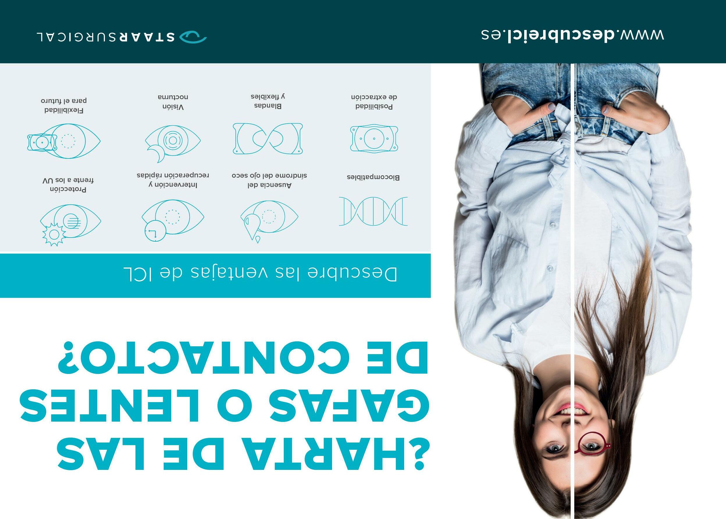
IN
DEPTH
Femtosecond laser has increased the accuracy and safety of cataract surgery.
Aiming to progress in the understanding
In recent years, one of the main advances in cataract surgical procedures has been the femtosecond laser, approved in 2021 for its use in this surgery, increasing the accuracy and safety of the intervention. Another paramount innovation in this field is intraocular lenses, which in recent years have achieved an exceptional quality and benefit level, providing good visual acuities at all distances.
As for the most common pathologies related to the progressive ageing of the population, main risk factor for multiple ocular alterations, the most widespread is cataracts, first cause of reversible blindness in the world. Its surgery is the most common in developed countries: every year approximately 30 million interventions are performed worldwide, over a million in Spain.
myopia, hyperopia, astigmatism or presbyopia.
t is estimated that 52% of the world’s population will have myopia in 2050. In fact, it is already the main cause for moderate and severe visual impairment in developed countries. Thus, in recent years, research has focused in refractive surgery to correct or minimise refractive defects, among these are, as well as
IN DEPTH
THE FUTURE OF OPHTHALMOLOGY IS LINKED TO RESEARCH
IThe 21st century has seen important advances in health research and, specifically, in the field of ophthalmology. This is due to several factors, among which we can highlight the development of pathologies related to an increase in the use of near vision without rest (studying, reading, using screens, etc.) and resulting decrease in activities that allow far vision.
Dr. Luis Fernández-Vega
22
Cueto-Felgueroso
Before strabismus it is paramount to detect the existence of amblyopia or lazy eye, as current treatments are only useful up to the age of 8/9, and if not undertaken on time at this age it is possible that the child will not develop their vision to 100% in one of their eyes. Finally, if that strabismus is not corrected with glasses and presents a significant magnitude the child will very likely need surgery, it will be the paediatric ophthalmologist who will decide when is the best time to perform it.
During childhood strabismus works differently than in adults as they do not refer double vision. If we detect strabismus in a child we need to take them to an ophthalmologist to undergo a full examination of their vision, as in most cases the cause is an uncorrected refractive defect such as hyperopia or myopia. After birth, the paediatrician performs a full examination to detect congenital eye diseases such as cataracts of glaucoma
Child Strabismus, Different From Adults
the most common malign intraocular tumour in children under 3, and is a risk for the vision and the life of the child. While survival rate is approximately 95%, early detection is paramount. The most common symptom is the appearance of a white spot in the pupil (leukocoria). Clinical suspicion of retinoblastoma by a paediatric ophthalmologist implies the need to be immediately transferred to a centre of national reference. Despite most of them being spontaneous (60%), 40% are genetically predisposed and are usually more aggressive and affect both eyes. IN DEPTH
On the other hand, retinoblastoma is
Two of the most severe problems that can appear in the post-natal stage and before the age of 2/3 are retinopathy of prematurity and retinoblastoma. The first disease appears in premature babies and those born underweight. The doctor needs to examine the eye fundus to avoid the apparition of injuries in the retina, this should happen a month after the birth of the baby. A later examination could lead to the apparition of a retinal detachment that can cause blindness.
the Most Severe Ailments in Babies
and Retinoblastoma,
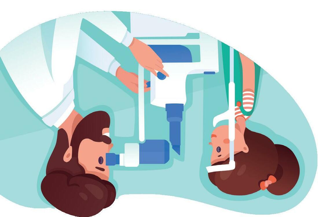
21
Strabismus is the eye subspecialty that refers to the motility of the eye, i.e., when the eyes are not aligned in the same direction when they look at a far or near object. This can occur due to different reasons and in different stages of life. When it happens in adulthood its main symptom is diplopia or double vision of objects. If this is acute, on most occasions it is indicated to undergo an image test and a subsequent urgent neurologic examination. If double vision remains and does not resolve, it is likely that the patient will need to undergo interventional treatments such as injections with botulin toxin or surgery.
Retinopathy of Prematurity
Another eye pathology with great repercussion on vision is congenital cataract. These are opacities of the crystalline present since birth. On many occasions the cause is unknown, but it is sometimes associated to infectious diseases or systemic alterations. The presence of this opacity hinders the child’s vision; an early surgery is needed to, together with a subsequent rehabilitation, achieve a good visual acuity in that eye.
It is recommended that children who have not had previous problems visit the ophthalmologist approximately at the age of three. It is true that after birth, the paediatrician performs a full examination of the eye morphology to detect congenital eye diseases (cataracts, glaucoma, strabismus) and it is advisable to remain vigilant for any changes in the child’s behaviours (starts falling often, alteration in the position of the eye, the child gets too close to objects to look at them, appearance of a white spot in the eye), a time at which we have to consult a paediatric ophthalmologist without delay.
the left, Dr Javier Fernández-Vega Sanz, Dr Lucía Fernández-Vega Sanz and Dr Belén Sánchez Cañal.
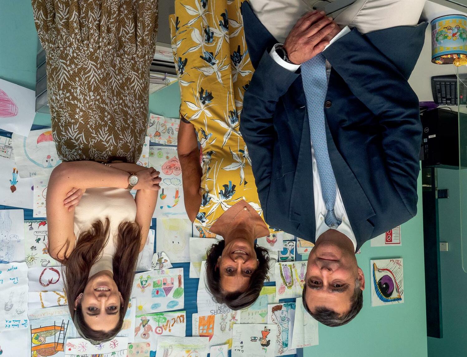
If there are no apparent visual problems, the first ocular revision should be at the age of three
PAEDIATRIC
20 IN DEPTH
OPHTHALMOLOGY
Paediatric ophthalmology treats eye ailments from birth to adulthood. It is very important that eye disorders in children are detected on time to be treated as soon as possible, thus avoiding visual problems and guaranteeing good vision throughout development and the rest of their lives. The success of the treatments greatly depends on the early diagnosis of ocular diseases. TREATS EYE AILMENTS
PREVENTS AND
IN CHILDREN
The advance of the diagnostic and therapeutic techniques in ophthalmology, both in clinical and surgical procedures has lead to the creation of medical subspecialties that aim to treat the most complex, specific eye diseases in some parts of the eye or in certain types of patients. The Instituto Oftalmológico Fernández-Vega, in its effort to maintain an excellent care, has a unit specialised in Paediatric Ophthalmology, headed by Dr Javier Fernández-Vega Sanz and with Dr Lucía Fernández-Vega Sanz and Dr Belén Sánchez Cañal in the team of ophthalmology specialists.
From
3. National Eye Institute. Presbicia (2022). Retrieved 27 July 2022, from https://www. nei.nih.gov/espanol/aprendasobre-la-salud-ocular/ enfermedades-y-afeccionesde-los-ojos/presbicia
2. Consejo General de Ópticos y Optometristas. Libro Blanco de la Salud Visual en España 2019 https://www.cgcoo.es/ libro-blanco-salud-visual-enespana-2019
1. Sociedad Española de Oftalmología (SEO). Presbicia - Oftalmoseo. (2022). Retrieved 27 July 2022, from https://www.oftalmoseo. com/patologias-frecuentes-2/ presbicia/
References:
Multifocal lenses (or diffractive) correct most refractive defects offering a high visual performance and a stable optical correction over time. There are increasingly more advanced materials and platforms that offer great clarity at all distances.
There is currently a new type of expanded view intraocular lens that offer the possibility of dispensing with glasses for virtually all daily activities. These lenses provide a full visual range, allowing the patient to recover a functional far, intermediate and near vision, and a very low visual discomfort.
IN DEPTH
What does surgery to correct presbyopia entail?
What types of intraocular lenses are there to correct presbyopia? What are the latest advances?
Surgery is one of the most highly recommended solutions in most cases for those patients who wish to stop using glasses because it offers long-lasting results, especially with intraocular lenses, which are for life. Furthermore, it enables to correct at the same time other refractive problems or cataracts definitively.
Implant and refractive surgery, which is the most common surgery, consists on replacing the crystalline with an intraocular lens, thus completely restoring visual function. This new lens will correct certain conditions or problems that the patient might have had before the surgery, depending on the type of intraocular lens used. By substituting the crystalline, this surgery avoids the development of cataracts in the future.
What are its advantages?
Corrective intraocular lenses for presbyopia have advanced technologies and materials that offer a highresolution image at all distances: far, intermediate and near. These can be subdivided into multifocal lenses (trifocal are more common) and progressive lenses.
The ophthalmologist will analyse each case in a personalised manner, performing an exhaustive study both at an anatomical and functional level, and taking into account the preferences and lifestyle of the patient to offer the alternative that better adapts itself to the patient’s situation and needs.
How is it determined which is the most appropriate therapeutic option for each patient?
The latest surgical advances allow to correct presbyopia in a safe, fast and definitive manner.
corneal 1implantation
Another option is surgery. There are currently different techniques to correct presbyopia, such as laser surgery, surgery to replace the crystalline with intraocular lenses or
There are different treatments to improve the symptoms of presbyopia. The first is the optical correction through the use of glasses or contact 1lenses
What are the options to correct presbyopia?
both in one same surgical procedure, as the implant and refractive surgery allows to replace the affected crystalline and re-establish visual function.
“Cataract and presbyopia have some common symptoms, such as blurry vision. It is currently possible to solve both in one same surgical procedure”
19
18
Both ailments affect the crystalline of the eye and are associated to ageing, so their incidence will increase due to the longevity of the population. Even though they are different conditions, they do have some common symptoms, such as blurry vision. It is currently possible to solve
Can presbyopia be prevented?
Presbyopia is an age-related refractive defect that causes blurry vision and difficulty to see near objects. Over time, the crystalline, the natural lens of the eye, looses its flexibility and ability to focus light, mainly in short 1distances This phenomenon is commonly known as “eye 1fatigue”
Among its most common symptoms are the gradual loss of the ability to focus objects that are nearby, the need to move the book or the phone away to see better, the feeling that the letters “dance” or are blurry, headaches when focusing on reading for a long time and eye fatigue.
Presbyopia is an unavoidable process caused by the natural ageing of the eye1. It appears from the age of 402 and is characterised by a gradual loss of the ability to focus near objects, which is commonly known as eye fatigue.
Is there any connection between cataracts and presbyopia? “THE LATEST ADVANCES IN SURGERY ENABLE THE SAFE, FAST AND DEFINITIVE CORRECTION OF PRESBYOPIA”
What is presbyopia? What are its symptoms?
IN DEPTH
Professor Luis Fernández-Vega Sanz
Presbyopia is a degenerative process caused by the unavoidable ageing of the eye. It is generally developed from the age of 240 and, with age, all individuals will develop presbyopia to some 3extent
Fortunately, there are currently efficient, fast and safe solutions that enable the correction of this ailment permanently and therefore removal of eyeglasses at all distances. Professor Luis Fernández-Vega Sanz, medical director at Instituto Oftalmológico Fernández-Vega explains the main treatments for presbyopia.
Lastly, pneumatic retinopexy is a less aggressive technique that enables to treat some types of detachments without surgery, just injecting a gas bubble and positioning. With the appropriate treatment, most retinal detachments can be solved; the key is to select the most appropriate one for each patient.
placing a gas bubble at the end of the surgery. The aim of the gas bubble is to apply pressure and block the tear, avoiding another detachment while the scarring of the laser occurs. Buckle is a silicone band that is placed around the eye, sutured, to bring closer the wall of the eye and the retina, thus facilitating that the retina returns to its position.
IN DEPTH
Each of these techniques has advantages and disadvantages. Vitrectomy, the most commonly used nowadays, consists of entering the eye through micro-incisions, removing the vitreous, draining the fluid behind the retina so that it returns to its original position, using laser around the tears and
What do they -en tail?
There are three different techniques: vitrectomy, buckle and pneumatic retinopexy. One of these techniques will be better indicated depending on the characteristics of the detachment and the patient.
How is retinal -deta chment treated?
Can retinal -detach ment be treated?
It consists of making a series of small burns around the retinal tear, which will quickly create an adherent scar around the tear sealing it and avoiding the flow of fluid under the retina. Thus, it would work as a weld around the retinal tear, avoiding the progression of a tear to a retinal detachment. Some patients who have injuries that predispose them to retinal detachments (such a thinner areas), can be treated with laser around this injuries but this does not avoid 100% retinal detachment, so the best prevention is for patients to understand the symptoms of when the vitreous is coming off and visit the specialist as soon as possible.
What is laser photocoagulation?
this situation, where we not only have a tear, but also a retinal detachment, the visual prognosis is worse and treatment is surgical.
Currently they can be successfully treated, thanks to high technology and sophisticates techniques used by retinal surgeons. The main objective of this surgery is to return the retina to its original place so that it can recover its function. However, depending on the type and its progression time it may be that, despite a successful surgery, there is a permanent visual deficit as the cells of the retina that have died cannot recovered. It is therefore paramount to visit the specialist as soon as the patient presents symptoms.
17
“Photocoagulation with laser of tears can prevent its development”
On many occasions it can, if the patients goes to the ER and a tear is observed before the retinal detachment has occurred, its development can be halted by photocoagulating with laser this tear. When there is a retinal detachment as well as the tear, the patient will observe a curtain or black spot in an area of the periphery of their visual field that slowly advances towards the centre until completely hindering vision. In
Usually a retinal detachment occurs following a tear/ hole in the retina. The gel that fills up the interior of the eye, the vitreous, has a paramount role in this -pro cess. When we are young, the vitreous is adhered to the retina throughout its surface, but as we age the vitreous separated from the retina causing a detachment. This is the normal -age ing process of the vitreous, which will normally cause no problem in most -in dividuals. However, in a small -percen tage, the vitreous might pull while coming apart from the retina and tear it at a given point, causing a retinal tear. If this happens, the more fluid part of the vitreous can flow behind the retina through that tear removing it from its posterior wall, thus causing a detachment.
How does a -deta chment occur?
What are its symptoms?
The separation of the sensorineural retina from the posterior wall, to which it should be adhered. The retina needs to be attached to it to be able to capture images, as it enables its correct feeding and functioning, so much so that if it’s separated the retina starts to degenerate and the cells that form it die, causing a visual loss that can be severe.
What is a retinal detachment?
It is a tissue -consis ting of neurons that form a layer that covers the interior of the eye, as if it were the paper wall of a room. The -op tical system of the eye is thought so that the images we see are focused in the retina (the same as the objective of a camera focuses the images in the film or sensor), which is then responsible for collecting those images and sending them to the brain, where they will be interpreted.
What is the retina?
common pathology in patients with high myopia and prevention is therefore important. Dr Álvaro Fernández-Vega González, ophthalmologist at the Department of Retina and Vitreous at Instituto Oftalmológico Fernández-Vega explains this pathology in this interview.
detachment
Dr. Álvaro Fernández-Vega González
Can it be prevented? “MOST RETINAL DETACHMENTS CAN BE SUCCESSFULLY TREATED BY A SPECIALISED SURGEON”
16 IN DEPTH
The most -charac teristic symptom that the retina is detaching is the sudden appearance of “floaters”, which are multiple, -diffe rent-shaped black stains (flies, dots, webs), that appear in the visual field. This can be -asso ciated to seeing a cloud that moves and glare or flashes of light. These -symp toms mean that the vitreous is coming off the retina; the patient needs to be able to identify them to urgently have their retina checked, as this is when retinal tears can occur.
Retinal
is a relatively
One of the areas in which this pigment can settle is the iridocorneal angle, which is where the natural drain of the aqueous humour of the eye occurs. When this filtration area is obstructed, liquid does not flow properly causing a slow increase in intraocular pressure. This is the mechanism that, when prolonged, can potentially cause glaucoma.
We undoubtedly recommend that you visit the ophthalmologist for an eye check-up. What you are describing is the common symptom associated to pigmentary dispersion syndrome, more common in young myopic males. This does not mean that you are going to have this dispersion, but we really believe it should be ruled out. This syndrome is produced when the iris, due to incorrect positioning, brushes against the anterior part of the crystalline, freeing pigment in the interior of the anterior chamber and potentially settling in one of the tissues in the front part of the eye.
I am a 35-year-old male with 3 diopters of myopia and I routinely use glasses. After doing sports I have blurry vision, is this normal or do I need to check my eyesight?
Ocular trauma is a very common cause of ocular high blood pressure, which if not promptly detected, can cause a progressive atrophy of the optic nerve over time.
It is very important, following an eye trauma, to carry out an exhaustive eye check-up that includs a gonioscopy and to watch out for different signs and symptoms that can develop. When the eye suffers trauma there can be corneal erosions, an intense increase in intraocular pressure due to potential damage in the angular structures, a recession of the angle that can cause pressure problems over the years (that’s why we recommend a longterm follow-up in these cases), hyphema (blood in the anterior chamber of the eye), intraocular pressure or uveitis, which can cause inflammatory glaucoma, cataract formation or even retinal detachment.
Good morning doctor, I am a young woman who loves paddle and the other day, while playing, I got hit in an eye with a ball. What eye problems can I have in the future due to this trauma?
THE PATIENTS’ QUESTIONS
First and foremost, the most important thing would be to recommend you go in for a check-up to see the current status of your glaucoma, as there are drugs that we use to reduce ocular pressure that cannot be used while pregnant. However, there are other options backed by literature that are known to be safe alternatives such as different hypotensive drops or even some types of lasers if necessary, depending on the level of glaucoma you have. With regards to your question, glaucoma does have a hereditary component and it is believed that congenital glaucoma affects 1 in 30 000 live births so it would be convenient for you to check your baby when s/he is born for the most common symptoms: photophobia, epiphora, blepharospasm or bigger and more opaque eyes than normal. However, 80-90% of congenital glaucoma cases are sporadic and approximately 10% are those that exhibit autosomal recessive heredity with incomplete penetration, which would mean that it would be necessary for the gen to be present in both parents (carriers) to develop the disease.
Hello, I am a 32-year-old woman and I am considering being a mother. I was diagnosed with glaucoma a few years ago, could my child have it too?
Dr. Andrés Fernández-Vega Cueto-Felgueroso
Individuals under 20 can develop two types of glaucoma: congenital and childhood. Childhood glaucoma has an incidence of 1 in every 300 000 individuals under 20, although it usually appears between the ages of 5 and 18 and is usually diagnosed in young myopic patients.
It is worth noting that myopia is a significant risk factor for developing glaucoma, especially in the case of high myopia or myopia magna. It is true that the risk is present in all myopic patients but the risk increases with the level of myopia. Therefore, even though you shouldn’t be worried a priori, we do recommend regular eye check-ups to all young individuals with myopia for the early diagnosis and treatment of glaucoma or other pathologies to which myopic patients are more predisposed.
Good afternoon, I am an 18-year-old with 6 diopters of myopia, my parents do not have glaucoma but I have read that I am more at risk because I have myopia. Is this true?
PATIENTS’ QUESTIONS ON GLAUCOMA AND MYOPIA
PATIENTS’ QUESTIONS
THE
14
The council Residents of the San Pablo Greater Forum recognised his exceptional contribution to the Spanish society.
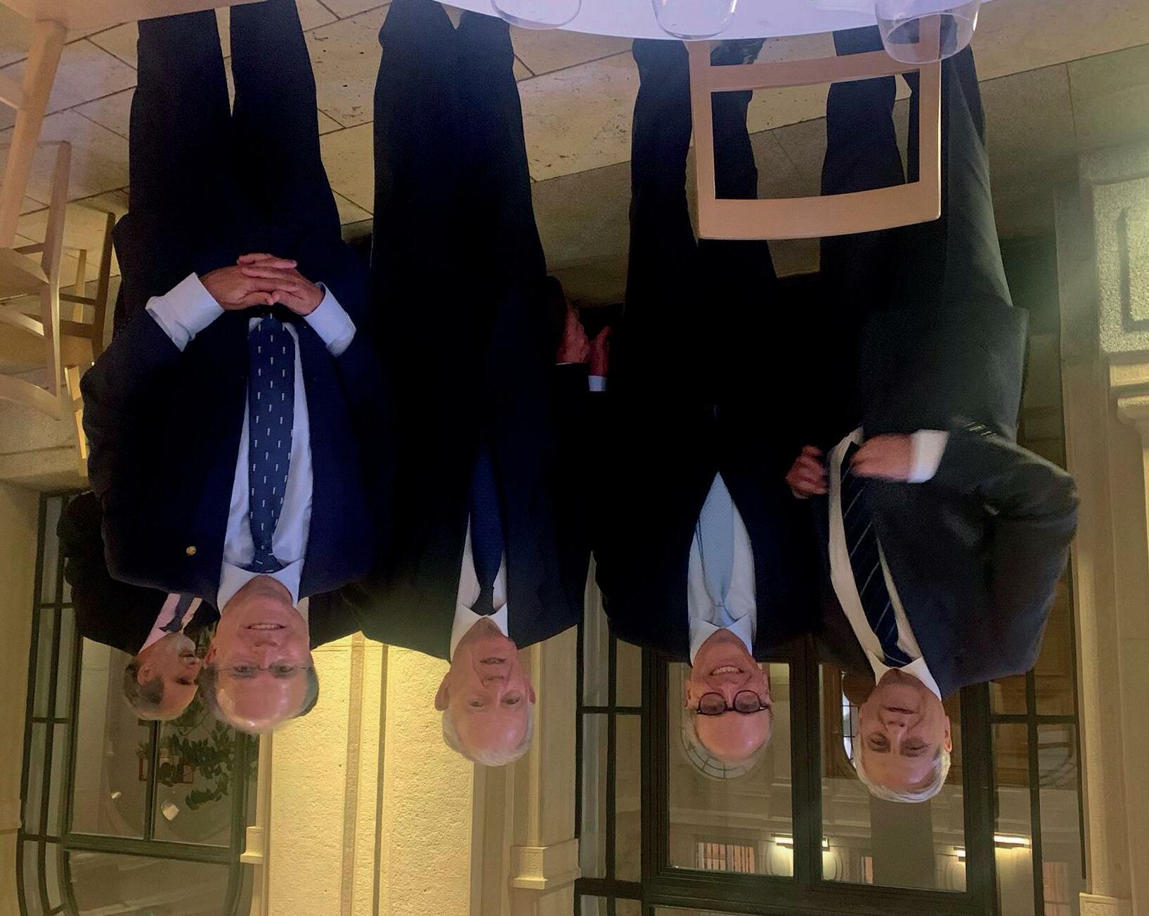
MAKING HEADLINES
Andrés
From the left, Andrés Contreras, Prof Luis Fernández-Vega Sanz, José Ramón Álvarez Rendueles and Javier López-Galiacho.
Members of the managing board of the Forum Pascual Cervera, Juan Carlos Fouz, Alfonso Pérez, Pablo FernándezCanedo, Aitor Errasti and David Rojo -promoter of the initiative of the awardsattended the event. Furthermore, Carlos Gregorio Hernández and José Manuel Varela -director and deputy director of the Residency Hall -, former residents and renowned members of the medical team at Instituto Oftalmológico Fernández-Vega, as well as several friends and family of the attendees went to the event.
After a few welcoming words by the president of the Greater Forum, Andrés Contreras, the Professor was introduced by former residents Ramón Álvarez-Rendueles, former governor of Banco de España and president of Fundación Princesa de Asturias, and Javier LópezGaliacho, former head of the Hall. Both highlighted the academic and professional merits of the medical director of the Institute, as well as the values represented by the Pauline spirit. Drs Luis y Andrés Fernández-Vega Cueto-Felgueroso, ophthalmologists from the fifth generation of the family, sons of the professor and members of the Residence during their six years of career, attended the act in which he was given the graduation band and a commemorative plaque.
he council Residents of the San Pablo Greater Forum has appointed Professor Luis FernándezVega Sanz, medical director of the Instituto Oftalmológico Fernández-Vega, “Resident of Honour”. During the ceremony they recognised his exceptional contribution to the Spanish society, his significant link to the values of the Pauline spirit and his exceptional trajectory as an international referent in ophthalmology.
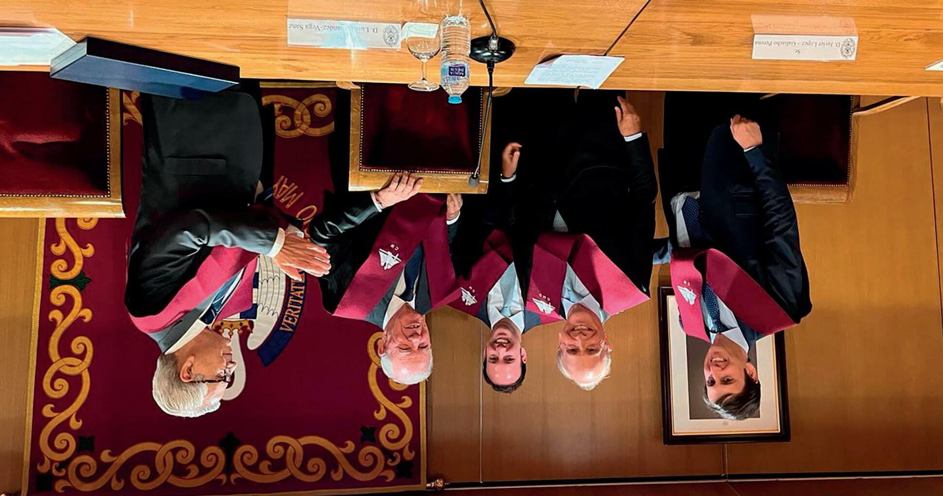
12 MAKING HEADLINES PROFESSOR LUIS FERNÁNDEZ-VEGA SANZ, APPOINTED ‘RESIDENT OF HONOUR’ BY THE SAN PABLO FORUM
T
Prof. Luis Fernández-Vega Sanz, together with his sons and José Ramón Álvarez Rendueles and Andrés Contreras Salido.
José
Ramón Álvarez Rendueles and Javier LópezGaliacho highlighted the academic and professional merits of the Professor.
He stressed his commitment, and that of his family, with Asturias, but he also mentioned the importance of having the necessary -condi tions to compete on equal terms with the most advanced communities of Spain.
used to receiving recognition as a doctor than as a businessman.
Paniceres didn’t forget to stress the professional and civic commitment of professor Luis Fernández-Vega, of whom he said “one can always count on him”. Luis Fernández-Vega, upon receiving his distinction, said “if I had an early medical vocation, my business one came along later and unexpectedly”, as it derived from a need to become a company when he turned a traditional practice into a prestigious, renowned in the field Institute, able to attend all types of eye pathologies, with exceptional professionals and the best technology. He is therefore more
the economy of a medical entity that cares for over one hundred thousand patients each year and performs approximately 10 000 surgeries, and which also helps to project a quality image that reverberates in different areas.
The winners, at the end of the event.
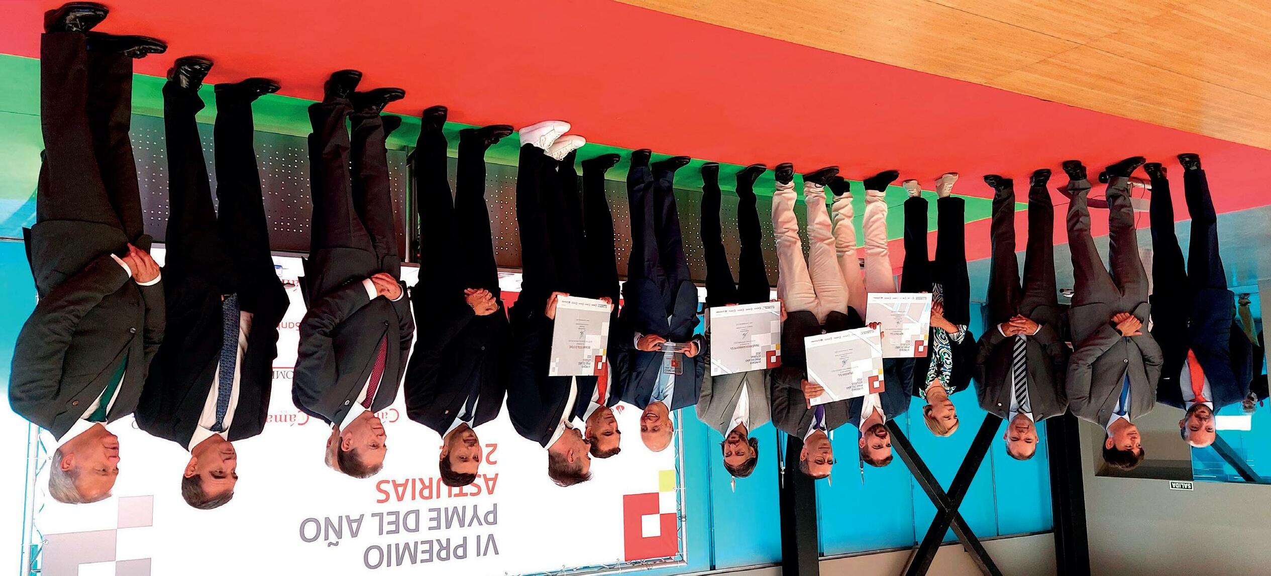
MAKING HEADLINES
11
The IOFV will represent Asturias at a national level in this initiative, which will take place in the first term of the next year.
Carlos Paniceres, president of the Chamber of Oviedo, was in charge of glossing the trajectory of a family – with five generations of ophthalmologists –and the impact in
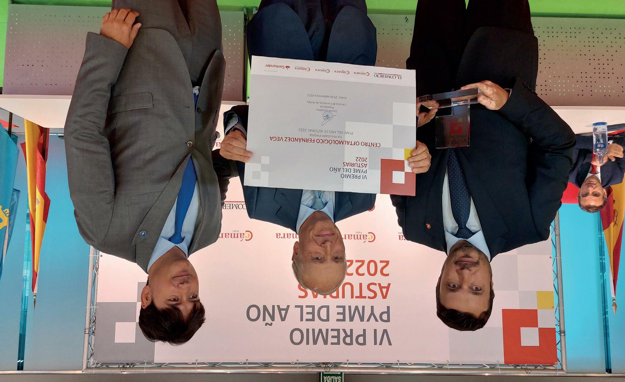
ther companies with accessits due to good behaviour in areas such as innovation, digitalisation or external projection were recognised during the awarding ceremony that took place in Avilés before individuals from the economic and political fields, as well as citizen from the area. The Instituto Oftalmológico Fernández-Vega was recognised as the best SME in Asturias in 2022. Cámara España and Grupo Vocento support this initiative and, therefore, the Institute will represent Asturias at the national event that will take place during the first term of the next year.
The award, given by the Asturian Chamber of Commerce, especially valued his contribution to the economy and the better image of the Principality.
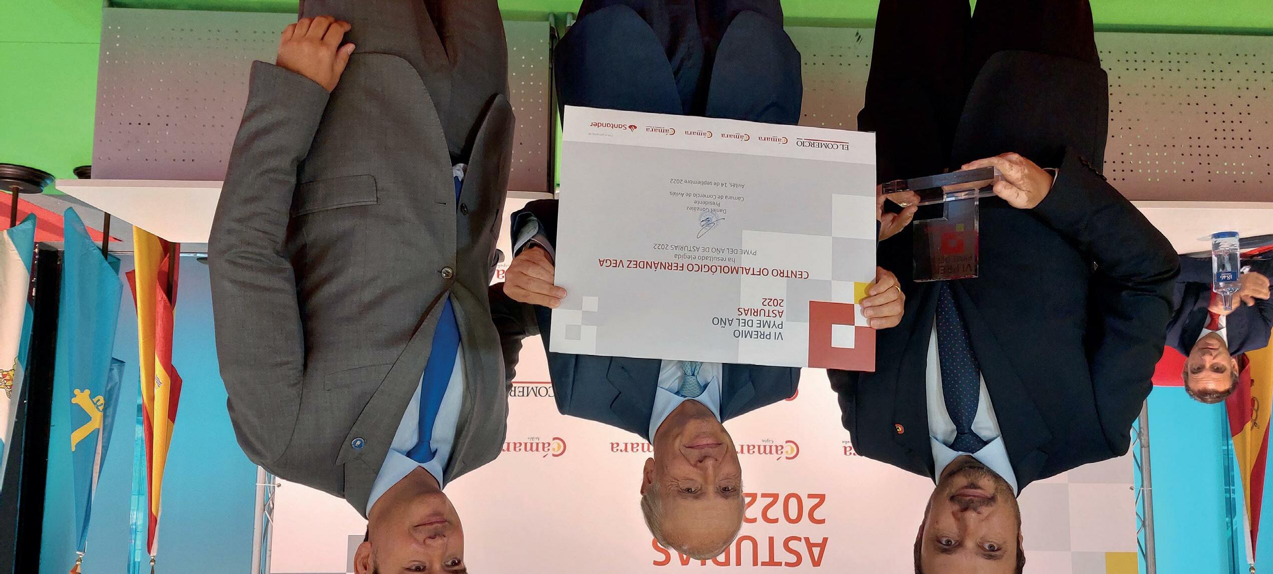
O10 MAKING HEADLINES
Daniel González, president of the Chamber of Commerce of Avilés; Professor Luis Fernández-Vega Sanz, medical director at IOFV, and Borja Sánchez, Science, Innovation and University Councillor of the Principality of Asturias.
THE INSTITUTE, SME OF THE YEAR IN ASTURIAS
Luis Fernández-Vega finished his speech with a renewed civic commitment to society and a strengthened stimulus to keep heading the Instituto Oftalmológico which, together with the Fundación Princesa de Asturias, and despite being in different levels, are according to surveys the most valued entities and institutions of the Principality.
During the moving awarding ceremony they mentioned his presidency at the Fundación Princesa de Asturias during a very satisfactory period also marked by the terrible pandemic that forced to innovate and modify several activities traditionally carried out during the award ceremony that takes place in October in the Asturian capital.
MAKING HEADLINES
Fernández-Vega attributed his vocation to family tradition and to his commitment to combine, together with other members of his family, the clinical activity with the research and teaching activities, which currently makes the Instituto Oftalmológico Fernández-Vega a reference caring for over 100 000 patients every year, with specialists among over their 200 professionals capable of looking after any eye pathology.
Luis Fernández-Vega felt honoured and thankful for this distinction – which he dedicated to his wife Victoria Cueto-Felgueroso – and proud and responsible for being now part of a group that gathers, in the medical and research section, personalities such as Severo Ochoa, Santiago Grisolia, Segovia Arana o Grande Covián among others.
Raimundo Abando, a renowned Asturian businessman, was responsible for highlighting the human and professional circumstances of the honouree, with whom he maintain a close friendship since they were young, and who highlights his commitment to the citizens while stressing the importance of this medical practice from an economic point of view, as it entails the 5% of GDP of the Asturian capital.
9
On this occasion, as is customary, they have granted their Medal to that individual who according to them represents the knowledge, commitment, values and professional career that are hallmark of the Foundation that promotes this initiative, and this year it has been awarded to professor Luis Fernández-Vega, president of the Eye Institute that carries his family name.
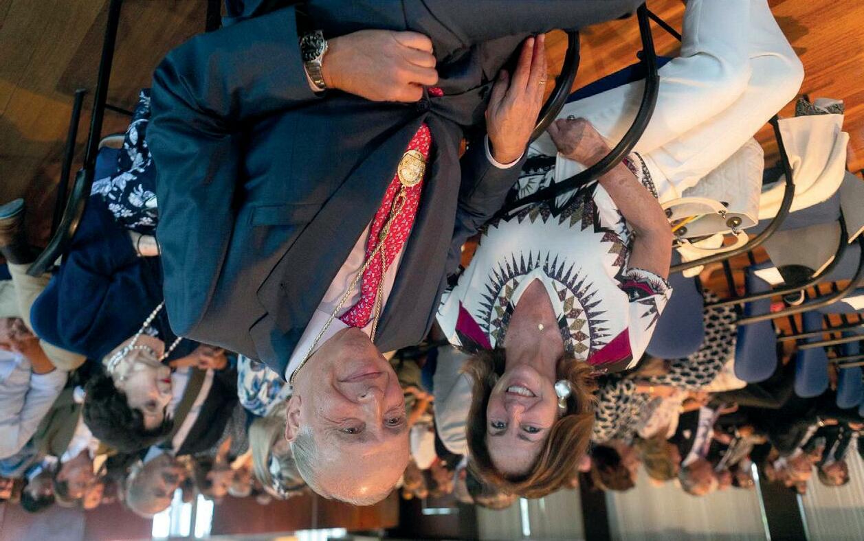
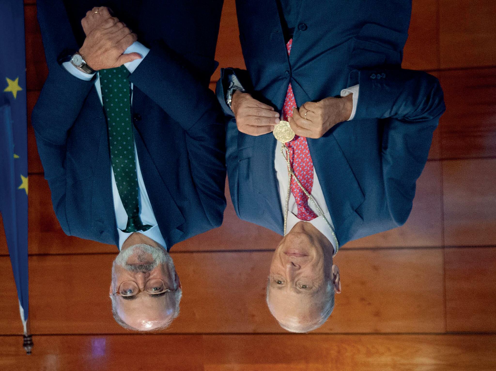
Historians, researchers, artists, writers, politicians, etc. undertake the courses of La Granda, headed by Benigno Pendás, who also heads the Spanish Royal Academy of Moral Sciences and Politics, so none of these disciplines are foreign to them.
a Granda is an exquisite location in the centre of -Astu rias. Arcelor Mittal has an old house there that has been used over time for countless business meeting as well as for these sessions that, for 44 years, have gathered in August most, if not the best, of the -Spa nish intellectuals, who have shared and disclosed their knowledge in a calm environment with a spirit close to that sought back in the day by the -so-ca lled Group from the Asturian University or, more currently, the one praised by Ortega and his school.
LA GRANDA COURSES AWARD ITS MEDAL TO LUIS FERNÁNDEZ-VEGA SANZ
LProfessor Luis Fernández-Vega Sanz and Benigno Pendás, president of La Granda Courses.
MAKING HEADLINES
Professor Luis Fernández-Vega Sanz and his wife, mrs Victoria Cueto-Felgueroso Botas, during the act.
8
Photo property of LNE
Over 6526 -inter views to physicians, nurses, patients’ associations, health journalists, hospital managers, nurse managers, members of the health public administration, -indi viduals responsible for hospital -phar macies and leaders from -pharmaceuti cal companies have been performed to carry out the Health Reputation -Moni tor 2022, the most prestigious -inde pendent ranking of its kind. The HRM assesses over 3010 objective -indica tors of 2912 clinical departments that provide a -perspec tive of the quality of care of each hospital, observing activity data such as provision of care, the patient’s experience and the techniques used or the results obtained.
The Instituto -Oftal mológico -Fernán dez-Vega stands out for its care quality and for its strong commitment to research and teaching thanks to Fundación de Investigación -Oftal mológica (FIO) and Instituto -Universita rio Fernández-Vega. Furthermore, it has the Fundación -Fer nández-Vega, which carries out an -im portant social work providing eye care to disadvantaged groups nationally and internationally.
Oftalmológico Fernández-Vega. “It is undoubtedly an honour for all of us to be consolidated as national and international referent”, adds the professor.
“This recognition encourages us to invest in excellence and commit to provide patients an ophthalmology service of the highest quality”, says Prof Luis Fernández-Vega Sanz, medical director at Instituto
ophthalmology centres in Spain in the Health Reputation Monitor (HRM) carried out by MERCO every year.
The general ranking consists of both the Ophthalmology -de partments in public hospitals and -priva te centres devoted to this specialty. Specifically, the Instituto -Oftalmoló gico -Fernández-Ve ga ranks in the top 3, following public hospitals.
7 FRONT PAGE
he Instituto Oftalmológico Fernández-Vega has been recognised for second year in a row as the best private ophthalmology centre in Spain. Its commitment to patients, research, training and high quality care have earned it the first position in the classification of the private
TThanks to its commitment to research, training and high quality care, the Instituto Fernández-Vega was ranked in first position in the classification of the private ophthalmology centres in Spain in the Health Reputation Monitor (HRM) carried out by MERCO.
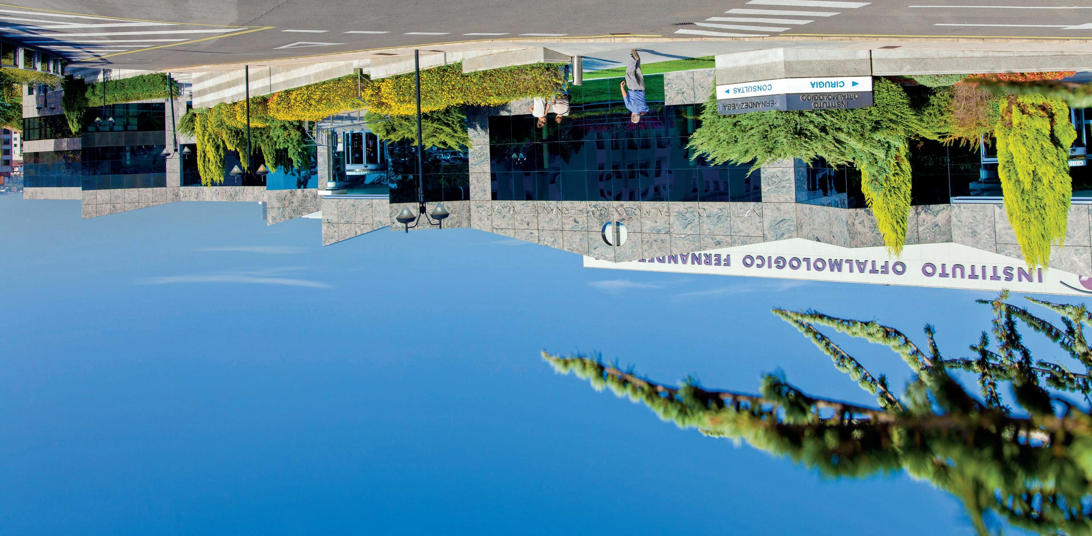
8TH EDITION OF THE HEALTH REPUTATION MONITOR THE INSTITUTO OFTALMOLÓGICO FERNÁNDEZ-VEGA, ONCE AGAIN CHOSEN AS THE PRIVATE OPHTHALMOLOGY CENTRE WITH THE BEST REPUTATION IN SPAIN
FRONT PAGE
6

SUMARIO MAKING HEADLINES La Granda courses award its medal to Luis Fernández-Vega Sanz 8/ THE PATIENTS’ QUESTIONS Patients’ Questions on Glaucoma and Myopia 14/ IN DEPTH Álvaro Fernández-Vega González: “Most Retinal Detachments Can Be Successfully Treated By A Specialised Surgeon” 16/ INVESTIGATION In 20% of the cases, skin rash due to monkeypox can affect the eyes 26/ SOLIDARITY ACTIVITIES Fundación Fernández-Vega visits Asociación Nora 28/ CONGRESS ACTIVITIES Physicians Luis Fernández-Vega Cueto-Felgueroso and Belén Alfonso Bartolozzi, Among the Best Young Ophthalmologists in Europe 31/ WE TAKE CARE OF OURSELVES Who Invented Cataract Surgery? 35/ OTHER SPECIALTIES Healthy Habits and Monitoring Blood Levels, Key to Preventing Cardiovascular Diseases 37/ THEY LOOK KINDLY UPON US Carlos Fitz-James Stuart y Martínez de
The greatest challenge of the Casa de Alba is to hold its heritage to the highest standards so that society participates in and benefits from the full legacy”. 38/ WE RECOMMEND Oviedo is music 39/ CROSSING OF GLANCES Ana de Armas 40/ FRONT PAGE The Instituto Oftalmológico Fernández-Vega, Once Again Chosen As The Private Ophthalmology Centre With The Best Reputation In Spain 6/ 5
Irujo:
ASTURIAS Avda. Dres. Fernández‑Vega, 34 33012 OVIEDO T. 985 240 141 MADRID Príncipe de Vergara, 131 28002 MADRID T. 91 577 99 59 email: instituto@fernandez-vega.com www.fernandez-vega.com Direction and coordination, content writing and editorial management: Atlántica Empresas Advertising: SGANMEDIOS 678 684 457 Design: Eteria, Marketing y Comunicación D.L. AS 5668 2007 MAGAZINE OF THE INSTITUTO OFTALMOLÓGICO FERNÁNDEZ-VEGA 4
As in previous years, these -ack nowledgements compel us and hold us responsible in our duty, and we are deeply grateful.
Furthermore, in the scientific field, Dr Luis -Fernández-Ve ga -Cueto-Felgue roso and Dr Belén Alfonso Bartolozzi were distinguished among the best European Young Ophthalmologists during the latest meeting celebrated in Milan, the Staar Experts Meeting. In that same meeting, Dr José Alfonso and Dr Carlos Lisa also received special mentions. All this confirms that our continuous bet on the excellent -trai ning of our -physi cians is a reality and firm hope for the best future. All this reinforced by the awards received by Dr Ignacio -Rodrí guez Uña in the section on scientific photography and panel -communica tion at the meeting of the Spanish Society of -Ophthal mology (Sociedad Española de -Oftal mología) that took part in Pamplona.
For its part, the Asturian Chambers of Commerce chose our Institute as SME of the year in the Principality for its direct contribution to Oviedo’s and the region’s GDP, as well as the image of excellence -projec ted to the exterior, which translates in general economic benefits of unquestionable importance.
The “Cursos de La Granda” – -acade mic entity of great tradition and prestige – was kind enough to award me with its Medal in view of what they -conside red a unique track record in the medical area as well as in the university and citizen areas, for a continued commitment – in this latter area – in which one can highlight the presidency of the Fundación Princesa de Asturias.
This is relevant because we have started the third term of the year with three awards of very different characteristics that I believe highlight much of what we have done, what we are and what we want to be in the near future.
The last one is the recognition as the private eye centre with the best -repu tation in Spain -ac cording to the study carried out by -MER CO among health professionals and managers - through over six thousand surveys -; MERCO has a well-deserved prestige within the field.
These are of a very diverse nature and originate in entities almost across Spain. But if they have -so mething in common is the generosity of those who award them, and that they become an -incen tive to go forward, to go beyond in our profession and care increasingly better for our thousands of patients. We obviously add to this a deep feeling of gratitude of all those who belong to this team, the true recipient, who thrives each day to be worthy.
There have been numerous -distinc tions to the -Fernán dez-Vega’s -oph thalmology practice received throughout so many decades and five generations practicing it.
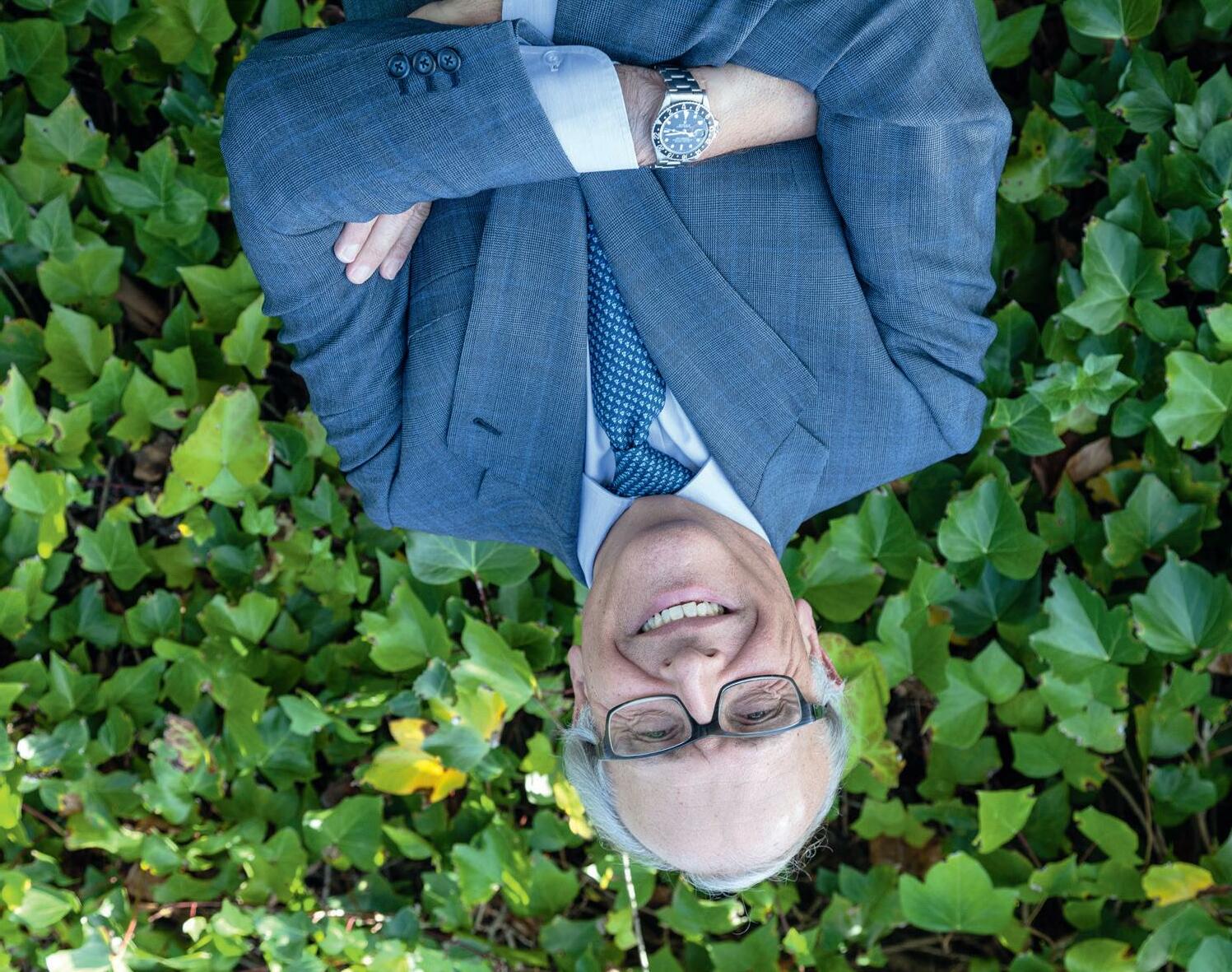
ACKNOWLEDGEMENT AND GRATITUDE
EDITORIAL
3

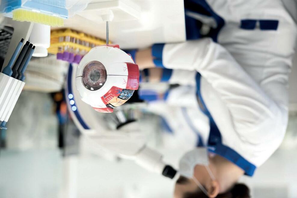
Collaborate with Fundación
Investigación
and promote
that enlightens
the code donation: 10000
de
Oftalmológica
science
www.fio.fernandez-vega.com To
INSTITUTO OFTALMOLÓGICO FERNÁNDEZ-VEGA, BEST EYE PRIVATE CENTRE IN SPAIN (MERCO 2022) FREE ISSUE Nº43 31
Physicians Luis Fernández-Vega Cueto-Felgueroso and Belén Alfonso Bartolozzi, Among the Best Young Ophthalmologists in Europe
38 er3 quarter 2022
Carlos Fitz-James Stuart y Martínez de Irujo: The greatest challenge of the Casa de Alba is to hold its heritage to the highest standards so that society participates in and benefits from the full legacy”.
