
eleflex Incorporated (Wayne, PA, USA; www.teleflex. com) has entered into a de finitive agreement to acquire Standard Bar iatrics, Inc. (Cincinnati, OH, USA: www.standardbariatrics. com) which has commercialized


INTERNATIONAL ® Vol.40 No.3 • 8-9/2022 ISSN 0898-7270

New Stent System Helps Endovascular Treatments

P
Cont’d on page 13Cont’d on page 6
Cont’d on page 18
Cont’d on page 17 INSIDE GLOBETECHMEDIA>>><<< International Calendar . . 22 Industry News . . . . . . . . . 21 News Update . . . . . . . . . . 7 Product News 6-8 News Update . . . . . . . . . 17 Product News 16-20 News Update . . . . . . . . . 11 Product News . . . . . 10-14 COVID-19 Update . . . . 3

an innovative powered stapling technology for bariatric surgery. Under the terms of the agree ment, Teleflex will ac quire Standard Bariat rics for an upfront cash payment of USD 170 million at closing, with additional consideration of
A
epsis, the body’s exaggerat ed response to infection, can cause widespread inflammation and organ failure. In a medical field like critical care, where time can mean life or death, a sepsis di agnosis is like the final buzzer. Tra ditionally identifying patients most
T
MRI Technique Predicts Sudden Cardiac Death
S
n innovative peripheral selfexpanding stent system is designed to improve implanta tion procedures for endovascular treatments.Biotronik Inc.’s (Lake Oswe go, OR, USA; www.biotronik. com) Pulsar-18 T3 stent system uniquely combines three tech nologies. A 4-French low-profile delivery system may decrease the risk of access site complications
S
Fits Inside Needle
udden cardiac death (SCD) accounts for 50% of all car diovascular deaths in the US, claiming more than 300,000 American lives annually, accord ing to the American Heart Asso ciation. Currently, an cardioverter-defibrillatorimplantable(ICD)

AI Diagnostic Tool Identifies Sepsis Within 12 Hours After Hospitalization


Ultra-Thin Endoscope
hotoacoustic imaging works by shining pulses of light on to absorbing structures in the body such as red blood cells or DNA. The structures then gen erate acoustic waves that can be detected by ultrasound sensors and used to form images that can resolve molecular, structur al and functional information from below the tissue surface. By combining light and sound to create 3D images, photoacoustic
at risk has relied on a clinician’s own discretion and experience treating sepsis. Now, a diagnostic tool leverages artificial intelligence to identify a patient’s likelihood of developing sepsis and how severe it will be as soon as 12 hours after their hospital admission.

Teleflex Acquires Bariatric Stapling Innovator If your subscription is not renewed every 12 months your Free Subscription may be automatically discontinued Renew / Start your Free ProductInstantDigitalAccessSubscriptionInteractiveMagazineOnlineInformation: Identify LinkXpress ® codes of interest as you read magazine Click on LinkXpress.com to reach reader service portal Mark code(s) of interest on LinkXpress ® inquiry matrix321 READERVISITSERVICE PORTAL LINKXPRESS COM ® See article on Page 5 High-Tech Vest Can Expedite Discharge of ICU Patients 3D Technologies Enable Fast and Precise SurgicalPreparation3DTechnologies Enable Fast and Precise Surgical Preparation WORLD’S CLINICAL NEWS LEADER OUR 4 OUR YEAR 0 th YEAR P atients with severe respira tory diseases require inten sive treatment and their lung function needs to be monitored on a continuous basis. For over 200 years, the stethoscope has been a standard tool for doctors Cont’d on page 12 L atest techniques for generating realistic 3D models of affected organs are providing surgeons with surgicalningabilitiesunprecedentedtowardplan-andpreparingforprocedures.
Cont’d on page 21
We diagnosticsmakethatmatter
Clinical Chemistry • Point-of-Care • Enzymes • Pre-Analytic Systems © 2022 SEKISUI Diagnostics, LLC. All rights reserved. Because every result matters
Because we both understand that there is a patient behind every answer—and that’s what matters most.
trademark of SEKISUI Diagnostics, LLC. 102HMI-09-22LINKXPRESS COM

At SEKISUI Diagnostics, what matters to you matters to us. Our whole purpose is to partner with you to provide intelligent solutions that enable you to make a timely difference.
For more information about our assays and systems, please visit sekisuidiagnostics.com or email us at questions@sekisui-dx.com is a
For the first time, using functional MRI with in haled xenon gas, researchers at Western University (Ontario, Canada; www.westernu.ca) have shown that the debilitating symptoms of what is now called ‘long-COVID’ such as brain fog, breathlessness and fatigue are related to microscopic abnormalities which affect how oxygen is exchanged from the lungs to the red blood cells. By having the study participants inhale polarized xenon gas while inside the MRI, the researchers could see in real-time the function of the 300-500 million tiny alveolar sacs, which are about 1/5 of a mm in diameter and re sponsible for delivering oxygen to the blood. Further
CT scans pointed to ‘abnormal trimming’ of the vascular tree, indicating an impact on the tiny blood vessels that deliver red blood cells to the alveoli to be oxygenated. There also appeared to be no difference in severity of this abnormality between patients who had been hospitalized with COVID-19, and those who recovered without hospitalization, according to the study.
A
CT Imaging Study Finds Vaccination Reduces Risk of COVID-19 Associated Pulmonary Embolism
An international team of scientists led by the Uni versity of Vienna (Vienna, Austria; www.univie.ac.at) has developed and validated an algorithm that can help healthcare professionals identify who is most at risk of dying from COVID-19 when admitted to a hospital. The tool, which uses artificial intelligence (AI), could help doctors direct critical care resources to those who need them most, and will be especially valuable to resource-limited countries. To develop the tool, the scientists used biochemical data from routine blood draws performed on nearly 30,000 pa tients hospitalized in over 150 hospitals in Spain, the US, Honduras, Bolivia and Argentina between March 2020 and February 2022. The resulting algorithm called COVID-19 Disease Outcome Predictor (CO DOP) - uses measurements of 12 blood molecules that are normally collected during admission. This means the predictive tool can be easily integrated into the clinical care of any hospital.
s hospitals and healthcare institutions around the world rush to order large volumes of COVID-19 remedies, the hospital/medical devices industry continues its unprecedented expansion to meet exploding global demand. The report that follows provides a survey of news and advances from June 1, 2022 until August 30, 2022. For a recap of earlier developments, the reader is invited to refer to previous issues of HospiMedica or visit www.HospiMedica.com
Immunomodulation Technology Rescues Patients from Cytokine Storm
3 HospiMedica International August-September/2022
COVID-19 Update
A retrospective review by researchers at the University of Utah (Salt Lake City, UT, USA; www. utah.edu) that examined the incidence of pulmonary embolism (PE) in COVID positive patients during computed tomography pulmonary angiography (CT PA) exams in the emergency department (ED) found that vaccination reduces the risk of COVID-19 associated PE. The study showed a difference in the incidence of PE in ED patients infected with the ancestral COVID-19 variants and those infected with the Delta and Omicron COVID-19 variants. Though the findings do not reach statistical significance, they suggest that patients infected with the Delta or Omicron COVID-19 variants may have a lower incidence of pulmonary embolism. Additionally, the researchers also found that vaccination with at least two doses does significantly reduce the risk of COVID-19 associated PE.
AI Algorithm Identifies Hospitalized Patients at Highest Risk of Dying From COVID-19
SeaStar Medical’s (Denver, CO, USA; www. seastarmedical.com) novel, simple-to-use Selective Cytopheretic Device (SCD) immunomodulation technology can quickly rescue patients from a cy tokine storm, regardless of how it was triggered, and restore reparative functionality to the body and potentially reverse injury. COVID-19, acute respi ratory distress syndrome, acute kidney injury, and many other health issues can result in a dangerous cytokine storm. By giving critically ill patients the potential to eliminate dialysis dependency, get out of the ICU faster, and restore the lives they were so close to losing, the SCD is positioned to become the new standard of care in the ICU. Because the SCD is an extracorporeal immunomodulator that
Lung-Imaging MR Technique Shows Cause of Long-COVID Symptoms
uses the body’s immune system to heal, healthcare professions remain in complete control of an effective adjuvant therapy that can be used without the worry of severe contraindications. The SCD can be easily added to extracorporeal therapies, such as dialysis or CRRT, seamlessly fitting into the workflow.

BENELUX, FRANCE Hasselt, Belgium
COVID-19 Vaccine Patch Fights SARSCoV-2 Variants Better Than Needles
Simone.Ciolek@globetech.net Tel: (49) 9771-1779-007
USA, UK Miami, FL 33280, USA
A GLOBETECH PUBLICATION
Publishers of: HospiMedica International • TradeMed.com LabMedica International • LabMedica en Español HospiMedica.com • HospiMedica.es • MedImaging.net LabMedica.com • LabMedica.es • BiotechDaily.com
Carolyn.Moody@globetech.net Tel: (1) 954-686-0838
Nadia.Liefsoens@globetech.net Tel: (32) 11-22-4397
Dr Jutta
Vol 40 No 3 • Published, under license, by Globetech Media LLC Copyright © 2022. All rights reserved. Reproduction in any form is forbidden without express permission.
INTERNATIONALADVERTISINGSUBSCRIPTION0898-7270INFORMATIONSALESOFFICESHOWTOCONTACTUS Subscriptions: Send Press Releases to: Advertising & Ad Material: Other Contacts: www LinkXpress info@globetechads@globetechHMNews@globetechcom.netnetnet
Marc Gueron Founder & Editorial Director
A large-scale study by researchers at the Icahn School of Medicine at Mount Sinai (New York, NY, USA; www.icahn.mssm.edu) has found that the estimate of blood viscosity was more strongly associated with mortality in COVID-19 patients than other commonly used risk stratification mea sures. The first study to evaluate blood viscosity in the prediction of mortality in COVID-19 patients found that hospitalized patients who had high blood viscosity had a 60% higher death rate with blood viscosity measured under high flow conditions such as the arteries and 32% higher mortality with blood viscosity measured at low flow such as the microcirculation (blood circulation in the smallest vessels), than patients with a low blood viscosity. The study concluded that that a simple calculation of blood viscosity was more robust in the identi fication of hospitalized patients at risk for dying from COVID-19 complications when compared to common measures of inflammation and the blood clotting biomarker D-dimer.
Total-Body PET Imaging Can Assess Immunological Response to COVID-19 Infections
Regional Director Regional Director Reader Service Manager 4HospiMedica International August-September/2022
A new study by researchers at the Oxford Uni versity Hospitals NHS Foundation Trust (Oxford, UK; www.ouh.nhs.uk) has found that the SARS-COV-2 Omicron variant is associated with fewer and less severe changes on chest CT compared with the Delta variant. The study also found that patients with Omicron had greater frequency of bronchial wall thickening but lower clinical severity and improved hospital outcomes than those with Delta. In a small series of hospitalized patients who had tested RT-PCR positive for SARS-CoV-2 with CT pulmonary angiog raphy performed within seven days of admission, the researchers found that the Omicron variant infection is less likely to be associated with SARS-CoV-2 pneu monia, and that, when pneumonia does occur, it is less severe on chest CT. In agreement with chest CT patterns, Omicron infection was associated with reduced clinical and biochemical markers of severity and improved hospital outcomes.
Regional Director Director
HospiMedica is published 4 times a year and is circuIat ed worldwide (outside the USA and Canada), without charge and by written request, to medical department chiefs and senior medical specialists related to critical care, surgical techniques and other hospital-based specialties; hospital directors/administrators; and major distributors/dealers or others allied to the field.
www . hospimedica . com
Teknopress Yayıncılık ve Ticaret Ltd. Şti adına İmtiyaz Sahibi: M. Geren • Yazı işleri Müdürü: Ersin Köklü Müşir Derviş İbrahim Sok. 5/4, Esentepe, 34394 Şişli, İstanbul P. K. 1, AVPIM, 34001 İstanbul • E-mail: Teknopress@yahoo.com Baskı: Postkom A.Ş. • İpkas Sanayi Sitesi 3. Etap C Blok • 34490 Başakşehir • İstanbul Yerel süreli yayındır. Yılda dört kere yayınlanır, ücretsiz dagıtılır.
COVID-19 Update DanSanjitGueronDutt
Raymond Jacobson, PhD
NewsNewsNewsPublisherEditorEditorEditor
Chest CT Scans of COVID-19 Patients Could Help Distinguish between SARS-CoV-2 Variants

AI Device Predicts When Critically Ill Patients Can Be Safely Removed From Ventilator

Blood Viscosity Testing Can Predict Risk of Death in Hospitalized COVID-19 Patients
JAPAN Tokyo, Japan Katsuhiro.Ishii@globetech.net Tel: (81) 3-5691-3335
Jerry Slutzky, PhD
For change of address or questions on your subscription, write to: HospiMedica lnternational, Circulation Services at above address or visit: www LinkXpress com
ISSN
CHINA Shenzhen, Guangdong, China Parker.Xu@globetech.net Tel: (86) 755-8375-3877
Carolyn Moody, RN
UC Davis (Davis, CA, USA; www.ucdavis.edu) which collaborated with United Imaging Healthcare Co., Ltd. (Shanghai, China; www.united-imaging. com) to develop the world’s first total-body PET/ CT scanner, uEXPLORER, has found that high sensitivity PET imaging using a specific T cell radiotracer offers a new methodology to track tissue concentrations of T cells without invasive sampling. Guided by the uEXPLORER scanner along with a new labeled radiotracer with high affinity to T cells (89Zr-Crefmirlimab-Berdoxam), the scientists unveiled the first high-resolution PET image of total-body CD8+ T-cell distribution after COVID-19 infection. The image could shed light on the pathological mechanism and immune response to COVID-19 infection.
A new study in mice conducted by researchers at the University of Queensland (Brisbane, Austra lia; www.uq.edu.au), in partnership with Vaxxas (Brisbane, Australia; www.vaxxas.com), suggests that a needle-free vaccine patch could better fight COVID-19 variants, such as Omicron and Delta, than a traditional needle vaccine. The ongoing emergence of variants of concern has resulted in decreased vaccine efficacy necessitating booster im munizations. Currently-available vaccines may not be as effective because of the constantly emerging new variants of COVID-19, and this has left research ers at the crossroads. The researchers tested the Hexapro SARS-CoV-2 spike vaccine using the Vaxxas high-density microarray patch (HD-MAP) technology and found the patch was far more effective at neu tralizing COVID-19 variants.
The Ottawa Hospital (Ontario, Canada; www. ottawahospital.on.ca) has become the first hospital in the world to evaluate an innovative medical device that uses artificial intelligence (AI) to predict when patients who are critically ill due to COVID-19 or surgery, can breathe on their own. The device, called the Extubation Advisor, constantly monitors and analyzes a patient’s vital signs, including blood pressure, oxygen levels, breathing rhythms and heart rate during their ventilation. Then, it uses AI to provide doctors with a specific read of when the patient can be safely removed from the ventilator. The system was used for three months at the bedside of ventilated patients in The Ottawa Hospital’s Intensive Care Unit (ICU), with permission from their families. After the successful initial evaluation, the metrics are looking promising, and the feedback received from physicians was very positive.
ALL OTHER COUNTRIES Contact USA Office ads@globetech.net Tel: (1) 954-686-0838
Joffre.Lores@globetech.net Tel: (1) 954-686-0838
GERMANY, SWITZ , AUSTRIA Bad Neustadt, Germany
Regional
KatsuhiroParkerCiolekXuIshiiDavidGueron
Joffre Lores
Regional Director
‘Covid Computer’ Uses AI to Detect COVID-19 from Chest CT Scans
Researchers from the University of Leicester (Leices ter, UK; www.le.ac.uk) have created a new AI tool that can detect COVID-19. The software analyses chest CT scans and uses deep learning algorithms to accurately diagnose the disease. With an accuracy rate of 97.86%, it is currently the most successful COVID-19 diagnostic tool in the world. The researchers will now further develop this technology in the hope that the Covid computer may eventually replace the need for radiol ogists to diagnose COVID-19 in clinics. The software, which can even be deployed in portable devices such as smart phones, will also be adapted and expanded to detect and diagnose other diseases (such as breast can cer, Alzheimer’s Disease, and cardiovascular diseases).
To all others: Paid Subscription is available for a twoyear subscription charge of US$ 100. Single copy price is US$ 20. Mail your paid subscription order accompa nied with payment to Globetech Media, LLC, P.O.B. 800222, Miami, FL 33280-0222, USA.
T HospiMedicaInternationalTo view this issue in interactive digital magazine format visit www.HospiMedica.com
“Modern technologies such as virtual reality, augmented reality and 3D printing offer previously untapped potential to improve surgery plan ning and execution as well as training,” emphasized Professor Rainer Malaka, Managing Director of the Technology Center Informatics and Information Technology (TZI) at the University of Bremen.
SunStrongerNuclearSunNuclearandCIRSarenowone,aspartofMirionMedical.
Learn more: sunnuclear.com/cirs
Placental MRI Method Detects Complications in Early Pregnancy
Cont’d
With the help of special AR glasses, surgeons can view the pa tient-specific 3D model as a “hologram” during surgery, using gesture control to rotate and turn it or place it manually. Before the procedure, you can already view the effects of an incision on the liver, which has a large blood supply, thus allowing you to estimate how much tissue will no longer be functional afterward. A physical 3D model, in combination with a training system, also allows the practice of complex interventions and stress situations. The prototypes from the VIVATOP project have passed clinical trials.

5 HospiMedica International August-September/2022
In the VIVATOP research project, scientists from the Universities of Bremen (Bremen, Germany; www.uni-bremen.de) and Oldenburg (Oldenburg, Germany; www.uol.de) and their collaborative partners have now developed 3D technologies that enable the medical team to assess the situation before and during surgery much more accurately. As a result, they expect a better assessment of surgical options and an associated higher success rate, especially in difficult cases. The joint project aimed to develop innovative and interactive 3D technologies for clinical use. The project consortium focused primarily on the liver, but due to the pandemic, added imaging of lungs to aid in the diagnosis of COVID-19 illnesses. The 3D visualization of an organ in virtual or aug mented reality (VR/AR) offers significant advantages over the two-di mensional images from computer tomography or magnetic resonance imaging (CT/MRI) that have been common in the past.
Image: A surgeon plans a procedure using a 3D-printed liver and VR glasses (Photo courtesy of University of Bremen)
3D Technologies Enable Fast and Precise Surgical Preparation


on page 8
If a tumor is located too close to important blood vessels, its surgical removal may prove to be dangerous or even impossible. Surgeons now have the ability to create realistic 3D models of affected organs that can be both digitally visualized and made physically tangible via 3D printing.
The researchers have also included a “multiuser” functionality that allows several people to work with the model at the same time. It doesn’t matter whether the participants are together in the same room or not – experts from other continents can also be dialed in via AR telephony. For remote experts participating from the operating room via livestream, various representations are being tested in order to show these models as realistically as possible and to give them a realistic impression of what is happening in the operating room. In preliminary meetings, however, the actual models from the 3D printer also prove their strengths, because they serve as visual objects without the use of technology.“Withthe help of the 3D models, we can capture the complex vas cular and organ anatomy much faster,” added Professor Dirk Weyhe, a visceral surgeon from Pius Hospital Oldenburg. “CTs and MRIs require a composition from two levels.”

A

he placenta plays a key role in fetal development and pregnancy morbidity, as well as neonatal, pediatric and even lifelong health. As the primary source of oxygen and nutrients for a developing fetus, abnormal placental development can be dangerous and has been linked to many adverse outcomes, including abnormalities in fetal growth, preeclampsia, which is the development of high blood pressure during pregnancy, preterm labor and stillbirth. The placenta is a dynamic organ that evolves over the course of pregnancy to support fetal development, so poor placental function early in pregnancy can become an ongoing and increasing health concern both to mother and baby. Despite the detrimental
105HMI-09-22LINKXPRESS COM
Cont’d from cover
With complementary and proven product portfolios, we share a commitment to easing technology adoption, optimizing Quality Management, and ensuring Patient Safety.
ore than 90% of patients with acute ischemic stroke (AIS) receive magnetic resonance imaging (MRI) in addition to computed tomography (CT) with few data to determine whether there is an associated benefit with patient outcomes. Now, a new study of patients hospitalized with AIS has found that a diagnostic imaging strategy of initial CT alone was noninferior to initial CT with additional MRI with regard to the clinical outcomes of death or dependence at hospital discharge or prevention of stroke or death at one year after discharge.
BATTERY-POWERED PORTABLE X-RAY MINXRAY
participants with a median age of 68 years and 131 being men (53%). The researchers found that death or dependence at discharge occurred more often in patients with additional MRI (59 of 123 [48%]) than in those with CT alone (52 of 123 [42.3%]); meeting the −7.50% criterion for noninferiority. Stroke or death within one year after discharge determined for 225 of 235 (96%) survivors occurred more often in patients with additional MRI (22 of 113 [19.5%]) than in those with CT alone (14 of 112 [12.5%]), meeting the 0.725 relative risk criterion for noninferiority.
The all-new web based PaxeraRIS is an intuitive workflow management platform that enables com plete control of all radiology data and simplifies the workflow across the entire radiology department.

lular energy source, in the hearts of 46 people prior to getting an ICD for primary prevention. The cardiac ATP levels were measured on clin ical MRI scanners using a magnetic resonance spectroscopy (MRS) technique developed at Johns Hopkins Medicine, to determine which patients had abnormal ATP metabolism. All patients were followed up every three to six months for an average of 10 years to determine which patients had appropriate ICD firings for life-threatening arrhythmias.
The study by researchers at the Duke University Medical Center (Durham, NC, USA; www.dukehealth.org) involved 246
Based on the findings of the propensity score-matched cohort study of patients hospitalized with AIS, the researchers
Cont’d from cover
In the small, but rigorous study, research ers at the Johns Hopkins University School of Medicine (Baltimore, MD, USA; www.hopkins medicine.org) measured the levels of adenosine triphosphate (ATP), the primary chemical cel
NeuViz Prime CT system scanner has spectral im aging capabilities, providing the ability to perform image acquisition and processing at multiple energy levels to improve visualization for CT and enhancing patient care.


Cont’d on page 7
for Acute Ischemic Stroke Patients
M
The TR90BH battery-powered portable X-ray gen erator is easy to use and quickly programmable for storage of up to five exposure tech niques. It is lightweight, ultra-dura ble, and powerful with a long-lasting battery life.

201HMI-09-22LINKXPRESS COM 202HMI-09-22LINKXPRESS COM 203HMI-09-22LINKXPRESS COM
- a small, battery-powered device placed in the chest to detect and stop irregular heart rhythms - is the primary means of preventing SCD in high-risk patients. The device continu ously monitors the heart rhythm and delivers electric shocks, when needed, to restore a regular heart rhythm. The battery life of an ICD is typically between five to seven years. Now, a new study has found that adults with abnormal heart metabolism are up to three times more likely to experience life-threatening arrhythmias (an irregular heart rhythm), and magnetic resonance imaging (MRI) techniques could be used to detect the condition and pre dict future SCD.
CT Imaging Alone Noninferior to CT/MR Combo
concluded that a diagnostic imaging strategy of initial CT alone was noninferior to initial CT plus additional MRI with regard to clinical outcomes at discharge and at one year. Further research is needed to determine which patients hospitalized with acute ischemic stroke benefit from MRI, according to the researchers.
approaches and lead to better predictions for who is most likely to need, or not need an ICD. However, they stress, that more studies are needed to assess different and larger pop ulations.“We believe this is the first time that impaired cardiac metabolism in people has been linked to an increased risk of life-threat ening arrhythmias or sudden cardiac death,” said study senior author Robert Weiss, M.D., professor of medicine at the Johns Hopkins
PAXERAMED
CT SCANNER NEUSOFT MEDICAL SYSTEMS

MRI Technique Predicts Sudden Cardiac Death
To receive prompt and free information on products, log on to www LinkXpress com or fill out reader service form located on last page, or scan the QR code on your mobile devicePRODUCT NEWS


RADIOLOGY WORKFLOW MANAGEMENT PLATFORM
6HospiMedica International August-September/2022
Results showed that people with low car diac ATP levels (impaired metabolism) had a three-fold higher risk of sudden cardiac death (if not saved by ICD intervention) compared to those with normal ATP metabolism. This was still the case when adjusted for low left ventricular ejection fraction, the metric currently used to determine the need for a primary prevention ICD. The researchers say the study findings could complement current
University School of Medicine. “This could open a window for a whole new approach, a metabolic approach for treating or preventing severe arrhythmias, which is something that is not currently available in cardiology.”
Attendees will be able to learn more about relevant, timely topics such as artificial intelligence (AI), COVID-19 and health equity and engage in meaningful networking opportunities throughout the week. Popular meeting features like the Image Interpretation Session, Case of the Day and Fast 5 sessions are returning for 2022. Also back is the Discovery Theater, where attendees can recharge with lively presentations and entertainment. New this year, the Learning Center Theater will offer engaging, interactive presentations throughout the week.
MRI
The Technical Exhibits, featuring cutting-edge imaging technologies and software applications, are a highlight of the RSNA annual meeting. More than 490 companies have already reserved 348,000 square feet of exhibition space. Making a return will be the AI Showcase and Theater, with new technologies, practical demonstrations and industry presentations. RSNA 2022 Virtual Access is also available for individuals who are impacted by institutional, corporate or national travel restrictions due to COVID-19. Virtual Access will feature nearly 100% of all programming."Weverymuch look forward to an exciting RSNA meeting in 2022," said RSNA president Bruce G. Haffty, M.D. "There is continued enthusiasm for attendance in person, and virtual access to the full program will also be available."
educational sessions, scientific sessions that demonstrate the latest research, and the exciting new technology available in our exhibition halls continue to make the RSNA annual meeting a highly valued (626) Rise in Abstract Submissions Ahead of Annual

T Need to check the performance of X-ray machines? Then the Radcal Touch meter is your tool of choice. Radcal Touches the World! Features: • Simple to use – Accurate and reliable • Customizable Touch Screen • Wi-Fi and USB Computer Connectivity • Report Generation DAP For further details: contact us at +1


"The keynote plenary speakers will focus on our theme for this year's meeting, "Empowering Patients and Partners in Care," with a goal of raising awareness of the value of the radiological sciences to our patients and multidisciplinary partners in health care," Dr. Haffty said. "Our large portfolio of quality
Under the slogan “Empowering Patients and Partners in Care”, RSNA 2022 will showcase the important role of radiology in multidisciplinary patient care. The RSNA annual meeting is the world's leading annual radiology forum, and RSNA 2022 will provide a multitude of research presentations and educational opportunities in all subspecialties.
he Radiological Society of North America (RSNA, Oak Brook, IL, USA; www.rsna.org) has announced that nearly 10,400 scientific and educational abstracts have been submitted for the Society's 108th Scientific Assembly and Annual Meeting. RSNA 2022 will be held from November 27 to December 1, 2022, at McCormick Place in downtown Chicago.
Image: RSNA`s annual meeting is the world`s largest medical imaging confer ence (Photo courtesy of RSNA)

7 HospiMedica International August-September/2022


Meeting
“Over seven years, 60-70% of these devices never discharge to save a life,” added T. Jake Samuel, Ph.D., first author of the study and fellow in cardiology at Johns Hopkins Medicine. “We’re spending billions of dollars a year on ICDs that are implanted and have procedural and post-procedural risks. There is a need for non-invasive approaches to better assess risk for who needs or doesn’t need an ICD to prevent sudden cardiac death in people.” Predicts Sudden Cardiac Death

357-7921 sales@radcal.com • www.radcal.com Visit us at ECMP Dublin, Ireland, 8/17-8/20 vc HospiMedica_clr_4.125x5.375_Eng22Jul20_11733.pdf 1 7/19/22 1:18 PM RSNA 2022 Sees
Cont’d from page 6

Placental MRI Method Detects Complications in Early Pregnancy
toward the ultrasound stickers could be made into wearable imaging products that patients could take home from a doctor’s office or even buy at a pharmacy.
REMOTE CONTROLLED RADIOGRAPHY TABLE VILLA SISTEMI MEDICALI

Image: Engineers have developed stickers that can see inside the body (Photo courtesy of MIT)
“We envision a few patches adhered to different locations on the body, and the patches would communicate with your cellphone, where AI algorithms would analyze the images on demand,” said the study’s senior author, Xuanhe Zhao, professor of mechanical engineering and civil and environmental engineering at MIT. “We believe we’ve opened a new era of wearable imaging: With a few patches on your body, you could see your internal organs.”
be replicated on virtually all modern MRI scanners. With quick data analysis, researchers noted that the imaging method could be easily adopted by Researchersclinicians.atOregon Health & Science University (OSHU, Portland, OR, USA; www.ohsu.edu) conducted a study to explore how an MRI could be used to give clinicians a more detailed look at placental health than the traditional ultrasound provides, and to better understand an MRI’s effectiveness in detecting placental abnormalities during pregnancy. Their ultimate goal is to reduce fetal
Cont’d on from page 5
The Eclipse & Eclipse 3D Probe Cover is a soft, dis posable probe cover for medical ultrasound probes/ transducers, pre-gelled inside with Aquasonic 100 Ultrasound Trans mission Gel.

Stamp-Sized Ultrasound Sticker Produces Clear Images of Internal Organs
ANALOG MOBILE X-RAY MACHINE SIEMENS HEALTHINEERS
ltrasound imaging is a safe and noninvasive window into the body’s workings, providing clinicians with live images of a patient’s internal organs. To capture these images, trained technicians manipulate ultrasound wands and probes to direct sound waves into the body. These waves reflect back out to produce highresolution images of a patient’s heart, lungs, and other deep organs. Currently, ultrasound imaging requires bulky and specialized equipment available only in hospitals and doctor’s offices. Now, a new design might make the technology as wearable and accessible as buying Band-Aids at theApharmacy.teamof engineers at MIT (Cambridge, MA, USA; www.web.mit. edu) has designed a new ultrasound sticker a stamp-sized device that sticks to skin and can provide continuous ultrasound imaging of internal organs for 48 hours. The researchers applied the stickers to volunteers and showed the devices produced live, high-resolution images of major blood vessels and deeper organs such as the heart, lungs, and stomach. The stickers maintained a strong adhesion and captured changes in underlying organs as volunteers performed various activities, including sitting, standing, jogging, and biking.
Cont’d on page 9
The current design requires connecting the stickers to instruments that translate the reflected sound waves into images. The researchers point out that even in their current form, the stickers could have immediate applications: For instance, the devices could be applied to patients in the hospital, similar to heart-monitoring EKG stickers, and could continuously image internal organs without requiring a technician to hold a probe in place for long periods of time. If the devices can be made to operate wirelessly a goal the team is currently working
The Polymobil Plus is a powerful entry-level analog mobile X-ray machine with simple to use, proven features for streamlined operation. It offers high image quality with its 20-kW generator and maximum tube current of 300 mA.
PROBE COVER PARKER LABORATORIES
The Apollo remote controlled radiography table allows users to get the best from the R/F room by offering a range of fast and accurate move ments that ensure extraordinary application capability and reduce preparation times.
204HMI-09-22LINKXPRESS COM 205HMI-09-22LINKXPRESS COM 206HMI-09-22LINKXPRESS COM 8HospiMedica International August-September/2022
impact of abnormal placental development, existing methods for evaluating placental function are often ineffective and limited in their ability to reliably detect risks during pregnancy. In prenatal care settings, most clinicians rely on ultrasound to take measurements of fetal growth and blood flow, but this method is limited in scope. Now, researchers have developed a new imaging method to measure the health of a placenta, which could help clinicians identify complications early in a pregnancy. The researchers used magnetic resonance imaging, commonly known as MRIs, and could
U
To receive prompt and free information on products, log on to www LinkXpress com or fill out reader service form located on last page, or scan the QR code on your mobile devicePRODUCT NEWS


where AI assistance reduced radiograph reading times by 6.3 seconds per“Itpatient.isvery
revolutionary AI software designed to assist radiologists and emergency clinicians in the diagnosis of skeletal fractures uses advanced algorithms to detect and localize lesions on X-rays –graphically highlighting areas of interest before submitting the images to radiologists for validation.
A

AI Software Integrated into X-Ray Imaging Helps Identify Bone Trauma at POC
of Mirion
Diagnostic Imaging Innovative QA SolutionsSunNuclearandCIRSarenowpart

“Having Fujifilm on board has been crucial in the development of this product, and to get it in front of the clinicians that need it. AI technol ogies are becoming increasingly established in the medical sector, and really proving their value. Any medical innovation is about improving the care of the patient, and BoneView promises to do just that,” added Christian Allouche, CEO at Gleamer.
Placental MRI Method Detects Complications in Early Pregnancy
109HMI-09-22LINKXPRESS COM
“Any research that helps us find ways to improve prenatal care is crucial,” said Victoria HJ Roberts, Ph.D., research associate professor in the Division of Developmental and Reproductive Sciences at OHSU’s Oregon National Primate Research Center, who co-led the study. “Pregnancy can be extremely taxing, both emotionally and physically, especially for someone who is experiencing a complicated pregnancy. It’s exciting that this research has identified a more effective way to detect complications early in pregnancy, so clinicians are able to provide the best care to the mother and developing baby.”
exciting to be part of this partnership. BoneView is an extraordinary tool that will assist busy radiographers, radiologists and clinicians to better manage patients at the point of care, adding value for both staff and the patients,” said Richard Cahalane, Product Manager Digital Modalities, Fujifilm Europe GmbH.
With complementary and proven product portfolios, we share a commitment to easing technology adoption, optimizing Quality Management, and ensuring Patient Safety.
ImagingMedicalTo view this issue in interactive digital magazine format visit www.HospiMedica.com 9 HospiMedica International August-September/2022
Medical, a growing group within the Mirion Technologies family.
Learn more: sunnuclear.com/diagnosticimagingqa

Fujifilm (Tokyo, Japan; www.fujifilm.com) has equipped its X-ray systems with a new image processing box called EX-Mobile enabling them to connect with Gleamer’s (Paris, France; www.gleamer.ai) BoneView software. Results are available within 30 seconds at the point of care, providing healthcare professionals with additional support to help improve patient management. The user-friendly software can be seamlessly integrated into Fujifilm’s comprehensive X-ray modality lineup, making it perfectly suited to small or remote clinics, pop-up medical centers, nursing homes, up to large multi-function institutions. This will aid medical staff in the rapid identification of patients with suspected fractures, triaging them for further investigation to ease workflows and enhance patient care pathways.
A clinical trial involving appendicular skeletal fractures found that BoneView reduced the number of false positives by 41.9 %, and im proved fracture detection sensitivity and specificity. These results are supported by another study involving additional anatomical locations,

Image: BoneView AI software will be integrated with Fujifilm X-ray systems (Photo courtesy of Fujifilm)

Cont’d from page 8 and maternal complications associated with placental dysfunction. The study gathered data from 316 pregnant women, including individuals considered both low-risk and high-risk for pregnancy complications.TheOHSUresearch
team developed and validated an MRI protocol that detects a signal in the blood that is linked to oxygen content. This readout is known as T2*, and T2* values provide key information about oxygen availability and placental blood flow. Oxygen is key for fetal growth and development, so if these values deviate from the normal range, it suggests that something might be wrong. T2* values outside of the normal range could indicate an issue related to the maternal blood supply of oxygen, compromised placental transport or fetal utilization of oxygen.Thestudy first established a baseline to determine what occurs throughout the course of an uncomplicated pregnancy. Participants underwent three MRI studies during weeks 10 through 40 of pregnancy. Researchers then looked at the ability of MRI to successfully identify complications in pregnancy using the T2* readings produced from the procedures. The study results suggest that even data from early on in pregnancy - 10 to 20 weeks - can be effective in the identification of at-risk pregnancies. Additionally, the MRIs in the study were performed using the imaging protocol developed by the OHSU team that could be implemented on virtually all modern MRI scanners, and data analysis is quick to perform, indicating that this method may be easily adopted and expanded for use across prenatal health care settings.
To receive prompt and free information on products, log on to www LinkXpress com or fill out reader service form located on last page, or scan the QR code on your mobile devicePRODUCT NEWS


The
207HMI-09-22LINKXPRESS COM 208HMI-09-22LINKXPRESS COM 209HMI-09-22LINKXPRESS COM 10HospiMedica International August-September/2022
denoising for ultra-low-dose CT images. Trained with over one million patient images containing varying degrees of noise for different body parts, its Clarity Engine separates image noise selectively while enhanc ing underlying structures; thus, providing clarity restored images. The patented Deep Learning Clarity Engine preserves natural image texture while clearing quantum noises, thereby providing comfort observation with enhanced image clarity.
INCUBATOR PROGETTI MEDICAL
Digital Radiography Detector Wins Frost & Sullivan Innovation Award
ADVANCED MECHANICAL VENTILATOR GETINGE Servo-u is an Advanced Mechanical Ventila tor features unique decision support tools such as Transpulmonary pressure, Open Lung Tool, Servo Compass, Stress Index, and Edi – the electrical activi ty of the diaphragm.

Weighing just around 2.25 kg including the battery the sleek 35 cm x 43 cm Lux 35 Detector with its glass-free sensor is Carestream’s lightest detector to date. The lightweight, glass-free wireless detector ergonomical ly designed with the comfort of patients and radiographers in mind, makes it easier for radiographers to transport while making rounds and perform ing bedside exams. Also, the Lux 35 Detector has additional ergonomic features that reduce stress and fatigue for radiographers, allow for easier positioning, and make the exam process more comfortable for patients.
The Lux 35 Detector supports a variety of image-processing options to assist radiologists, including Tube and Line Visualization, Pneumothorax Visualization, EVP Plus, Bone Suppression and many more. It uses Care stream’s ImageView Software powered by Eclipse for high image quality and also features the X-Factor, so the detector can be shared with other compatible DRX equipment. Additionally, the Lux 35 Detector battery is
C
User definable and scalable-clarity settings allow users to maintain im age clarity at reduced-dose settings, helping users to achieve a consistent decision in quantitative and qualitative analyses. Done in real-time, the clinical workflow of the ClariCT.AI is simple and comprehensive. En hanced image clarity allows users to optimize the trade-offs between im age quality and radiation dose, and helps users achieve the best practice in low-dose CT imaging. It can be a standalone solution running from a desktop PC or be built into different modalities such as CT machines and PACS system. The technology also supports cloud-based distribution and deployment. ClariCT.AI is both FDA-cleared and CE marked, and is available through the Nuance AI Marketplace, which connects more than 9,000+ facilities in the US.
C
backwards compatible it works with DRX Plus Detectors and uses the same battery charger.
ClariPi’s ClariCT.AI utilizes its Clarity Engine to perform CT image
arestream Health (Rochester, NY, USA; www.carestream. com) was the first to introduce to the market a wireless cas sette-sized detector. Now, the Carestream Lux 35 Detector has earned the 2022 Frost & Sullivan Global New Product Innovation Award in the digital radiography detector sector. Frost & Sullivan has noted that the Lux 35 Detector “encompasses all of the attractive features of differ ent digital radiography (DR) detectors in a single solution, making it the industry’s best DR detector.”
The MORPHEUS M anesthesia machine is suitable for administration of oxygen – air – nitrous oxide –halothane – enflurane – isoflurane – sevoflurane – desflurane mixtures on adult, children and newborn patients.


AI Technology Uses Noise Reduction to Increase CT Image Clarity
“The Frost & Sullivan Award is another validation of our research and development team’s continued success at developing solutions that incorporate cutting-edge technology to improve radiology workflow and enhance patient care,” said Dharmendu Damany, Chief Technology Officer at Carestream. “With our first cesium iodide, glass-free Lux 35 Detector for the medical space, we provide a solution that is lightweight and provides superb resolution and better detail, and a reduced exposure as compared with gadolinium detectors.”
T scans are vital for screening, diagnosing, monitoring and treating different conditions, although radiation concerns raise doubts among patients and clinicians over the medical necessity of this kind of imaging. Now, an FDA-cleared and CE-marked CT denoising solution is addressing the unmet need for higher quality imaging on low-dose CT scans, which means patients and clinicians no longer have to choose between quality CT imaging and radiation safety.ClariCT.AI from ClariPi Inc. (Seoul, South Korea; www.claripi.com) is an AI-powered low-dose CT denoising solution that offers superb clarity of CT images scanned with substantially reduced radiation dose. Using deep learning to remove thousands of different noise patterns, ClariCT.AI produces images with the diagnostic clarity comparable to that of high-dose CT images. The vendor-agnostic software solution has been trained with world's largest image dataset over one million, and its deep-learned Clarity Engine clears image noise safely while enhancing underlying structures to a level of regular dose images from the noisy ultra-low-dose CTs.
ANESTHESIA MACHINE SIARE
The PG NEST incubator features an air circuit that supplies a quiet and comfortable ambient that reduc es external noise, while the inner temperature is monitored and con trolled on various points to ensure accuracy and safety.

Researchers from Pritzker School of Molecular Engineering (PME) at The University of Chicago (Chicago, IL, USA; www.uchicago.edu) have developed a flexible, stretchable computing chip that processes informa tion by mimicking the human brain. The device aims to change the way health data is processed. The future of healthcare that many envision includes wearable biosensors to track complex indicators of health in cluding levels of oxygen, sugar, metabolites and immune molecules in people’s blood. One of the keys to making these sensors feasible is their ability to conform to the skin. As such skin-like wearable biosensors emerge and begin collecting more and more information in real-time, the analysis becomes exponentially more complex. A single piece of data must be put into the broader perspective of a patient’s history and other health parameters.
able technology with AI and machine learning to create a powerful device which can analyze health data right on our own bodies,” said Sihong Wang, a materials scientist and Assistant Professor of Molecular Engineering. “Integration of AI with wearable electronics is becoming a very active landscape. This is not finished research, it’s just a starting point.”
11 HospiMedica International August-September/2022

oday, getting an in-depth profile about one’s health requires a visit to a hospital or clinic. But in the future, people’s health could be tracked continuously by wearable electronics that can detect disease even before symptoms appear. Unobtrusive, wearable computing devices are one step toward making this vision a reality. Now, a brainy Band-Aid, a smart watch without the watch, marks a leap forward for wearable health technologies.
The researchers assembled polymers into a device that allowed the AI-based analysis of health data. Rather than work like a typical computer, the chipcalled a neuromorphic computing chip -functions more like a human brain, able to both store and analyze data in an integrated way. To test the utility of their new device, the researchers used it to analyze electrocardiogram (ECG) data representing the electrical activity of the human heart. They trained the device to classify ECGs into five categories healthy or four types of abnormal sig nals. Then, they tested it on new ECGs. Whether or not the chip was stretched or bent, they showed it could accurately classify the heartbeats. More work is needed to test the power of the device in deducing patterns of health and disease. But eventually, it could be used either to send patients or clinicians alerts, or to automatically tweak medications. The researchers are already planning new iterations of the device to both expand the type of devices with which it can integrate and the types of machine learn ing algorithms it uses.
111HMI-09-22LINKXPRESS COM
T
Skin-Like Wearable Chip Analyzes Health Data with Brain-Mimicking AI

Image: Stretchy computing device feels like skin but analyzes health data with brain-mimicking AI (Photo courtesy of The University of Chicago)
“With this work we’ve bridged wear
Today’s smart phones are not capable of the kind of complex anal ysis required to learn a patient’s baseline health measurements and pick out important signals of disease. However, cutting-edge artificial intelligence (AI) platforms that integrate machine learning to identify patterns in extremely complex datasets can do a better job. But send ing information from a device to a centralized AI location is not ideal. The PME research team set out to design a chip that could collect data from multiple biosensors and draw conclusions about a person’s health using cutting-edge machine learning approaches. Importantly, they wanted it to be wearable on the body and integrate seamlessly with skin. For this, the researchers turned to polymers, which can be used to build semiconductors and electrochemical transistors but also have the ability to stretch and bend.

MULTI-PARAMETER PATIENT MONITOR BIOCARE ELECTRONICS

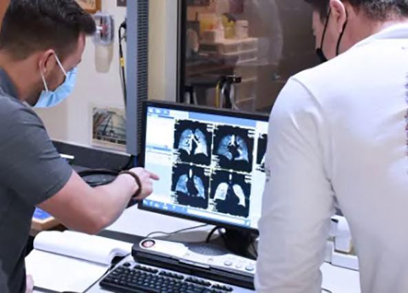
As part of the Pneumo.Vest project, re searchers at Fraunhofer IKTS (Dresden, Ger many; www.ikts.fraunhofer.de) have devel oped a textile vest with integrated acoustic sensors, presenting a high-performance ad dition to the traditional stethoscope. Piezoc eramic acoustic sensors have been incorpo rated into the front and back of the vest to register any noise produced by the lungs in the thorax, no matter how small. A software program records the signals and electroni cally amplifies them, while the lungs are de picted visually on a display. As the software knows the position of each individual sensor, it can attribute the data to its precise loca tion. This produces a detailed acoustic and optical picture of the ventilation situation of all parts of the lungs. But what is particularly special about the system is that it collects and stores the data permanently, examina tions can take place at any given time and in the absence of hospital staff.
at different points with a stethoscope, a number of sensors are used simultaneously. Alongside the acoustic sensors, the software is at the core of the vest. It is responsible for storing, depicting and analyzing the data. It can be used by the doctor to view the acous tic events in specific individual areas of the lungs on the display. The use of algorithms in digital signal processing enables a targeted evaluation of acoustic signals. This means it is possible, for example, to filter out heart beats or to amplify characteristic frequency ranges, making lung sounds, such as rustling or wheezing, much easier to hear.
Image: Visual representation shows different lung areas and their ventilation situation (Photo courtesy of Fraunhofer IKTS)

“Pneumo.Vest is not looking to make the stethoscope redundant and does not replace the skills of experienced pneumol ogists,” explained Ralf Schallert, project manager at Fraunhofer IKTS. “However, auscultation or even CT scans of the lungs only ever present a snapshot at the time of the examination. Our technology provides added value because it allows for the lungs to be monitored continuously in the same way as a long-term ECG, even if the patient is not attached to machines in the ICU but has instead been admitted to the general ward.”
ECG HOLTER & SOFTWARE ASPEL

A team of researchers has developed a technology whereby noises in the lungs are recorded using a textile vest with integrated acoustic sensors. The signals are then con verted and displayed visually using software. In this way, patients can be discharged early from the intensive care unit (ICU) but can still be monitored continuously. The technol ogy increases the options for diagnosis and improves the patient’s quality of life.
Pneumo.Vest also indicates the status of the lungs over a period of time, for example over the previous 24 hours. Needless to say, traditional auscultation can also be carried out directly on the patients. However, in stead of carrying out auscultation manually
The HLT HOLCARD-712 v.301ALFA (ECG Holter+ Software) with 12-channel digital recorder allows quick analysis of ECG examination (arrhythmias and ST segment). It enables simultaneous arrhythmia detection in all the three channels.
The PM-900 multi-parameter patient monitor features a 12.1 inches high resolution color TFT screen with a user-friendly interface design. It offers powerful data review with 480 hours graphic and tabular trends of all parameters.
NCPAP (NASAL CONTINUOUS POSITIVE AIRWAY PRESSURE) STEPHAN
The EasyFlow nCPAP is precisely tailored to the needs of premature and newborn infants for providing gentle, non-invasive ventilation. Available in various easily adjust able sizes with adjustable length and/or height.

who use them to listen very accurately to heartbeats and the lungs and, as a result, to diagnose illnesses. Now, the stethoscope is getting some help.
On top of this, the researchers are now developing machine learning algorithms. In the future, these will be able to structure and classify complex ambient noises in the thorax. Then, the pneumologist will carry out the final assessment and diagnosis. Patients can also benefit from the digital sensor alternative. When wearing the vest, they can recover without requiring constant observation from medical staff. They can transfer to the general ward and possibly even be sent home and move about more or less freely. Despite this, the lungs are moni tored continuously, and any sudden deterio ration can be reported to medical personnel straight away. The technology was initially designed for respiratory patients, but it also works well for people in care facilities and for use in sleep laboratories. It can also be used to train young doctors in auscultation. The first tests have shown that the concept is successful in Pneumo.Vestpractice.isaproduct that is cut out for the increasingly strained situation at hos pitals. Millions of patients with respiratory or lung diseases require inpatient treatment every year, with most of them connected to a ventilator for more than 24 hours. This does not take into account the current
To receive prompt and free information on products, log on to www LinkXpress com or fill out reader service form located on last page, or scan the QR code on your mobile devicePRODUCT NEWS


Cont’d from cover
High-Tech Vest Could Speed Discharge of ICU Patients
increase in respiratory patients due to the COVID-19 pandemic. As a result of increas ing life expectancy, the medical industry also expects the number of older patients with breathing problems to increase. With the help of the new technology, the burden on hospitals and, in particular, costly ICUs can be relieved as their beds will no longer be occupied for quite as long.
210HMI-09-22LINKXPRESS COM 211HMI-09-22LINKXPRESS COM 212HMI-09-22LINKXPRESS COM 12HospiMedica International August-September/2022
“There will be genomic diagnostic devices that we’ll be able to use right at the bedside in the hospital very soon,” added Brakenridge. “This is really the first time that we’ve been able to move genomic technology to a point-of-care application and take something very ex citing at the scientific bench, translate it into a highly insightful biologic metric, and see it used in patients.”


CriticalCareTo view this issue in interactive digital magazine format visit www.HospiMedica.com 13 HospiMedica International August-September/2022 Size: 2400 x 1200 mm (3 mm thick)100% Silicone YOUR GLOBAL SOURCE FOR STERILIZATION ACCESSORIES Thermo-Resistant ( 60 °C to 300 °C) Fully Washable & Flexible Suitable for central sterilization services Sterilizable STERILIZABLE INSTRUMENT & WORK-SURFACE MATS Front Back WASHING TRAYS MAT Heavy Silicone Cover & Transport Tablet TURBO MACHINESWASHINGTRAYS SILICON INSTRUMENT MAT Front Back MICRO INSTRUMENT MAT Exchangable Net Exchangable Nets INVITEDTOAPPLYDISTRIBUTORS Up to 37 cm in length GLOVESRESISTANTTHERMO Front Back WASHING TRAYS MAT NEW! VICOTEX Place de la Gare 1 • 1009 Pully • Switzerland Tel: (41) 21-728-4286 • Fax: (41) 21-729-6741 E-Mail: contact@vicotex.com www.vicolab.com S.A. COVERANDSILICONETABLETSTEELNETS


Cont’d from cover
Researchers at the University of Florida Health (Gainesville, FL, USA; www.ufhealth.org) evaluated the tool that relies on an algorithm that helps practitioners quickly discern which patients are most at risk. In the event that sepsis is not recognized early and managed promptly, septic shock ensues, resulting in multiple organ failure and death. Of those who survive sepsis, only half will completely recover. The rest will either die within one year or be encumbered by long-term disabilities, according to the WHO. The earlier sepsis is detected, the greater the likelihood of a full recovery. Rapid determination and early intervention is the key to treating it. The new algorithm marks an instance where technology can better identify how patients’ genetics can influence their response to treatment plans, and has more than halved the time it takes doctors to get information they need to make decisions before it’s too late.
eart disease is so deadly in part because the heart, unlike other organs, cannot repair itself after injury. That is why tissue engineering, ultimately including the wholesale fabrication of an entire human heart for transplant, is so important for the future of cardiac medicine. To build a human heart from the ground up, researchers need to replicate the unique structures that make up the heart. This includes recreating helical geometries, which create a twisting motion as the heart beats. It has been long theorized that this twisting motion is critical for pumping blood at high volumes, but proving that has been difficult, in part because
Biofabrication Breakthrough Could Pave Way for Bioengineered Hearts


H

“There is no consistent way of recognizing and triaging critically ill patients when they’re admitted to the ICU,” said Lyle L. Moldawer, Ph.D., director of the Sepsis and Critical Illness Research Center, and emeritus director of the UF Laboratory of Inflammation Biology and Surgical Science. “While this may not pose a problem at large academ ic institutions with dedicated specialists, it can be harder for places where tertiary care is less developed. The worst thing you can do is have a patient sit in the ICU for 72 hours or even 96 hours without an “Sepsisintervention.”isavery heterogeneous disease,” said Scott Brakenridge, first author and currently a trauma surgeon at the University of Wash ington. “People’s immune systems react in different ways to infection and display different levels of illness. In fact, one of the main reasons that finding effective therapeutics to treat sepsis has been so challeng ing is due to this variation among patients.”

Clinicians who treat critically ill patients must contend with two questions: Firstly, will the patient have a difficult clinical trajectory, re quiring more aggressive interventions and supervision? And secondly, if that’s the case, then how can clinicians determine the best type of treatment uniquely suited to them? Physiological responses to sepsis run the gamut. Someone can be septic from something as simple as a urinary tract infection, receive antibiotics and be discharged within three days. Another patient with the same diagnosis can go down a much more clinically complex path due to things like age, disease history and comorbidities. The new tool lends a precision medicine perspective, allowing clinicians to tailor their care to the individual and the drugs they will respond best to before it’s too late.
Image: A new AI diagnostic tool can identify a patient’s likelihood of devel oping sepsis (Photo courtesy of Pexels)
Cont’d on page 14
113HMI-09-22LINKXPRESS COM


AI Diagnostic Tool Identifies Sepsis Within 12 Hours After Hospitalization

The BR-502 is a dual wavelength total bilirubin meter that is easy to operate, thanks to advanced electronic circuits like the company’s propri etary auto-zero system and allows easily handling of hemolysis and turbidity.

Image: A FRJS spun dual chambered ventricle (Photo courtesy of SEAS)
To receive prompt and free information on products, log on to www LinkXpress com or fill out reader service form located on last page, or scan the QR code on your mobile devicePRODUCT NEWS


creating hearts with different geometries and alignments has been challenging. Now, a team of bioengineers has developed the first biohybrid model of human ventricles with helically aligned beating cardiac cells, and has shown that muscle alignment does, in fact, dramatically increase how much blood the ventricle can pump with eachThiscontraction.advancement was made possible using a new method of additive textile manufacturing, Focused Rotary Jet Spinning (FRJS), which enabled the high-throughput fabrication of helically aligned fibers with diameters ranging from several micrometers to hundreds of nanometers. Developed at the Harvard John A. Paulson School of Engineering and Applied Sciences (SEAS, Cambridge, MA, USA; www.seas.harvard.edu), FRJS fibers direct cell alignment, allowing for the formation of controlled tissue engineered structures.
HEMODYNAMIC MONITORING SYSTEM MASIMO

213HMI-09-22LINKXPRESS COM 214HMI-09-22LINKXPRESS COM 215HMI-09-22LINKXPRESS COM 14HospiMedica International August-September/2022
Biofabrication Breakthrough Could Pave Way for Bioengineered Hearts
Over the centuries, physicians and scientists have built a more com prehensive understanding of the heart’s structure but the purpose of the spiral-like arrangement of heart muscles has remained frustratingly hard to study. In 1969, Edward Sallin, argued that the heart’s helical alignment is critical to achieving large ejection fractions - the percent age of how much blood the ventricle pumps with each contraction. To test Sallin’s theory, the SEAS researchers used the FRJS system to control the alignment of spun fibers on which they could grow cardiac cells.The first step of FRJS works like a cotton candy machine - a liquid polymer solution is loaded into a reservoir and pushed out through a tiny opening by centrifugal force as the device spins. As the solution leaves the reservoir, the solvent evaporates, and the polymers solidify to form fibers. Then, a focused airstream controls the orientation of the fiber as they are deposited on a collector. The team found that by angling and rotating the collector, the fibers in the stream would align and twist around the collector as it spun, mimicking the helical structure of heart muscles. The alignment of the fibers can be tuned by changing the angle of theUnlikecollector.3Dprinting, which gets slower as features get smaller, FRJS can quickly spin fibers at the single micron scale – or about 50 times smaller than a single human hair. This is important when it comes to building a heart from scratch. Take collagen for instance, an extra cellular matrix protein in the heart, which is also a single micron in diameter. It would take more than 100 years to 3D print every bit of
BILIRUBIN METER APEL
researchers compared the ventricle deformation, speed of elec trical signaling and ejection fraction between ventricles made from heli cal aligned fibers and those made from circumferentially aligned fibers. They found on every front, the helically aligned tissue outperformed the circumferentially aligned tissue. The team also demonstrated that the process can be scaled up to the size of an actual human heart and even larger, to the size of a Minke whale heart (they didn’t seed the larger models with cells as it would take billions of cardiomyocyte cells).“The human heart actually has multiple layers of helically aligned muscles with different angles of alignment,” said Huibin Chang, a post doctoral fellow at SEAS. “With FRJS, we can recreate those complex structures in a really precise way, forming single and even four cham bered ventricle structures.”

The M300/M500 is a stable and reliable syringe pump that offers multi-CPU monitoring and high-precision injection. It offers up to 10 injection modes, including time, weight, se quence, cascade mode, etc.

collagen in the human heart at this resolution. FRJS can do it in a single day. After spinning, the ventricles were seeded with rat cardiomyocyte or human stem cell derived cardiomyocyte cells. Within about a week, several thin layers of beating tissue covered the scaffold, with the cells following the alignment of the fibers beneath. The beating ventricles mimicked the same twisting or wringing motion present in human hearts.The
The LiDCO Hemodynamic Monitoring System pro vides beat-to-beat advanced hemodynamic monitoring to support informed decision-mak ing in high-acuity care areas such as the operating room.
Cont’d
SYRINGE PUMP COMEN MEDICAL
“This work is a major step forward for organ biofabrication and brings us closer to our ultimate goal of building a human heart for transplant,” said Kit Parker, the Tarr Family Professor of Bioengineering and Applied Physics at SEAS. from page 13
M
ike the hardiest weed, glioblastoma al most always springs back - usually within months after a patient’s initial brain tumor is surgically removed. That is why survival rates for this cancer are just 25% at one year and plummet to 5% by the five-year mark. One of the challenges of treating this disease is that surgeons can’t always remove every bit of tumor or glioma stem cells that might linger in the brain. Now, a powerful immuni ty-boosting postoperative treatment could transform the odds for patients with glioblastoma.
The OPTI B-Lac Cassette can be used exclusively on the OPTI CCATS2 portable blood gas and electrolyte analyzer which uses OPTI Med

Single Cassette for Measurement of Lactate and Blood Gas Aids Rapid Sepsis Diagnosis
ical’s optical fluorescence technology. This unique, patented technology does not use electrodes or contact points, thus eliminating the need for costly electrode maintenance. The OPTI CCA-TS2 analyzer provides lab-quality results with zero standby cost. The single-use consumable is only used during a measurement and safely locks waste inside. To help meet exact testing needs, the OPTI CCA-TS2 analyzer has a variety of cassette configurations available. Each cassette comes individually wrapped, in cartons of 25, reducing the potential for consumable waste.
Cont’d
Image: B-Lac Cassette can be an important diagnostic tool for severe cases of COVID-19 (Photo courtesy of OPTI Medical)

The OPTI B-Lac Cassette from OPTI Medical Systems (Roswell, GA, USA; www.optimedical.com) offers measurement of lactate and blood gas on one cassette for rapid results in just minutes. The OPTI B-Lac Cas sette measures lactate, pH, PCO2, PO2, tHb, and SO2 in whole blood, to assist in quick diagnosis at the point of care. It offers true measurement of total hemoglobin using optical reflectance technology. The individu ally wrapped, disposable, single-use cassette contains all of the elements needed for calibration, sample measurement, and waste containment.
easurement of lactate levels has assumed significant impor tance for the diagnosis of severe cases of COVID-19. Being a useful marker of sepsis, elevated lactate levels can indicate how serious the septic shock is. An early diagnosis of sepsis has a substantial impact on the patient’s outcome, and continuous measurement of lactate levels provides useful information about the individual's condition when treating sepsis with a concomitant COVID-19 infection. Now, an indi vidually wrapped, disposable, single-use cassette that measures lactate, pH, PCO2, PO2, tHb, and SO2 in whole blood could be an important diagnostic tool for severe cases of COVID-19.
115HMI-09-22LINKXPRESS COM CriticalCareTo view this issue in interactive digital magazine format visit www.HospiMedica.com 15 HospiMedica International August-September/2022
A key characteristic of glioblastoma is the ag gressive nature of the tumor cells that infiltrate the surrounding tissues. As a result of this, surgeons are unable to clearly feel the boundaries between the tumor and normal tissue. The surgeons cannot remove as much as possible because all the tissues in the brain are vital. Hence, the tumor comes back again, sharply decreasing the survival rate after treat ment. Now, scientists at the University of Wiscon sin–Madison (Madison, WI, USA; www.wisc.edu) have developed a hydrogel that can be injected into the brain cavity left behind by the excised tumor. The hydrogel delivery method works well because it completely fills the brain cavity, slowly releases the medicine into the surrounding tissue, and promotes the cancer-killing immune response.
The hydrogel is packed with nanoparticles de signed to enter and reprogram certain types of im mune cells called macrophages. These immune cells normally clean up infectious invaders in the body, but in the tumor environment, they can change into a form that instead suppresses the immune system and promotes cancer growth. And because of the inflammation created by surgery, these rogue mac rophages flock to the surgical site, potentially fueling
Injectable Gel Can Help Patients with Brain Tumor Recover After Surgery
L on page 16
Image: Sponge electrodes in a variety of thicknesses (Photo courtesy of ACS Nano 2022)
SURGICAL PANEL EIZO DISPLAY TECHNOLOGIES

T
The 2100 Series HD FloNavi Fluorescence Endoscop ic Imaging System enhances visualization of tissue perfusion in real-time and enables users to switch between white light, standard FL, color scale FL and multi-display modes.
ENDOSCOPIC IMAGING SYSTEM OPTOMEDIC
OT LIGHT DR. MACH
cancer relapse.
“It provides hope for preventing glioblasto ma relapse,” said Quanyin Hu, an assistant pro fessor in the University of Wisconsin–Madison School of Pharmacy’s Pharmaceutical Sciences Division. “We prove that it can actually eradi cate these glioma stem cells, which can even tually prevent the glioblastoma from coming back. We can significantly improve survival.”
216HMI-09-22LINKXPRESS COM 217HMI-09-22LINKXPRESS COM 218HMI-09-22LINKXPRESS COM
the treatment approach could also be applied to other aggressive solid tumors, including breast cancer.
The Surgical Panel SP1-24 with an integrated monitor as well as IT and video management components is a digital image viewing system designed for use in the operating room. The 24-inch monitor offers maximum user-friendliness.

Cont’d from page 15
16HospiMedica International August-September/2022
o monitor heart rhythms and muscle function, doctors often attach elec trodes to a patient’s skin, detecting the electrical signals that lie beneath. These impulses are vital to the early diagnosis and treatment of many disorders, but currently available electrodes have limited function or are expensive to manufacture. Researchers, however, have now developed a low-cost, spongy version with improved signal detection that’s made with a surprising template - a sugar cube.The current gold-standard electrodes for electrophysiologic monitoring rely on a silver disc that contacts the skin through a conduc tive gel. These electrodes are critical tools for detecting abnormal electrical signals linked to health issues, such as heart attacks, brain disorders or neuromuscular diseases. These devices are not without their drawbacks, however. They are rigid and cannot conform well to the skin, particularly when the patient is physically active, reducing signal quality. In addition, the conductive gel dries quickly, pre venting long-term monitoring and rare-event detection. Addressing these challenges, re searchers at Washington University in St. Lou
If effective in humans, the hydrogel treat ment could eliminate the need for postsurgical chemotherapy or radiation, reducing toxic side effects while also improving patient outcomes. The next step is testing the hydrogel in larger animal models and also monitoring long-term efficacy and toxicity beyond the four to sixmonth period he previously studied. While the researchers initially focused on glioblastoma,
Image: Quanyin Hu’s lab has developed an injectable gel that offers promise for tough-totreat brain tumors (Photo courtesy of Univer sity of Wisconsin–Madison)

The LED 300DF SC OT-light features powerful LED technology and helps to keep a cool head by reduc ing heat radiation to a barely per ceptible minimum while its faceted multiple lens system ensures lowest shadowing and homogeneity.

Injectable Gel Can Help Patients with Brain Tumor Recover After Surgery
Low-Cost Sponge Electrodes Improve Signal Detection for Medical Monitoring
is (St. Louis, MO, USA; www.wustl.edu) have designed soft electrodes that better conform to the skin, as well as microneedle-based ver sions that physically penetrate the skin, but these are expensive to manufacture, limiting their widespread use. So, the researchers wanted to develop a low-cost sponge-like elec trode that would offer more consistent and resilient skin contact.
To receive prompt and free information on products, log on to www LinkXpress com or fill out reader service form located on last page, or scan the QR code on your mobile devicePRODUCT NEWS


micropores with a conductive thin film to form the electrode. Because the micropores allowed the spongy material to have increased contact area with the skin, the new device showed strong signal intensity and reduced noise when compared with standard electrodes. The micropores also helped the device carry more conductive gel, which kept them from drying out as quickly and losing signal, compared to standard versions. The gel also acted as a shock absorber, reducing the negative impacts of patient movement on skin-electrode contact and ensuring signal detection.
The researchers tested the ability of the sponge device to monitor uterine contractions during labor and found it performed as well as, or better than, a conventional electrode. As a low-cost, flexible alternative, sponge electrodes expand the possibilities for wearable health care applications, including use in medi cal exams that require patients to move, or for long-term monitoring of people at home or at work, say the researchers.
To make the new device, researchers start ed with commercially available sugar cubes, which they molded into a template that was dipped into liquid polydimethylsiloxane (PDMS). The PDMS became a solid structure after a curing step. They then dissolved the sugar with hot water and coated the sponge’s
Cont’d from cover
"I was immediately impressed by the new concept of the Pulsar-18 T3 system," said Dr. Koen Deloose, Head of the Department of Vascular Surgery AZSint Blasius Hospital Dendermonde. "The combination of having a very ergonomic handle combined with a tri-axial system and also everything in a 4-French concept was, for me, quite unique."

"The Pulsar-18 T3 stent system is an innovative solution that de livers clinically proven performance – providing effective therapy that is easy to use for physicians while minimizing metal burden and may
17 HospiMedica International August-September/2022
The Pulsar-18 T3 stent system is indicated for use to improve luminal diameter in patients with symptomatic de novo, restenotic or occlusive lesions located in the superficial femoral or proximal popliteal arteries, with reference vessel diameters from 3.0 to 6.0 mm and total lesion lengths up to 190 mm. BIOTRONIK will offer the Pulsar-18 T3 in up to a 200 mm stent length for treatment of long lesions.

The trial conducted by researchers at Mayo Clinic (Rochester, MN, USA; www.mayoclinic. org) tested needle ablation using in-catheter, heated, saline-enhanced, radio frequency en ergy, also known as SERF, to substantially in crease heat transfer, compared to conventional ablation methods. The new process produces deeper, controllable lesion scars at sites inside the heart muscle. The catheter can accurately control the ablation size and treat tissue that is deeper in the heart wall, which is where life-threatening arrhythmias that cause ventric ular tachycardia are often found.
T
New Stent System Helps Endovascular Treatments
Image: Pulsar-18 T3 self-expanding peripheral stent system has received FDA approval (Photo courtesy of BIOTRONIK)
Ablation Technique Shows Promise for Hard-to-Treat Ventricular Tachycardia

herapies of medication and traditional ablation, which uses heat or cold to scar small areas of heart tissue, may not be enough to prevent ventricular tachycardia, an abnormally rapid heart rhythm that is a leading cause of sudden cardiac death worldwide. People with ventricular tachycardia live with the potential of sudden cardiac death from irregular electrical signals in the heart. There fore, many patients also have an implantable cardioverter-defibrillator (ICD) to address dangerous arrhythmias. While an ICD shock corrects the heart's rhythm, it does not prevent arrhythmia. Corrective shocks are also stressful for patients and can reduce quality of life, especially if needed frequently. Now, a new ablation technique for patients with ventricular tachycardia has been used in a first-in-human multicenter trial that involved several methods to directly eliminate abnormal heart tissue which causes life-threatening rhythm.
117HMI-09-22LINKXPRESS COM
and reduce the need for closure devices when compared to 6-French devices. The tri-axial system with braided shaft design facilitates stable and accurate implantation of the clinically proven Pulsar stent. With its thin struts and low chronic outward force (COF), the Pulsar-18 stent contributes to a reduced mean area of restenosis.
In the trial, 32 participants from six centers underwent needle electrode ablation. Each had experienced multiple episodes of ven tricular tachycardia that did not respond to drug therapy after an ICD was implanted and standard ablation was done. These patients had about 45 shocks from their defibrillator in the six months before their procedure. For 31 of these 32 patients, their clinical ventricular tachycardia was eliminated immediately at the end of the procedure. Device therapies, such
The redesigned Pulsar-18 T3 stent system offers physicians an intui tive and ergonomic wheel-operated handle for one-handed stent release enhancing the ability to control deployment. While the stent system has been redesigned with new features to enhance deployment, it builds on the established Pulsar stent performance. Data highlights the long-term safety and efficiency of the Pulsar stent, showing freedom from target lesion revascularization rate of 89.3% and no major target limb amputa tions at 24 months.
reduce the risk of restenosis for patients," stated David Hayes, M.D., Chief Medical Officer, BIOTRONIK, Inc.
as shock or pace regulation, were reduced by 89% during the five-month follow-up period. Five patients in this high-risk group had complications, primarily in the earlier procedures. The next step in this research is a larger clinical trial with approximately 150 patients to prove the findings and demonstrate the safety of the new SERF technology.
"This preliminary trial is important because it suggests that there is a new way to treat problematic ventricular tachycardia by reducing or eliminating the shocks that ICDs deliver. It appears to be effective in achieving this goal," said Douglas Packer, M.D., a cardiac electrophysiol ogist at Mayo Clinic, first author and principal investigator of the study. "For clinicians, it provides hope that treatment in the electrophysiology lab may be effective, even if other treatments failed. For patients, it pro vides hope that their quality of life can be improved."
Cont’d on page 19
Image: A photoacoustic imaging endoscope probe can fit inside a medical needle (Photo courtesy of King’s College London)
NEAR-PATIENT SURGICAL DISPLAY BARCO

SURGICAL TABLE MAQUET
imaging can provide important clinical infor mation, but until now the instruments have been either too bulky or too slow for practical use as forward-viewing endoscopes. Now, researchers have created a photoacoustic im aging endoscope probe that can fit inside a medical needle with an inner diameter of just 0.6
patient’s risk of sustaining long-term damage. Up until now, surgeons have commonly used a pull wire to manually navigate the catheter tip through the convoluted network of blood vessels. The drawback of this method is that, since the tip can only be moved in two directions, the complex procedure takes a long time and requires a great deal of skill and experience. Now, a new type of catheter steered by remote control
Themillimeters.ultra-thin endoscope developed by researchers at King’s College London School of Biomedical Engineering & Imaging Sciences (London, UK; www.kcl.ac.uk) consists of two optical fibers each roughly the diameter of a human hair. Although fiber-based photoacous tic endoscopy probes have been developed, they usually require a bulky ultrasound detec tor or have a low imaging speed. In the new work, the researchers overcame both of these challenges by combining wavefront-based beam shaping with light-based ultrasound detection and a fast algorithm for controlling the device. This unique combination allowed them to create an extremely small probe with out sacrificing imaging speed.
“The imaging speed of this photoacoustic endomicroscopy probe is two orders of magni tude higher than those previously reported,” added Xia. “It could eventually allow 3D char acterization of tissue during various minimally invasive procedures such as tumor biopsies. This could help clinicians pinpoint the right area to sample, which would increase the diagnosis accuracy.”
The Maquet Otesus surgical table is the evolution of the proven Maquet Alphamaquet 1150 OR system which has been continuously updat ed to meet the comfort, safety, and flexibility requirements of multiple surgical disciplines.

VIDEO LARYNGOSCOPE INFINIUM MEDICAL
The ClearVue VLXL video laryngoscope adopts stateof-the-art image technology and the advantage of a 2.0-megapixel full view camera with 8-inch high-resolution monitor. Its HD optics offer anti-fog and high-res olution images.
Magnetically Steerable Catheter
Cont’d from cover to Speed Up Stroke Treatment
S
The new probe uses two thin optical fibers one for delivering the pulsed light used to generate the photoacoustic waves and the other for ultrasound detection. For the exci tation light, a high-speed digital micromirror
To demonstrate the new device, the re searchers used it to acquire high-resolution images of mouse red blood cells covering an area 100 microns in diameter. The research ers point out that imaging performance was not substantially degraded when the probe was scanned, suggesting that it isn’t affected by modest fiber bending. However, as a step toward clinical use, they will fur ther investigate how complex fiber bending or semi-rigid configurations affect imaging performance. They also say that artificial intelligence could be used to increase the imaging
the King’s College London School of Biomedi cal Engineering & Imaging Sciences. “Our new thin endoscope can resolve subcellular-scale tissue structural and molecular information in 3D in real-time and is small enough to be integrated with interventional medical devices that would allow clinicians to characterize tissue during a procedure.”
The AMM240ED is a 24-inch near-patient surgical display purpose-built for endoscopy imaging with LED backlights and 1920x1200 res olution. Video and images can be placed in Picture-by-Picture mode with independent adjustments.

device is used to scan a tightly focused light spot. This device has nearly one million tiny mirrors that can be independently flipped at tens of thousands of frames per second to change the wavefront of the light so that it can be focused and scanned quickly. For the ultrasound detection, the researchers developed an optical microresonator – a tiny structure made for confining light - that could be fabricated on the tip of an optical fiber. When sound waves hit the microresonator, its thickness changes, which, in turn, modifies the amount of light that is reflected back into the fiber, allowing optical detection of the acoustic waves.
219HMI-09-22LINKXPRESS COM 220HMI-09-22LINKXPRESS COM 221HMI-09-22LINKXPRESS COM 18HospiMedica International August-September/2022
To receive prompt and free information on products, log on to www LinkXpress com or fill out reader service form located on last page, or scan the QR code on your mobile devicePRODUCT NEWS


Ultra-Thin Endoscope Fits Inside Needle
trokes are the world’s second most common cause of both death and long-term disability. One in four adults over the age of 25 will suffer a stroke in their lifetime. If a blood clot in the brain cannot be removed using drugs, those affected require emergency surgery, which sees the surgeon maneuver a catheter through an artery past the heart into the brain to clear away the disturbance of the blood supply. The sooner and faster the procedure is carried out, the lower the
“Traditionalspeed.light-based endoscopes can on ly resolve tissue anatomical information on the surface and tend to have large footprints,” said Wenfeng Xia, leader of the research team from

F TradeMed provides a sophisticated yet easy-to-use global B2B platform for sourcing medical equipment. TradeMed connects buyers and sellers worldwide through a safe, secure and dy namic network. Solely dedicated to medical products, TradeMed is the premier choice for med ical suppliers, hospital decisionmakers and buyers worldwide, regardless of size or budget. Connecting Buyers with Suppliers Worldwide Reach new sources of supply Identify latest products and technologies Send inquiries directly to suppliers Receive latest product alerts Chat live with suppliers SIGN UP FOR FREE! WORLD’S MEDICAL PRODUCT MARKETPLACE DESIGNNEW

Fluorescence Open Surgery Imaging Provides Surgeons an Improved View
The FloNavi 3100 Series fluorescence open surgery imaging system is suitable for various applications and leads the traditional endoscopic system industry into a new era of functional endoscopic systems. Some of its applications include liver transplant surgery, open hepatectomy surgery, skin flap transplantation, locating sentinel lymph node of mam mary cancer, plastic surgery, and soon.
one of the three founders of Nanoflex. “Even surgeons with less experi ence should be able to treat strokes with our system.”
Magnetically Steerable Catheter to Speed Up Stroke Treatment
“With our system, it will be possible to carry out procedures from a distance by remote control and on a screen,” added Silvia Viviani, who studied robotics at ETH Zurich and has worked at Nanoflex since 2021.
For surgeons to be able to work with the magnetic catheter, patients must lie next to a magnetic navigation system that creates a directed magnetic field. In contrast to commercially available equipment that is permanently installed in the operating theatre, Nanoflex’s magnetic field generator is significantly lighter and thus, has a wider range of applications. It can be wheeled into the operating theatre as needed and requires only power and water. The smaller and more flexible magnetic navigation system will be significantly cheaper than competing products. The researchers’ vision is for every big hospital to have one of their mag netic navigation systems in future. Until then, however, they still have a lot of work to do. The aim is to obtain approval for the U.S. market in two years. To this end, they must not only assess every conceivable risk and establish ways to minimize them, but also present a solid plan for how to manufacture the system in a standardized way and how to operate it with maximum reliability. The researchers are confident that the system will enter the market, and that the technology will lend itself to other fields as well, such as heart and eye surgery, gastroscopy and fetal“Notsurgery.only can the catheter tip be bent in any direction thanks to a magnetic head; it’s also smaller, more maneuverable and safer due to the softness of the material,” explained ETH alumnus Christophe Chautems,
The FloNavi 3100 Series fluorescence open surgery imaging system is as a unique product from OptoMedic Technologies Inc. (Guangdong, China; www.optomedic.com) that features Color Scale FL Mode which can better show the fluorescence concentration and tumor boundary with ultra-high sensitivity. It uses industry-leading 4CMOS technology to en hance visualization of tissue perfusion in real-time. Additionally, the 3100 series camera head comes with a white light source and a NIR source.
and computer via a magnetic field is set to change all that.
Cont’d from page 18
Image: The FloNavi 3100 Fluorescence Open Surgery Imaging System (Photo courtesy of OptoMedic)
To view this issue in interactive digital magazine format visit www.HospiMedica.com TechniquesSurgical


19 HospiMedica International August-September/2022

luorescence imaging has become a hot word in the field of surgery. A growing number of surgeons are using fluorescence imaging technology to identify the target area during proce dures. Fluorescence imaging systems benefit both surgeons and patients, although not many fluorescence camera heads for open surgery are available in the market.

Researchers from ETH Zurich (Switzerland; www.ethz.ch) who developed the magnetic catheter with its spin-off Nanoflex Robotics (Zurich, Switzerland; nanoflexrobotics.com) believe that its precision steering will speed up and simplify procedures compared to conventional catheters in the future. Since hospitals are currently short of catheter specialists, the team is hopeful that their system will give more patients access to prompt stroke treatment. Having a soft and more maneuverable catheter should also reduce the risk of accidental vessel damage. Another advantage is that the surgeon steers the magnetic catheter by remote control, which means they don’t have to be at the patient’s side during the procedure. This shields doctors from the radiation of the Xray machine that allows them to get their bearings inside the patient’s body. The researchers are hoping that, in future, stroke patients will be operated on as quickly as possible at the nearest local hospital by a specialist who doesn’t even have to be on the premises, thus saving precious time.

To receive prompt and free information on products, log on to www LinkXpress com or fill out reader service form located on last page, or scan the QR code on your mobile devicePRODUCT NEWS


VIDEO COLPOSCOPE ADVANCED INSTRUMENTATIONS
The VC-1000 video colposcope with acetic and acid iodine reaction test timer, single aperture light source, and electronic green filter enhances vascular image display. It offers in tegrative management of captured display images.

ELECTROSURGICAL GENERATOR SYMMETRY SURGICAL
T
DIGITAL VIDEO LARYNGOSCOPE SINO-HERO BIO-MEDICAL ELECTRONICS

to improve patient care and diagnosis, healthcare and medical institutions are currently looking for newer techniques and technological breakthroughs in surgery and imaging management systems. This is one of the key drivers of the growth of the surgical imaging market. Additionally, the increase in the elderly population is leading to rising prevalence of cardiovascular and neurological problems, thereby driving a higher demand for surgical treatments and imagingMoreover,technologies.sincecontact, collision, and adventure sports often have a higher risk of injury, sports medicine clinics are increasingly utilizing small C-arms and other diagnostic imaging modalities. This is expected to further fuel market growth. Geographically, Asia-Pacific is expected to offer lucrative growth prospects for players in the surgical imaging market due to its large population, growing interest in surgical imaging, expanding healthcare infrastructure, and rising demand for minimally invasive procedures.
I
he global surgical imaging market is projected to grow at a CAGR of more than 3% from an estimated USD 1.2 billion in 2022 to USD 1.4 billion by 2028, driven by an increase in the number of minimally invasive surgical procedures performed worldwide, rise in hybrid operating rooms, growing demand for integrated imaging systems, and technological developments in surgical imaging systems. Additionally, an increase in surgeries, rising frequency of sports injuries, increasing government expenditure on healthcare, and a growing elderly population will also contribute to the market growth. These are the latest findings of Valuates Reports (Bangalore, India; reports.valuates.com), a provider of market research and business forecastingSurgeonsreports.must master the utilization of image-guided procedures as minimally invasive surgery becomes the norm in the field of neurosurgery. The introduction of image-guided neurosurgery has significantly advanced the microsurgical management of intracranial diseases such as tumors and vascular abnormalities. Hence, the increasing use of imaging in neurosurgery is expected to drive the growth of the surgical imaging market.The surgical imaging market is expected to be driven further by the increasing use of imaging in orthopedic surgeries. X-ray images are essential for minimally invasive procedures used by orthopedic surgeons. When setting fractures, real-time imaging is essential for metal placement and fracture reduction. Orthopedic surgeons employ Xrays in a variety of ways. As a first-line diagnostic tool, this is their primary and most
222HMI-09-22LINKXPRESS COM 223HMI-09-22LINKXPRESS COM 224HMI-09-22LINKXPRESS COM 20HospiMedica International August-September/2022
The events will again take place under Informa’s AllSecure health and safety requirements. The enhanced measures include 35 guidelines covering all aspects of cleaning and hygiene, social distancing measures, screening, and a track and trace in conjunction with local authorities.
Based on application, the orthopedic segment is expected to be the most lucrative in the surgical imaging market due to increased use of C-arms during orthopedic implantation, increased availability of surgical imaging equipment for orthopedic surgery, and higher adoption of intraoperative surgical imaging.Inorder
Two Top Asian Shows to Resume Events, Move Venue to Bangkok
“We are delighted to be relaunching Medlab Asia and Asia Health as a co-located event in Thailand. Both events have been integral to developing the healthcare and laboratory sectors in the ASEAN region over the last decade,” said Tom Coleman, Exhibition Director for Informa Markets.
significant utility. Without a radiograph of the affected area, no orthopedic assessment can be deemed complete. During orthopedic surgeries, X-rays are utilized to locate implants and validate the correctness of surgical operations such as the insertion of pedicle screws in the spine.
The two meetings are to include a series of CME, CMTE, CNEU, and CPD accredited conferences. Medlab Asia is scheduled to host six conference tracks, including Laboratory Management, Clinical Chemistry, Clinical Microbiology, Molecular Diagnostics & Therapeutics, Hematology, and COVID-19 Updates. The congress focus for Asia Health will include Hospital Procurement, Sterilization, Decontamination & Patient Safety, Healthcare Business in Thailand, and Total Radiology. The conferences will be supported by workshops featuring latest applications in the ASEAN region healthcare sector.
The Smoke Shark II 120V is an ultra-quiet generator for effective smoke and particulate removal with a modern, lightweight, compact design that allows for simplified use, storage and stack-ability.
The SmartScope LV Pro is a digital video laryn goscope made from high quality material with an ti-shatter protection and features an ergonomic and durable design. It comes with disposable blades to avoid cross infection.

nforma Markets (London, UK; www. informamarkets.com) has announced that its two top Asian shows, Medlab Asia and Asia Health, will resume their annual events in October of this year, and move their venue to Thailand. The exhibitions, to take place on October 19-21, 2022, at the Impact Exhibition Center in Bangkok, are expected to draw more than 4,200 attendees during the 10th edition of both events, featuring more than 300 exhibitors from over 20 countries. Both events are once again expected to be supported by a host of government entities, industry leaders and influencers in their respective sectors.
Global Surgical Imaging Sector to Surpass USD 1.4 Billion by 2028
Teleflex Acquires Bariatric Stapling Innovator
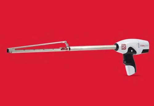
eration we are very pleased to have finally submitted our De Novo request for approval to the FDA. We have made an investment of GBP 2.8 million in the project which has all been expensed,” said Paul Swinney, Chief Executive of Tristel plc. “The United States is the largest ultrasound market in the world and our competitors will be the same as those we compete with in all our other markets world wide. To our knowledge no new high-level disinfectant has been approved by the FDA and introduced into the United States since 2011, other than variants of previously ap proved products. Duo ULT is recognized as a leading high-level disinfectant throughout the rest of the world, and our entry into the Unit ed States market will be a significant inflection point for the company.”
“Teleflex’s strategy is to invest in innovative products and technologies that can meaningful ly enhance clinical efficacy, patient safety and comfort, reduce complications, and lower the overall cost of care,” said Liam Kelly, Chair man, President and Chief Executive Officer.
“After five years of testing and data gen


“The acquisition of Standard Bariatrics adds an exciting and differentiated product serving the large and growing sleeve gastrectomy mar ket, which we estimate to be approximately 120,000 procedures annually in the U.S. In addition, the deal enables Teleflex to leverage our strength in our existing bariatric surgeon call point, with a differentiated product that
Image: Titan SGS’ first-of-its-kind design en sures more consistent staple formation (Photo courtesy of Standard Bariatrics)
Tristel is a UK-based manufacturer of infec tion prevention products. Ultrasound probe disinfection is one of Tristel’s most important areas of focus within the hospital infection pre vention market, accounting for approximately 40% of the company’s global revenues. Parker is a leading manufacturer in the US market for the conductive gels and sheaths that are used in all ultrasound procedures, with a nationwide distribution network.
To view this issue in interactive digital magazine format visit www.HospiMedica.com IndustryNews

Cont’d on page 21
Tristel has submitted its De Novo request for approval to the US FDA for Tristel Duo ULT, a high-level disinfectant foam that can be used on ultrasound probes used for intra-cav itary and skin surface diagnostic procedures. To Tristel’s knowledge, Tristel Duo ULT is the first disinfectant to make a De Novo request to the FDA, reflecting the novelty of the product. Duo ULT is widely used throughout Europe, the Middle East, Asia, and Australasia. During the company’s current financial year, the prod


P
complements many of our key surgical prod ucts, including our ligation portfolio, MiniLap Percutaneous Surgical System and Weck EFx Fascial Closure Portfolio.”
Parker Labs in Alliance for Ultrasound Probe Disinfectants
uct will have been used in over eight million disinfection procedures of ultrasound probes worldwide.TheFDA’s De Novo review and approval process stipulates a 150-day decision time frame. Additional data requests are common place, and the FDA website states that the aver age duration for review and approval is approx imately 11 months. Tristel is also proceeding with the state registration of another version of Duo which has been approved by the US EPA for the disinfection of general medical surfaces. The company has received 24 state approvals and anticipates that all others will have been received by the end of 2022. Tristel will manu facture and distribute both Duo products via its commercial partnership with Parker.
arker Laboratories Inc. (Fairfield, NJ, USA; www.parkerlabs.com) and Tris tel plc (Newmarket, UK; www.tristel. com) have entered into a commercial partnership in which Tristel will manufacture and distribute its high-level ultrasound probe disinfectants.

up to USD 130 million payable upon the achievement of certain commercial milestones.


The Titan SGS from Standard Bariatrics ad dresses unmet needs in sleeve gastrectomy by offering surgeons the longest continuous staple cutline of 23 centimeters. This first-of-its-kind stapler may help users achieve more consistent and symmetrical sleeve pouch anatomy, set ting their patients up for optimized outcomes. While every patient’s anatomy is different, the Titan SGS long staple line enables surgeons to plan and place staples in one firing, minimizing variations sometimes associated with the use of multiple overlapping short-cartridge staple firings. Additionally, the design may result in a
more secure staple line and fewer chances of leaks, as evidenced with higher burst pressures.
21 HospiMedica International August-September/2022


OCTOBER
14th World Stroke Congress – World Stroke Organi zation. Oct 26-29; Singapore; worldstrokecongress. org
Medical Japan 2022 Tokyo– International Medical and Elderly Care Expo. Oct 12-14; Tokyo, Japan; medical-jpn.jp
NOVEMBER
RSNA 2022 - Annual Meeting of the Radiological Society of North America. Nov 27 - Dec 1; Chicago, IL, USA; rsna.org
ATTENTION: PANDEMIC,
ESPE 2022 - 60th Annual Meeting of the European Society for Paediatric Endocrinology. Sep 15-17; Rome, Italy; eurospe.org
RANZCR 2022 – Annual Scientific Meeting of The Royal Australian and New Zealand College of Ra diologists. Oct 27-30; Adelaide, Australia; ranzcr.com
APSR 2022 – 26th Congress of the Asian Pacific Society of Respirology. Nov 17-20; Seoul, Korea; apsr2022.org
45th World Hospital Congress of the International Hospital Federation (IHF). Nov 9-11; Dubai, UAE; worldhospitalcongress.org
Medicall Expo 2022 – 30th Edition. Sep 17-19; New Delhi, India; medicall.in
4th MedExpo Ethiopia 2023. Jan 19-21; Addis Aba ba, expogr.com/ethiopia/medexpo
EAPS 2022 – 9th Congress of the European Acad emy of Paediatric Societies. Oct 7-11; Barcelona, Spain; eaps2022.kenes.com
EUSOBI 2022 – Annual Scientific Meeting of the European Society of Breast Imaging. Sep 29 - Oct 1; Malmo, Sweden; eusobi.org
AOCR 2022 – 20th Asian Oceanian Congress of Ra diology. Sep 20-24; Seoul, Korea; aocr2022.org
ESSO 41 – 41st Congress of the European Society of Surgical Oncology. Oct 19-21; Bordeaux, France; esso41.org
UEG Week 2022 – United European Gastroenterolo gy. Oct 8-11; Vienna, Austria; ueg.eu/week
Pan e Ibérico Lima 2022 – 14th Congress of the Pan
18th EuGMS Congress – European Geriatric Medi cine Society. Sep 28-30 London, UK; eugms.org
many events are being rescheduled for a later date, converted into virtual venues, or altogether cancelled. Please check with the event organizer or website prior to planning for any forthcoming event 22HospiMedica International August-September/2022
ESC Asia 2022 with APSC & AFC – European Society of Cardiology – Asian Pacific Society of Cardiology. Dec 1-3; Singapore; escardio.org
Zdravookhraneniye 2022 – Russian Health Care Week. Dec 5-9; Moscow, Russia; zdravo-expo.ru Medicall Expo 2022 – 31st Edition. Dec 9-11; New Delhi, India; medicall.in
UAA 2022 – 19th Urological Association of Asia Congress. Oct 5-8; Sydney, Australia; uaa2022.org
TurkRad 2022 – 43rd Turkish National Radiology Congress. Nov 1-6; Antalya, Turkey; turkrad2022.org
For a free listing of your event, or a paid advertisement in this section, contact: International Calendar, HospiMedica International E-mail: info@globetech.net
MEDICA 2022. Nov 14-17; Dusseldorf, Germany; medica-tradefair.com
ESCR Annual Scientific Meeting – European Society of Cardiovascular Radiology. Oct 27-29; Rome, Italy; escr.org
Congress of Radiology and Diagnostic Imaging. Sep 1-3; Flori anopolis, Brazil; congressocbr.com.br
ERS International Congress 2022 – European Respiratory Society. Sep 4-6; Barcelona, Spain; erscongress.org
CMEF 2022 – China International Medical Equip ment Fair. Nov 23-26; Shenzhen, China; cmef. com.cn
ASUM 2022 – Conference of the Australasian Society for Ultrasound in Medicine. Sep 16-18; Ade laide, Australia; asum2022.com.au
ExpoMedical 2022. Sep 21-23; Buenos Aires, Argen tina; expomedical.com.ar
ECISM LIVES 2022 – Annual Congress of European Society of Intensive Care Medicine. Oct 22-26; Paris, France; esicm.org
Or, Circle LinkXpress Numbers of Interest on Reader Service Card and email Card to: subs@globetech.net Every advertisement or product item in this issue contains a LinkXpress ® number as shown below: FREEINFORMATIONPRODUCT 999HMI-09-22LINKXPRESS COM Renew / Start your Free InstantSubscriptionOnlineProductInformation Identify LinkXpress ® codes of interest as you read magazine Click on LinkXpress.com to reach reader service portal Mark code(s) of interest on LinkXpress ® inquiry matrix321 CalendarInternational HMI-09-22 HOSPIMEDICA INTERNATIONAL Reader Service Form PLEASE COMPLETE THE FOLLOWING A TYPE OF ESTABLISHMENT (Check only one box) (1) o General Hospital (incl. Univ. Hosp.) (2) o Specialized Hospital/Clinic Please Specify . . . . . . . . . . . . . . . . . . . . . . . . (3) o Distributor/Dealer/Manufacturer (4) o Research/Univ. Education (5) o Govt. Agency/Hosp. Mgmt. Org. (6) o Consulting/Planning/Design (7) o OtherPlease Specify . . . . . . . . . . . . . . . . . . . . . . . . B YOUR FUNCTION (Check only one box) (1) o Hospital Director (2) o Chief Medical Director (3) o Business (Executive/Manager) (4) o Department Chief (5) o Medical Practitioner (6) o OtherPlease Specify . . . . . . . . . . . . . . . . . . . . . . . . C YOUR DEPT OR SPECIALTY (Check only one box) (a) o General/Internal Medicine (b) o Cardiology (c) o Critical Care/Anesthesiology (d) o Ob/Gyn (e) o Orthopedics/Rehabilitation (f) o Pediatrics (g) o Radiology/Imaging (h) o Surgery (i) o Urology (j) o Patient Care (k) o Administration/Purchasing (l) o Engineering/Technical (m) o General Management (Business) (n) o Marketing/Sales (Business) (z) o OtherPlease Specify . . . . . . . . . . . . . . . . . . . . . . . . D Are you an M D (Dr Med )? o YES E How many other readers share this copy of HospiMedica International? . . . . . . . . . . . The publisher reserves the right to qualify requests VI CIRCLE LINKXPRESS NUMBERS OF INTEREST TO RECEIVE FREE INFORMATION For EXPRESS service: visit www .LinkXpress .com or Scan and Email this form to: subs@globetech .net Yes, I wish to receive free copies of HospiMedica International SIGNATURE (REQUIRED) DATE: DAY MONTH YEAR 101 102 103 104 105 106 107 108 109 110 111 112 113 114 115 116 117 118 119 120 121 122 123 124 125 126 127 128 129 130 131 132 133 134 135 136 137 138 139 140 141 142 143 144 145 146 147 148 149 150 151 152 153 154 155 156 157 158 159 160 161 162 163 164 165 166 167 168 169 170 171 172 173 174 175 176 177 178 179 180 181 182 183 184 185 186 187 188 189 190 191 192 193 194 195 196 197 198 199 200 201 202 203 204 205 206 207 208 209 210 211 212 213 214 215 216 217 218 219 220 221 222 223 224 225 226 227 228 229 230 231 232 233 234 235 236 237 238 239 240 241 242 243 244 245 246 247 248 249 250 251 252 253 254 255 256 257 258 259 260 261 262 263 264 265 266 267 268 269 270 271 272 273 274 275 276 277 278 279 280 281 282 283 284 285 286 287 288 289 290 291 292 293 294 295 296 297 298 299 300 Tel: ( )( ) @ E-MAIL (REQUIRED): Renewals)Allfor(NeededCodeSubscriberIndividualofNameDepartmentandPositionInstitutionofNameAddressMailingProvinceCity,CountryCodePostal WRITE CLEARLY IN BLOCK LETTERS or AFFIX YOUR SUBSCRIBER LABEL APPEARING ON COVER CBR22SEPTEMBER2022–51stCongressoftheBrazilian

50th National Congress of the Italian Society of Medical Radiology (SIRM). Oct 6-9; Rome, Italy; sirm.org
ISS 49th Annual Meeting – International Skeletal Society. Oct 23-28; Barcelona, Spain; international skeletalsociety.com
49th COMMEC Congress 2022 – Mexican College of
ISUOG World Congress 2022 - International Society of Ultrasound in Obstetrics & Gynecology. Sep 1618; London, UK: isuog.org
2023
EASD 2022 – 58th Annual Meeting of the European Association for the Study of Diabetes. Sep 19-23; Stockholm, Sweden; easd.org
Critical Care Congress 2023 – 52nd Annual Meeting of the Society of Critical Care Medicine (SCCM). Jan 15-18 New Orleans, LA, USA; sccm.org
REHACARE 2022 – International Trade Fair for Rehabilitation and Care. Sep 14-17; Dusseldorf, Germany; rehacare.com
ISETJANUARY2023–International Symposium on Endovas cular Therapy. Jan 16-19; Miami Beach, FL, USA. iset.org
EANM 2022 – 35th Annual Congress of the Euro pean Association of Nuclear Medicine. Oct 15-19; Barcelona, Spain; eanm.org
ACEP22 – Scientific Assembly of the American College of Emergency Physicians. Oct 1-4; San Fran cisco, CA, USA; acep.org
MICCAI 2022 – 25th International Conference on Medical Image Computing and Computer Assisted Intervention. Sep 18-22; Singapore; miccai2022.org
CIRSE 2022 – Annual Congress of the Cardiovascular and Interventional Radiological Society of Europe. Sep 10-14 Barcelona, Spain; cirse.org
EUSEM 2022 – 16th European Emergency Medicine Congress. Oct 15-19; Berlin, Germany; eusem.org Asia Health 2022. Oct 19-21; Bangkok, Thailand; medlabasia.com
ESGO 2022 – 23rd European Congress on Gynae cological Oncology. Oct 27-30; Berlin, Germany; congress.esgo.org
American and Iberian Federation of Critical Med icine and Intensive Therapy. Oct 31 - Nov 5; Lima, Peru; paneiberico-lima2022.com
Medic West Africa 2022. Sep 7-9; Lagos, Nigeria; medicwestafrica.com
ESVS 2022 – Annual Meeting of the European Society for Vascular Surgery. Sep 20-23; Rome, Italy; esvs.org
CBMI 2022 – 27th Brazilian Congress of Intensive Care Medicine. Nov 10-12; Brasilia, Brazil; amib. org.br
DECEMBER
NASCI 2022 – Annual Meeting of the North Ameri can Society for Cardiovascular Imaging. Sep 10-13; Cleveland, OH, USA; nasci.org
ASTRO 2022 – 64th Annual Scientific Meeting of the American Society for Radiation Oncology. Oct 2326; San Antonio, TX, USA; astro.org
Africa Health 2022. Oct 26-28; Johannesburg, South Africa; africahealthexhibition.com
ESMO Congress 2022 - European Society for Medi cal Oncology. Sep 9-13; Paris, France; esmo.org
Critical Medicine. Oct 10-15; Guadalajara, Mexico; commec.org
Due to the CORONAVIRUS
Global Health Saudi Exhibition 2022. Oct 9-11; Ri yadh, Saudi Arabia; globalhealthsaudi.com
JFR 2022 - Journées Francophones de Radiologie. Oct 7-10; Paris, France; jfr.radiologie.fr
ANESTHESIOLOGY 2022 – Annual Meeting of the American Society of Anesthesiologists. Oct 22-26; New Orleans, LA, USA; asahq.org
AAOS 2023 Annual Meeting – American Academy of Orthopaedic Surgeons. Mar 7-11; Las Vegas, NV, USA; aaos.org
EULAR 2023 – European Congress of Rheumatology. May 31 - Jun 3; Milan, Italy; congress.eular.org
60th ERA Congress – European Renal Association. Jun 15-18; Milan, Italy; era-online.org
Africa 2023. Feb 16-18; Dar-es-Salaam, Tanzania; expogr.com/tanzania/ medexpo
2023
2023 - World Congress on Osteo porosis, Osteoarthritis and Musculoskeletal Diseas es. May 4-7; Barcelona, Spain; wco-iof-esceo.org
ESTRO 2023 – Annual Congress of the European So ciety of Radiology & Oncology. May 12-16; Vienna, Austria; estro.org
SPR 2023 – Annual Meeting of the Society for Pediatric Radiology. May 16-20; Austin, TX, USA; pedrad.org
EFORT Congress 2023 – 24th Annual Congress of European Federation of National Associations of Orthopaedics and Traumatology. May 24-26; Vien na, Austria; congress.efort.org
AAEM23 – 29th Annual Scientific Assembly of the American Academy of Emergency Medicine. Apr 2125; New Orleans, LA, USA; aaem.org
SIR 2023 –Annual Meeting of the Society of Inter ventional Radiology. Mar 4-9; Phoenix, AZ, USA; sirmeeting.org
APSCVIR 2023 – 17th Annual Meeting of the Asia Pacific Society of Cardiovascular and Interventional Radiology. Apr 12-15; Seoul, Korea; apscvir.com
EHA 2023 - Annual Congress of the European Hematology Association. Jun 8-11; Frankfurt, Ger many; ehaweb.org
123rd Congress of the Japan Surgical Society (JSS). Apr 27-29; Tokyo, Japan; jssoc.or.jp
2023 – European Society of An aesthesiology. Jun 3-5; Glasgow, UK; esaic.org
ECRMARCH2023–European Congress of Radiology. Mar 1-5; Vienna, Austria; myesr.org
MedtecLIVE 2023. May 23-25; Nuremburg, Germa ny; medteclive.com
Hospitalar 2023. May 23-26; Sao Paulo, Brazil; hospitalar.com
International Medical and Elderly Care
12th Asian Conference on Emergency Medicine (ACEM). Apr 20-23; Manila, Philippines; asiansem. org
ECIO 2023 – European Conference on Interven tional Oncology. Apr 16-19; Stockholm, Sweden; ecio.org
ATTENTION: Due to the CORONAVIRUS PANDEMIC, many events are being rescheduled for a later date, converted into virtual venues, or altogether cancelled. Please check with the event organizer or website prior to planning for any forthcoming event 23 HospiMedica International August-September/2022
143rd Annual Meeting of the American Surgical Association (ASA). Apr 20-22; Toronto, Canada; americansurgical.org
2023 ISMRM & ISMRT Annual Meeting & Exhibition – International Society for Magnetic Resonance in Medicine. Jun 3-8; Toronto, Canada; ismrm.org
91st EAS Congress 2023 – European Atherosclerosis Society. May 21-24; Mannheim, Germany; eas-so ciety.org
24thFEBRUARYTanzaniaMedExpo
KIHE 2023 – Kazakhstan International Healthcare Exhibition. May 17-19; Almaty, Kazakhstan; kihe.kz
Nordic Congress of Radiology 2023. May 24-26; Helsinki, Finland; ncr2023.fi
EUROSON 2023 – 34th Congress of European Fed eration of Societies for Ultrasound in Medicine and Biology. May 25-27; Riga, Latvia; euroson2023.com
ESGAR 2023 – 34th Annual Meeting of the Euro pean Society of Gastrointestinal and Abdominal Radiology. Jun 13-16; Valencia, Spain; esgar.org
SIIM 2023 – Annual Meeting of the Society for Imaging Informatics in Medicine. Jun 14-16; Austin, TX USA; siim.org
DCK 2023 – 139th Congress of the German Society for Surgery (DGCH). Apr 25-28; Munich, Germany; dgch.de
FIME 2023 – Florida International Medical Expo. Jun 21-23; Miami, FL, USA; fimeshow.com
AUA 2023 – Annual Meeting of the American Urological Association. Apr 28 - May 1; Chicago, IL, USA; auanet.org
Orlando, FL, USA; aium.org
CARS 2023 – Computer Assisted Radiology and Sur gery. Jun 20-23; Munich, Germany; cars-int.org
AIUM 2023 – Annual Meeting of the American Institute of Ultrasound in Medicine. Mar 25-29;
CRITICARE 2023 - 29th Annual Conference of the Indian Society of Critical Care Medicine (ISCCM). Feb 22-26; Indore, India; isccm.org
50th Annual Meeting of the Japanese Society of Intensive Care Medicine (JSICM). Mar 2-4; Kyoto, Japan; jsicm.org
42nd ISICEM Brussels – International Symposium on Intensive Care & Emergency Medicine. Mar 2124; Brussels, Belgium; isicem.org
SAGES 2023 – Annual Meeting of Society of Ameri can Gastrointestinal and Endoscopic Surgeons. Mar 29 – Apr 1; Montreal, Canada; sages.org
SEACare 2023 – 23rd Southeast Asian Healthcare & Pharma Show. Apr 19-21; Kuala Lumpur; Malaysia; abcex.com
CAR 2023 Annual Scientific Meeting – Canadian Association of Radiologists. Apr 27-30; Montreal, Canada; car-asm.ca
ATS 2023 – International Conference of the Amer ican Thoracic Society. May 19-24; Washington, DC USA; thoracic.org
23rd MEDEXPO Africa 2023. Jun 21-23; Nairobi, Kenya; expogr.com/kenyamed
82nd Annual Meeting of Japan Radiological Society (JRS). Apr 13-16; Yokohama, Japan; radiology.jp
HIMSS 2023 – Healthcare Information and Man agement Systems Society. Apr 17-21; Chicago, IL, USA; himss.org
JPR 2023 – 53rd Sao Paulo Conference on Radiology & 25th Meeting of the Latin American Congress of Pediatric Radiology. Apr 27-30; Sao Paulo, Brazil; spr.org.br
ECE 2023 – 25th European Congress of Endocrinolo gy. May 13-16; Istanbul, Turkey; ese-hormones.org
2 E asy W ays To Continu e / Start Your FREE Subscription 21 BY EMAIL • Fill-out all required data on Read er Service Card including signature and date (incomplete or unsigned cards cannot be processed). • Circle inquiry numbers of interest to receive free information • Scan card and Email to: subs@globetech.net ONLINE Visit LinkXpress .com to enter your subscription data and reader inquiries. ATTENTION: IF YOUR APPLICATION IS NOT RECEIVED AT LEAST ONCE EVERY 12 MONTHS YOUR FREE SUBSCRIPTION MAY BE AUTOMATICALLY DISCONTINUED READER SERVICE PORTAL LINKXPRESS COM ® VISIT InternationalCalendar Provided as a service to advertisers. Publisher cannot accept responsibility for any errors or omissions. Advertising Index Inq No . Advertiser Page 124 COMEN ............................. 24 – ECR 2022 ............................ 3 – IHF Dubai 2022 21 115 Mayo Clinic 15 117 OptoMedic 17 107 Radcal 7 102 Sekisui 2 105 Sun Nuclear 5 109 Sun Nuclear ........................... 9 – TradeMed ............................ 19 111 Undis ............................... 11 113 Vicolab .............................. 13 Vol. 40 Issue 9/20223InternationalHospiMedica
Medical Japan Osaka–Expo. Jan 18-20; Osaka, Japan; medical-jpn.jp
KIMES 2023 – Korea International Medical & Hos pital Equipment Show. Mar 23-26; Seoul, Korea; kimes.kr
MedicalAPRILFair
Arab Health 2023. Jan 30 - Feb 2; Dubai,
Brasil. Apr 4-6; Sao Paulo, Brazil; medi calfair-brasil.com.br
ARRS 2023 Annual Meeting – American Roentgen Ray Society. Apr 16-20; Honolulu, HI, USA; arrs.org
Medical Fair India 2023. Apr 27-29; New Delhi, In dia; medicalfair-india.com
WCO-IOF-ESCEOMAY
ESOC 2023 – 9th European Stroke Conference. May 24-26; Munich, Germany; eso-conference.org
EuroAnaesthesiaJUNE
UAE; arab healthonline.com
SIOP Europe 2023– 4th Annual Meeting of the Eu ropean Society for Paediatric Oncology. May 8-12; Valencia, Spain; siopeurope.eu Vietnam Medi-Pharm Hanoi 2023. May 10-13; Ha noi, Vietnam; vietnammedipharm.vn
ACC.23 – American College of Cardiology’s 70th Annual Scientific Session & Expo. Mar 4-6; New Or leans, LA, USA; accscientificsession.acc.org
EMIM 2023 – 18th European Molecular Imaging Meeting. Mar 14-17; Salzburg, Austria; e-smi.eu ExpoMED Eurasia 2023. Mar 16-18; Istanbul, Turkey; expomedistanbul.com
WCN 2023 – World Congress of the International Society of Nephrology (ISN). Mar 30 - Apr 2; Beijing, China; theisn.org
ESTS 2023 – 31st Meeting of the European Society of Thoracic Surgeons. Jun 4-6; Milan, Italy; ests.org
SAR 2023 – Annual Scientific Meeting of the Society of Abdominal Radiology. Feb 26 - Mar 3; Austin, TX, USA; abdominalradiology.org
124HMI-09-22LINKXPRESS COM
