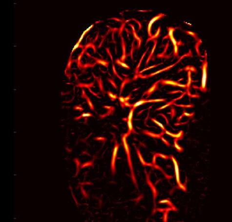
Known as high-definition microvasculature imaging (HDMI), the new technique noninvasively captures images of minute vessels within tumors and automatically categorizes the masses .

Technique

Gallbladder cancer (GBC) surgery is a technically demanding procedure that requires significant expertise to meet high-quality surgical standards. Despite this, subpar surgeries are common, highlighting the need for specific quality benchmarks. The quality of surgery significantly affects both immediate and long-term postoperative outcomes, and is a critical factor in studies evaluating surgical
Tumor-Destroying System Uses
Cancer remains one of the leading causes of death worldwide, with over 18 million new cases reported annually. Despite more than half of these cases being detected at an early stage, curative measures have not kept pace. Presently, surgery is the primary curative option for solid


Robotic System Allows Surgeons 4-Handed
Scientists have developed the first-ever surgical sys tem that facilitates four-arm lap aroscopic surgery by allowing surgeons to control two sup plementary robotic arms using haptic foot interfaces. This remarkable achievement inte(Lausanne, Switzerland; www. epfl.ch) has devised a surgical

Gallbladder
Cancer Surgery Guidelines
Cont’d on page 18 Cont’d on page 20 Cont’d on page 10 INSIDE GLOBETECH MEDIA >>> <<< International Calendar 22 . . . . . . . . . 21 3 HospiMedica EXPO . . . 6-10 . . . . . . . . . 17 HospiMedica EXPO 18-20 11 HospiMedica EXPO . . 12-16
Laparoscopic
If your subscription is not renewed every 12 months your Free Subscription may be automatically discontinued Renew / Start your Free Subscription Access Interactive Digital Magazine Instant Online Product Information: Identify LinkXpress ® codes of interest as you read magazine Click on LinkXpress.com to reach reader service portal Mark code(s) of interest on LinkXpress ® inquiry matrix 1 2 3 VISIT READER SERVICE PORTAL LINKXPRESS COM ® See article on Page 7
Ultrasound
Advances Cancer Diagnostics
Ultrasound
Advances Cancer Diagnostics
Procedures
AI-Powered
AI-Powered
Technique
Needle
Smart
INTERNATIONAL ® Vol.41 No.3 • 8-9/2023 ISSN 0898-7270
Know sooner. Decide faster.
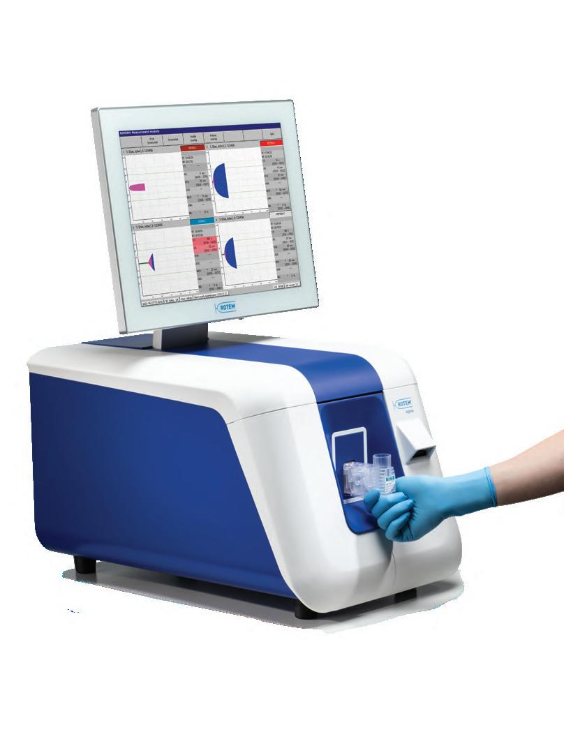
Now at the point of care, ROTEM sigma provides real-time results in critical bleeding situations.

Introducing a simple, fully automated thromboelastometry system that supports faster decision-making at the point of care. Using proven technology, ROTEM sigma delivers real-time results, with A5 parameter for early clot-strength assessment. Easy-to-interpret TEMograms provide valuable clinical information to guide transfusion and other management decisions in critical bleeding situations for improved patient outcomes.
To learn more about ROTEM sigma, visit info.Werfen.com/ROTEM.
Patient Blood Management
ROTsigPrintAd NA REV 00 12.22 Results from ROTEM sigma should not be the sole basis for a patient diagnosis; results should be considered along with a clinical assessment of a patient’s condition and other laboratory tests. Not Health Canada-licensed. Not available in all countries. ©2022 Instrumentation Laboratory. All rights reserved.
NEW 102 HMI-09-23 LINKXPRESS COM
Tau PET Scans: New Gold Standard for Predicting Cognitive Decline?

lzheimer’s disease, a common neurodegenerative disorder, results in a gradual deterioration of memory and autonomy. This condition is characterized by the buildup of neurotoxic proteins, specifically amyloid plaques and tau tangles, within the brain. Given the silent progression of these pathological changes over decades, early diagnosis is vital to initiate possible interventions. Now, researchers have demonstrated that tau PET, an innovative imaging method for visualizing the tau protein, can predict cognitive decline in patients with superior accuracy compared to conventional imaging techniques. These are the findings of a study published on August 9, 2023 in Alzheimer’s & Dementia: The Journal of the Alzheimer’s Association, supporting the rapid integration of tau PET into clinical practice for providing early and individualized interventions.
A


Positron emission tomography (PET), a key diagnostic tool for Alzheimer’s disease, employs injected tracers to visualize specific brain pathologies. While amyloid plaques may not invariably correlate with cognitive or memory impairment, the presence of tau aligns closely with clinical symptoms and significantly influences whether a patient remains stable or deteriorates rapidly. Visualizing tau using imaging techniques has posed challenges due to its intricate structure and lower concentration.
Flortaucipir, a radiotracer approved by the U.S. Food and Drug Administration (FDA) in 2020, binds with the tau protein. This tracer facilitates the detection of tau accumulation and its spatial distribution in the brain, enabling precise evaluation of its role in the clinical manifestation of the disease.
Scientists from the University of Geneva (UNIGE, Geneva, Switzerland; www.unige.ch) and the Geneva University Hospitals (HUG, Geneva, Switzerland; www.hug.ch) embarked on a study to determine which imaging technique—amyloid PET, glucose metabolism PET, or tau PET—best predicted future cognitive decline in Alzheimer’s disease. The study revealed that while all PET measures correlated with the presence of cognitive symptoms, validating their role as strong indicators of Alzheimer’s disease, tau PET exhibited the greatest predictive capacity for cognitive decline rates, even in individuals with minimal symptoms. These findings support incorporating tau PET into routine clinical assessments to assess individual prognosis and select the most appropriate therapeutic plan for each patient.
“This breakthrough is crucial for better management of Alzheimer’s disease. Recently, drugs targeting amyloid have shown positive results. New drugs targeting the tau protein
also look promising,” said Associate Professor Valentina Garibotto who directed the research. “By detecting the pathology as early as possible, before the brain is further damaged, and thanks to new treatments, we hope to be able to make a greater impact on patients’ future and quality of life.”

HospiMedica International To view this issue in interactive digital magazine format visit www.HospiMedica.com 3 HospiMedica International August-September/2023
Image: Tau imaging with 18F-Flortaucipir PET in Alzheimer’s disease (Photo courtesy of UNIGE)
MYESR.ORG
INTERNATIONAL
Groundbreaking AI-Based Method Accurately Classifies Cardiac Disease Using Chest X-Rays

Valvular heart disease, a leading cause of heart failure, is commonly diagnosed using echocardiography. However, this technique demands specialized expertise, leading to a shortage of proficient technicians. Chest radiography, on the other hand, is a widely used diagnostic method for identifying primarily lung diseases. Even though the heart is visible in chest radiographs or chest X-rays, its potential to detect cardiac function or disease has been largely unexplored until now. Given their widespread use, rapid execution, and high reproducibility, chest X-rays could serve as a supplementary tool to echocardiography for diagnosing cardiac conditions if they could accurately determine cardiac function and disease. Now, an innovative artificial intelligence (AI) tool uses chest X-rays to classify cardiac functions and identify valvular heart disease with unprecedented accuracy.
Scientists at Osaka Metropolitan University (Osaka, Japan; www.omu.ac.jp) have developed an AI-based model capable of accurately classifying cardiac functions and diagnosing valvular heart diseases using chest X-rays. Given the potential for bias and resultant low accuracy if AI is trained on a single dataset, the team collected a multi-institutional dataset comprising 22,551 chest X-rays and corre-
sponding echocardiograms from 16,946 patients across four facilities between 2013 and 2021. The AI model was trained using chest X-rays as input data and the corresponding echocardiograms as output data, enabling it to learn the features connecting the two datasets.
The AI model succeeded in precisely classifying six selected types of valvular heart disease, with the Area Under the Curve (AUC is a rating index denoting an AI model’s capability with a value range from 0 to 1—the closer to 1, the better) ranging from 0.83 to 0.92. The AUC was 0.92 at a 40% cut-off for detecting left ventricular ejection fraction—an essential metric for monitoring cardiac function.
“It took us a very long time to get to these results, but I believe this is significant research,” stated Dr. Daiju Ueda from Osaka Metropolitan University who led the research team. “In addition to improving the efficiency of doctors’ diagnoses, the system might also be used in areas where there are no specialists, in night-time emergencies, and for patients who have difficulty undergoing echocardiography.”
Image: An artificial intelligence-based model classifies cardiac functions from chest radiographs (Photo courtesy of Osaka Metropolitan University)
First-of-Its-Kind Technology Detects ‘Invisible’ Risk of Heart Disease from Routine Cardiac CT Scans
Coronary Artery Disease (CAD), the leading cause of death globally, often remains undetected until it’s too late. For many years, cardiologists have understood that inflammation is a key factor driving the onset of CAD and plaque rupture. However, existing diagnostic tests do not reliably detect it. Now, a cutting-edge image analysis technology can identify and measure the invisible signs of inflammation in routine CT heart scans.
Caristo Diagnostics’ (Oxford, UK; www. caristo.com) CaRi-Heart technology can detect coronary inflammation as well as atherosclerosis (plaque) using standard cardiac CT scans. The CaRi-Heart report quantifies coronary inflammation through a unique and patented biomarker called the Fat Attenuation Index Score (FAI Score), which is specific to each patient and measured for each coronary artery. Additionally, the CaRi-Heart report
Cont’d on page 5
Publishers of: HospiMedica International • LabMedica International LabMedica en Español • HospiMedicaExpo.com • LabMedicaExpo.com HospiMedica.com • HospiMedica.es • MedImaging.net LabMedica.com • LabMedica.es
HOW TO CONTACT US
Subscriptions:
Send Press Releases to:
Advertising & Ad Material:
Other Contacts:
www LinkXpress com HMNews@globetech net ads@globetech .net info@globetech net
ADVERTISING SALES OFFICES
USA, UK Miami, FL 33280, USA
Carolyn.Moody@globetech.net
Joffre.Lores@globetech.net
Tel: (1) 954-686-0838
Tel: (1) 954-686-0838
GERMANY, SWITZ , AUSTRIA Bad Neustadt, Germany
Simone.Ciolek@globetech.net Tel: (49) 9771-1779-007
BENELUX, FRANCE Hasselt, Belgium
Nadia.Liefsoens@globetech.net Tel: (32) 11-22-4397
JAPAN Tokyo, Japan
Katsuhiro.Ishii@globetech.net Tel: (81) 3-5691-3335
CHINA Shenzhen, Guangdong, China
Parker.Xu@globetech.net Tel: (86) 755-8375-3877
ALL OTHER
SUBSCRIPTION INFORMATION
HospiMedica is published 4 times a year and is circuIated worldwide (outside the USA and Canada), without charge and by written request, to medical department chiefs and senior medical specialists related to critical care, surgical techniques and other hospital-based specialties; hospital directors/administrators; and major distributors/dealers or others allied to the field.
To all others: Paid Subscription is available for a twoyear subscription charge of US$ 100. Single copy price is US$ 20. Mail your paid subscription order accompanied with payment to Globetech Media, LLC, P.O.B. 800222, Miami, FL 33280-0222, USA.
For change of address or questions on your subscription, write to: HospiMedica lnternational, Circulation Services at above address or visit: www LinkXpress com
ISSN 0898-7270
Vol 41 No 3 • Published, under license, by Globetech Media LLC Copyright © 2023. All rights reserved. Reproduction in any form is forbidden without express permission.
Teknopress Yayıncılık ve Ticaret Ltd. Şti adına
İmtiyaz Sahibi: M. Geren • Yazı işleri Müdürü: Ersin Köklü Müşir Derviş İbrahim Sok. 5/4, Esentepe, 34394 Şişli, İstanbul P. K. 1, AVPIM, 34001 İstanbul • E-mail: Teknopress@yahoo.com Baskı: Postkom A.Ş. • İpkas Sanayi Sitesi 3. Etap C Blok • 34490 Başakşehir • İstanbul Yerel süreli yayındır. Yılda dört kere yayınlanır, ücretsiz dagıtılır.
COUNTRIES Contact USA Office ads@globetech.net Tel: (1) 954-686-0838 A GLOBETECH PUBLICATION
www . hospimedica . com COVID-19 Update Dan
David Gueron Sanjit
Carolyn
RN Simone Ciolek Parker Xu Karina Tornatore Publisher Managing Editor News Editor Regional Director Regional Director Regional Director Reader Service Manager 4 HospiMedica International August-September/2023 Marc Gueron Founder & Editorial Director HospiMedica International To view this issue in interactive digital magazine format visit www.HospiMedica.com
Gueron
Dutt
Moody,
Neonatal MRI System Designed for NICUs Revolutionizes Access for Fragile Newborns

Most infants in neonatal intensive care units (NICUs) cannot undergo magnetic resonance imaging (MRI) due to their fragility and the risk involved in moving them from the NICU to the radiology department. Now, an innovative neonatal MRI system tailored specifically for infant anatomy can be directly installed in the NICU and employs a 3 Tesla (3T) magnet, a feature previously absent in neonatal MRI systems.
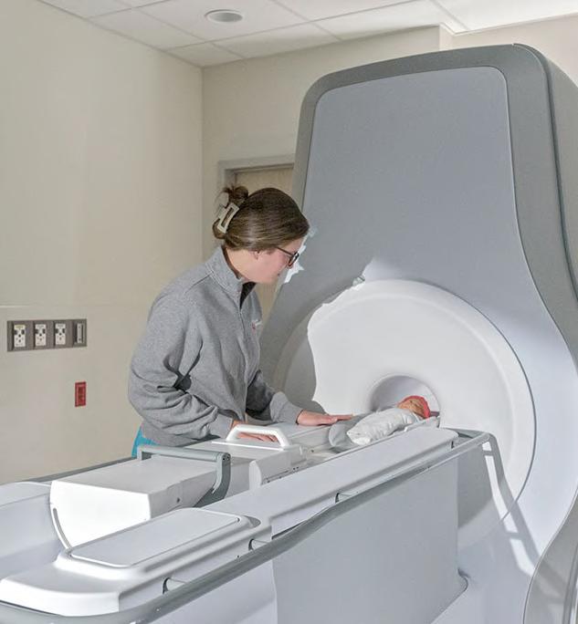
The Ascent3T is a groundbreaking neonatal MRI system from Eyas Medical Imaging (Cincinnati, OH, USA; www.eyasmri.com) that addresses a significant accessibility issue for infants by enabling installation directly within the neonatal unit. This eliminates the risks associated with transporting delicate newborns to separate MRI sites within the hospital. The Ascent3T’s compact design and innovative engineering cater specifically to infant anatomy. The system features a 3T magnet, which provides a more thorough and precise diagnostic tool, enabling healthcare professionals to receive detailed imaging of vital organs, such as the brain, lungs, heart, and abdomen. A 3T MRI system offers unparalleled accuracy in neonatal diagnostics, enabling the development of improved treatment plans and enhanced patient outcomes for vulnerable infants.
This innovation marks a major advancement in neonatal imaging and is anticipated to revolutionize the way healthcare professionals assess newborns. While the Ascent3T has not yet received clearance from the U.S. Food and Drug Administration (FDA) for commercial use in the United States, the company hopes to apply for FDA 510(k) clearance soon, thus enabling healthcare providers and neonatal patients to take advantage of this technology.
“The Ascent3T brings an unprecedented level of MR imaging and access to our smallest patients,” said MR physicist Charles Dumoulin, Ph.D., founder and CTO of Eyas who conceived the system. “By reinventing MRI technology, doctors can now visualize and understand neonatal health conditions using state-of-the-art MR imaging tailored to infants. It is a tremendous opportunity for better-informed medical decisions and improved neonatal care.”
First-of-Its-Kind Technology Detects ‘Invisible’ Risk of Heart Disease from Routine Cardiac CT Scans

provides the CaRi-Heart Risk, which evaluates the overall eight-year risk of a fatal heart attack (based on coronary inflammation status, plaque, and clinical risk factors). Research studies have demonstrated that abnormal FAI is linked to a 6-9 times higher risk for fatal heart attacks and a 5 times higher risk for non-fatal heart attacks.

CaRi-Heart’s newest product feature, the CaRi-Plaque module, is a web-based software medical device designed for trained operators to analyze cardiac computed tomography angiography (CCTA) data for the characterization and quantification of coronary plaque components. Trained operators can use the CaRi-Plaque module to generate a report describing the physical characteristics of coronary plaque powered by artificial intelligence (AI) algorithms. By combining proprietary measurements of coronary artery inflammation and plaque characteristics, with other clinical risk factors, CaRi-Heart can accurately predict an individual’s risk of heart attack to guide therapy.
“Inflammation plays an important part in the development of atherosclerosis and is a strong predictor of cardiovascular disease progression and events,” said Professor Keith Channon, Caristo Diagnostics Chief Medical Officer, “Previously, chest pain clinics have returned most patients back to primary care without a defined prevention or treatment pathway. With coronary inflammation and plaque evaluation provided by the CaRi-Heart analysis, the clinical team will be able to use the additional information to identify at-risk patients more effectively and optimize their treatment, so future cardiac events can be prevented.”
Image Quality Phantom



5 HospiMedica International August-September/2023
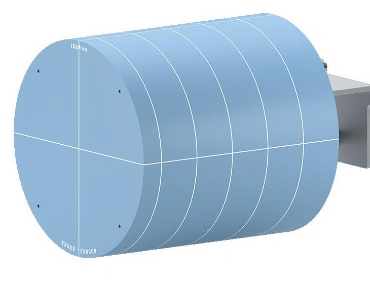
Introducing IQphan™ Comprehensive
Image: The Ascent3T Neonatal MRI System is designed specifically for infant anatomy (Photo courtesy of Eyas Medical Imaging)
With IQphan, a single phantom addresses QA across the range of specifications of different CT scanners, enabling you to gain more QA information. High-Contrast Resolution Module Slice Thickness & Geometric Evaluation Module Low-Contrast Detectability Module HU Module Uniformity Module sunnuclear.com Learn more: sunnuclear.com 105 HMI-09-23 LINKXPRESS COM
Cont’d from page 4
X-Ray Imaging Technique to Significantly Improve Breast Cancer Detection
n 2020, breast cancer emerged as the most frequently diagnosed cancer globally, with over two million recorded cases. It represented 24.5% of cancer diagnoses in women and 15.5% of cancerrelated deaths. In many developed nations, mammography screening programs serve as a key early detection strategy, contributing to reduced mortality rates. However, the complexity of reading mammograms, even for experts, presents a challenge. The low contrast of breast tissue under X-ray and the often unclear representation of the breast's complex interior by two-dimensional imaging complicate the process. Additionally, the mandatory compression of the breast for X-ray examination can cause discomfort or even pain, deterring some women from undergoing screenings. Now, researchers have successfully enhanced mammography, an X-ray imaging technique used for early-stage tumor detection, leading to significantly improved reliability and a less distressing experience for patients.
A research team that included scientists from the Paul Scherrer Institute (PSI, Aargau, Switzerland; www.psi.ch) has extended conventional computed tomography (CT) to yield significantly higher image resolution while maintaining the same radiation dose. This improvement could facilitate the earlier detection of small calcium deposits or microcalcifications, potential indicators of breast tumors, thus improving the survival prospects for affected women. The experts anticipate the swift clinical implementation of this X-ray phase contrast-based technique. Phase-contrast X-ray imaging improves tumor diagnostics by incorporating additional physical data. This allows for the utilization of an effect image creation, generally overlooked in conventional X-rays, that captures the information contained in signals produced when X-rays refract and scatter upon

contact with biological tissue. This is due to electromagnetic waves, including X-rays and visible light, undergoing not only attenuation but also refraction and diffraction when traversing structures of varying densities. This information can be leveraged to enhance image contrast and resolution, enabling easier identification of minuscule objects.
The researchers employed grating interferometry (GI), a technique used to measure physical systems, for developing their method. In this approach, X-rays pass through not only the object under examination but also through three gratings with a line spacing of a few micrometers, making the additional information visible. The team published their work in the journal Optica, presenting several images that illustrate the superior resolution and contrast of GI computed tomography compared to traditional X-rays. The X-rays can originate from a standard source, delivering a radiation dose similar to conventional CT breast scans. Moreover, the new screening approach should increase patient comfort during the procedure. Patients can lie face down on a table with chest-area gaps while the shielded tomograph underneath rotates around the breasts to construct a threedimensional image. The team aims to initiate clinical trials in collaboration with their clinical partners by the end of 2024, by which time they expect to have a prototype device ready for initial patient examinations.
“The phase-contrast X-rays reveal fine details of the tissue,” said Rahel Kubik-Huch, Director of the Department of Medical Services at Baden Cantonal Hospital (KSB) and Chief Physician for Radiology, who was involved in the research work. “This translational project is meant to explore the potential of this technique for detecting breast cancer in its early stages. We hope that one day our patients will be able to benefit from these advances.”
SPECT/CT Can Guide Treatment Decisions in Prostate Cancer Patients
Approved by the US FDA in 2022, 177Lu-PSMA is an effective treatment for metastatic castration-resistant prostate cancer and is currently administered using a standardized dosing interval. However, responses to this treatment can vary significantly, with some patients showing earlier signs of progression than others. Now, a new study has shown that early stratification with 177Lu-SPECT/CT allows patients responding positively to take a “treatment holiday” and offers non-responsive patients the chance to switch to an alternative treatment. By monitoring early-response biomarkers in patients undergoing 177Lu-PSMA prostate cancer treatment, physicians can customize dosing intervals, greatly enhancing patient outcomes.
In the study involving 125 men who received six weekly doses of 177Lu-PSMA in a clinical program, researchers at St. Vincent’s Hospital (Sydney, Australia; www.svhs.org.au)
Cont’d on page 8


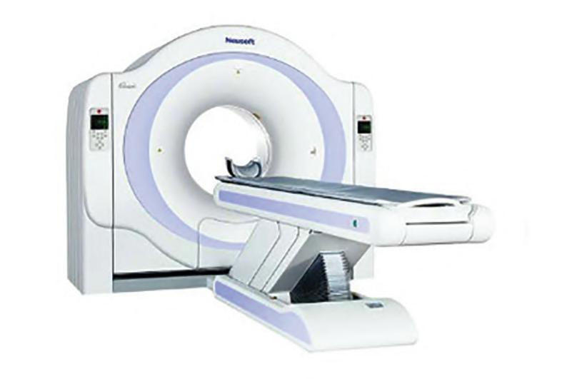
 64-SLICE CT SCANNER NEUSOFT MEDICAL SYSTEMS
The NeuViz 16 Classic is a 64-slice CT platform featuring a powerful tube design enabling continuous scanning for longer periods. The efficient design of its 32-row detector results in a high X-ray conversion
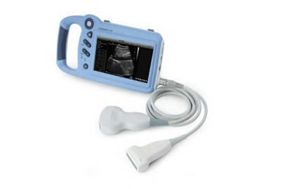
HANDHELD POC ULTRASOUND LANDWIND MEDICAL
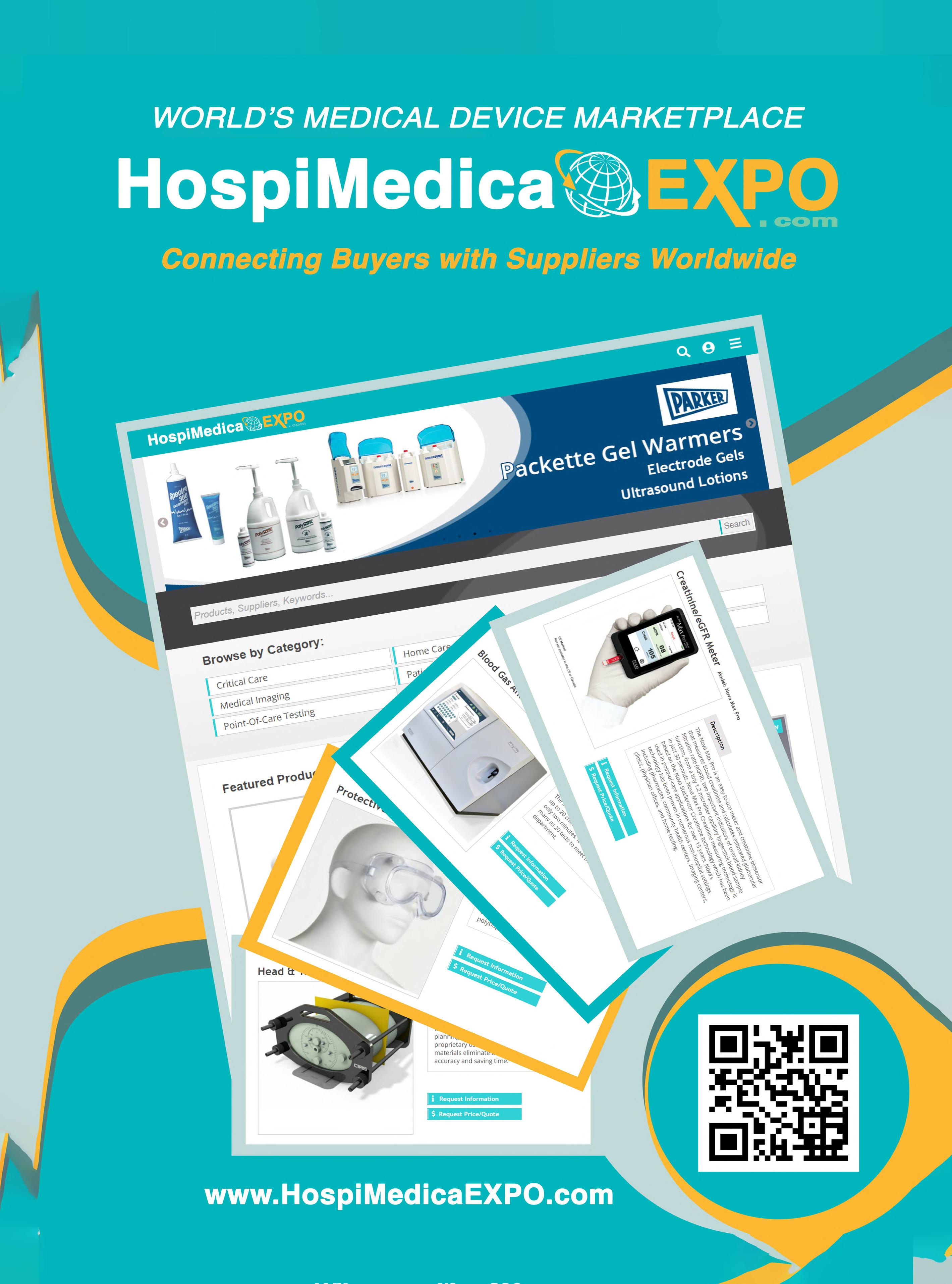
202 203 HMI-09-23 LINKXPRESS COM 6 HospiMedica International August-September/2023 WORLD’S MEDICAL DEVICE MARKETPLACE
P09 is a handheld visualization tool especially designed for point-of-care ultrasound diagnostics. The powerful ultrasound technology can be used in clinics, hospitals or practice settings and easily taken from room to room.
To receive prompt and free information on products, log on to www.linkXpress.com or scan the QR code on your mobile device
I
Image: A new method extends conventional CT to yield significantly higher image resolution for the same radiation dose (Photo courtesy of Paul Scherrer Institute)
Technique
Advances Cancer Diagnostics


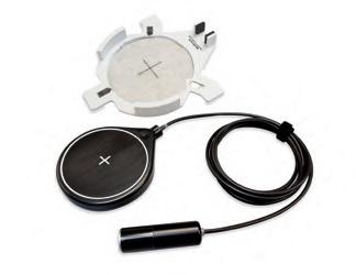


ach year, over 40,000 new thyroid cancer cases are reported. While 60-80% of patients with thyroid tumors undergo biopsies, the financial and potential physical toll of these procedures may be unnecessary for those with benign tumors. It is presently challenging for medical practitioners to accurately gauge the severity of a tumor, with different doctors having divergent opinions on a tumor’s threat level.
Standard ultrasound methods, which generate images of tissues and organs based on the sound waves they reflect, are efficient at identifying thyroid tumors. However, the technology can struggle to distinguish the minute sounds emitted from small blood vessels, or microvasculature, from those of the surrounding tissue, despite microvasculature providing vital clues about a mass’s cancerous nature. Although the introduction of contrast agents (chemicals easily visualized and commonly used in medical imaging procedures) allows ultrasound to display detailed images of tumor microvasculature, these substances need to be injected into patients and sometimes cause adverse side effects. While more recent ultrasound techniques can offer clearer nodule images, the ultimate evaluation still relies on the physicians’ subjective judgment.
Researchers at the Mayo Clinic College of Medicine and Science (Rochester, MN, USA; www.mayoclinic.org) have demonstrated that a pioneering cancer diagnostic method, which combines advanced ultrasound techniques with artificial intelligence (AI), can effectively diagnose thyroid cancer. This method — referred to as high-definition microvascu lature imaging, or HDMI — noninvasively captures images of the minute vessels within tumors and, based on vessel characteristics, automatically categorizes the masses. The researchers believe that HDMI could poten tially resolve the long-standing diagnostic challenge of assessing thyroid tumors in a clinical setting.
The researchers developed HDMI in an effort to develop an afford able, noninvasive imaging solution for evaluating thyroid tumors that could deliver quantifiable results and minimize errors. This system uses machine learning, a subset of AI, to assess high-resolution images of tumor microvasculature. The technique has already shown potential in accurately assessing breast tumors. In a recent study published in the journal Cancers, the team tested HDMI on thyroid tumors in 92 patients. They captured images of the tumors using HDMI and analyzed a dozen features related to the size and shape of the microvasculature in the images, including their density and branching points. All patients in the study, in consultation with their physicians, chose to have their tumors biopsied to confirm their malignancy status. Those with tumors deemed cancerous underwent surgery for the removal of the mass.
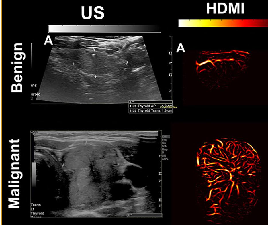
The researchers provided their machine learning algorithms with 70% of their imaging data from the patient tumors, along with the malignancy status, to teach algorithms how to interpret various features. Through a process of trial and error, the algorithms constructed predictive models, which were then used to determine the status of tumors imaged in the remaining 30% of the data. HDMI’s classifications were accurate 89% of the time, based on the clinical assessments of the biopsies and surgeries. These results suggest that HDMI could be a more reliable diagnostic method than traditional techniques and could spare numerous patients from unnecessary surgeries in the future. The researchers are now refining the method to enhance its accuracy even further. They plan to investigate its performance in diagnosing other types of cancer and whether it can assist in monitoring the effectiveness of chemotherapy on cancerous growths.
“Because HDMI allows you to objectively differentiate benign nodules from malignant ones, it could greatly improve diagnostic accuracy and reduce the number of unnecessary surgeries being done now,” said study author Azra Alizad, M.D., a professor of radiology and biomedical engineering at Mayo Clinic.
Image: A combination of advanced ultrasound and AI could upgrade cancer diagnostics (Photo courtesy of Mayo Clinic)

7 HospiMedica International August-September/2023 Medical Imaging To view this issue in interactive digital magazine format visit www.HospiMedica.com To view this issue in interactive digital magazine format visit www.HospiMedica.com
E
AI-Powered Ultrasound
DAP vc HospiMedica_clr_Eng 3.75 x 7.5 due 7/20/2023 Radcal Touches the World! Features: • Simple to use – Accurate and reliable
Customizable Touch Screen
Wi-Fi and USB Computer Connectivity
Report Generation Need to check the performance of X-ray machines? Then the Radcal Touch meter is your tool of choice. For further details: contact us at +1 (626) 357-7921 sales@radcal.com • www.radcal.com vc HospiMedica_clr_3.75x7.5_Eng23July20_12368.pdf 1 7/6/23 3:26 PM 107 HMI-09-23 LINKXPRESS COM
•
•
•
To receive prompt and free information on products, log on to www.linkXpress.com or scan the QR code on your mobile device


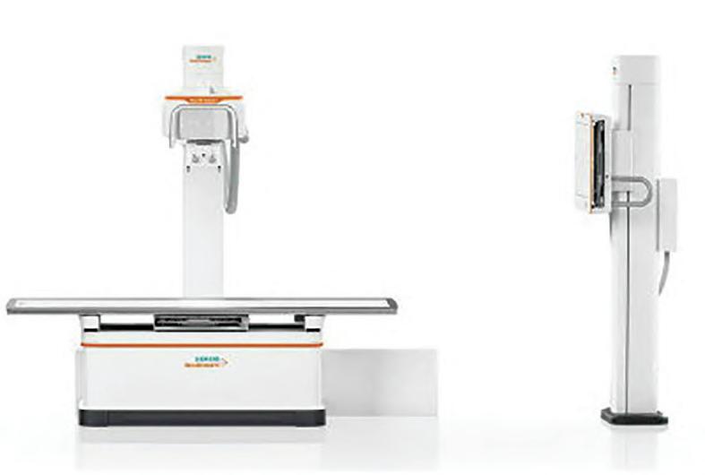
FLOOR-MOUNTED X-RAY MACHINE SIEMENS HEALTHINEERS
SPECT/CT Can Guide Treatment Decisions in Prostate Cancer Patients
Cont’d from page 6

evaluated progression-free survival and overall survival of different dosing intervals. After each dose, 177Lu-SPECT/CT imaging was performed on the men. Post the second dose, the researchers analyzed the men’s prostate-specific antigen (PSA) levels and the 177Lu-SPECT response to guide further management. Patients were categorized based on their level of response. Response Group 1, constituting 35% of participants, showed a significant reduction in PSA level and partial response on 177LuSPECT, and was advised to stop treatment until PSA levels increased. Response Group 2, comprising 34% of the participants, exhibited stable or reduced PSA and stable disease on SPECT imaging; these men continued their six-week treatment plan until it was no longer clinically beneficial. Men in Response Group 3 (31%) witnessed a rise in PSA levels and had
progressive disease on SPECT imaging. These patients were offered the chance to try an alternative treatment.

A reduction of more than 50% in PSA levels was observed in 60% of patients. On average, participants in the study had a median PSA progression-free survival of 6.1 months and a median overall survival of 16.8 months. The median PSA progression-free survival for Response Groups 1, 2, and 3 was 12.1 months, 6.1 months, and 2.6 months, respectively. The overall survival was 19.2 months for Response Group 1, 13.2 months for Response Group 2, and 11.2 months for Response Group 3. Furthermore, for those in Response Group 1 who took a “treatment holiday,” the median treatment-free period was 6.1 months.
“Personalized dosing allowed one-third of the men in this study to have treatment breaks while still achieving the same progression-free
and overall survival outcomes they would have if they received continuous treatment,” said Andrew Nguyen, a senior staff specialist in the Department of Theranostics and Nuclear Medicine at St. Vincent’s Hospital. “It also allowed another one-third of men who had early biomarkers of disease progression the opportunity to try a more effective potential therapy if one was available.”
The study was presented at the Society of Nuclear Medicine and Molecular Imaging 2023 Annual Meeting in Chicago, IL, USA on June 27, 2023
New Imaging Capabilities Drive Expansion in Global Mobile C-Arm Systems
Mobile C-arm systems offer a host of advantages for healthcare professionals and patients alike. These systems are heavily relied upon by interventional cardiologists, radiologists, and surgeons across various specialties, including orthopedics, gastroenterology, and urology. They are utilized in operating rooms, surgical suites, and outpatient procedures. The key benefit of this fluoroscopy technology is its mobility around the patient, achieving optimal image acquisition and thereby enhancing patient outcomes. Factors like advancements in 3D imaging and flat panel detectors, an aging population with cardiovascular and orthopedic conditions, and emphasis on reducing radiation exposure and procedure time, are driving the use of mobile C-arm systems. Consequently, the global mobile C-arm systems market is projected
to grow at a CAGR of 4.7%, from USD 1.43 billion in 2023 to USD 1.80 billion by 2028.
These are the latest findings of Mordor Intelligence (Hyderabad, India; www. mordorintelligence.com), a market research firm.
The growth of the mobile C-arm systems market is expected to be boosted by factors like the rising prevalence of chronic diseases, advancements in imaging technology, and the introduction of new products. The increasing global burden of chronic diseases and surgeries, particularly heart procedures, are expected to further fuel market growth. Moreover, efforts by the market players to enhance the efficiency and workflow of mobile C-arm systems through technological advancements will also contribute to market growth. However, challenges like high procedural and equipment costs and a shortage of skilled professionals could hamper
the market’s growth during the forecast period. By product type, the mini C-arm segment is expected to witness significant growth in the coming years due to factors such as the rising prevalence of road injuries, trauma, and orthopedic diseases, advances in medical imaging, and the advantages offered by mini Carms. Mini C-arms are ideal for fluoroscopy of extremities at minimized dose levels and their lightweight design ensures ease of handling in small spaces and operating rooms.
Geographically, North America holds the major share in the global mobile C-arm systems market. This can be attributed to factors such as the growing prevalence of chronic diseases, FDA approval of advanced products, well-established healthcare infrastructure, and strategic initiatives taken by key market players in the region.
MULTIX Impact E is a versatile floor-mounted X-ray machine featuring an easy, intuitive system operation that allows users to detect anatomical details at low doses. It has been purposefully designed to improve access to care.
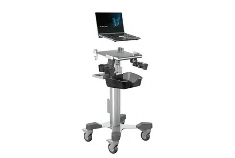
LAPTOP ULTRASOUND CHISON MEDICAL IMAGING
205 206 HMI-09-23
HospiMedica International August-September/2023 WORLD’S MEDICAL DEVICE MARKETPLACE
SonoAir 70 is the world’s lightest and thinnest laptop ultrasound, powered by a new Air platform. It combines high-definition image quality and intelligent features to help achieve a fast and reliable diagnosis
LINKXPRESS COM
Image: Personalized dosing in prostate cancer treatment improves patient outcomes (Photo courtesy of Freepik)

ULTRASOUND TRANSMISSION GEL PARKER LABORATORIES

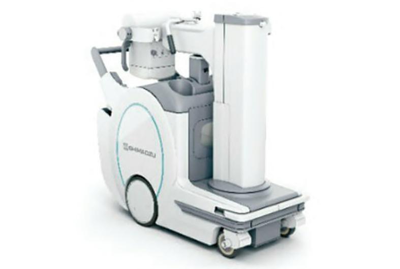
MOBILE X-RAY SYSTEM SHIMADZU MEDICAL SYSTEMS
World’s First Bedside Gamma Camera
gamma-optical camera developed by Serac Imaging Systems (London, UK; www. seracimagingsystems.com ) forms images showing the distribution of an administered radiopharmaceutical within a patient’s body, assisting in diagnosing or monitoring diseases. The device uses a microcolumnar CsI(Tl) crystal scintillator to convert gamma photons into optical ones detected by a semiconductor. The compact camera head, with a 6-inch (15 cm) diameter and weighing less than 5 kg, offers easy portability throughout the hospital by a single operator. Its plug-and-play connection ensures readiness within minutes, anywhere in the hospital. Its small size enables close proximity to the patient, enhancing image resolution and acquisition times. Moreover, its fully articulated arm facilitates easy positioning of the camera head to ensure patient comfort.
The device features a pinhole collimator, which allows operators to determine the optimal balance of image resolution, acqui-
sition speed, and field of view for individual cases. Automatic aperture size adjustment without changing parts ensures quick, flexible solutions for various clinical applications. The built-in optical detector shares the same field of view, regardless of imaging distance or angle, allowing for overlaying of optical and gamma images with no parallax and real-time image streaming to the control PC.
By offering an alternative to conventional gamma/SPECT cameras for imaging organs and structures within a small, portable footprint, Seracam enhances the capacity within a nuclear medicine department without the need for an additional camera room and facilitates point-of-care molecular imaging throughout the hospital. Seracam is currently undergoing clinical testing in a six-month study involving 25 patients. Potential applications include imaging small organs such as the thyroid, bone, renal, infection imaging, lymphatic imaging, and sentinel lymph node localization.
“We believe that the combination of the

co-aligned gamma-optical hybrid imaging capability, alongside the compact size and portability of the camera, have real potential to improve and expand nuclear imaging options, diagnosis and outcomes for patients,” said Mark Rosser, Chief Executive Officer of Serac Imaging Systems.


Algorithm Halves Lumbar MRI Time
does not expose patients to ionizing radiation. However, it is impaired by lengthy acquisition times and the need for parameter adjustments to enhance image quality, which can further extend scan times. Over recent years, artificial intelligence (AI), specifically deep learning (DL), has made significant strides in various imaging areas, including image classification, segmentation, denoising, super-resolution, and image synthesis/transformation. Nevertheless, the impact of AI algorithms on routine whole MRI lumbar spine protocol acquisition has yet to be explored.
In a new study, researchers at Sant’Andrea University Hospital (Rome, Italy; www. ospedalesantandrea.it) compared quantitative and subjective image quality, scanning time, and diagnostic confidence between a novel
deep learning-based reconstruction (DLR) algorithm and the standard MRI protocol for the lumbar spine. By using the DLR algorithm, researchers were able to cut the duration of lumbar MRI exams by half. Furthermore, these improved scan times did not compromise image quality but rather enhanced the signal-to-noise ration (SNR).
For this study, GE Healthcare’s (Chicago, IL, USA; www.gehealthcare.com) FDA-approved AIR Recon DL algorithm was applied to the exams of 80 healthy volunteers who underwent a 1.5T MRI examination of the lumbar spine between September 2021 and May 2023. Both the DLR algorithm and standard protocols were utilized to complete sequences, which were later assessed by two radiologists who were unaware of the


reconstruction techniques used.
The DLR algorithm yielded a notable reduction in protocol scanning time, reducing it from almost 13 minutes to just over 6 minutes. The blinded radiologists reported that the reconstruction algorithm provided a higher SNR across all sequences and superior contrast-tonoise (CNR) for axial and sagittal T2-weighted fast spin echo images. Both readers rated the overall image quality for all sequences as superior with the DLR, leading the research team to suggest that the DLR protocol can be safely integrated into clinical practice. The team also noted additional benefits of shortening lumbar MRI protocols, including cost-effectiveness and enhanced patient compliance, especially for those who are claustrophobic or experiencing severe physical pain.
The MobileDaRt Evolution MX8 Version v type mobile X-ray system features the V series glass-free Flat Panel Detector (FPD). It is lighter than conventional glass-based FPDs and features a smaller pixel size of just 99μm.
208 209 HMI-09-23 LINKXPRESS COM 10 HospiMedica International August-September/2023 WORLD’S MEDICAL DEVICE MARKETPLACE To receive prompt and free information on products, log on to www.linkXpress.com or scan the QR code on your mobile device
The Sterile Aquasonic 100 ultrasound transmission gel is the world standard for sterile ultrasound gel. It is completely aqueous and acoustically correct for the broad range of frequencies used in medical
Cont’d from cover
Image: Seracam is a truly portable system for point of care molecular imaging (Photo courtesy of Serac Imaging)
Cont’d from cover
Blood Filter Device Removes Circulating Tumor Cells in Pancreatic Cancer Patients
Pancreatic Ductal Adenocarcinoma (PDAC), a type of pan creatic cancer, is predicted to be the second most common cause of cancer-related fatalities by 2030. Circulating Tumor Cells (CTCs), cancer cells that break away from a primary tumor and enter the bloodstream, are responsible for metastatic cancer. This form of cancer is often resistant to standard treatments such as chemotherapy, radiation, and immunotherapy, making it particularly difficult to treat. The presence of CTCs in blood correlates with the outcomes of metastatic cancer. Preliminary methods that extract CTCs from the bloodstream have shown promising results in animal trials, implying that reducing CTCs in the blood might slow or pre vent metastasis. Now, an extracorporeal blood filter procedure can remove CTCs from the bloodstream of patients suffering from PDAC, offering an important tool for treating this lethal cancer with a low 5-year survival rate.

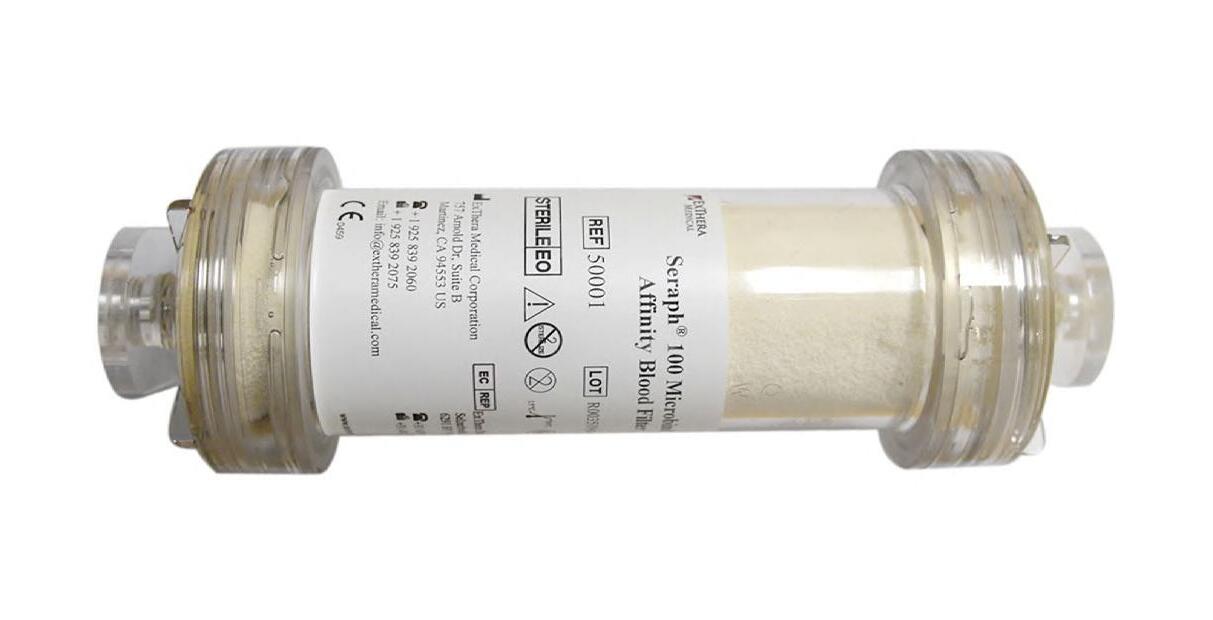
ExThera Medical’s (Martinez, CA, USA; www.extheramedical. com ) OncoBind extracorporeal blood filter procedure, based on its Seraph 100 Microbind Affinity Blood Filter, has proven successful in removing metastatic circulating tumor cells in several in vitro models. As the patient’s blood flows through the OncoBind Blood Filter, it passes over beads with receptors that imitate those on human cells that are targeted by pathogens and CTCs for attachment and colonization. The CTCs are then captured and adsorbed to the beads’ surface, effectively removing them from the bloodstream. Like the Seraph 100, the OncoBind system does not introduce anything foreign into the blood. OncoBind, similar to Seraph’s adsorption media (the beads), represents a versatile platform that uses immobilized (chemically bonded) heparin due to its proven blood compatibility and its unique ability to bind CTCs. The U.S. Food and Drug Administration (FDA) has approved an Investigational Device Exemption (IDE) application for ExThera’s OncoBind extracorporeal blood filter procedure.


“Seraph technology has an excellent safety record in more than 2,500 clinical treatments performed globally, of bacterial, viral, and fungal bloodstream infections,” said Bob Ward, Ph.D. (h)., NAE, the CEO & a founder of ExThera Medical. “We are pleased to begin an important new clinical trial, and ecstatic that FDA has approved our recent IDE application. If ExThera’s technology captures CTCs in the clinical setting, as it has in the laboratory, the potential is enormous, adding another much-needed tool to the arsenal for treating PDAC, a lethal cancer with a low 5-year survival rate.”
Clinical and Research Applications


11 HospiMedica International August-September/2023 111 HMI-09-23 LINKXPRESS COM
Image: The OncoBind procedure is based on the Seraph 100 Microbind Affinity Blood Filter (Photo courtesy of ExThera)
ENTHERMICS MEDICAL SYSTEMS


Multifunctional Device Combines Hemorrhage Treatment and Monitoring

Numerous products exist for controlling hemorrhages such as cotton or gauze bandages, powders, and tourniquets. These are typically used for shallow wounds of uniform shape, however, they lack the ability to simultaneously detect bleeding and control hemorrhage. Recent advancements have introduced shapememory sponges that are highly absorbent and retain their structure even when treating deep, irregularly shaped wounds, facilitating blood coagulation. While cellulose-based shape-memory sponges are effective, they are not biodegradable unlike their gelatin-based counterparts, and both lack hemorrhage detection capabilities. Now, scientists have engineered a multi-purpose device for treating deep, non-compressible, and irregularly-shaped wounds. This device offers prompt hemorrhage management, minimal inflammation, infection control, adjustable biodegradation rates for both internal and external use, and sensing capabilities for long-term hemorrhage tracking. This device can be valuable for timely alerts and managing bleeding from surgical wounds, traumatic injuries, and critical illnesses.
Scientists at the Terasaki Institute for Biomedical Innovation (TIBI, Los Angeles, CA, USA; www.terasaki.org) created the innovative hemorrhage management device using silk fibroin, a protein from the Bombyx mori silkworm. This biodegradable material possesses excellent anti-inflammatory and mechanical properties and can be engineered into highly absorbent, porous, memory-shaped sponges that promote coagulation and tissue regeneration. The adjustable degradation rates of these sponges allow for longer in-body use and the potential to incorporate sensors to monitor bleeding. Leveraging these capabilities, the scientists designed a unique, all-in-one hemorrhage management device that integrates two silver nanowire layers positioned above and a hemostatic sponge layer below. The nanowires serve as both hemorrhage detection sensors and antibacterial agents.
The device underwent a thorough evaluation, including tests for mechanical properties, biocompatibility, and biodegradation. Mechanically, the silk fibroin sponges demonstrated excellent elasticity, absorptive capacity, and high retention of pore size and shape against water and blood exposure, along with maximum hemorrhage control achieved by adjusting silk fibroin concentrations to match the mechanical properties of the surrounding wound tissue. The sponge and nanowire layers demonstrated favorable biocompatibility and minimal anti-inflammatory responses, as proven by the successful cell viability and proliferation tests using connective tissue samples. Further
ANESTHESIA SYSTEM

testing indicated that the sponge’s degradation rate could be slowed by increasing the silk fibroin concentration and incorporating a methanol wash step during the manufacturing process.
Following this evaluation, the device and a commercial gelatin-based anti-hemorrhage device were implanted under the skin in rat models for comparison. After four weeks, the commercial sponge had completely degraded while the silk fibroin nanowire device remained intact. The implanted sponge triggered minimal inflammation and had no negative impact on the rats’ organs or behavior. Moreover, the silk fibroin device surpassed the commercial sponge in hemorrhage control tests. In addition to managing hemorrhage, the device features a nanowire-based capacitive sensor for detecting bleeding. As the sponge absorbs blood during bleeding, its capacitance increases without any change in shape. This increase in capacitance is measurable and directly corresponds to the amount of absorbed blood, thereby enabling real-time bleeding monitoring.
“This multifunctional device offers many attractive features for hemorrhage control and wound monitoring and is highly adaptable for different types of wounds and tissues,” said Ali Khademhossein, Ph.D., TIBI’s Director and CEO. “And the hemorrhage monitoring feature also opens up several possibilities for integrative biosensing and additional therapeutics.”
The ivNext fluid warmer features Enthermics’ new IV inventory management system offering clinicians a simple way to monitor and precisely warm IV bags. It can warm fluids to recommended temperatures using
MINDRAY PATIENT MONITORING & LIFE SUPPORT
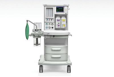
The WATO EX-30 anesthesia machine provides clinical users with basic monitoring measurements on a colorful screen with up to three waveforms including Paw, TVe, MV, Ppeak, and Pmean, along with graphics at the same time.
211
12 HospiMedica International August-September/2023 WORLD’S MEDICAL DEVICE MARKETPLACE
212 HMI-09-23 LINKXPRESS COM
To receive prompt and free information on products, log on to www.linkXpress.com or scan the QR code on your mobile device
Image: The new technique offers improved diagnostic precision and potential for personalized therapy for a common arrhythmia (Photo courtesy of Terasaki Institute)
Ultra-Thin
Microcatheter

with Fiber Optic Sensors: Game Changer in Heart Disease Detection
There is growing evidence that points to the importance of accurate assessments of the coronary microvasculature (the constriction of the smallest heart vessels) in tailoring heart disease therapies. This is especially relevant for women and diabetic patients who are more susceptible to microvascular dysfunction. Accurate information about a person's heart health can enable doctors to make more informed treatment decisions, such as prescribing medication, deciding on surgical intervention, or discontinuing medication. However, traditional X-rays (angiograms) used by cardiologists to image the heart's larger arteries do not adequately display these tiny blood vessels. Consequently, the current clinical practice may lead to overdiagnosis, resulting in potentially unnecessary invasive procedures such as stent insertion, which carry risks and require recovery time. Now, a new diagnostic technology uses tiny fiber optic sensors to detect the causes of heart disease, more quickly and accurately than existing methods.
Scientists at University College London (UCL, London, UK; ac.uk) have developed a new device named iKOr that uses an ultra-thin microcatheter integrated with fiber optic sensors, enabling doctors to examine both blood pressure and blood flow around the heart as well as identify signs of artery narrowing and thickening, common indicators of heart disease. The slimline probe is especially suitable for detecting microvasculature. The iKOr device incorporates a temperature and pressure sensor merely 0.2mm wide — twice the thickness of a human hair — which is inserted through the patient's blood vessels on an ultra-thin catheter.

The iKOr device measures the flow rate around the heart by emitting a brief pulse of light upstream of the vessels being investigated, thereby slightly warming the blood by approximately one degree. The sensor records the time taken for the downstream temperature change, allowing the device to determine whether the flow is being obstructed by the narrowing of vessels. However, before the device can gain widespread use by doctors, researchers must verify its safe and easy application in patients. Initial patient tests confirm its safety, ease of use, and functionality. These will be followed by a more extensive clinical trial to further confirm the device's safety and superior performance compared to existing tests. Researchers suggest that the iKOr device could potentially assist numerous patients experiencing cardiovascular symptoms like chest pains, whose cause remains unidentified by current techniques.
“The iKOr device is responding to a clinical need – to significantly improve how blood flow in the heart is measured,” said lead iKOr developer, Professor Adrien Desjardins from the UCL Medical Physics & Biomedical Engineering. “Our microcatheter
provides concurrent pressure and flow measurements from inside coronary arteries – this is unique and makes the tiny blood vessels more measurable, compared to traditional X-rays. This will help to significantly improve diagnosis and treatment for a large group of patients; those with obstructive
Critical Care To view this issue in interactive digital magazine format visit www.HospiMedica.com
13 HospiMedica International August-September/2023 113 HMI-09-23 LINKXPRESS COM
To receive prompt and free information on products, log on to www.linkXpress.com or scan the QR code on your mobile device


CALIBRATION SYRINGE
Revolutionary Tool Diagnoses Heart Disease Due to Invisible Blockages in Small Blood Vessels
Microvascular disease primarily impacts the inner linings and walls of minute blood vessels branching off from the coronary arteries. While these minuscule vessels may not contain plaque, damage due to microvascular disease can induce spasms and restrict the supply of oxygenated blood to the heart muscle. This disease, more prevalent in women than men, can result in symptoms such as chest discomfort, breathlessness, and fatigue. Standard treatment includes specific medications, unlike those used for larger vessel heart disease, and lifestyle modifications like exercise and a nutritious diet. However, diagnosing microvascular disease accurately has been challenging due to its poor visibility during cardiac catheterization, unlike atherosclerotic plaque in the primary coronary arteries. Now, an innovative method can evaluate the health of smaller arteries within the heart and effectively detect microvascular disease which had remained a
diagnostic challenge until now.
The Coroventis CoroFlow Cardiovascular System from Abbott (Lake Forest, IL, USA; www.abbott.com) is a revolutionary diagnostic tool that allows physicians to identify coronary microvascular dysfunction resulting from invisible blockages in the smallest arteries of the heart. Used in conjunction with Abbott's PressureWire X Guidewire, this wireless device produces hemodynamic data that measures the functioning of the epicardium (the heart's outermost lining), tiny blood vessels, and ventricles (the heart's largest pumping chambers). Designed for use during coronary angiography, the CoroFlow system assists in evaluating crucial heart function parameters and diagnosing or ruling out small vessel cardiac disease in individuals with symptoms such as chest discomfort, particularly those without significant blockages in the major coronary arteries.
Chest pain frequently prompts visits to

Handheld Device Determines Correct Nasogastric Feeding Tube Placement

Annually, nurses are tasked with inserting over a million feeding tubes into patients' stomachs for the delivery of nutrition, hydration, and medication. It is reported that inaccurate placement of nasogastric tubes (NGTs) occurs in 1-3% of all insertions, which can lead to severe consequences including treatment delays and, in some cases, death. The majority of NGTs are inserted 'blind', as the procedure does not involve any form of visualization to confirm correct positioning before use. Traditional methods, in use for the past 20 years, are inherently flawed. The first technique to confirm NGT placement involves pH testing of the NGT aspirate. If a pH level of 5.5 or lower is
achieved, the NGT is deemed safe, otherwise, chest X-rays are required. However, pH strips can fail in up to 45% of cases due to difficulty in obtaining aspirate, necessitating an X-ray for the patient and causing an average treatment delay of approximately eight hours. Now, a new electronic device specifically designed for correct NGT placement could eliminate the problem of incorrectly inserted feeding tubes.

NGPod Global (Cheshire, UK; www. ngpodglobal.com) has developed the NGPOD system, a device designed to tackle the risks associated with traditional nasogastric placement confirmation techniques. The portable NGPod device employs a fiber optic sensor that is introduced directly into the stomach via the

INFANT TRANSPORT INCUBATOR NOVOS MEDICAL SYSTEMS
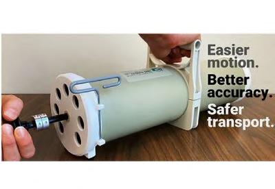
Image: The CoroFlow Cardiovascular System is an advanced platform to measure physiological indices (Photo courtesy of Abbott)
primary care physicians, cardiologists, or emergency rooms. Without a definitive diagnosis, patients often repeatedly seek medical attention, searching for the root cause of their discomfort. With the CoroFlow system, doctors can precisely diagnose the presence or absence of microvascular disease, identify the causes of a patient's symptoms, and determine the best treatment to alleviate discomfort and improve heart health. For most individuals suffering from unexplained chest pain, the CoroFlow system can provide a previously elusive answer, confirming their symptoms as real — and treatable.
feeding tube, eliminating the need for aspirate while transmitting light signals back to a handheld device. The NGPOD system also eliminates the need to aspirate gastric contents from the patient and reduces the risk of clinical interpretation errors by providing a safe bedside test that delivers a clear 'yes' or 'no' result. Furthermore, the device lowers the requirement for unnecessary X-rays to confirm placement, contributing to patient safety and cost-efficiency.Using the product is straightforward and would require a maximum of 20 minutes of training. The device is currently in 12 hospitals in the UK with another eight scheduled in the next six months.
The KT 1000 infant transport incubator provides a stable microclimate for safe and efficient transport of newborns in hospital settings. Its advanced technology and superior features enable easy control of various parameters.
214
14 HospiMedica International August-September/2023 WORLD’S MEDICAL DEVICE MARKETPLACE
Hans Rudolph Calibration Syringes, now available with an easy-to-install handle for 3L syringes, deliver a known calibrated air volume as a standard for reliable calibration and measurement of respiratory volume
215 HMI-09-23 LINKXPRESS COM
Sensor Patch Offers Non-Invasive Leak Diagnosis and Sealing of Post-Abdominal Surgery Sutures



ollowing abdominal surgery, there is a potential risk of leaks occurring at the sutured areas, leading to the contents of the digestive tract flowing into the abdomen. While the concept of sealing sutures in the abdominal cavity with plaster has been introduced in operating rooms, clinical success is not always guaranteed due to variations in tissue adhesion. The problem lies in the fact that the patches, made of protein-containing material, dissolve too quickly upon contact with digestive juices. Until now, healthcare professionals have had to rely on physical symptoms experienced by patients or delayed laboratory tests to identify leaking sutures, which often provide insufficient and delayed information. A new and innovative solution in the form of a hydrogel-polymer patch addresses this challenge. This new patch prevents highly acidic digestive juices and bacteria-laden food residues from escaping the digestive tract, thus averting peritonitis or potentially life-threatening blood poisoning (sepsis). What sets this patch apart is the integration of non-electronic sensors that sound an “alarm” before digestive juices can leak into the abdominal cavity.
Researchers at Empa (Zurich, Switzerland; www.empa.ch) and ETH Zurich (Zurich, Switzerland; www.ethz.ch) have developed the sensor patch from a synthetic hydrogel, consisting of four different acrylic substances. To enable the hydrogel patch to detect leaks, the team collaborated with hospitals in Switzerland and international research partners to devise this innovative solution. The novel material possesses “vision” through its sensitive response to changes in pH value and the presence of specific proteins near the wound. Depending on the location of the leak, the reaction occurs within minutes or a few hours. The sensor patch detects escaping digestive fluid due to its composite structure. For instance, when acidic

ECG-AI Algorithm to Aid Physicians in Earlier Identification of Cardiac Amyloidosis



Cardiac amyloidosis is a severe, progressive, and often underdiagnosed rare disease leading to heart failure. In those affected, stiff heart walls hinder the left ventricle’s function, disrupting proper blood flow in and out of the heart. Early diagnosis is crucial for timely interventions that can enhance patient outcomes. However, the disease’s rarity, coupled with its nonspecific and varied symptoms such as shortness of breath, knee pain, bilateral carpal tunnel syndrome, kidney disease, and gastrointestinal issues, make early diagnosis challenging. While standard ECGs are usually obtained to assess these symptoms, human interpretation often overlooks subtle feature combinations that may signify cardiac amyloidosis. Now, an artificial intelligence electrocardiogram (AI-ECG) solution can detect patterns in ECG signals that are usually imperceptible to humans to provide an early warning for cardiac amyloidosis.
Anumana, Inc. (Cambridge, MA, USA; www.anumana.ai) has developed an AI-ECG algorithm to facilitate the early detection of cardiac amyloidosis. The AI-powered software can interpret signals from ECGs that might be missed by human analysts. Given the widespread use of the noninvasive ECG test, AI-ECG algorithms have the potential to reach a larger patient population at an earlier stage. Anumana is presently working on transforming this algorithm into a Software-as-a-Medical-Device (SaMD), with an aim to integrate this solution seamlessly into existing clinical workflows. This AI-ECG solution has also received the Breakthrough Device Designation from the U.S. Food and Drug Administration (FDA), ensuring patients and healthcare providers have timely access to this algorithm.
“The ubiquitous nature of the painless, non-invasive Electrophysiology tests gives ECG-AI algorithms the potential to reach a larger number of patients earlier, something clinicians have long hoped for,” said Venky Soundararajan, PhD, Co-founder, and Chief Scientific Officer of Anumana. “Receiving the FDA Breakthrough Device Designation for our Cardiac Amyloidosis ECG-AI Algorithm recognizes the significant potential of this tool to detect disease early.”
gastric juice interacts with the sensor material, gas bubbles emerge within the patch’s matrix. These bubbles can then be visualized using ultrasound.






In the latest development, the researchers have further enhanced the patch’s capabilities. The sensor response now includes a visible change during patient examinations using computed tomography (CT). If a leak is present at the surgical site, contrast deviations in ultrasound and CT images indicate its occurrence. The new material composition of the integrated sensor aids in detecting leaks, as it can be shaped into a form that stands out in imaging processes. Upon contact with digestive fluid, the material changes its shape from circular to ring-shaped.
According to the researchers, a sensor with a distinctive shape compared to anatomical structures in CT and ultrasound images could reduce ambiguity in diagnosing impending leaks. Moreover, the material possesses the necessary properties for wound closure, including a strong bond to tissue, the ability to form networks and stability against digestive fluids. As a result, this cost-effective and biocompatible “super glue,” consisting predominantly of water, has the potential to not only reduce the risk of complications following abdominal surgery but also shorten hospital stays and save healthcare costs.

Critical Care To view this issue in interactive digital magazine format visit www.HospiMedica.com 115 HMI-09-23 LINKXPRESS COM
Singu20 Nucleic Acid Extractor AccuRa-32 Real-Time PCR System (32 samples) Singu20 Nucleic Acid Extractor AccuRa mini Real-Time PCR System (8-16 samp es) ≤30 minutes qPCR Detection Kits (PCR fluorescence method): Respiratory Diseases: COVID-19, fluA, fluB, AdV, TB and mu tiplex test Blood Diseases: HBV, HCV, HIV and multiplex test Sexually Transmitted Diseases: HPV CT NG UU and multip ex test Viral Zoonotic Diseases: MPV Vector-borne Diseases: PF, ZIKV Genetic Diseases: MTHFR Animal Diseases: Swine/Avian/Aquatic Animal/Ruminant/Companion Animal Diseases 5-Part Hematology Analyzer Hematology + CRP +SAA Joint Analyzer (Auto Sampling) ≤35 minutes
For small and medium labs, @SINGUWAY is your answer.
15 HospiMedica International August-September/2023
F
Image: The sensor patch can withstand several times the pressure in the abdominal cavity (Photo courtesy of Empa)
Lab-on-a-Patch Enables


Real-Time Monitoring of Multiple Disease Biomarkers

Advancements in continuous glucose monitors (CGMs) have significantly improved the management of diabetes, however, they come with a major shortcoming: CGMs are incapable of measuring any diagnostic target other than glucose. Now, DNA-based sensing has emerged as a breakthrough technology that overcomes this limitation with its ability to enable continuous and real-time measurements of any diagnostic target.
Nutromics’ (Melbourne, Australia; www.nutromics.com) is developing a "lab-in-a-patch" that harnesses DNA sensor technology to track various targets such as disease biomarkers and difficult-to-administer drugs within the human body. Currently undergoing in-human trial testing, Nutromics' DNA-based sensor platform is poised to bring about a significant shift in diagnostic methods. By integrating multiple DNA-based sensors with minimally invasive microneedles, the platform allows for continual and real-time tracking of various vital targets. With its aptamer platform, Nutromics stands to improve the prognosis for millions of patients in clinically critical conditions. By offering medical practitioners the ability to continuously monitor a wide array of analytes that presently cannot be analyzed in real-time, Nutromics’ "labin-a-patch could become a game changer in various clinical situations.
The initial product to come out from Nutromics’ stable is a wearable device equipped with a vancomycin sensor. Vancomycin, a potent antibiotic used for treating severe infections like sepsis, presents a dosage challenge due to its narrow therapeutic window, high toxicity, and limited patient blood level data available to doctors. DNA-based sensors can overcome these hurdles by providing continuous, real-time information on vancomycin concentration levels, enabling doctors to calibrate the antibiotic dosage to maintain the therapeutic range and avoid toxicity. Additionally, DNA-based sensors can significantly influence heart attack diagnosis. Troponin, a protein that markedly increases in the bloodstream upon heart muscle damage (like during a heart attack), can be continuously monitored to provide early trend data. This could drastically reduce heart attack diagnosis time to mere minutes, ultimately saving lives. Nutromics envisions a future where DNA-based biosensors extend beyond acute hospital settings to pointof-care, general lab testing, and even consumer use for the prevention of disease among healthy individuals.
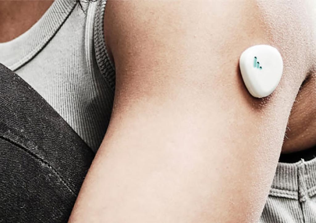
Image: A breakthrough technology combines multiple DNA sensors with microneedles (Photo courtesy of Nutromics)
Breakthrough Implant Marks Shift in Treatment of Gastroesophageal Reflux
Acid reflux, a condition characterized by the regurgitation of the stomach’s acid content into the esophagus, affects approximately 400 million individuals daily, making it the second-largest treatment field globally. This condition can progress into a chronic disease known as gastroesophageal reflux disease (GERD) when acid reflux episodes become frequent. Traditional surgical treatments have been associated with numerous complications, such as difficulties in swallowing and an inability to belch or vomit. This is largely due to these methods encircling the food passage to bolster the closing ring muscle. Now, a novel implant based on a unique invention offers a solution to acid reflux without interfering with the food passageway.
Implantica AG’s (Zug, Switzerland; www.implantica.com) RefluxStop is a CE-marked implant designed to prevent gastroesophageal reflux. Unlike drug therapy, RefluxStop not only manages acid reflux symptoms but also eliminates stomach fluid regurgitation. This innovative, non-active implant is positioned on the upper section of the stomach through laparoscopic (keyhole) surgery. The RefluxStop device acts as a mechanical barrier that prevents the lower esophageal sphincter (LES) from shifting into the thorax. With abdominal pressure, the LES can function normally. Recently, RefluxStop won the Medtop Tech Award for the most innovative medical device among all competitors.
 ARIA 150 C is a high-end lung ventilator for intensive care based on an artificial intelligence algorithm for adult, pediatric and neonatal patients, offering high-performance non-invasive ventilation modes (NIV
TRANSPORT PATIENT MONITOR NIHON KOHDEN
Life Scope PT is a high perception smart transport monitor designed with leading edge technology for continuous monitoring of patients even during transit. It provides complete modular flexibility with the Smart Cable System.
16 HospiMedica International August-September/2023 WORLD’S MEDICAL DEVICE MARKETPLACE
217 218 HMI-09-23 LINKXPRESS COM
To receive prompt and free information on products, log on to www.linkXpress.com or scan the QR code on your mobile device
Virtual Reality Visualization Helps Surgeons Plan Procedures in Advance
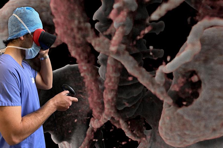
Virtual reality (VR), now a highly accessible technology, offers a myriad of opportunities, particularly for visualizing and maneuvering through intricate three-dimensional data—a capability that holds significant potential for scientific applications. Now, a novel surgical planning solution is using VR to provide surgeons with detailed patient avatars for improving procedure preparation. Created instantaneously from CT scans or MRI images using proprietary technology that leverages human-data interaction and machine learning research, these patient avatars offer invaluable resources for pre-operative planning and can also be utilized during surgeries.
AVATAR MEDICAL’s (Paris, France; www.avatarmedical.ai) VR surgical planning solution converts patients’ medical images into 3D avatars within an interactive VR environment. These lossless, real-time 3D avatars provide clinicians with an accurate and instant way to understand their patients’ medical imagery. AVATAR MEDICAL’s unique technology utilizes VR and Bayesian methods to facilitate ultra-fluid navigation within medical images. The technology is compatible with standard MRI and CT scans, does not require pre-processing of data, and enables physicians to modify patient avatars in real time within the VR environment.
So far, over 100 surgeons from 20 distinct hospitals and universities have benefited from this solution. It has been employed for case studies, student education, and patient engagement, leading to six medical publications. AVATAR MEDICAL’s VR surgical planning solution has also received 510(k) clearance from the U.S. Food and Drug Administration (FDA).

Pen-Like Device for Real Time Tissue Analysis Can Revolutionize Surgical Pathology





For over a century, cancer tissue analysis has depended on long-established workflows, with traditional histopathology results typically taking 40 minutes or more. In this process, tissue is biopsied, sent to a lab, frozen, sliced, stained, and examined by a pathologist under a microscope. While awaiting results, the patient remains under anesthesia, increasing their risk of infection and incurring additional costs for both the patient and hospital. Now, a groundbreaking device incorporating cutting-edge mass spectrometry and artificial intelligence (AI) can identify cancerous tissue—in vivo and ex vivo— within just 10 seconds, empowering surgeons, oncologists, and healthcare providers with real-time decision-making capabilities.
The MasSpec Pen System from MS Pen Technologies (Houston, TX, USA; www.mspentechnologies.com) is a proprietary, user-friendly, disposable, and biocompatible device that facilitates rapid tissue analysis. This non-invasive pen-like instrument grants medical professionals access to sophisticated technology that was previously available only to specialists. The MasSpec Pen System ensures fast, precise and highly accurate tissue identification, both in vivo and ex vivo. The more cancer is removed, the less likely tumors will recur and the less likely patients will need adjuvant treatments.

With the MasSpec Pen System, medical professionals can characterize tissues without leaving the operating room or extracting tissue from the patient. This portable, battery-operated machine can be relocated as needed—whether to a patient room or just inches from the operating table. Due to its quick and non-invasive nature, the process can be repeated multiple times if suspicious tissue remains. By eliminating the need for repeated invasive pathology results, the MasSpec Pen System significantly reduces procedure times, decreases extended anesthesia use, lowers the risk of infection, and minimizes financial burdens for both patients and hospital systems.


117 HMI-09-23 LINKXPRESS COM
Image: A surgeon visualizing a kidney cancer CT scan to plan his partial nephrectomy (Photo courtesy of AVATAR MEDICAL)
Size: 2400 x 1200 mm (3 mm thick) 100% Silicone YOUR GLOBAL SOURCE FOR STERILIZATION ACCESSORIES Thermo-Resistant (- 60 °C to 300 °C) Fully Washable & Flexible Suitable for central sterilization services Sterilizable STERILIZABLE INSTRUMENT & WORK-SURFACE MATS Front Back WASHING TRAYS MAT Heavy Silicone Cover & Transport Tablet TURBO WASHING MACHINES TRAYS SILICON INSTRUMENT MAT Front Back MICRO INSTRUMENT MAT Exchangable Net Exchangable Nets INVITEDTOAPPLY DISTRIBUTORS Up to 37 cm in length THERMO RESISTANT GLOVES Front Back WASHING TRAYS MAT NEW! VICOTEX Place de la Gare 1 • 1009 Pully • Switzerland Tel: (41) 21-728-4286 • Fax: (41) 21-729-6741 E-Mail: contact@vicotex.com www.vicolab.com S.A. SILICONE TABLET AND STEEL COVER NETS 17 HospiMedica International August-September/2023
Robotic Systems Allows Surgeons 4-Handed Laparoscopic Procedures

system that lets surgeons control two robotic arms in addition to their own natural arms, using haptic foot interfaces featuring five degrees of freedom. Each hand handles a surgical instrument, while one foot controls an endoscope/camera and the other foot directs an actuated gripper. One of the groundbreaking elements of this system is the shared control between the surgeon and the robotic assistants. The control framework designed by the researchers enables collaborative work between the surgeon and the robots within a concurrent workspace, while satisfying the stringent precision and safety requirements of laparoscopic surgery.
The foot pedals incorporate actuators that provide haptic feedback to the user, steering the foot toward the target and limiting force and movement to prevent patient endangerment due to erroneous
foot movements. This system broadens the scope for surgeons to execute four-handed laparoscopic operations, enabling a single surgeon to undertake a task typically requiring two to three individuals. This concept, known as shared control, sometimes allows the robotics to preemptively direct the surgeon’s instrument control, anticipating the surgeon’s intended movement. For instance, when tying a knot, the endoscope aligns itself in the correct position and the gripper may reposition to avoid obstruction.
The research team conducted a detailed user study involving practicing surgeons to assess the system’s user-friendliness and efficacy. The study demonstrated that the system has the potential to lessen the workload of surgeons while enhancing precision and safety. The shared-control strategies integrated into the system were shown to reduce task
Quick Healing Bone Scaffold Could Be Game-Changer in Tissue Regeneration

Bone tissue loss, which can result from trauma, osteoporosis, and metastatic bone diseases, impacts 20 million individuals globally every year, making bone the second most frequently transplanted tissue. Established techniques such as autografting, allografting, and xenografting are commonly used, yet they pose challenges like infection risk and rejection that impede their clinical applications. Bone tissue engineering, a method that combines scaffolds, hydrogels, and occasionally stem cells to create bone tissue, is a potential solution. Despite its potential, the technique still hasn’t reached its full capabilities, mainly due to the lack of biomaterial systems that accurately replicate native bone complexity. Now, researchers at Technical University
of Denmark (DTU, Kongens Lyngby, Denmark; www.dtu.dk) have advanced tissue regeneration by developing a multi-leveled scaffold that replicates the properties of native bone on nano, micro, and macro scales. In their study, they observed near-complete bone healing in a rat model within eight weeks using their scaffold, without the need for growth factors. Additionally, the scaffold is combinatorial, capable of releasing several essential bone minerals while meeting the mechanical properties required to match the compressive strength of human cancellous bone. The team is now
Cont’d on page 19
load, enhance performance, boost fluency, and improve coordination during laparoscop-


Cont’d on page 19


4K
VIDEO ROUTING & RECORDING SOLUTION NDS SURGICAL IMAGING
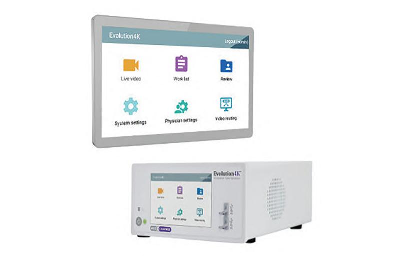
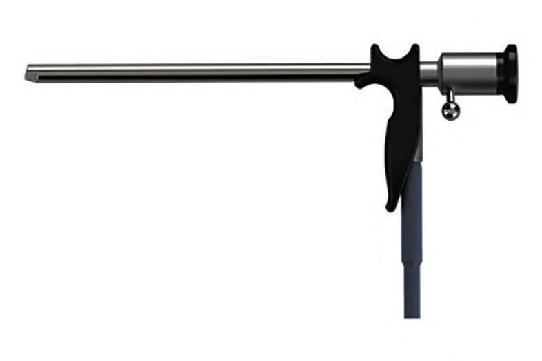
The Evolution4K IP is an all-in-one medical grade 4K video routing and recording solution. Simple, effective and powerful, it enables 4K surgical video routing, patient workflow, streaming video, and hospital sys-
LARYNGOSCOPE XION MEDICAL GMBH
220 221 HMI-09-23 LINKXPRESS COM 18 HospiMedica International August-September/2023 WORLD’S MEDICAL DEVICE MARKETPLACE To receive prompt and free information on products, log on to www.linkXpress.com or scan the QR code on your mobile device
The XION laryngoscope portfolio includes both standard laryngoscopes with fixed magnification and zoom laryngoscopes with two magnification levels. In both levels, the control lever provides easy focusing.
Cont’d from cover
Image: The first four-arm laparoscopic surgical device opens up new possibilities for surgeons (Photo courtesy of EPFL)
New Technology Reduces Bacteria and Deadly Infections in Medical Implants
lated infections lies in inhibiting biofilm formation. The rise of antibiotic-resistant bacterial strains has spurred the need for the development of indiscriminate antibacterial coatings and cutting-edge solutions. Now, a pioneering technology significantly lowers the risk of harmful bacterial biofilm formation on post-operative medical implants without resorting to antibiotics or harmful chemicals.
DeBogy Molecular Inc. (Battle Creek, MI, USA; www.debogy. com) has developed a proprietary technology that can alter molecular surface structures to electrostatically eliminate viruses, bacteria, and fungi upon contact. The DeBogy platform has proven effective across a diverse range of materials, including those used in the medical industry. Recent research has confirmed the efficiency and safety of DeBogy technology in eliminating harmful bacteria that thrive on the surface of post-operative medical implants. This groundbreaking in vivo study revealed that bacterial biofilm in animals with DeBogy-treated implants was reduced by 99.9% compared to a control group, seven days post-surgery. The infection rate in surrounding tissue in animals with DeBogy-treated implants was lowered by 99.8%. Overall, the animals implanted with DeBogy-treated devices were healthier than the control group, displaying noticeable reductions in inflammation, fibrosis, vascularization, and necrosis.
Robotic Systems Allows Surgeons
4-Handed Laparoscopic Procedures
Cont’d from page 18
ic procedures. Even as the system undergoes further testing and refinements, these results confirm its capability to facilitate four-arm surgical tasks without the need for intensive training. The system has already been successfully employed to train specialists, and clinical trials are currently underway.
“Controlling four arms simultaneously, moreover with one’s feet, is far from routine and can be quite tiring,” said Professor Aude Billard, head of the Learning Algorithms and Systems Laboratory (LASA). “To reduce the complexity of the control, the robots actively assist the surgeon by coordinating their movements with the surgeon’s through active prediction of the surgeon’s intent and adaptive visual tracking of laparoscopic instruments with the camera. Additionally, assistance is offered for more accurate grasping of the tissues.”
Quick Healing Bone Scaffold Could Be Game-Changer in Tissue Regeneration
Cont’d from page 18
aiming to reduce the healing time to four weeks and achieve near-immediate tissue regeneration without using endocrine factors and cells as well as investigating potential applications for other tissues.
This research has huge implications. By incorporating stem cells, bioactive components like collagen and gelatin, coatings that encourage native cell migration into the scaffolds, and electromagnetic stimulation, this could potentially accelerate the healing process for soldiers with severe musculoskeletal fractures or civilians with traumatic injuries. These individuals typically face months of hospitalization and a lengthy recovery. Notably, the newly developed scaffold is primarily composed of glass, alginate, and nano silicate, materials already approved by the FDA. This approval significantly reduces regulatory obstacles, allowing the scaffold to be confidently and efficiently used in clinical settings, thereby expediting development and improving patient outcomes.
“The promise of a new antibacterial technology that can fight infection without antibiotics, chemicals, or temporary coatings is truly transformational,” said Wayne Gattinella, CEO of DeBogy Molecular. “The possibilities of the DeBogy platform to improve the quality of life and reduce the cost of care for millions of people are enormous.”
“The DeBogy technology has the potential to revolutionize the way we prevent and treat surgical site infection,” added Dr. Houssam Bouloussa, spine surgeon and cofounder, DeBogy Molecular.
Image: New technology kills dangerous bacteria proliferating on surface of medical implants post surgery (Photo courtesy of DeBogy)

19 HospiMedica International August-September/2023 To view this issue in interactive digital magazine format visit www.HospiMedica.com Surgical Techniques
Cont’d from cover
CRYOSURGICAL SYSTEM

Tumor-Destroying System Uses Smart Needle to Treat Cancer
Cont’d from cover tumors, but patients are often ineligible due to the tumor’s difficult-to-reach location or proximity to critical anatomy, posing high risks of damage to surrounding organs. Now, a precision-guided needle, capable of accurately destroying tumors without affecting surrounding healthy tissue, could enable physicians to precisely destroy pathological tissue while preserving surrounding healthy structures.
Current Surgical Inc. (Washington, DC, USA; www.currentsurgical.com) is a needlebased technology wherein doctors need only position a needle, which subsequently delivers energy to the tumor via thermal ablation to destroy it. The company’s smart surgical needle technology integrates high-resolution sensors and real-time image analysis, offering physicians enhanced control and immediate feedback during tumor destruction. Consequently, surgeons could treat any solid tumor, regardless of its location, enabling millions of patients to benefit from curative surgery without the traditional constraints.
The needle technology incorporates a


Cont’d from cover


novel, miniaturized class of ultrasound tech nology. The micro-machine electromechan ical sensors embedded in the needle offer superior imaging capabilities compared to conventional ultrasound technology. Cur rent Surgical is working on reducing the size of its technology to make it fit within a needle. By automating crucial surgical steps, the company’s technology allows doctors to concentrate on selecting the most effective treatment for their patients. In addition to treating even the most resistant diseases, the technology holds potential for diverse applications, including the treatment of cardiac arrhythmias, hypertension, and other chronic pain sources.
“Technological advancements now allow us to detect cancers earlier and earlier, but the capability to treat those tumors hasn’t improved,” said Current Surgical CEO Al Mashal. “We are bridging this gap through our smart needle technology. We want to give doctors the ability to curatively treat cancers in hardto-reach places — without the harmful, debilitating side effects of other treatment options.”
“We’re excited about this technology not only for its curative potential, but also because of how easy it is to put into the hands of the surgeon,” added Current Surgical CTO Chris Wagner. “Since the technology is integrated into the needle, it doesn’t take a large, expensive operating room to actually help patients.”
Image: The ‘smart’ needle is being designed to treat cancerous tumors (Photo courtesy of Current Surgical)

Gallbladder Cancer Surgery Guidelines
results. Benchmarking is one method for developing precise quality measures. It’s a process of quality improvement where optimal outcomes are identified to serve as a reference point for comparing performance.
For the first time, researchers from Boston University School of Medicine (Boston, MA, USA; www.bumc.bu.edu) and the University of Texas MD Anderson Cancer Center (Houston, TX, USA; www.mdanderson.org) have established a benchmark for GBC surgery. This benchmark could help identify the best treatment hospitals for patients with gallbladder cancer and enhance the overall
quality of care. The team analyzed data from over 900 patients who underwent GBC surgery between 2000 and 2021 at 13 different hospitals across seven countries and four continents. From this data, a benchmark group was chosen. This group included 245 low-risk patients who underwent standard GBC surgery at established centers of excellence, programs that are distinguished by their high levels of expertise and associated resources. This group was used as a standard for comparison.
The researchers used the benchmark group to establish best practice guidelines related to surgical outcomes, such as the du-
ration of surgery, rates of morbidity, severe morbidity, blood loss, transfusion, positive margin, and the number of lymph nodes retrieved. When compared to the non-benchmark group, the benchmark group displayed significantly better overall survival rates.
The researchers believe that the benchmark values they’ve established will enable surgeons and institutions to define the best possible outcomes for GBC patients and facilitate comparisons with other patient groups, thereby enhancing the quality of collaborative research. Their findings were published on May 6, 2023 in the journal Annals of Surgical Oncology.
The Wallach LLCO2 System is a single-trigger cryosurgical system that maximizes temperature and minimizes clogging and sputtering associated with CO2 gas. It is designed for use with medical-grade CO2 gas for optimal performance.
VIDEO PROCESSOR PENTAX
223 224 HMI-09-23 LINKXPRESS COM 20 HospiMedica International August-September/2023 WORLD’S MEDICAL DEVICE MARKETPLACE
The IMAGINA video processor comprises three new endoscopes – one video gastroscope and two video colonoscopies – and supports each step of the clinical pathway, from screening to diagnosis towards dayto-day therapy.
To receive prompt and free information on products, log on to www.linkXpress.com or scan the QR code on your mobile device
MEDICA 2023 to Serve as Showcase for Medtech Start-Ups

The internationally leading medical trade fair MEDICA (Düsseldorf, Germany; www.medica-tradefair.com) has long been recognized as a global platform for health sector startups. At the forthcoming MEDICA 2023, which will be running concurrently with the professional suppliers’ trade fair COMPAMED 2023 from 1316 November, hundreds of new developer teams will be present among more than 5,000 exhibiting companies to seek collaborations related to funding, production, product approval, marketing, and sales. The event boasts a number of program highlights geared towards startups, providing them with an optimal platform for introducing their innovative solutions to the international professional healthcare community. Notable program highlights include the 12th MEDICA Startup Competition, the 15th Healthcare Innovation World Cup, the MEDICA START-UP PARK, and numerous startup exhibitions at the MEDICA CONNECTED HEALTHCARE FORUM.
The MEDICA Startup Competition places emphasis on a wide array of healthcare innovations, ranging from artificial intelligence (AI) and health apps to laboratory diagnostics and medical robotics. This year, “Sustainability” is being introduced as a new category. The 2022 winner of the competition was a Spanish startup, Idoven, which has developed an AI-based platform for cardiology-as-a-service. Idoven’s proprietary AI enhances the accuracy and consistency of electrocardiograph (ECG) interpretations. Their participation in the competition offered the company immense global visibility and attracted around USD 20 million in funding for future projects and developments.
The 15th Healthcare Innovation World Cup could be another interesting opportunity for startups, scale-ups, and smaller mid-level companies. It focuses on smart Internet of Medical Things (IoMT) solutions, such as digital biomarkers, smart band-aids, or wearables with network connectivity. The 12 selected finalists will be invited to showcase their businesses at MEDICA 2023 on the MEDICA CONNECTED HEALTHCARE FORUM stage. Last year’s winner was ViewMind Inc., a company specializing in management of neurodegenerative diseases.
The MEDICA START-UP PARK has become a central hub for startup networking, already attracting a record 40 early registrations this year. A repeat participant is the Ukrainian start-up initiative “Up To Future,” promoting startups such as HandyUsound, which developed a portable ultrasound system. Also participating is Korea’s Megnosis, the creator of EEG helmets designed to detect dementia early and stim-
BD Sells Surgical Instrumentation Platform to STERIS
Becton, Dickinson and Company (BD, Franklin Lakes, NJ, USA; www.bd.com) has signed a definitive agreement to sell its Surgical Instrumentation platform to STERIS (Mentor, OH, USA; www. steris.com) for USD 540 million. The divestiture includes the transfer of V. Mueller, Snowden-Pencer, and Genesis branded products as well as three manufacturing facilities.
STERIS offers a wide range of infection prevention products and other procedural services, mainly aimed at healthcare, pharmaceutical, medical device, and dental clients. The acquisition of BD’s surgical instrumentation platform, which complements STERIS’s Healthcare segment, will empower STERIS to more effectively meet the product and service demands of hospitals and surgery centers. The platform has three dedicated manufacturing sites and about 20,000 SKUs in its portfolio. The divestment of its Surgical Instrumentation platform aligns with the “Simplify” objective of BD’s 2025 strategy and represents a significant move towards streamlining its product portfolio and manufacturing network.
ulate brain cells and neurons. Among German participants is AssistMe, which will introduce a smart sensor system for use in underwear for incontinence at MEDICA 2023.
“I was very surprised at how global this trade fair is!” said Rika Christanto, COO at Idoven, who is pleased with the effect of participating at MEDICA and the competition finals on the stage of the MEDICA CONNECTED HEALTHCARE FORUM.

21 HospiMedica International August-September/2023
To view this issue in interactive digital magazine format visit www.HospiMedica.com Industry News
Image: The final pitches of the MEDICA Start-up COMPETITION have been among the most popular program highlights (Photo courtesy of MEDICA)
2023
SEPTEMBER
6th World Congress on Regional Anaesthesia & Pain Medicine and 40th Annual Congress of the European Society of Regional Anaesthesia and Pain Therapy (ESRA). Sep 6-9; Paris, France; esraworld2023.com
ERS International Congress 2023 – European Respiratory Society. Sep 9-13; Milan, Italy; erscongress.org
EANM 2023 – 36th Annual Congress of the European Association of Nuclear Medicine. Sep 9-13; Vienna, Austria; eanm.org
CIRSE 2023 – Annual Congress of the Cardiovascular and Interventional Radiological Society of Europe. Sep 9-13 Copenhagen, Denmark; cirse.org
Medic East Africa 2023. Sep 13-15; Nairobi, Kenya; mediceastafrica.com
Medical Fair Thailand 2023. Sep 13-15; Bangkok, Thailand; medicalfair-thailand.com
REHACARE 2023 – International Trade Fair for Rehabilitation and Care. Sep 13-16; Dusseldorf, Germany; rehacare.com
EUSEM 2023 – 17th European Emergency Medicine Congress. Sep 17-20; Barcelona, Spain; eusemcongress.org
HIMSS23 APAC Health Conference & Exhibition – Healthcare Information and Management Systems Society. Sep 18-21; Jakarta, Indonesia; himss.org
ExpoMedical 2023. Sep 20-22; Buenos Aires, Argentina; expomedical.com.ar
19th EuGMS Congress – European Geriatric Medicine Society. Sep 20-22 Helsinki, Finland; eugms.org
HospiMedica International
KCR 2023 – 79th Korean Congress of Radiology. Sep 20-23; Seoul, Korea; kcr4u.org
CADI 2023 – Argentinian Congress of Diagnostic Imaging. Sep 21-23; Buenos Aires, Argentina; cadi2023.com.ar
IndoHealthCare 2023. Sep 21-23; Jakarta, Indonesia; indohealthcareexpo.com
Medical Fair Brasil. Sep 26-28; Sao Paulo, Brazil; medicalfair-brasil.com.br
Medic West Africa. Sep 26-28; Lagos, Nigeria; medicwestafrica.com
ESVS 2023 – Annual Meeting of the European Society for Vascular Surgery. Sep 26-29; Belfast, UK; esvs.org
EUSOBI 2023 – Annual Scientific Meeting of the European Society of Breast Imaging. Sep 28-30; Valencia, Spain; eusobi.org
UAA 2023 – 20th Urological Association of Asia Congress. Sep 28 - Oct 1; Dubai, UAE; uaanet.org
ESGO 2023 – 24th European Congress on Gynaecological Oncology. Sep 28 – Oct 1; Istanbul, Turkey; esgo.org
OCTOBER
ASTRO 2023 – 65th Annual Scientific Meeting of the American Society for Radiation Oncology. Oct 1-4; San Diego, CA, USA; astro.org
EASD 2023 – 59th Annual Meeting of the European Association for the Study of Diabetes. Oct 3-6; Hamburg, Germany; easd.org
34th Medicall Expo. Oct 7-9, New Delhi, India; medicall.in
MSK 2023 – Joint Meeting of Global Muscoloskeletal Societies. Oct 8-13; London, UK; internationalskeletalsociety.com
MICCAI 2023 – 26th International Conference on Medical Image Computing and Computer Assisted Intervention. Oct 8-12; Vancouver, BC, Canada; miccai.org
ACEP23 – Scientific Assembly of the American College of Emergency Physicians. Oct 9-12; Philadelphia, PA, USA; acep.org
15th World Stroke Congress – World Stroke Organization. Oct 10-12; Toronto, ON, Canada; worldstrokecongress.org
Medical Japan 2023 Tokyo– International Medical and Elderly Care Expo. Oct 11-13; Tokyo, Japan; medical-jpn.jp
CBR23 – 52nd Congress of the Brazilian College of Radiology and Diagnostic Imaging. Oct 12-14; Rio de Janeiro, Brazil; cbr.org.br
JFR 2023 - Journées Francophones de Radiologie. Oct 13-16; Paris, France; jfr.radiologie.fr
ANESTHESIOLOGY 2023 – Annual Meeting of the American Society of Anesthesiologists. Oct 13-17; San Francisco, CA, USA; asahq.org
UEG Week 2023 – United European Gastroenterology. Oct 14-17; Copenhagen, Denmark; ueg.eu/week
ISUOG World Congress 2023 - International Society of Ultrasound in Obstetrics & Gynecology. Oct 16-19; Seoul, Korea; isuog.org
Africa Health 2023. Oct 17-19; Johannesburg, South Africa; africahealthexhibition. com
RANZCR 2023 – Annual Scientific Meeting of The Royal Australian and New Zealand College of Radiologists. Oct 19-21; Brisbane, Australia; ranzcr.com
ESMO Congress 2023 - European Society for Medical Oncology. Oct 20-24; Madrid, Spain; esmo.org
ECISM LIVES 2023 – Annual Congress of European Society of Intensive Care Medicine. Oct 21-25; Milan, Italy; esicm.org
ESSO 42 – 42nd Congress of the European Society of Surgical Oncology. Oct 25-27; Florence, Italy; esso42.org
46th World Hospital Congress of the International Hospital Federation (IHF). Oct 25-27; Lisbon, Portugal; worldhospitalcongress.org
ESCR ESTI Joint Meeting 2023 – European Societies of Cardiovascular Radiology and Thoracic Imaging. Oct 26-28; Berlin, Germany; escr.org
CMEF Autumn 2023 – China International Medical Equipment Fair. Oct 28-31; Shenzhen, China; cmef.com.cn
Global Health Exhibition 2023. Oct 29-31; Riyadh, Saudi Arabia; globalhealthsaudi.com
NOVEMBER
WUFMB Congress 2023 – World Federation for Ultrasound in Medicine and Biology. Nov 4-7; Muscat, Oman; wfumb.info
BSIR 2023 – Annual Scientific Meeting of the British Society of Interventional Radiology. Nov 8-10; Newport, UK; bsirmeeting.org
MEDICA 2023. Nov 13-16; Dusseldorf, Germany; medica-tradefair.com
APSR 2023 – 27th Congress of the Asian Pacific Society of Respirology. Nov 16-19; Singapore; apsr2023.sg
ASUS 2023 – 6th Congress of the Asian Surgical Ultrasound Society. Nov 18-20; Seoul, Korea; asus2023.org
CBMI 2023 – 28th Brazilian Congress of Intensive Care Medicine. Nov 23-25; Florianopolis, Brazil; amib.org.br
RSNA 2023 – Annual Meeting of the Radiological Society of North America. Nov 26-30; Chicago, IL, USA; rsna.org
DECEMBER
ESEM23 - Emirates Society Emergency of Medicine Conference 2023. Dec 6-9; Dubai, UAE; esemconference.ae
35th Medicall Expo. Dec 8-10, Kolkata, India; medicall.in
65th ASH Annual Meeting and Exposition –American Society of Hematology. Dec 9-12; San Diego, CA, USA; hematology.org
International Calendar Provided as a service to advertisers. Publisher cannot accept responsibility for any errors or omissions. Advertising Index Inq No . Advertiser Page – AOCR 2024 . . . . . . . . . . . . . . . . . . . . . . . . . . 21 – ECR 2024 3 – HospiMedica 23 – HospiMedica EXPO . . . . . . . . . . . . . . . . . . . . . 9 124 Parker . . . . . . . . . . . . . . . . . . . . . . . . . . . . . . . 24 107 Radcal 7 115 Singuway . . . . . . . . . . . . . . . . . . . . . . . . . . . . 15 105 Sun Nuclear . . . . . . . . . . . . . . . . . . . . . . . . . . . 5 113 UNDIS 13 117 Vicolab . . . . . . . . . . . . . . . . . . . . . . . . . . . . . . 17 102 Werfen . . . . . . . . . . . . . . . . . . . . . . . . . . . . . . . 2 111 West Medica 11 – WFUMB 2023 . . . . . . . . . . . . . . . . . . . . . . . . 19 Vol 41 Issue 3 9/2023
For a free listing of your event, or a paid advertisement in this section, contact: International Calendar, HospiMedica International E-mail: info@globetech.net ATTENTION: IF YOUR APPLICATION IS NOT RECEIVED AT LEAST ONCE EVERY 12 MONTHS YOUR FREE SUBSCRIPTION MAY BE AUTOMATICALLY DISCONTINUED READER SERVICE PORTAL LINKXPRESS COM ® Every advertisement or product item in this issue contains a LinkXpress number as shown below: 999 HMI-09-23 LINKXPRESS COM Identify LinkXpress codes of interest as you read magazine Click on LinkXpress.com to reach reader service portal Mark code(s) of interest on LinkXpress inquiry matrix 1 2 3 Renew/ S tart Your Free Subscription Access Instant Online Product Information VISIT
JANUARY
2024
Medical Japan 2024 Osaka– International Medical and Elderly Care Expo. Jan 17-19; Osaka, Japan; medical-jpn.jp
Critical Care Congress 2024 – 53rd Annual Meeting of the Society of Critical Care Medicine (SCCM). Jan 21-24; Phoenix, AZ, USA; sccm.org
ISET 2024 – International Symposium on Endovascular Therapy. Jan 22-25; Miami Beach, FL, USA; iset.org
IRA 2024 – 76th Annual Conference of the Indian Radiological and Imaging Association. Jan 25-28; Vijayawada, India; iria2024.com
Arab Health 2024. Jan 29 - Feb 1; Dubai, UAE; arabhealthonline.com

FEBRUARY
AAOS 2024 Annual Meeting – American Academy of Orthopaedic Surgeons. Feb 12-16; San Francisco, CA, USA; aaos.org
36th Medicall Expo. Feb 16-18; Mumbai, India; medicall.in
ECR 2024 – European Congress of Radiology. Feb 28 - Mar 3; Vienna, Austria; myesr.org
MARCH
CRITICARE 2024 – 30th Annual Conference of the Indian Society of Critical Care Medicine (ISCCM). Mar 1-3; Kolkata, India; isccm.org
WCA 2024 – 18th World Congress of Anaesthesiologists. Mar 3-7; Singapore; wca2024.org
5th MedExpo Ethiopia 2024. Mar 6-8; Addis Ababa, Ethiopia; expogr.com/ethiopia/medexpo
EMIM 2024 – 19th European Molecular Imaging Meeting. Mar 12-15; Porto, Portugal; e-smi.eu Medical Fair India 2024. Mar 13-15; Mumbai, India; medicalfair-india.com
51st Annual Meeting of the Japanese Society of Intensive Care Medicine (JSICM). Mar 14-16; Sapporo, Japan; jsicm.org
KIMES 2024 – Korea International Medical & Hospital Equipment Show. Mar 14-17; Seoul, Korea; kimes.kr
SALMED International Medical Fair 2024. Mar 19-21; Poznan, Poland; salmed.pl
43rd ISICEM – International Symposium on Intensive Care & Emergency Medicine. Mar 1922; Brussels, Belgium; isicem.org
AOCR 2024 – Asian Oceanian Congress of Radiology. Mar 22-25; Taipei, Taiwan; aocr2024.org
SIR 2024 – Annual Meeting of the Society of Interventional Radiology. Mar 23-28; Salt Lake City, UT, USA; sirmeeting.org
APRIL
144th Annual Meeting of the American Surgical Association (ASA). Apr 4-6; Washington, DC, USA; meeting.americansurgical.org
39th Annual EAU Congress – European Association of Urology. Apr 4-7; Paris, France; uroweb.org
UltraCon 2024 – Annual Conference of the American Institute of Ultrasound in Medicine (AIUM). Apr 5-10; Austin, TX, USA; aium.org
37th Medicall Expo. Apr 5-7; Hyderabad, India; medicall.in
ACC.24 – American College of Cardiology’s 73rd Annual Scientific Session & Expo. Apr 6-8; Atlanta, GA, USA; accscientificsession.acc.org
83rd Annual Meeting of Japan Radiological Society (JRS). Apr 11-14; Yokohama, Japan; radiology.jp
WCO-IOF-ESCEO 2024 - World Congress on Osteoporosis, Osteoarthritis and Musculoskeletal Diseases. Apr 11-14; London, UK; wco-iof-esceo.org
WCN 2024 – World Congress of the International Society of Nephrology (ISN). Apr 13-16; Buenos Aires, Argentina; theisn.org/wcn
AAN 2024 – 76th Annual Meeting of the American Academy of Neurology. Apr 13-18; Denver, CO, USA; aan.com
SEACare 2024 – 24th Southeast Asian Healthcare & Pharma Show. Apr 17-19; Kuala Lumpur; Malaysia; abcex.com
24th MEDEXPO Africa 2024. Apr 17-19; Nairobi,

Kenya; expogr.com/kenyamed
SAGES 2024 – Annual Meeting of the Society of American Gastrointestinal and Endoscopic Surgeons. Apr 17-20; Cleveland, OH, USA; sages.org
ATS 2024 – International Conference of the American Thoracic Society. May 17-22; San Diego, CA, USA; conference.thoracic.org
124th Congress of the Japan Surgical Society (JSS). Apr 18-20; Tokoname, Japan; jp.jssoc.or.jp
DCK 2024 – 141st Congress of the German Society for Surgery (DGCH). Apr 24-26; Leipzig; Germany; dgch.de
Expomed Euroasia 2024. Apr 25-27; Istanbul, Turkey; expomedistanbul.com
AAEM24 – 30th Annual Scientific Assembly of the American Academy of Emergency Medicine. Apr 27 - May 1; Austin, TX, USA; aaem.org


ECTES 2024 – 23rd Congress of the European Society for Trauma & Emergency Surgery (ESTES). Apr 28-30; Lisbon, Portugal; estes-congress.org
ECIO 2024 – European Conference on Interventional Oncology. Apr 28 - May 1; Palma de Mallorca, Spain; ecio.org
MAY
APSCVIR 2024 – 18th Annual Meeting of the Asia Pacific Society of Cardiovascular and Interventional Radiology. May 3-5; Bangkok, Thailand; apscvir.com
AUA 2024 – Annual Meeting of the American
Urological Association. May 3-6; San Antonio, TX, USA; auanet.org
ESTRO 2024 – Annual Congress of the European Society of Radiology & Oncology. May 3-7; Glasgow, UK; estro.org
2024 ISMRM & ISMRT Annual Meeting & Exhibition – International Society for Magnetic Resonance in Medicine. May 4-9; Singapore; ismrm.org
ARRS 2024 Annual Meeting – American Roentgen Ray Society. May 5-9; Boston, MA, USA; arrs.org
Heart Failure 2024 – World Congress on Acute Heart Failure. May 11-14; Lisbon, Portugal; escardio.org
KIHE 2024 – 29th Kazakhstan International Healthcare Exhibition. May 15-17; Almaty, Kazakhstan; kihe.kz

Hospitalar 2024. May 21-24; Sao Paulo, Brazil; hospitalar.com
61st ERA Congress – European Renal Association. May 23-26; Stockholm, Sweden; era-online.org
EuroAnaesthesia 2024 – European Society of Anaesthesiology. May 25-27; Munich, Germany; euroanaesthesia.org
92nd EAS Congress 2024 – European Atherosclerosis Society. May 26-29; Lyon, France; eas-society.org
EFORT Congress 2024 – 25th Annual Congress of European Federation of National Associa-


tions of Orthopaedics and Traumatology. May 29-31; Hamburg, Germany; congress.efort.org
JUNE
CARS 2024 – Computer Assisted Radiology and Surgery. Jun 18-21; Barcelona, Spain; cars-int.org
FIME 2024 – Florida International Medical Expo. Jun 19-21; Miami, FL, USA; fimeshow.com

ICEM 2024 – 22nd International Conference on Emergency Medicine. Jun 19-23; Taipei. Taiwan; ifem.cc
SIIM 2024 – Annual Meeting of the Society for Imaging Informatics in Medicine. Jun 27-29; National Harbor, MD, USA; siim.org
JULY
ESHRE 2024 – 40th Annual Meeting of the European Society of Human Reproduction and Embryology. Jul 7-10; Amsterdam, Netherlands; eshre.eu Meditech 2024 – 8th International Health Fair. Jul 9-12; Bogota, Colombia; feriameditech.com Asia Health 2024. Jul 10-12; Bangkok, Thailand; medlabasia.com
SCCT 2024 – 19th Annual Scientific Meeting of the Society of Cardiovascular Computed Tomography. Jul 18-21; Washington, DC, USA; scct.org
AUGUST
International Surgical Week 2024 – 50th World Congress of the International Society of Surgery ISS/SIC. Aug 25-29; Kuala Lumpur, Malaysia; isw2024.org
International Calendar
23 HospiMedica International August-September/2023 PREMIER MULTIMEDIA
SERVING THE WORLD’S HOSPITAL / MEDICAL COMMUNITY Anytime, Anywhere, On the Go... PRINT MAGAZINE INTERACTIVE DIGITAL EDITION WEB PORTAL ENGLISH • SPANISH MOBILE VERSION PRINT MAGAZINE • INTERACTIVE DIGITAL EDITION WEB PORTAL • MOBILE VERSION • MOBILE APPS HOSPIMEDICA EXPO • E-NEWSLETTER E-MAIL MARKETING • ONLINE SOLUTIONS DAILY CLINICAL NEWS .com
PLATFORM


124 HMI-09-23 LINKXPRESS COM


 64-SLICE CT SCANNER NEUSOFT MEDICAL SYSTEMS
The NeuViz 16 Classic is a 64-slice CT platform featuring a powerful tube design enabling continuous scanning for longer periods. The efficient design of its 32-row detector results in a high X-ray conversion
HANDHELD POC ULTRASOUND LANDWIND MEDICAL


