Prof. Dr. Dr. h.c.
Roger Stephan
Dean & Head of the Institute for Food Safety and Hygiene

Prof. Dr. Dr. h.c.
Roger Stephan
Dean & Head of the Institute for Food Safety and Hygiene
I am delighted to introduce our Vetsuisse Faculty Report covering the years 2023 and 2024.
Interdisciplinary research plays a pivotal role in effectively addressing the challenges of our time. It promotes the exchange of knowledge and methods across disciplines, thereby expanding the boundaries of thought and action. Within the faculty, it promotes innovation by encouraging departments to collaborate on complex questions. However, to realize our full potential we must take a step beyond faculty boundaries.
Breaking away from traditional “research silos” is crucial. Such silos limit perspectives and often hinder the development of holistic solutions. When disciplines and institutions collaborate strategically, synergies emerge where the whole becomes greater than the sum of its parts.
Combining different approaches and perspectives reveals insights that isolated research cannot. It also creates a space for creativity and innovation, where societal, technological, and ecological challenges can be addressed in an interdisciplinary manner.
In this report, you will find inspiring stories about interdisciplinary research at our faculty and much more.
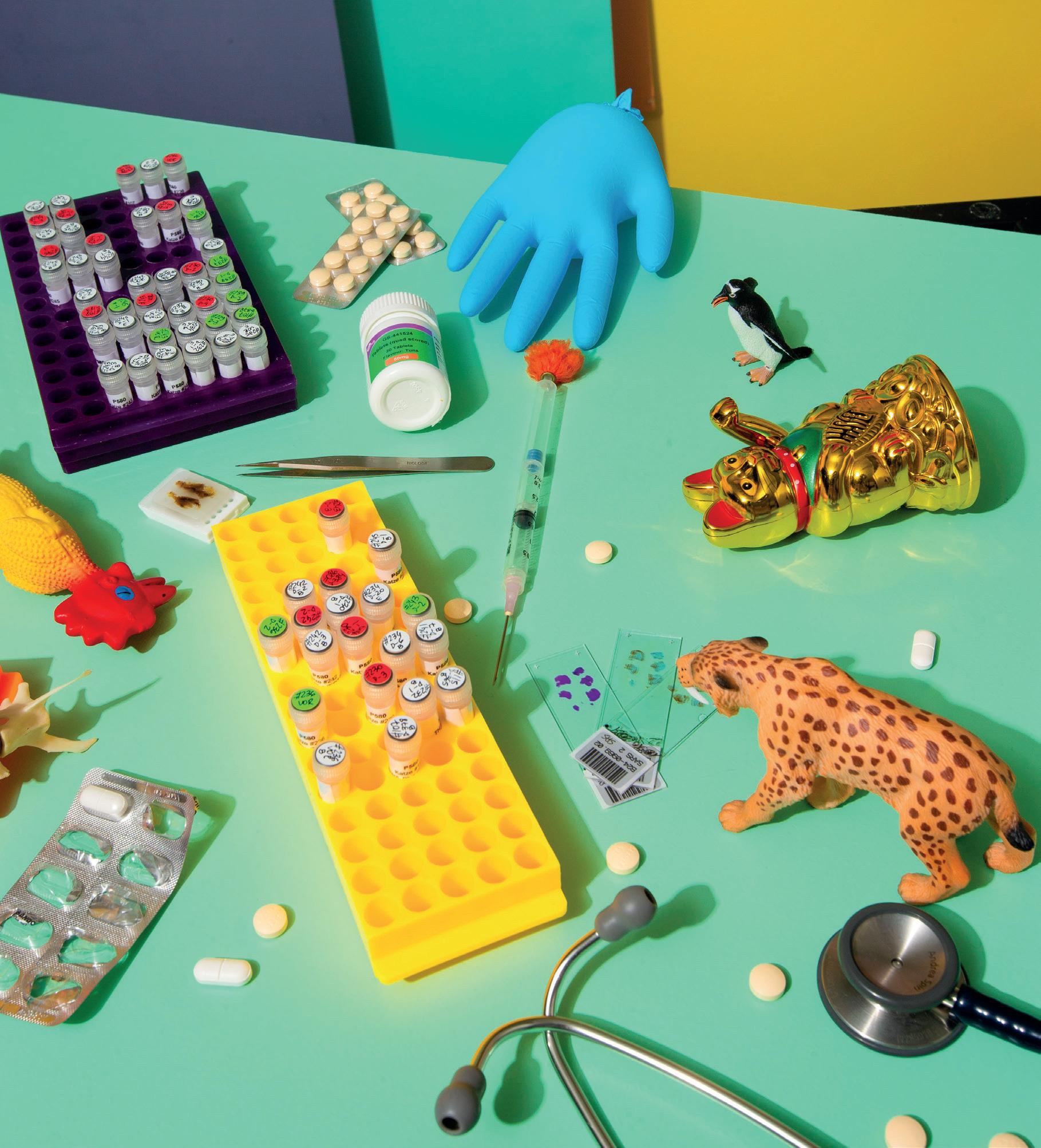
Taking the form of playful still lifes, the visuals throughout this report illustrate that an interdisciplinary approach is often needed to forge new research paths. Created by photographer Michelle Aimée Oesch, the images show how the unexpected can take root when different approaches are combined.
I hope you enjoy reading our report.
I would like to extend my gratitude to the faculty, staff, and students for all their efforts over the last two years!

Very best wishes,
Roger Stephan Dean & Head of the Institute for Food Safety and Hygiene
Management
Management and Secretariat
Human Resources
Controlling
Office of Student Affairs
E-Learning
Facility Management
Vetcom
IT Unit
Support
Finance ICT
Procurement
Reception, CRM & Marketing
Digital Communication
Care
Animal care small animals
Animal care large animals
Institute of Veterinary Anatomy
Department of Molecular Mechanisms of Disease
Musculoskeletal Research Unit (MSRU)
CABMM
Institute of Veterinary Physiology
Institute of Veterinary Pharmacology and Toxicology
Institute of Laboratory Animal Science
Section of Immunology
Institute for Food Safety and Hygiene
Veterinary Bacteriology
Poultry and Rabbit Diseases
Institute of Parasitology
Institute of Veterinary Pathology
Institute of Virology
Institute of Animal Nutrition and Dietetics
Section of Epidemiology
Clinic for Small Animal Internal Medicine
Clinic for Small Animal Surgery
Zoo Animals, Exotic Pets and Wildlife Hospital
Clinic for Radio-Oncology and Medical Oncology
Emergency Medicine Section
Neurology and Neurosurgery Section
Dermatology Section Clinic for Ruminants Clinic of Reproductive Medicine
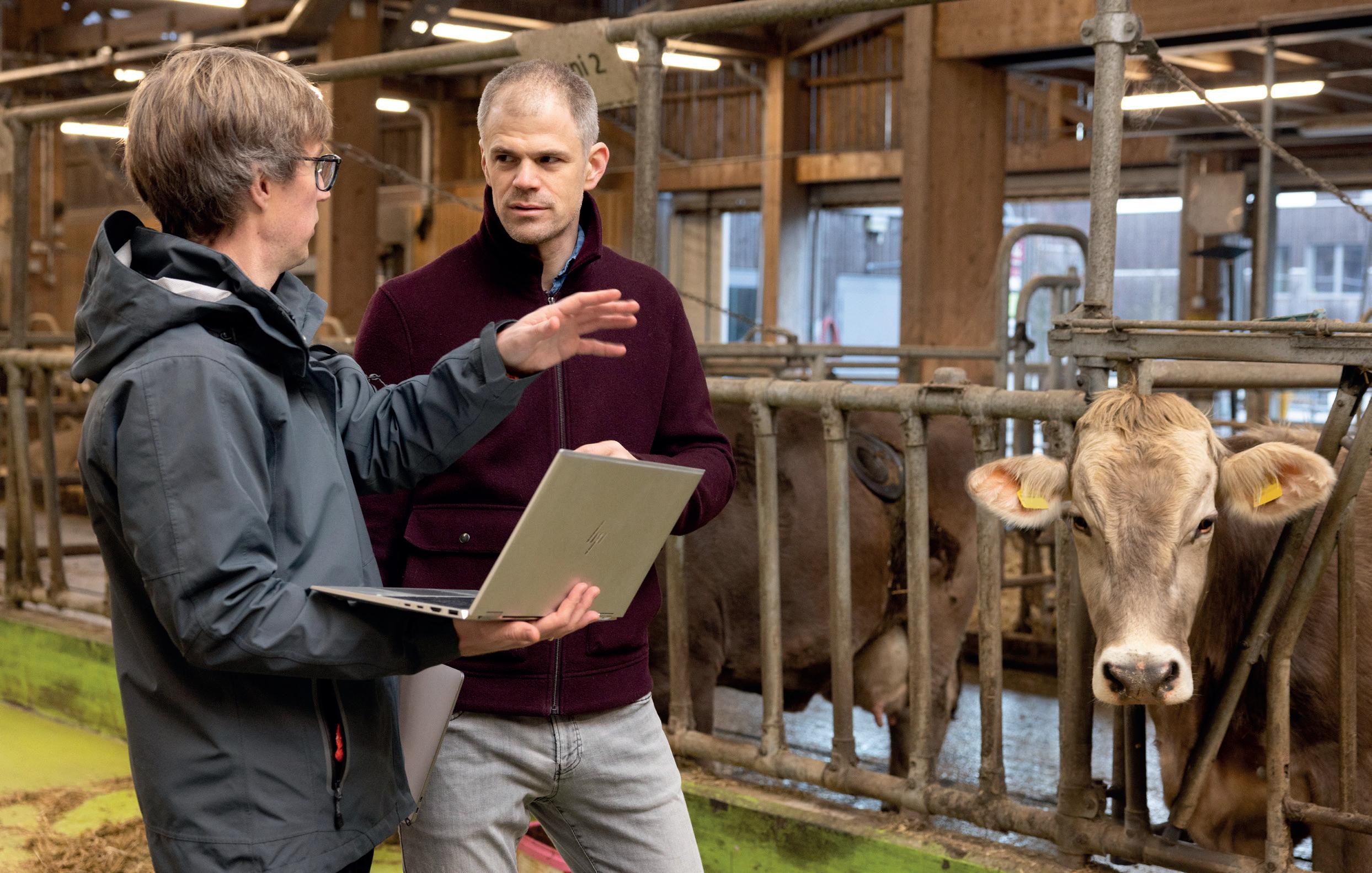
Farm Animal Field Service and Herd Medicine
Pig Medicine Section
Clinic for Equine Internal Medicine
Clinic for Equine Surgery
Sports Medicine Section
Anaesthesiology Section
Clinical Laboratory
Pharmacy
Ophthalmology Section Clinic for Diagnostic Imaging
28,741
Appointed and Advanced Professors 2023/2024
Manuela Schnyder
Associate professor ad personam for Veterinary Parasitology as of August 1, 2023
Alessio Vigani
Assistant professor for Emergency and Intensive Medicine and Extracorporeal Therapy in Small Animals as of October 1, 2023
Regula Bettschart-Wolfensberger
Full professor ad personam for Veterinary Aneasthesiology as of May 1, 2024
Ulrich Bleul
Full professor ad personam for Reproductive Medicine with a focus on Farm Animals as of May 1, 2024
Marcus Clauss
Full professor ad personam for Comparative Digestive Physiology, Nutrition and Biology of Zoo Animals, Pets and Wildlife as of May 1, 2024
International Awards 2023
The Faculty of Veterinary Medicine of Ghent University, Belgium, awarded Prof. Marcus Clauss the Sarton Medal 2023 for contributions to the history and philosophy of science.
The University of Agricultural Sciences and Veterinary Medicine of Cluj-Napoca, Romania, awarded PD Dr. sc. Dennis C. Turner an honorary doctorate, Dr.h.c.
The Institute of Medical Illustrators (IMI) awarded the work of our scientific photographer Michelle Aimée Oesch with two Gold, one Silver, and two Bronze medals.
Patricia Beer, PhD student at the Clinic for Small Animal Surgery, received the VÖK-prize of the
International Awards 2024
Maya Kummrow, attending physician and Co-director of the Clinic for Zoo Animals, Exotic Pets and Wildlife, was appointed president of the European Association of Zoo and Wildlife Veterinarians (EAZWV).
Patrick Kircher
Full professor for Diagnostic Imaging as of May 1, 2024
Salomé LeibundGut-Landmann
Full professor ad personam for Immunology as of May 1, 2024
Urs Meyer
Full professor for Veterinary Pharmacology as of May 1, 2024
Carla Rohrer Bley
Full professor for Clinical and Translational Oncology as of May 1, 2024
Paul Torgerson
Full professor for Veterinary Epidemiology as of May 1, 2024
Thomas Van Boeckel
Associate professor for One Health with a focus on Epidemiology as of August 1, 2024
Austrian Small Animal Veterinary Medicine Association (Vereinigung Österreichischer Kleintiermediziner VÖK).
Sandra Felten, attending physician at the Clinic for Small Animal Surgery, received the incentive award of the German Academy of Animal Health (Akademie für Tiergesundheit).
Mirja Nolff, privatdozent and chief attending physician soft tissue and oncological surgery at the Clinic for Small Animal Surgery, received the incentive award of the German Veterinary Medical Society (Deutsche Veterinärmedizinische Gesellschaft).
Roger Stephan, professor for Food Safety and Hygiene, was welcomed as a member of Leopoldina, the oldest existing academy of science in the world.
The Vetsuisse Faculty of the University of Zurich awarded an honorary doctorate to Dr. Monique Eloit. The award honors a dedicated scientist who has developed a sustainable health management system in the field of veterinary science and who is strongly committed to the prevention and control of epidemic animal diseases and zoonoses – for the benefit of animals, but also of humans.
The Vetsuisse Faculty awarded the Walter Frei Prize to Prof. Dr. Frank Møller Aarestrup for his research, which has contributed significantly to international standards for the detection and monitoring of antibiotic resistance and has had a major influence on the way antimicrobial agents are used worldwide.
The faculty further awarded the degree of honorary doctor to Dr. Polly Taylor in merit of her significant contribution to veterinary anaesthesiology and analgesia research, education, and animal welfare worldwide.
Each year, the Vetsuisse graduates elect their “best coaches”, giving recognition to those individuals that stood out for their teaching commitment during the students’ clinical rotational year. Veterinarians and animal caretakers from all clinics and the Institute of Veterinary Pathology are eligible. The award is sponsored by the company Virbac.
Virbac Best Coach and Carer Awards 2023
Farm Animal Clinic: Sascha Mark, Farm Animal Care, Gianna Räschle, Farm Animal Medicine
Equine Hospital: Armin Simmel, Equine Care, Carlotta Valetti, Equine Surgery
Virbac Best Coach and Carer Awards 2024
Farm Animal Clinic: Sascha Mark, Care, Sybilla Rzaca, Medicine
Equine Hospital: Moritz Linus Schiegg, Care, Elias Quiroz, Medicine
Small Animal Clinic: Jonas Zürrer, Veterinary Medical Practice Assistant, Pavlos Natsios, Medicine
The teacher of the year is designated by the Vetsuisse students from the first to the fourth year. We congratulate:
Annual Prize UZH
Dr. sc. nat. Erin Beebe received the University of Zurich’s 2023 Annual Prize for her dissertation “Comparative Multimodal Molecular Characterisation of Human and Canine Tumours and their Microenvironment”
Dr. med. vet. Kira Dassler received the 2024 Annual for her dissertation “Antimicrobial resistance: dissemination, prevention and control at the human-companion animal intersect”
Semester Prizes UZH
Ranja Steinhauer received a UZH Semester Prize for her master’s thesis “Evaluation of dried blood spot testing for serological monitoring of epizootic and zoonotic pathogens in wild boar”.
Natalie Miller-Collmann received a UZH Semester Prize for her master’s thesis “Prevalence of
Small Animal Clinic: Simone Euler, Small Animal Care, Claudia Kümmerle, Small Animal Medicine
Clinical Diagnostics and Services and Veterinary Pathology: Peter Hofer, Imaging, Eva Dervas, Pathobiology
Veterinary Pathology: Eva Dervas
Specialist/Clinical Diagnostics and Services: Vanessa Heitzmann
2023: Dr. med. vet. Frauke Seehusen, privatdozent, Veterinary Pathology
2024: Dr. med. vet. Hanna Marti, Veterinary Pathology
anatomical variations of the 6th and 7 th cervical vertebrae in clinically healthy horses”.
Daphne Zubler received a UZH Semester Prize for her master’s thesis “Detection of Chlamydia suis in pig manure using real-time PCR”.
Dr. rer. nat. Vincent Spegg received the 2023 Young Scientist Paper Award.
Dr. sc. nat. Martina Panatta received the 2024 Young Scientist Paper Award.
The team of the small and large animal receptions received the Vetsuisse Faculty Effort Award 2023.
The Vetsuisse Band received the Effort Award 2024.
Graduation Ceremony 2023
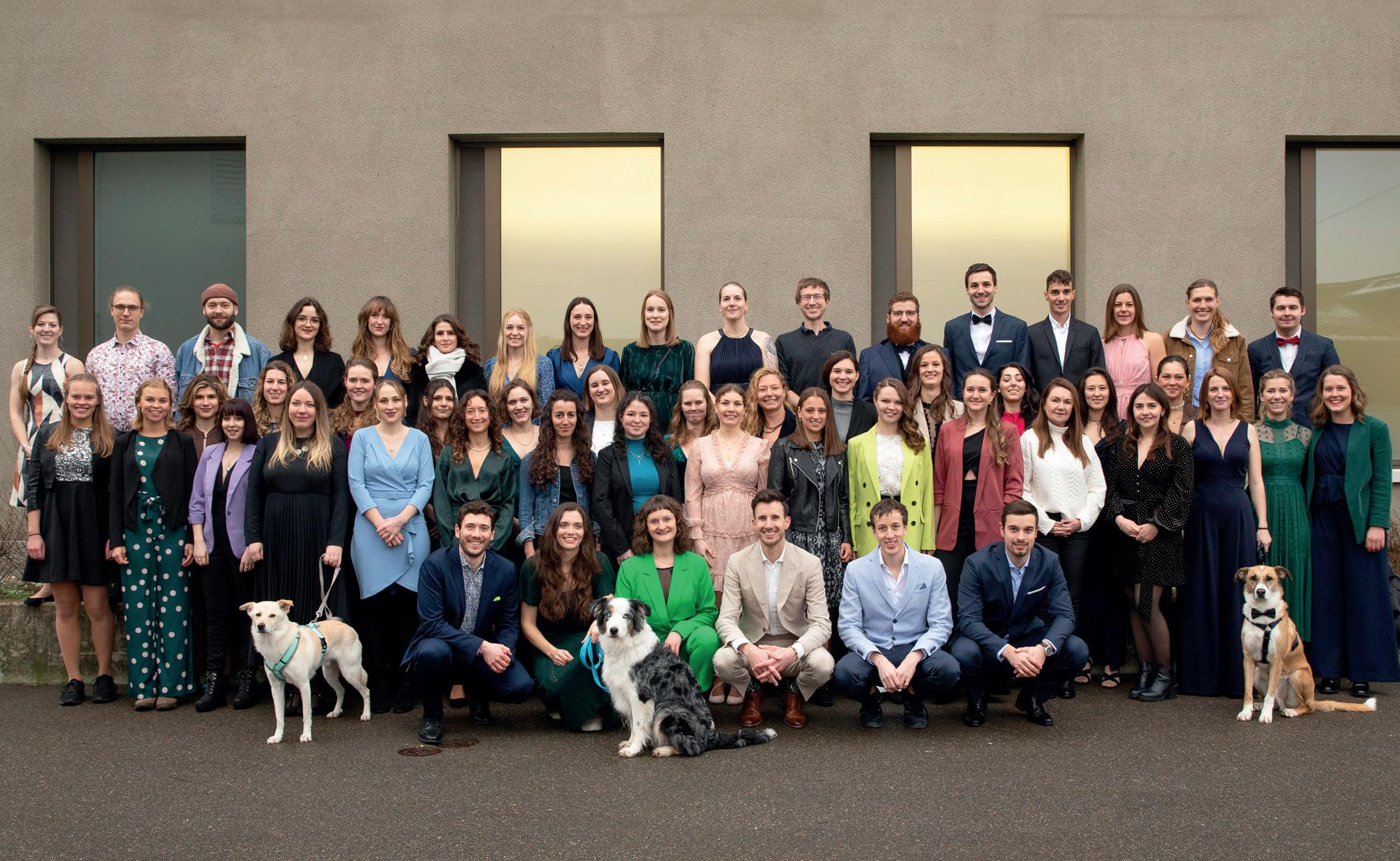

Another 60 new vets received their diplomas from the Vetsuisse Faculty at a celebratory event on April 18, 2024.
Page 24: When veterinarians and paleontologists join forces to unearth ancient clues to the health of our pets.

Page 28: How a professor of veterinary pathology is contributing to Long COVID research.
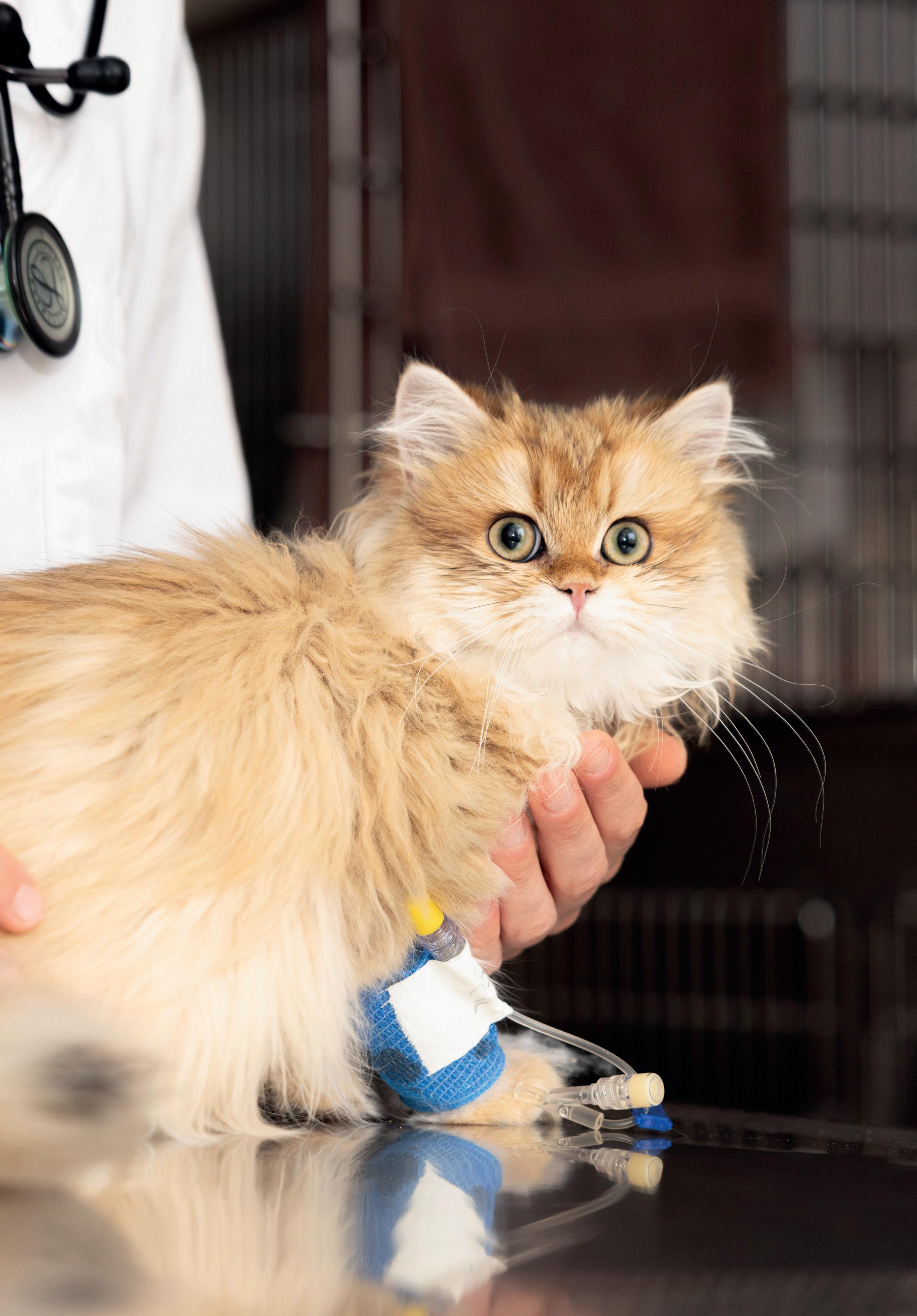
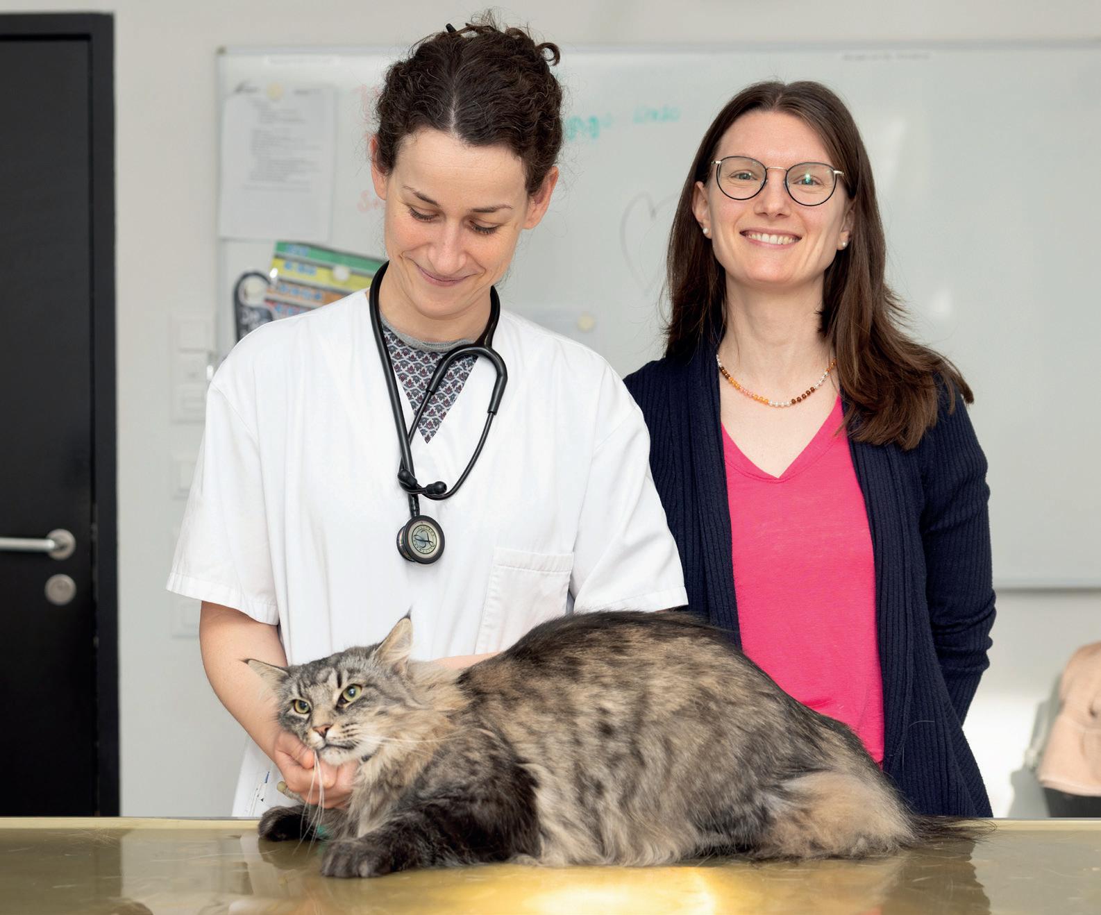
Feline Infectious Peritonitis (FIP) is often a death sentence for cats, with no approved treatment available in most European countries. A clinical study at the Vetsuisse Faculty Zurich is changing that. With a 90 percent success rate, it offers new hope to cat owners and veterinarians alike. Its findings not only improve treatment but also contribute to more precise medical prognoses for FIP. Text by Santina Russo
“It won’t be long before the anesthetic takes effect,” says Dr. med. vet Solène Meunier, attending clinician at the University Animal Hospital of the Vetsuisse Faculty. For now, Luna, a cute six-month-old British Longhair kitten, is awake and sprightly. But her rapid breathing is not just due to excitement: she has a liquid effusion in her chest, which exerts pressure on her left lung, preventing it from filling with air. That is why she needs a needle decompression in her thorax.
Fluid accumulation in the chest or abdomen are typical outcomes of Feline Infectious Peritonitis (FIP), the severe disease affecting Luna. FIP is fatal: untreated, cats die within days or weeks. The disease is caused by the feline
“ Only with these collaborations can we pursue our goals: to improve FIP diagnosis, monitor possible long-term side effects of the treatment and relapses, and establish reliable medical prognoses for affected cats.”
Andrea Spiri, Researcher, Clinical Laboratory, Vetsuisse Faculty, University of Zurich
coronavirus, which is transmitted through contact with feces.
A treatment hard to come by
Although the virus is widespread, thankfully only five to ten percent of infected cats develop the disease. “In feline cells, the virus can mutate into variants, which trigger the disease,” Meunier explains. While a few drugs are known to be effective against FIP, one in particular is known to cure affected cats: a compound named GS-441524 — a preform of the drug Remdesivir, which was developed for humans to treat Marburg virus disease and Ebola, and is also used against COVID-19.
However, the U.S. manufacturer holding the drug’s patent, Gilead Sciences, has no interest in initiating its approval for veterinary medicine. As a result, in most European countries, GS-441524 can only be administered through clinical studies. In Switzerland, two clinical studies are underway: one recently began at the Vetsuisse Faculty in Bern, while the other is being conducted by the Clinical Laboratory and Clinic for Small Animal Internal Medicine at the Vetsuisse Faculty Zurich. “Veterinarians tell us that they are extremely pleased that they can now offer a legal treatment option to patients and their owners through the
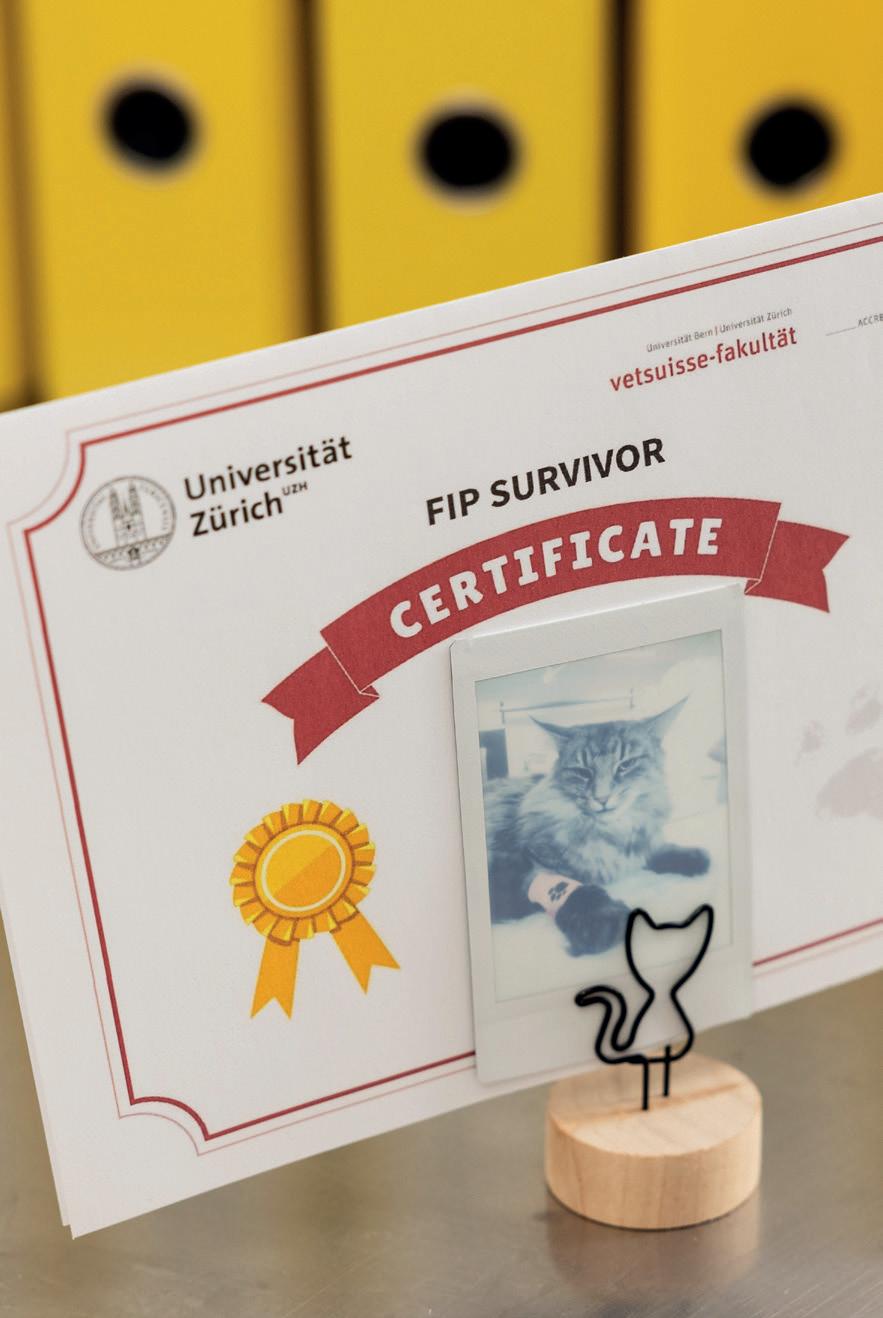
clinical study,” she says. By the end of 2024, the study had been running for two years and had saved the lives of over 160 cats.
One of them is Luna, who is now sedated and ready for the minor procedure on her thorax. Meunier places an ultrasound probe to Luna’s chest. “This is the heart, here is the liver and that’s the lung,” she says, pointing out the structures on the screen. But right next to the lung there is a large pitch-black area. ”This is the liquid we need to remove and send to the laboratory for immediate analysis.”
The veterinarian carefully inserts the syringe into the little cat’s chest and gently pulls out the plunger, extracting orange-yellow liquid. The procedure is repeated three times until the black area on the screen has disappeared — the lung can fill with air again. Which also means that Luna will be well enough to return home today. It is day five of her treatment with GS-441524, and her owners will continue to administer the medication as pills for a total of six weeks.
“One of the first findings of our clinical study was that a six-week treatment period is sufficient for all forms of FIP,” recounts Dr. sc. med. vet. Andrea Spiri, the researcher at the Vetsuisse Zurich Clinical Laboratory responsible for the laboratory part of the study. Earlier clinical studies from collaborators at the University of Munich had successfully reduced the previous 12-week treatment regimen to six weeks, but only in cats with fluid accumulation,
“Veterinarians are extremely pleased that they can now offer a legal treatment option to patients and their owners through the clinical study.”
Solène Meunier, attending physician, Clinical Infectiology and Preventive Healthcare, Vetsuisse Faculty, University of Zurich
which is one of several forms of FIP. “However, it quickly became clear that a six-week treatment has the same positive effect on all forms of FIP,” says Spiri. The shorter duration lowers costs and improves the cats’ quality of life. “The drug needs to be administered once a day at a specific time, so we recommend keeping outdoor cats indoors during the treatment period, and this can be very stressful for them.”
The study’s laboratory work includes tests to support FIP diagnosis and the analysis of samples from the cats in the study during and after treatment. Through these analyses the researchers can provide feedback on the course of the disease, determine prognostic markers and develop new diagnostic options. One such newly developed method is then used on Luna’s liquid to confirm the FIP diagnosis and to monitor the disease outcome.
Another of the study’s findings concerns co-infections. “Now that FIP is treatable, it is important to determine if and how FIP treatment influences — or is influenced by — other diseases,” Spiri explains. Indeed, diagnostic tests
on 100 of the study cats revealed that co-infections are common. Almost a third of the animals were co-infected with the feline calicivirus that causes painful oral ulcerations, for instance. The researchers observed that the cats carrying such co-infections responded to FIP treatment just as positively as the other cats.
An incredible success story
Overall, the study achieves a 90 percent success rate across all forms of FIP. By the end of 2024, the study had included 176 cats. “Most of the few study cats that died already had severe organ damage — in these cases, help came too late,” says Spiri. Others succumbed to non-FIP related conditions. According to the researcher, an important factor for success is the close clinical monitoring during and for two years after the FIP treatment, and the treatment of any concomitant illnesses. A key factor is the close interdisciplinary collaboration between the laboratory specialists and the clinicians. Infectiologists like Solène Meunier examine the cats and are in constant contact with their owners, whereas the lab specialists provide precise and fast diag-
“ Now that FIP is treatable, it is important to determine if and how FIP treatment influences — or is influenced by — other diseases.”
Andrea Spiri, Clinical Laboratory, Vetsuisse Faculty, University of Zurich
nostics to complement the clinical findings. “The rapid feedback from the laboratory on diagnosis and treatment response is essential for the study,” Spiri points out. Additionally, neurologists and ophthalmologists assist when cats exhibit neurological or ocular signs, which occur in some forms of FIP. Intensive care specialists are consulted for severely ill cats upon arrival at the hospital. The Red Blood Cell Group of the Institute of Veterinary Physiology conducts detailed analysis of the anemia observed in many of the FIP cats. Also, the study collaborates with the Institute of Veterinary Pathology to determine causes of death in the few cats that did not survive and to investigate possible reasons for treatment failure. "Only with these collaborations can we pursue our goals: to improve FIP diagnosis, monitor possible long-term side effects of the treatment and relapses, and establish reliable medical prognoses for affected cats,“ says Spiri. Back in the examination room, it is now Hamar’s turn. More than any other cat, the four-year-old Norwegian Forest Cat stands for the study’s success, as two years back he was its very first patient. The clinical study saved his life, and he has been well since. Today, Hamar will pass his final monitoring tests. For the last time, attending clinician Meunier has taken a blood sample, collected urine via needle aspiration from the bladder, and is now examining the cat using ultrasound. The stately tomcat endures every-
At the University of Zurich, experts from different disciplines are working together with international partners to research avian malaria. Their findings could not onl y help threatened bird species but also open new perspectives for human medicine. Text by Florian Bayer
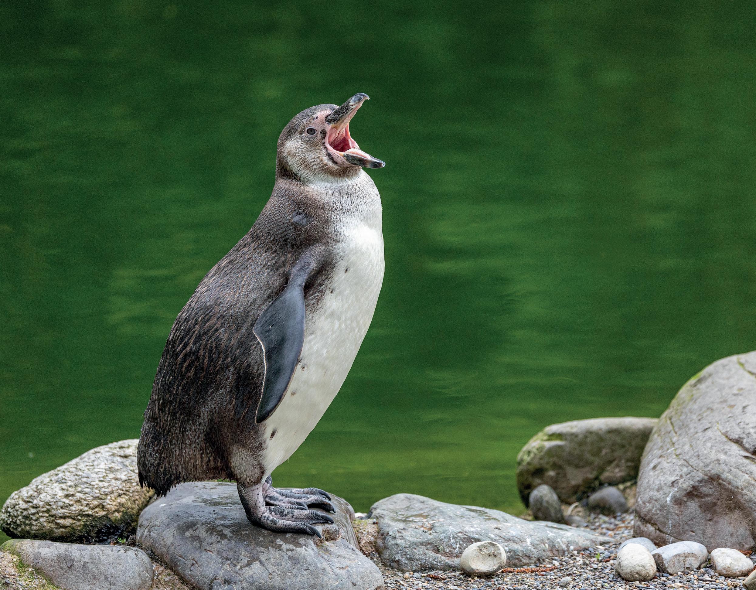
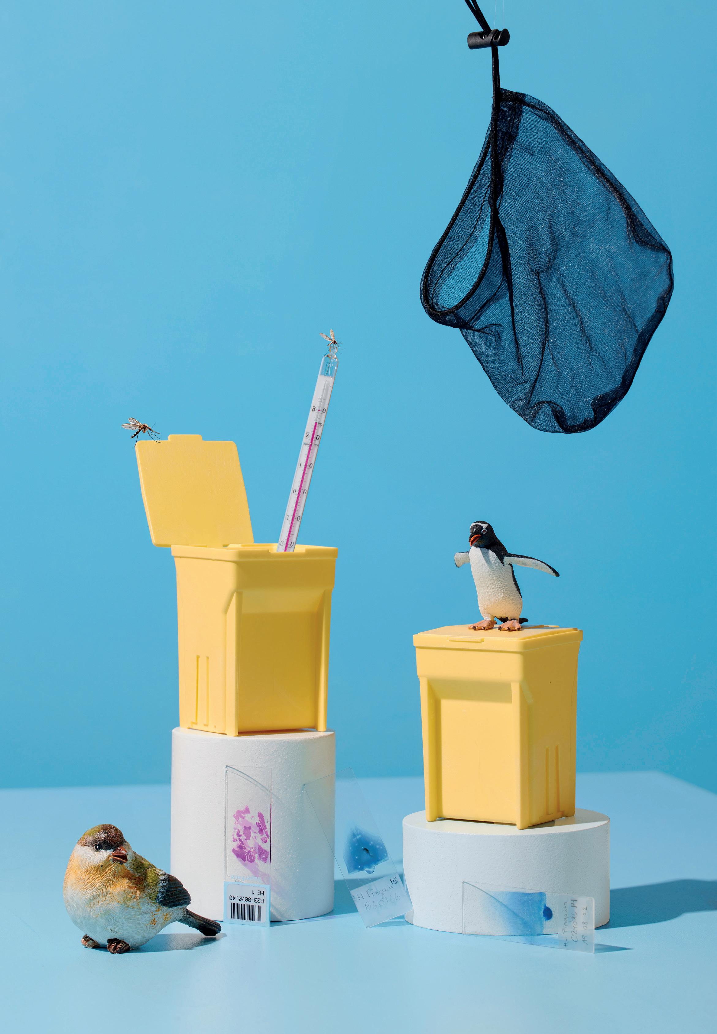
When people think of malaria, most think of a tropical human disease. However, malaria also affects birds – worldwide. It poses a particularly serious threat to non-adapted bird species. This is evident in zoo-kept penguins, where the disease is one of the main causes of death. But wild birds in our latitudes are also affected.
At the University of Zurich (UZH) an interdisciplinary team led by the Institute of Parasitology and involving the University Animal Hospital Zurich puts a focus on avian malaria. Besides basic research, the researchers are also investigating practical applications for prevention, diagnosis and treatment. To this end, UZH has established a research network of molecular biologists, parasitologists, veterinary physicians, pathologists, ecologists, zoo curators and other experts, which has been growing continuously since 2022.
History and overview
Research into avian malaria has a varied history. It experienced its heyday in the 1940s – at a time when the cultivation of human malaria parasites was not yet possible and other animal models were not yet available. Many fundamental insights and intervention approaches, including vaccine development, were originally gained from bird models. Even the first transgenic parasites were developed in a chicken malaria model (Plasmodium gallinaceum).
“ We investigate which parasites circulate between different bird species and how they behave within the host’s body.”
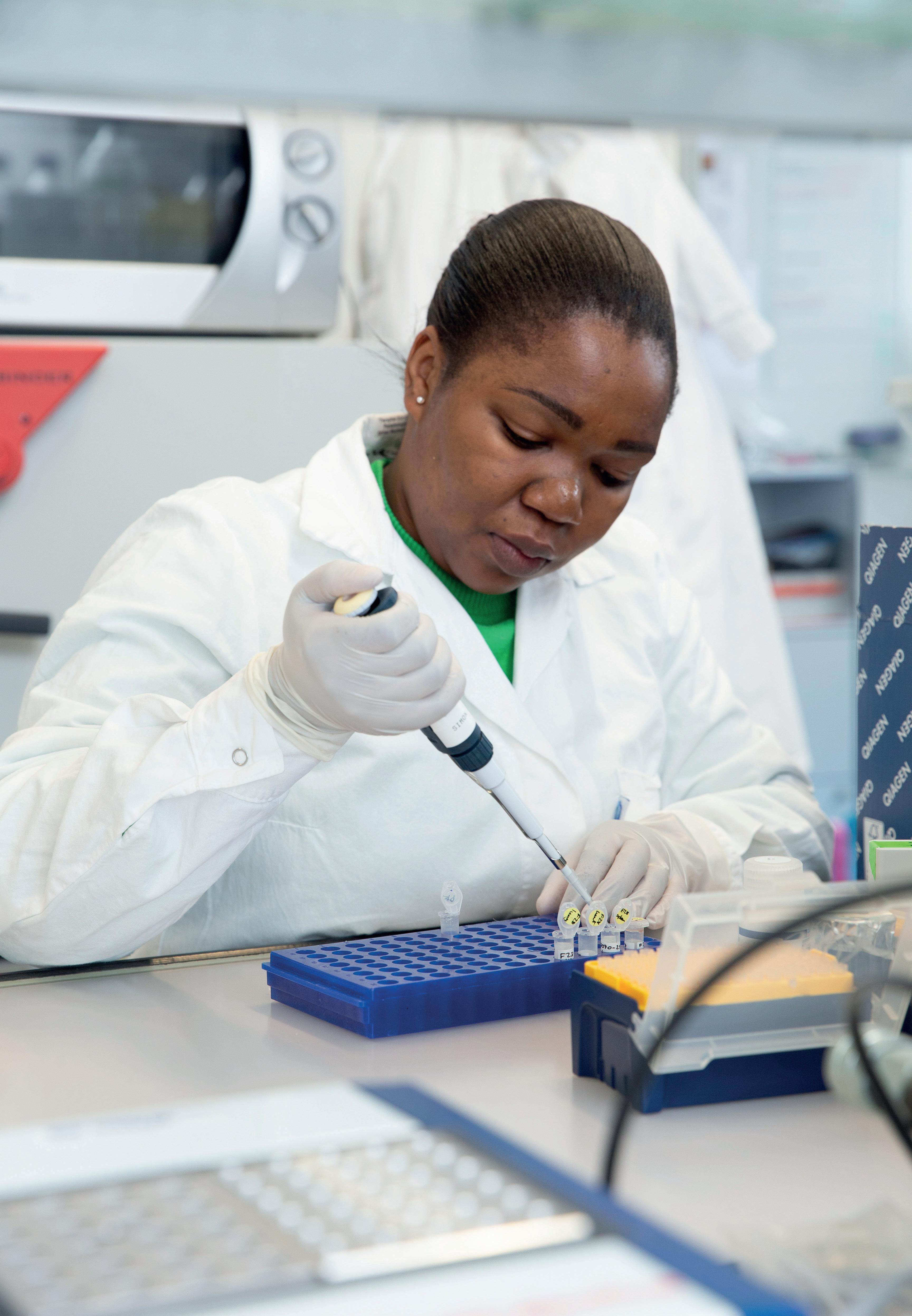
Doctoral student Gillian Muchaamba, Institute of Parasitology, Vetsuisse Faculty, University of Zurich
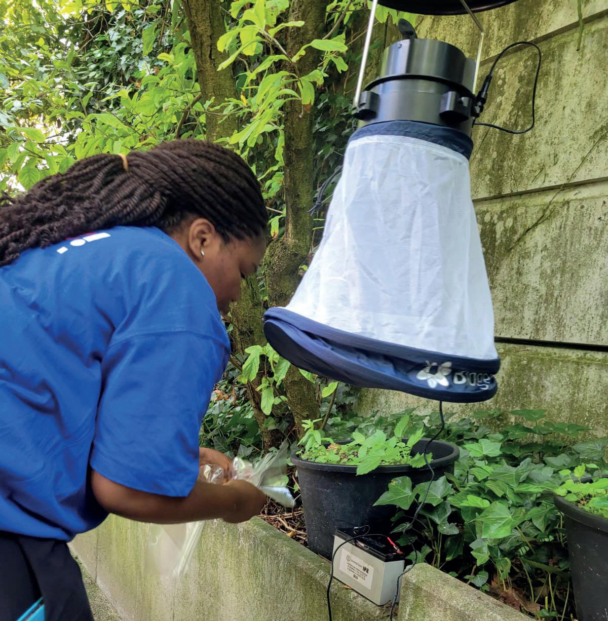
However, interest declined significantly after the discovery of rodent malaria parasites and the successful human malaria parasite in vitro culture in the late 1970s. While human malaria research is funded through various biomedical research foundations, avian malaria research is limited to ecology and evolution studies. The UZH team wants to change that, investigating both basic and translational research aspects of avian malaria in Switzerland. The main focus is on the transmission pathways of avian malaria within zoo set-ups. “We investigate which parasites circulate between different bird species and how they behave within the host’s body,” says Gillian Muchaamba, a doctoral student at the Institute of Parasitology. Results so far show that transmission mostly occurs through Culex
pipiens mosquitoes. Sparrows and blackbirds have been identified as important reservoir hosts in zoos in Switzerland, harboring the parasite without becoming ill themselves. Concerningly, the prevalence of avian malaria is high in these hosts. The researchers were also able to prove that the most common pathogens – Plasmodium matutinum and Plasmodium relictum – behave differently depending on the infected bird species. In penguins that are studied in zoos in Zurich, Bern and Basel, the parasites are observed mainly in the tissue of various organs, which can lead to severe inflammation in infected organs. In native birds like blackbirds, however, multiplication occurs primarily in the blood, allowing the parasite to produce stages for onward
The UZH team is developing new diagnostic procedures to detect avian malaria antibodies in penguins. Traditional bloodbased testing methods are proving challenging.
“ The combination of our experience from human malaria research with veterinary expertise creates completely new possibilities,”
Matthias Marti, Head of the Institute of Parasitology, Vetsuisse Faculty, University of Zurich
transmission to bird hosts via mosquitoes. Infection in deep tissue rather than blood cells in zoo-kept penguins complicated diagnostics of avian malaria, which is traditionally done by blood smear and PCR analysis. The team is therefore working on developing a novel diagnostic procedure to detect the presence of antibodies in penguins as a measure of exposure to the parasites. Currently, a recombinant protein is being produced to serve as the basis for a rapid development test for avian malaria.
Studying avian malaria in wild birds presents researchers with particular challenges. “With wild birds like sparrows or blackbirds, it’s difficult to obtain fresh samples for histological examination,” explains Professor Dr. Matthias Marti, who leads the Institute of Parasitology. Unlike larger birds such as raptors, injured small birds are rarely found and brought in for treatment.
Prof. Marti’s team has been studying human malaria for the past two decades, with funding from various basic and translational charities. Avian malaria research is understudied, in part due to difficulties in attracting funding. The team has obtained pilot funding for part of this project from the University, which will serve as a basis for more grants to sustain the research in future.
Far-reaching
Intensive construction activity destroys traditional breeding areas for wild birds, further complicating research. To bridge this gap, the team works closely with the bird rehabilitation center in Zurich and other partners such as the Zurich and Bern zoos. Experimental infection trials on canaries also help to better understand the disease progression observed in wild birds.
The UZH team is particularly proud of the international research network that junior researcher Muchaamba has built since fall 2022. For instance, the experimental infection studies are conducted with ecologists at the University of Lausanne. The project additionally benefits from close collaboration with the University of Glasgow, where UZH parasitologist Marti leads a second laboratory in malaria research. Bioinformatic analyses are mainly conducted there. Valuable expertise also comes from Lithuania, where a center of excellence for avian malaria has existed for 35 years, and from population genetics specialists in Singapore who study various human and animal malaria parasites in Asia. Histological examinations are
done in collaboration with the UZH and University of Bern institutes of pathology, as well as the Institute for Fish and Wildlife Health, University of Bern.
The practical significance of the research is particularly evident in the context of climate change. The alteration of habitats and migratory bird routes could lead to the introduction of new pathogens into previously unexposed bird populations. In Hawaii and New Zealand these pathogens have already led to dramatic declines in native bird species.
The situation in Switzerland is also changing. Climate change influences the distribution of carrier mosquitoes, and populations of important reservoir hosts like sparrows are declining in urban areas. This could alter the ecological balance between parasites and hosts and lead to new challenges.
An important goal is sequencing the first complete genome of avian malaria pathogens found in this region. The UZH researchers also plan to develop an in vitro culture system for avian malaria parasites. “That would be a real breakthrough,” explains Marti, “because it facilitates systematic functional studies across the life cycle of the parasite.” This step would open completely new possibilities for basic research, and could accelerate the development of new treatment strategies.
For the future, the research team also plans further epidemiological studies in Switzerland. “We want to better understand which parasite species occur here, what their genetic structure looks like, and how they spread between different bird populations and across geographical boundaries,” explains Muchaamba.
Interdisciplinary collaboration has proven to be key to success. “The combination of our experience from human malaria research with veterinary expertise creates completely new possibilities,” emphasizes parasitologist Marti. The insights gained should not only contribute to protecting endangered bird species but could also open new perspectives for combating human malaria.
Florian Bayer is a freelance journalist in Vienna. He was a health editor at The Standard and now writes for several newspapers in Germany, Austria, and Switzerland.

Page 14: How a clinical study in Zurich is changing the face of Feline Infectious Peritonitis.

Page 32: Where wildlife, livestock, and people intersect, interdisciplinary research could help tackle tuberculosis.
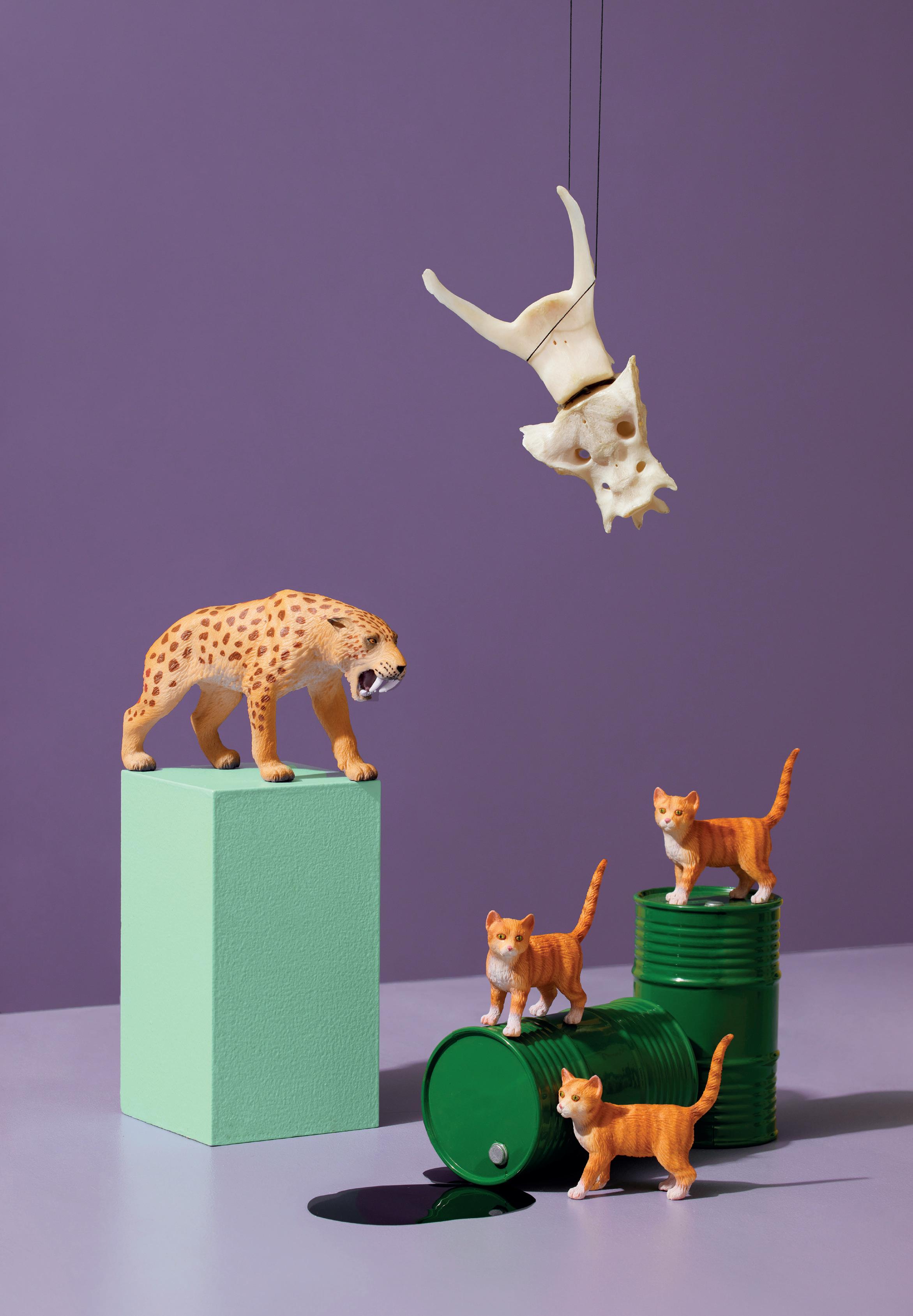
A collaboration between veterinarians and paleontologists shows that saber-toothed cats had spinal diseases similar to those of mode rn animals. This finding provides valuable lessons for species conservation and animal breeding. Text by Dr.
Hildegard Kaulen
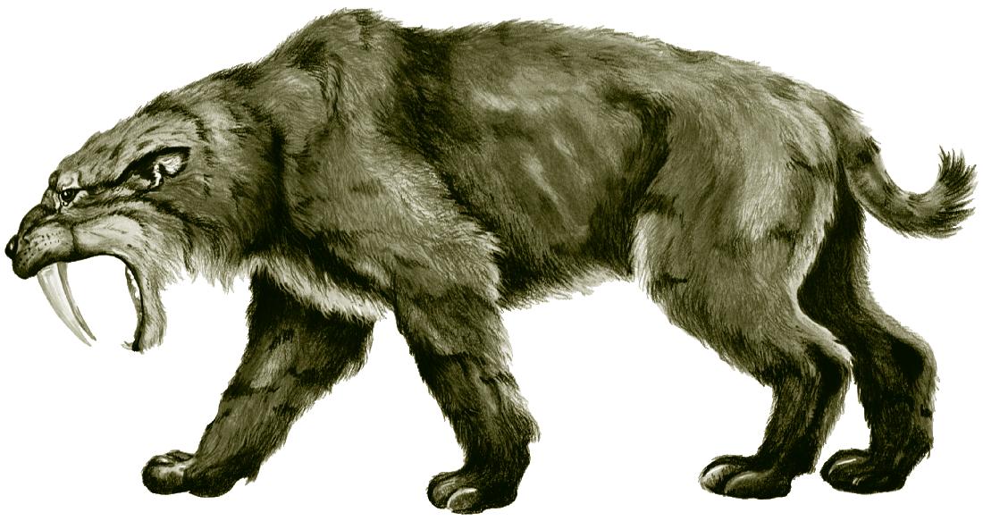
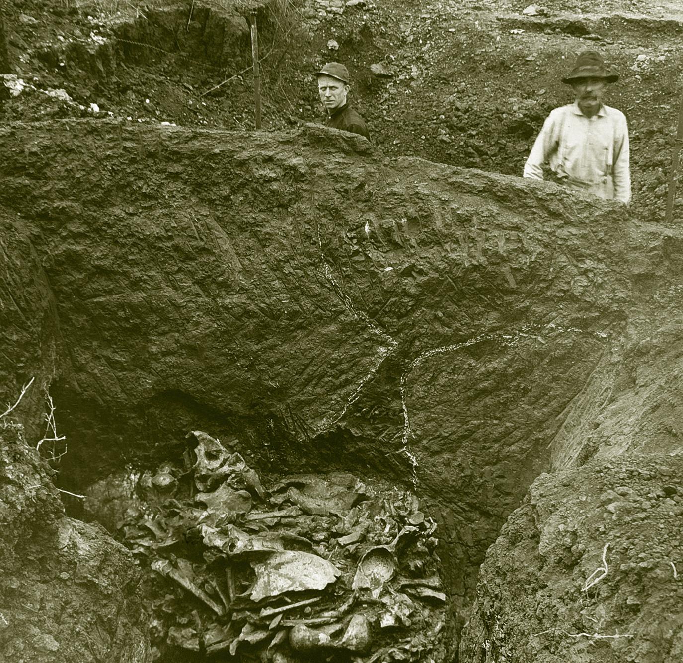
Dr. med. vet. Hugo Schmökel has always been interested in paleontology. When the small animal surgeon first picked up pathologically altered vertebral bones from saber-toothed cats Smilodon fatalis at the La Brea Tar Pits and Museum in Los Angeles, he immediately noticed how similar these changes were to lesions found in today’s feline patients. Yet saber-toothed cats became extinct 13,000 years ago and left no modern descendants. “I was immediately electrified,” says Schmökel, who works for Evidensia Academy in Stockholm and is affiliated with the Department of Paleontology at the University of Zurich, headed by Professor Marcelo Sánchez-Villagra, who was also involved in the research. “I wanted to
cause paleontologists are not trained in veterinary diagnostics. As a spinal surgeon for small animals, I see a lot of CT scans every day, but veterinary radiologists are much better at reliably and objectively diagnosing and classifying pathologies due to their broad range of expertise. This adds weight to a diagnosis.”
The collaboration was also a win-win situation for Del Chicca. “I previously knew very little about the diseases of extinct animals. But a bone is a bone,” she says. “It’s a very old system. Our research shows that the responses to illnesses have remained similar over the millennia. This is very interesting. By jointly looking at the fossil skeletons, we also learned a lot about today’s animal health regard-
Interdisciplinary collaboration is crucial in the scientific world. The specific areas of expertise have particular competence that can be used synergically. This is one of the keys of evolutionary medicine.
Francesca
know if the innate and acquired skeletal pathologies of saber-toothed cats were indeed the same as those of modern domestic animals.” Schmökel took CT scans of the most prominent vertebral abnormalities to create 3D reconstructed images that allow the skeletal samples to be viewed as if they were real.
Successful collaboration initiated
To confirm the diagnosis, he turned to veterinary radiologists Dr. med. vet. Francesca Del Chicca and Dr. med. vet. Regine Hagen from the Clinic for Diagnostic Imaging of the Vetsuisse Faculty. “It was clear to me that specialized veterinarians had to review my assessments,” Schmökel says. “There are incorrect diagnoses in paleontology be -
ing breeding and protection of species. This is what makes this collaboration so valuable.”
Remarkable number of malformations
What did Schmökel and Del Chicca discover? The doctors studied two types of skeletal pathologies: innate malformations such as block vertebrae and transitional vertebrae, and pathological bone growth. In a block vertebra, two vertebrae fail to form individual vertebra and stay merged; in a transitional vertebra, the vertebra exhibits features of two different vertebral types. Bone injuries from obvious trauma were not part of the research.
The study shows that the prehistoric predator had many more block and transitional vertebrae than modern ani-
“ Our interdisciplinary collaboration has led to the discovery of many new findings that other scientists had misinterpreted because they had not examined the fossils with a veterinary eye.”
Hugo Schmökel, Chief Veterinary Officer, Evidensia Academy, Sweden
mals or human beings. The doctors suspect that the high number of transitional vertebrae was caused by inbreeding, as the already high number of transitional vertebrae increased just before the saber-toothed cats went extinct. “There are also recent examples showing that inbred gray wolves and pumas have more malformed vertebrae. Mammoths also had transitional cervical vertebrae shortly before they became extinct,” says Schmökel.
Block vertebrae have a different cause. They can be traced back to faulty embryonic development caused by toxins. Schmökel and Del Chicca hypothesize that the crude oil in the La Brea Tar Pits area of Los Angeles, where the bones were found, could have triggered the large number of block vertebrae. “We know that crude oil causes malformations in birds and fish,” says Schmökel. “Crude oil was probably present everywhere in the landscape with tar pits, for example, in the drinking water or in the sabertoothed cats’ prey. It may be that they ingested a lot.” Both view this finding as an important reminder of the disastrous consequences of today’s oil pollution for all living creatures.
Did the lesions cause the saber-toothed cats to suffer?
What were the consequences of these deformities? “It is likely that not all these malformations had a direct impact on individual health,” says Del Chicca. “Modern dogs and cats also tolerate these deformities very well. But they may develop problems in old age or in association with other pathologies. It is therefore also possible that the sabertoothed cats were similarly affected as they got older and became less efficient in hunting.”
The doctors know much less about the possible causes of the unusual pathological bone growth found in the study. These special changes are much more common in sabertoothed cats than in modern pets, which rarely develop such large bone proliferations. “The reason might have been an infection, an autoimmune disease or poisoning,” says the veterinary radiologist. The bone growth might
also be related in part to the malformed vertebrae. Del Chicca rules out that the bone proliferations were caused by a fracture because of their protected position in the body and their distribution. However, she is sure that the pathological bone growth had a much greater impact on the predators’ health than the abnormal vertebrae. “Some proliferations look very severe. We don’t see that in our pets,” she says. “These bone growths probably pinched nerves, irritated soft tissue and reduced mobility. This very likely caused pain, so that the animals probably had difficulty finding prey and ended up looking for carcasses instead.”
What lessons can be learned? “One important lesson is that we need to be more aware of the problems caused by inbreeding in modern animal breeding and species conservation,” says Schmökel. “We can’t start conservation programs when there are only 50 or so animals left of a species. At that point, they are already badly harmed by inbreeding. We must give them more space and protect their environment early on. We cannot let the populations become so isolated and small.”
Schmökel and Del Chicca also question the current breeding practice. “There is a lot of screening these days,” says Del Chicca. This means that animals with a certain disease such as hip dysplasia or retinopathy are excluded from breeding. This is good in itself, but it also means that there are fewer animals available for breeding, so the population becomes small again. This increases the risk of inbreeding.” In addition, Schmökel favors placing breeding under the Swiss Animal Protection Act and the obligatory supervision of a breeding specialist who will ensure that the population is constantly refreshed. There are already 400 registered hereditary diseases in dogs.
Dr. Hildegard Kaulen is a molecular biologist and science and medical journalist who writes for daily newspapers and science magazines.
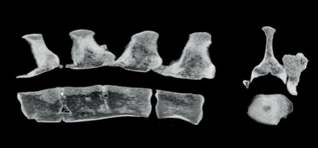
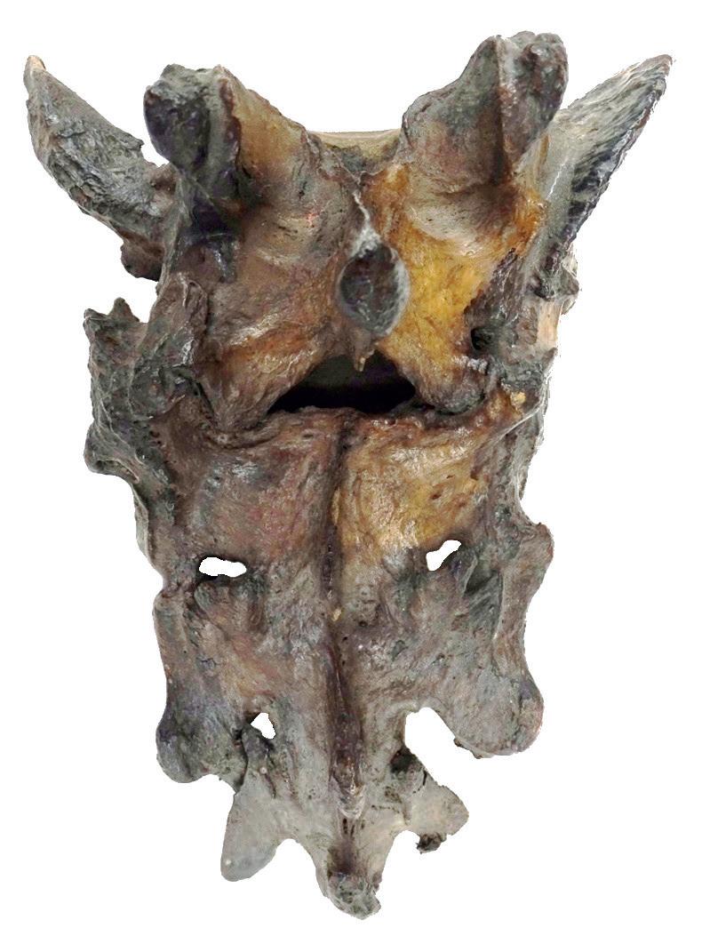
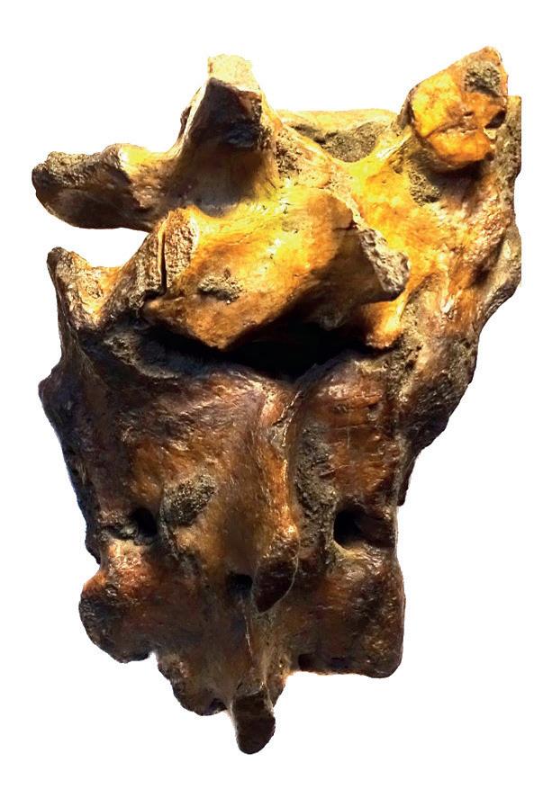
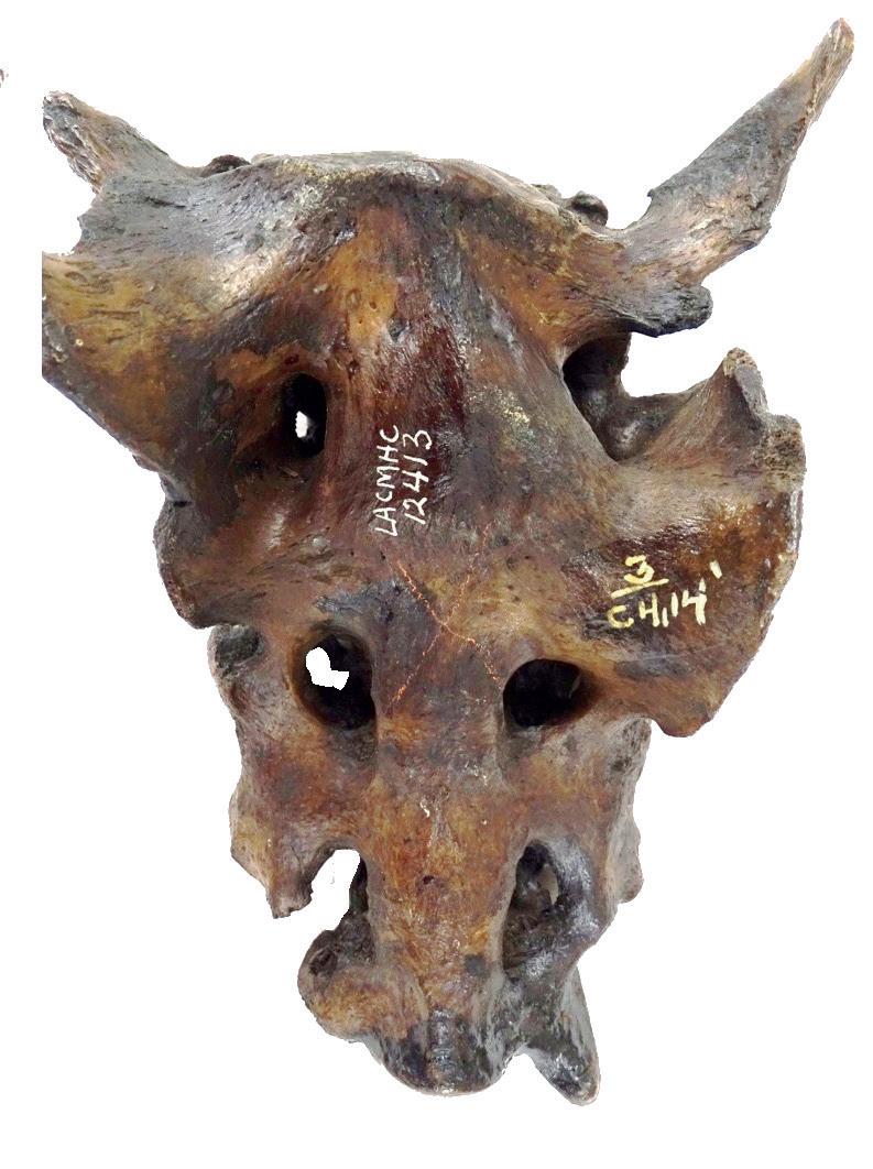
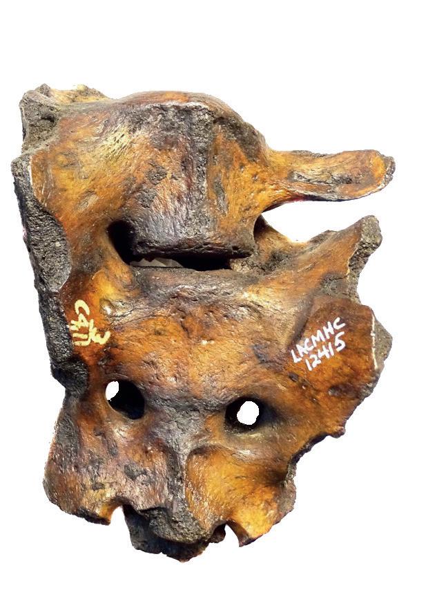
The CT images (with multiplanar reconstruction) show block vertebrae (three fused vertebral bodies) and bone proliferation around a synovial joint.
The bones depicted show severe lumbosacral transitional vertebrae.
Long after fever and other symptoms fade, COVID-19 can leave lasting damage. At the Vetsuisse Faculty, veterinary pathologist Anja Kipar and her team are part of a major European project investigating the origins of Long COVID. By analyzing tissue samples, they examine how the virus affects cells in the lungs and brain — work that c ould uncover relevant pathomechanisms and guide better treatments.
Text by Santina Russo
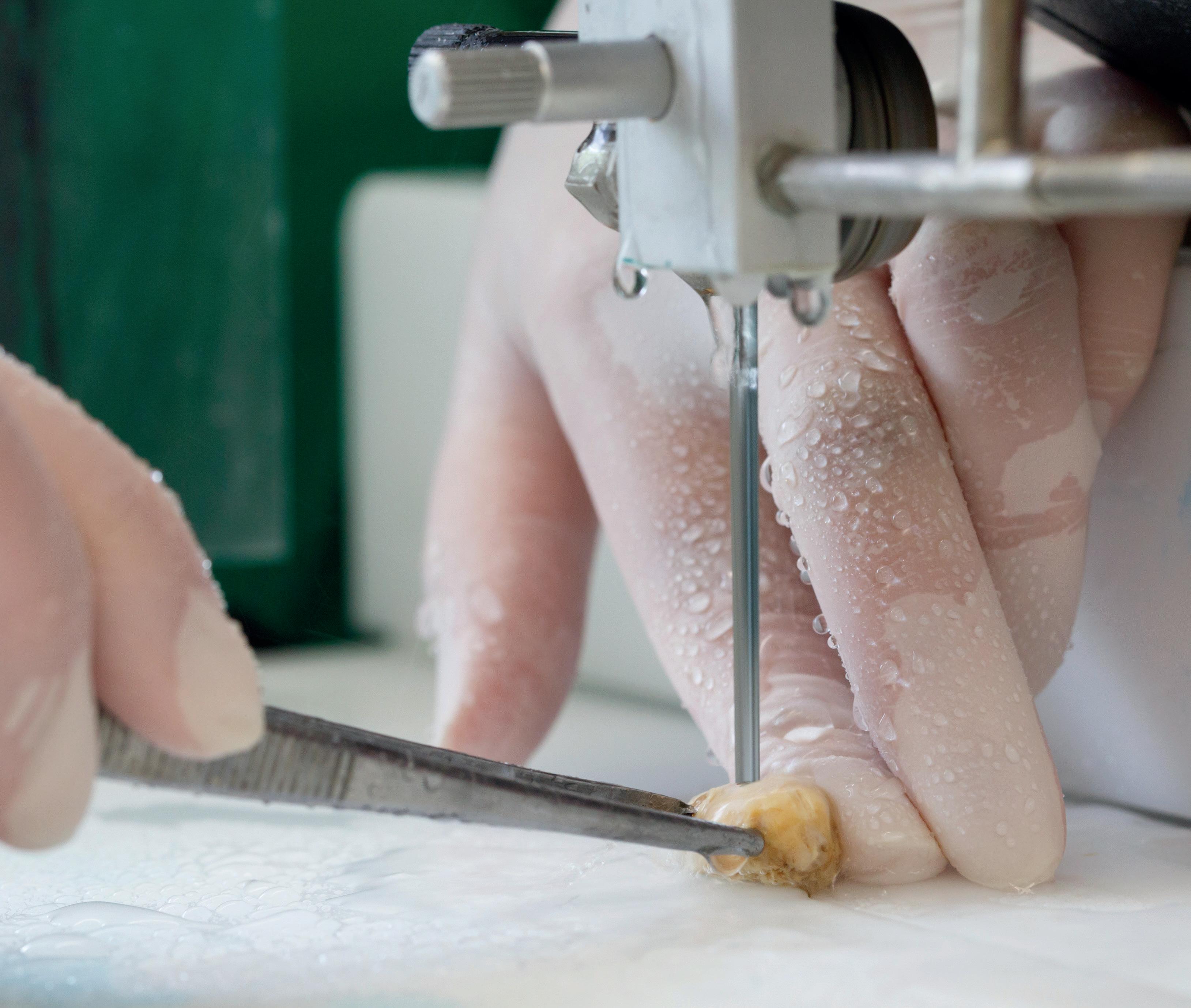
Belinda Senn carefully removes a thin layer from the sample she is holding. It is a paraffin block prepared from the left lung of a hamster that was infected with SARS-CoV-2, the virus that caused the COVID-19 pandemic in humans. With a cutting tool named a microtome, the biomedical analyst in the lab of Professor Dr. med. vet. Anja Kipar at the University of Zurich, slices away the protective paraffin to reach the tissue. For the microscopical analysis, the group needs a two-micrometer-thin slice of lung tissue — just two-thousandths of a millimeter.

A precise microsection is cut and immediately placed first in a cold-water bath, then in a warm-water bath to remove any creases from the delicate slice. The sample is one of many the research group is using to decipher exactly what happens in tissue during and after a SARS-CoV-2 infection — and to obtain insights into why some patients develop Long COVID and do not recover for months or years.
The work is part of a large European Horizon-funded project aimed at elucidating the predisposing factors and mechanisms behind Long COVID development. With their expertise in laboratory animal pathology and animal models of viral diseases, the group led by Kipar, Director of the Institute of Veterinary Pathology at the Vetsuisse Faculty, complements the project’s virology and clinical studies with mechanistic investigations.
“Only in controlled lab settings with samples from animal models can we truly investigate the effects of different SARS-CoV-2 variants in tissues and cells,” says Kipar. Mice infected with the Delta variant offer insights into its impact on the central nervous system. Meanwhile, hamsters exhibit disease progression more similar to humans when it comes to viral effects in the lungs.
In the lab, Senn has now affixed the smoothed lung section to a microscope slide, and proceeds to cut another one, its twin. Later, the samples will be stained — one with a standard stain to detect tissue damage and response, the other with a stain that will reveal the presence of the virus. Examined with a microscope, these tissue sections will show the morphology of each individual cell and uncover the consequences of the infection. In addition, RNA sequencing will provide detailed insights into the tissue’s transcriptional response.
Group leader Kipar was one of the first veterinary pathologists in Europe to study the mechanistic effects of viral infections in laboratory animals. She has been doing so for the last two decades — as a senior lecturer, then a professor of Veterinary Pathology at the University of Liverpool, later at the University of Helsinki and now at the Vetsuisse Faculty in Zurich. This gave her the expertise that later became crucial in studying SARS-CoV-2 when the pandemic hit.
Today, in the project studying Long COVID — also referred to as Post COVID-19 Condition by the World Health Organization — she is collaborating with the same research institutes where she previously worked. At the University of Liverpool, Professor James Stewart’s group set up the mouse models to provide Kipar with samples infected by different SARS-CoV-2 variants. Meanwhile, Dr. Giuseppe
“ In controlled lab settings with samples from animal models, we can truly investigate the effects of different SARS-CoV-2 variants in tissues and cells.”
Anja Kipar, Institute of Veterinary Pathology (IVPZ), Vetsuisse Faculty, University of Zurich
Balistreri’s group at the University of Helsinki contributes to the project with in vitro infection studies of human cells and organoids, which are small, simplified versions of organs. In addition, the Long COVID clinics at the university hospitals in Helsinki, Groningen (NL), and Basel provide clinical data and enable the investigation of human cerebrospinal fluid and blood samples. All these findings come together with Kipar’s mechanistic studies in the Long COVID project.
So far, the findings of Kipar’s group have shown that in mice, within three to four days of intranasal infection with the Delta variant, the virus is present and often widely distributed in the brain. The scientists also observed that the infected brain tissue attracts leukocytes — immune cells involved in inflammation. Of all the brain cells, only neurons are infected, yet they are not destroyed. While the cells
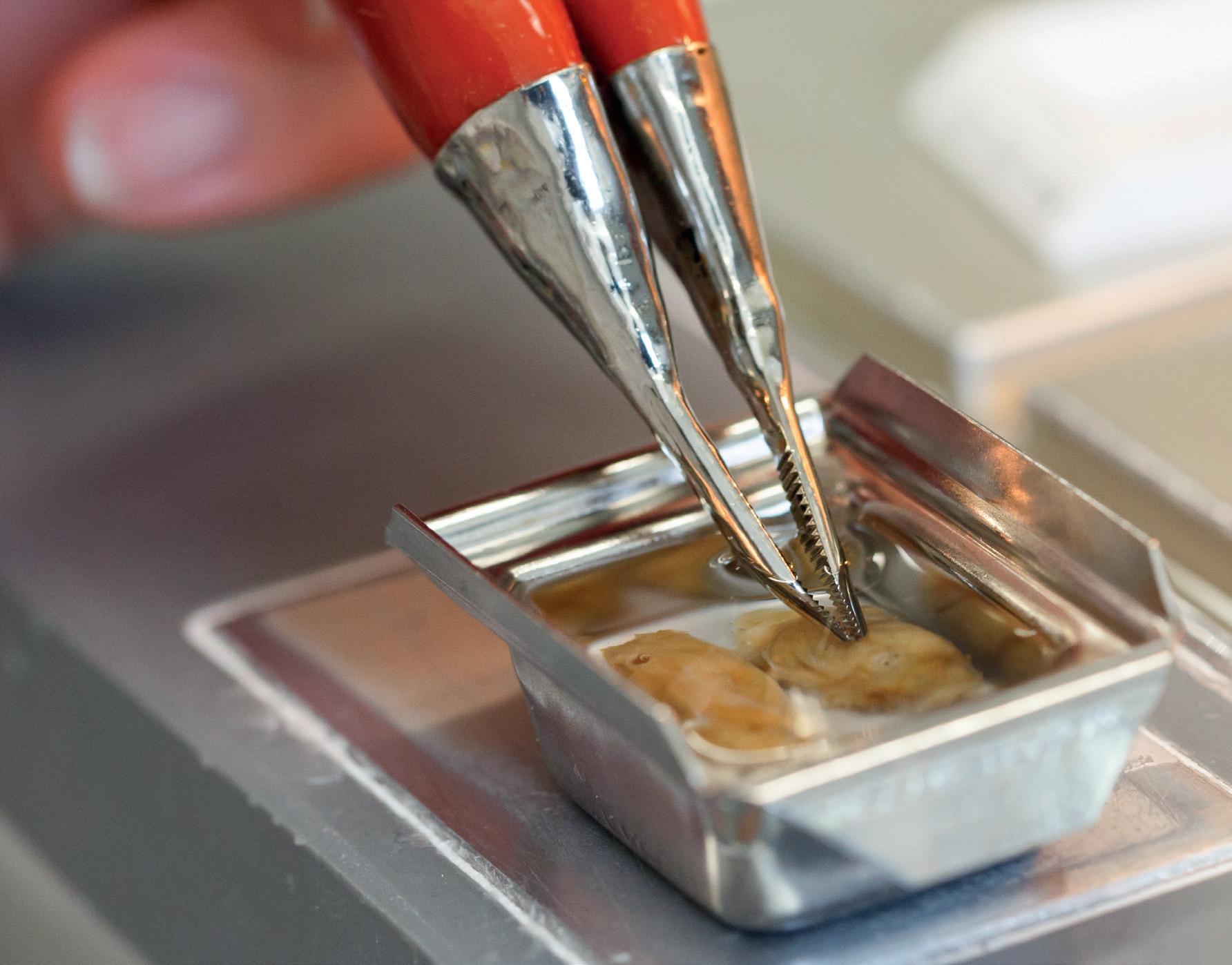
A close look at the two halves of a mouse brain in warm liquid paraffin, ready to be transformed into the paraffin block that will be cut at the microtome.
“ With our SARS-CoV-2 models, we are addressing questions that apply to many other viral diseases — questions that, so far, have not been systematically addressed.”
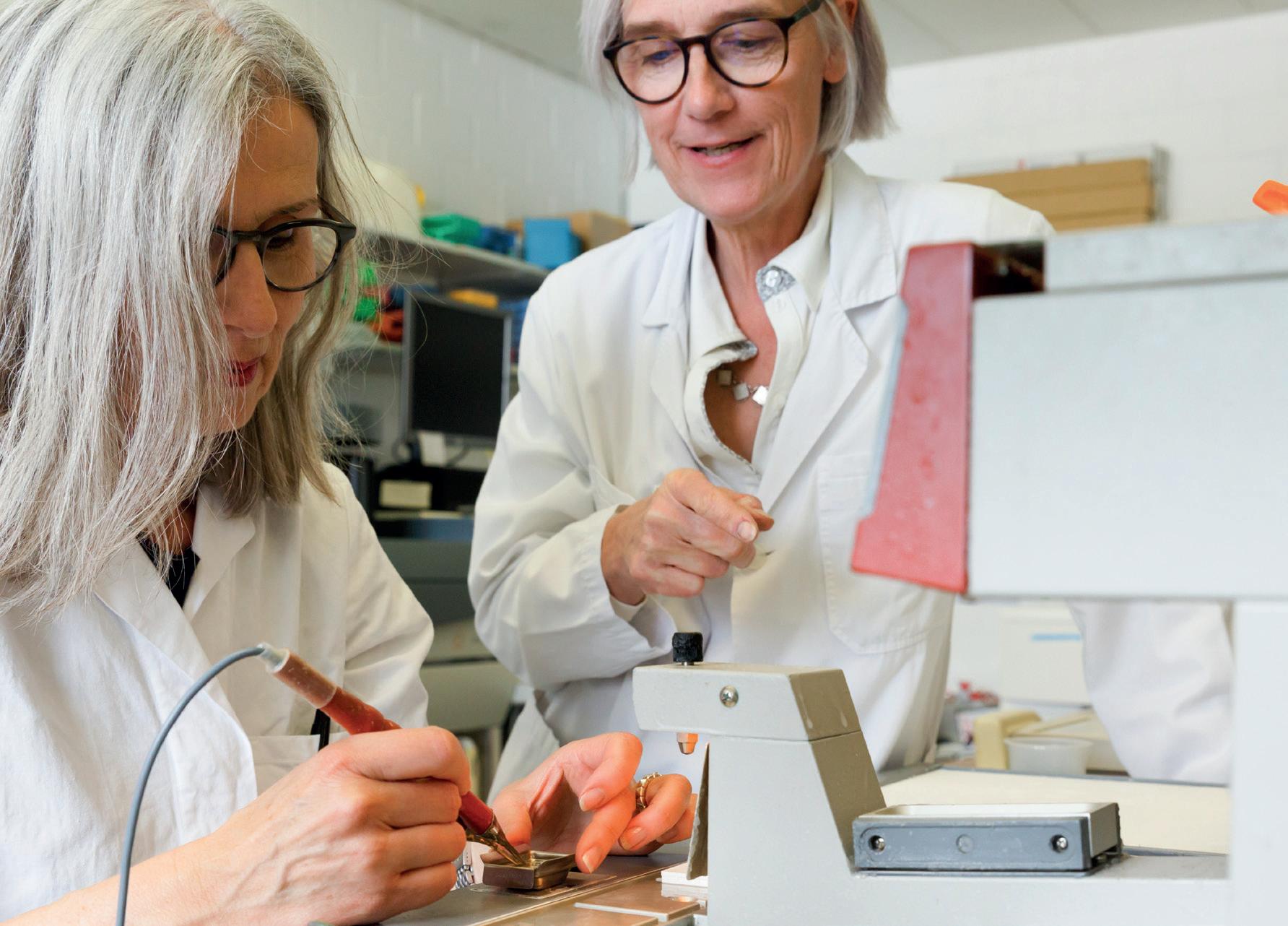
Anja Kipar (right) and head laboratory technician Sabina Wunderlin-Giuliani discuss how to best embed the decalcified skull of a mouse to ensure optimal histological specimens.

survive, they apparently induce a mild inflammatory response.
Complementary RNA sequencing analyses then revealed findings that might offer explanations for some neurological impairments seen in Long COVID patients. Indeed, recent preliminary results from mouse brains infected with the Delta variant provided evidence that the infection compromises various neurological processes, including neuron structure, the formation of connections between neurons, neural communication, and processes linked to behavior and learning.
To gain a broader understanding of the consequences of viral infection, the team plans to extend its investigations to analyze changes in small-molecule metabolites and lipids in the brain, which make up cell membranes and can act as fat-soluble metabolites. In another ongoing study, the group is examining the effects of certain antiviral drugs that were used in human patients. Moreover, the team is planning to employ a method called spatial transcriptomics. “With this method, RNA analysis will reveal for each individual cell for which cellular components the transcription is upregulated upon infection and, consequently, which cellular pathways are directly linked to the pathological processes,” Kipar explains.
In addition, the researchers will investigate the lung-brain axis. “It is possible that an infection in the lungs leads to the release of mediator molecules into the blood that can trigger neuronal dysfunction in the brain,” the professor explains. Applied to humans, this would suggest that patients with a SARS-CoV-2 infection in the lung could experience neurological symptoms even if the virus does not reach the brain. This could represent another link to Long COVID.
And the work doesn’t stop there: For Anja Kipar, these studies also contribute to understanding other viral infections that affect neurons, among them tick-borne encephalitis, or infections with other respiratory viruses. “Even with the flu, people often experience subtle symptoms of the central nervous system, such as headaches or temporary loss of taste,” she points out. She concludes: “With our SARS-CoV-2 models, we are addressing questions that apply to many other viral diseases — questions that, so far, have not been systematically addressed.”
Santina Russo, PhD, is a freelance health and science journalist based in Zurich, Switzerland.
Innovation in tuberculosis research is not only happening in ordered laboratories, or on the frontiers of computing. Interdisciplinary research in ecosystems where wildlife, livestock, and people intersect could lead the fight against the ever-evolving pathogen. Text by Janine
Stephen
Veterinarian and microbiologist Dr. Giovanni Ghielmetti has considerable respect for the disease he has spent much of his adult life researching. Mycobacterium tuberculosis complex bacteria are ancient enemies that have evolved alongside humans and animals for 70,000 years. Despite vast strides in surveillance and diagnostics, tuberculosis caused illness in over ten million people in 2023, and the disease inflicts a vast toll on wildlife and livestock.
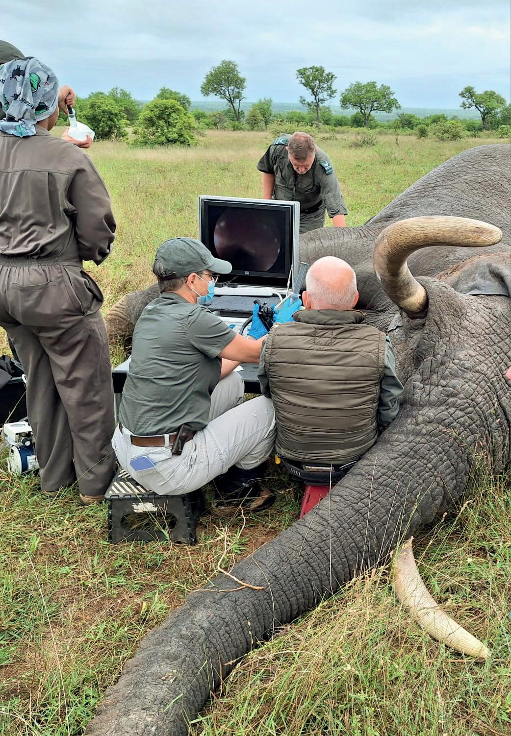
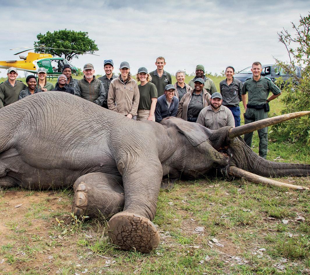
For almost ten years, Ghielmetti investigated mycobacterial disease in animals at the Swiss National Reference Laboratory for Bovine Tuberculosis. In 2023, he swapped the familiar setting for South Africa. Here, Mycobacterium bovis is endemic in wildlife, from African buffalo and lions to elephants. The disease also circulates in livestock: the cattle and goats that are an essential part of life in rural areas. And it infects human populations in areas where wildlife, livestock, and people overlap.
In this complex context, “a crucial research aspect is disease transmission,” Ghielmetti says. “We don’t know exactly how interspecies transmission occurs. It’s very important to investigate the entire ecosystem, rather than focus on a single host if we’d like to understand the disease a little better.”
Ghielmetti joined an interdisciplinary team of experts at Stellenbosch University’s Animal Tuberculosis Research Group [see box below], which falls under the medical faculty. Here, a “One Health” approach sees scientists from
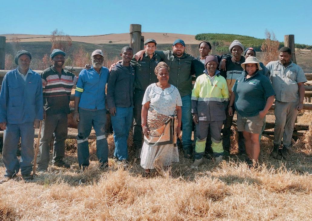
Dr. Giovanni Ghielmetti joined the Stellenbosch University Animal TB Research Group as a postdoctoral research fellow in March 2023. A research microbiologist and a diplomate of the American College of Veterinary Microbiologists, he focuses on surveillance, diagnostics, genome sequencing, bioinformatics and more. Individuals Dr. Ghielmetti worked closely with at Stellenbosch University include: Prof. Wynand Goosen, who also worked on the analysis of human DNA sputum samples and livestock sampling in KwaZulu-Natal, Dr. Debbie Cooke, who enabled the sampling of cattle in KwaZulu-Natal and helped team members work within local communities, and Microbiologist and PhD student Christoffel Opperman, who has a human medical background. Next-generation sequencing methods that the team had developed and validated on animal samples were successfully applied to human clinical samples. The technique found mycobacteria species from extrapulmonary sites that routine methods had not correctly identified. Additional research tackled nontuberculous mycobacteria mixed infections, highlighting the need for multidisciplinary teams to formulate personalized treatment strategies.
the human and animal health fields collaborate on studying multiple facets of the disease, “from pathogenesis to epidemiology, host immune responses, and the pathogen biology itself.”
“
It’s very important to
investigate the entire ecosystem, rather than focus on a single host if we’d like to understand the disease a little better.”
The fellowship underscored some of South Africa’s striking contrasts. Ghielmetti worked with “some of the best minds in the field” in a US$ 66-million, state-of-the-art Biomedical Research Institute at Tygerberg Hospital. But he also undertook logistically demanding field work, in remote rural and wildlife areas.
The Animal TB Research Group was able to utilize advances in culture-independent testing of DNA using whole genome sequencing methods. “It is a quickly evolving field and it’s improving rapidly,” Ghielmetti explains. “With a portable device the size of a mobile phone we can generate crucial data that will allow us to address epidemiological questions within hours or a few days. This wasn’t even thinkable a decade ago.” The developments will aid not only TB research, but outbreak investigations and phylogenetic studies in lower-resource settings.
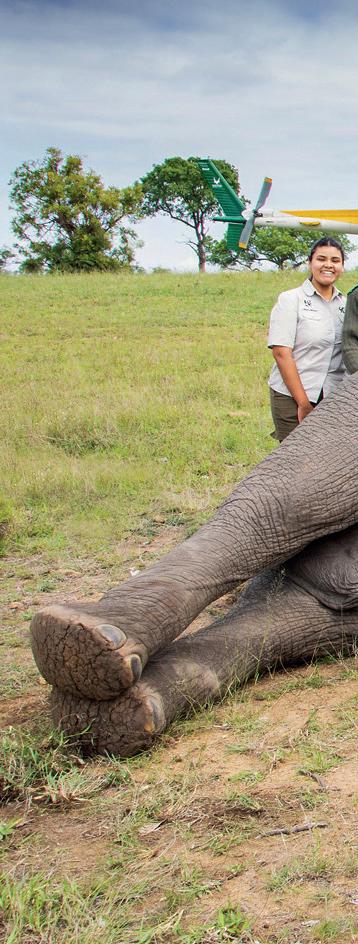
African elephants and lions in the Kruger National Park were the subjects of two studies Ghielmetti worked on. Ten elephants were darted as part of ongoing research led by South African Research Chair in Animal TB, Professor Dr. Michele Miller and conducted by PhD student Stacy Engel. Blood and bronchoalveolar lavage fluid samples were taken to investigate the animals’ immune responses to TB antigens and identify potential pathways of immune activation. “Her research is likely to lay a foundation for identifying biomarkers that can predict whether different life-threatening infections are present – not only TB,” says Ghielmetti. “There is also growing interest in other pathogens, including elephant endotheliotropic herpesvirus (EEHV).”
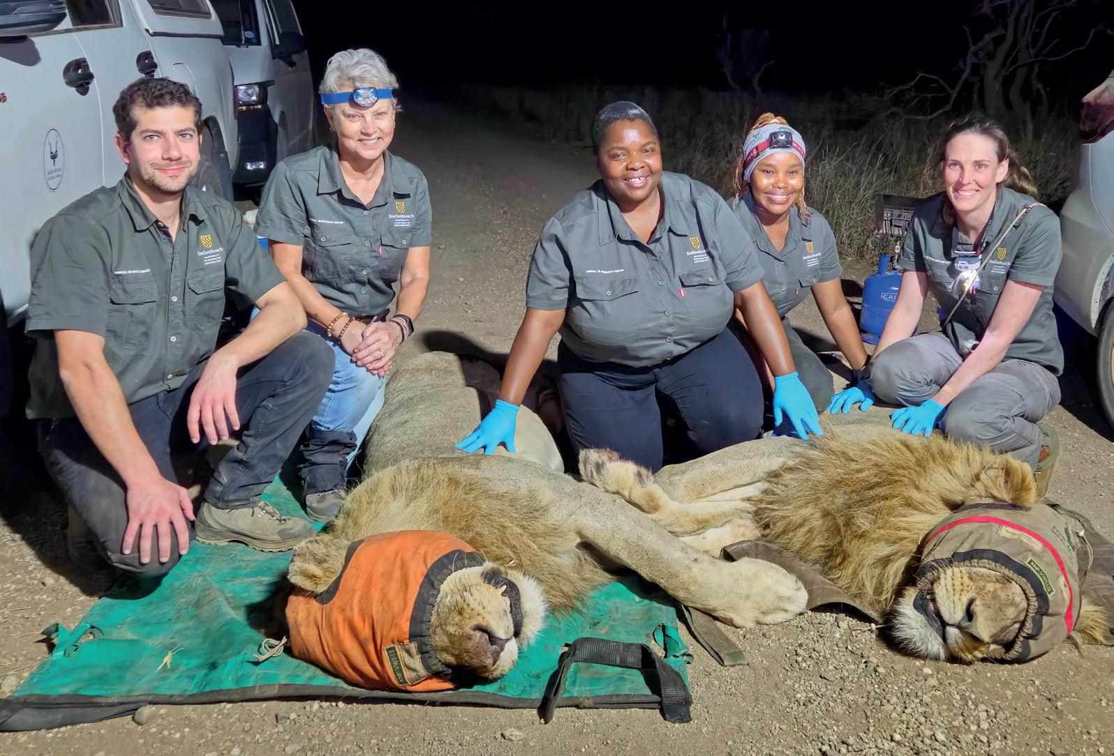
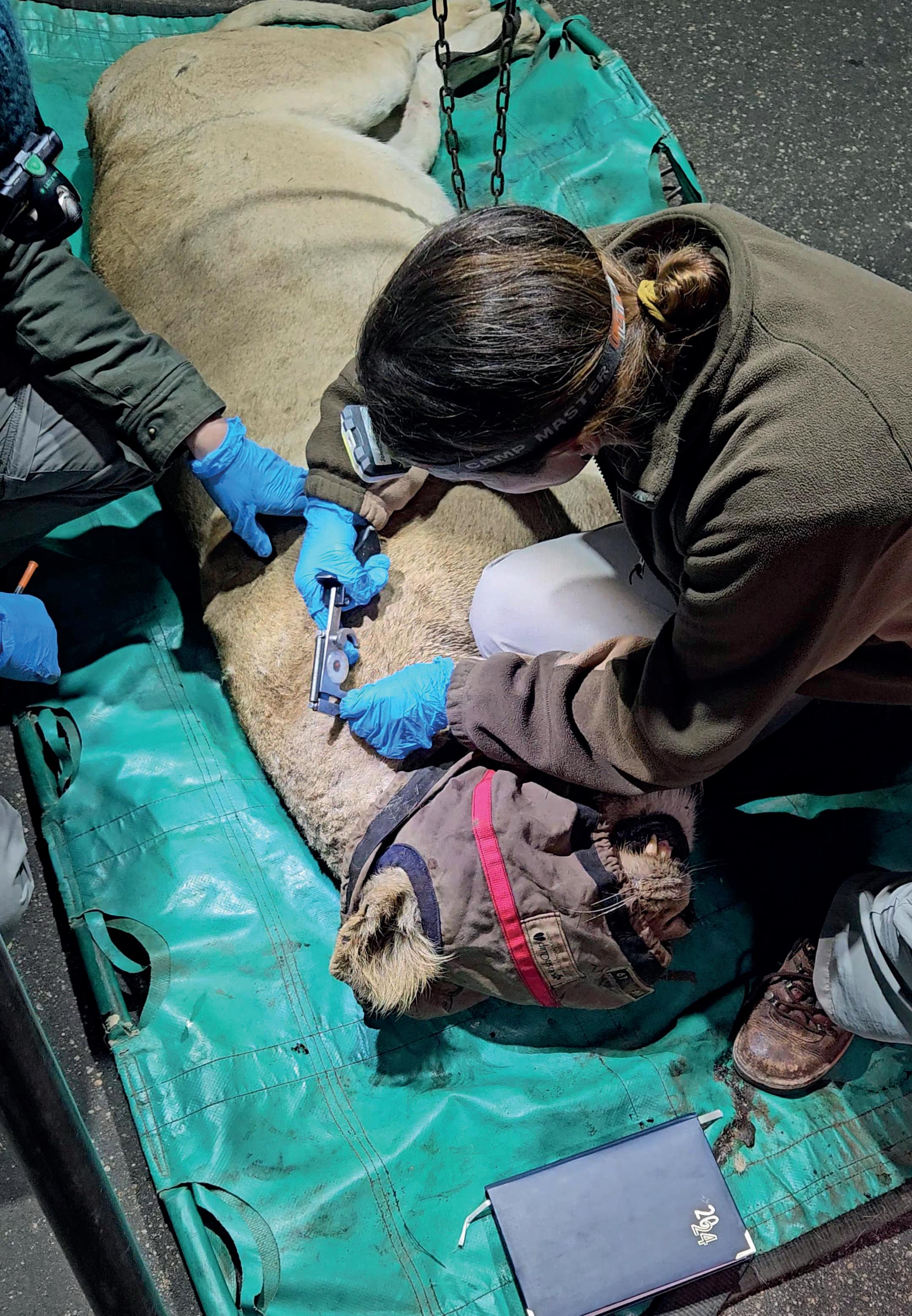
A study that sampled 80 African lions and involved multiple teams had a variety of working hypotheses. These ranged from diagnostics (whether X-ray imaging of immobilized lions in the field could detect TB disease) to a never-done-before attempt to identify host and pathogen biomarkers to facilitate identification between M. bovis infection and TB disease in naturally infected animals. Brand-new interdisciplinary research will also bring inter-
Members of the Animal TB Research Group, including principal investigator Michele Miller (second from left) and Giovanni Ghielmetti, with two immobilized male African lions after sampling and X-ray imaging in Kruger National Park. From right to left: Tanya Kerr, Sinomotfo Intimate Shongwe, Rachiel Gumbo.
national experts together to investigate microbial communities in the gut and respiratory microbiome and how they contribute to inflammatory and immune responses in lions – echoing work done in the human TB research field.
The animal-human interface
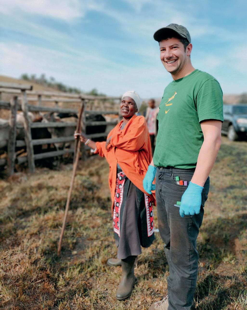
Another field trip saw Ghielmetti involved in sampling 100 cattle from communities around Pietermaritzburg in KwaZulu-Natal. This followed an earlier dramatic finding: a team including Ghielmetti had analyzed archived human sputum samples to prove that zoonotic M. bovis had infected people living around a wildlife park called Hluhlu-
“ It makes you really think about how important each experiment and each sample you collect is. ”
Giovanni Ghielmetti, Research Associate, Institute of Veterinary Bacteriology, Vetsuisse Faculty, University of Zurich
we-iMfolozi, further north in KwaZulu-Natal, (a first for South Africa). But how was transmission between cattle, wildlife, and humans occurring, and were the strains of M. bovis the same? Multiple sampling efforts of cattle in the region were needed.
“Our hypothesis was that where the disease is endemic in cattle, we would expect shedding of the pathogen through nasal secretion and feces,” says Ghielmetti. “This could lead to the contamination of the environment, including water sources, which are then accessible to other animals, and consumed by people as drinking water – a major source of infection.”
In what is likely a world first, the team was able to describe fecal shedding of viable Mycobacterium bovis bacteria into water sources. “We are now keen to look at the whole genome sequences of the M. bovis [strains] and compare them with those from other animal species, including buffalo, and once we put our hands on viable M. bovis from humans from the region, we will compare those as well.” The expectation is that the strains will be linked.
The discovery of M. bovis DNA in human samples in South Africa again highlighted the complexity of the South African context. The country’s high human immunodeficiency virus (HIV) prevalence rate exacerbates human tuberculo
“Addressing the disease holistically, across different species and the environment, is the only way forward. We can move away from siloed mentalities to more integrated solutions, where different stakeholders who are experts in their own fields share knowledge and we end up with a common product. ”
Giovanni Ghielmetti, Research Associate, Institute of Veterinary Bacteriology, Vetsuisse Faculty, University of Zurich
sis transmission. “HIV increases the risk of all zoonotic diseases,” warns Ghielmetti. “Current molecular methods and culture media that have been used for screening human patients for M. tuberculosis, are not optimized for the detection of M. bovis.” Missing or misdiagnosing M. bovis cases has wide implications for screening and treatment, and could contribute to growing resistance to available drugs.
Human and animal TB scientists from four continents and half a dozen institutions worked on the human sputum DNA study; they brought together expertise in molecular biology, genetics, immunology and infectious disease. Interdisciplinary collaboration, Ghielmetti believes, enables better use of resources and knowledge. “We share the same goal,” he says. “Addressing the disease holistically, across different species and the environment, is the only way forward. We can move away from siloed mentalities to more integrated solutions, where different stakeholders who are experts in their own fields share knowledge and we end up with a common product.”
An example is data sharing for infectious diseases. “This will require standardizing next-generation sequencing methods and bioinformatics pipelines.” The resulting data could then be compared across countries and continents, leading to reduced costs and more representative research outcomes, says Ghielmetti.
The interplay between human and animal TB research could also accelerate knowledge of the disease. “Sample processing, data generation, analysis of the data, and storage [in animal TB research] can be conducted using very similar methods as has been done in the last few years in the human field… there is huge potential to be derived from learning and using existing frameworks.”
After returning to Zurich, Ghielmetti found his experiences had changed his perspectives. Dramatic moments, such as the time a sedated 230-kilogram male lion woke unexpectedly as he was preparing to lift it into a vehicle to be transported to the X-ray station, have left their mark. So have the challenges of working in a socio-economic con-
text with limited resources. “It makes you really think about how important each experiment and each sample you collect is,” Ghielmetti says. “How precious the samples we collected, say from African lions, are.”
He expects to see more rapid change in his field. “Advances in sequencing technologies and artificial intelligence are transforming our ability to analyze complex genomic data,” Ghielmetti says. “The way biomarkers can be extrapolated from complex data sets, the potential for drug development, antibiotic-resistance prediction – these are a few examples where the new technologies in genomics and the integration of AI will change our world.”
But the South Africa experience will not be left behind.
“Working closely with what I’d call some of the leaders in the field of TB genomics, bioinformatics, and immunology was a privilege and learning from these experts will shape my academic journey for many years. I believe the collaborations initiated will evolve into a broader international network.”
Janine Stephen is a journalist based in South Africa who focuses on healthcare, energy and sustainability reporting. She has written for a variety of international and local publications, including High Life South Africa, the Sunday Times (South Africa) and the Daily Maverick.

Page 40: What the One Health Institute at the University of Zurich is doing to combat the global issue of antibiotic resistance.
Page 18: Why an international and interdisciplinary approach is necessary to fight avian malaria.
The One Health Institute (OHI) at the University of Zurich is t ackling one of the most pressing issues in global health: antibiotic resistance. Through innovat ive methods, interdisciplinary collaboration, and applied science, researchers are working to understand the spread of resistance in farm animals and its implications for human health. Text by Justus Krueger
Mapping antibiotic resistance: a global challenge
Professor Dr. Thomas Van Boeckel, who joined the OHI as its first professor in August 2024, brings extensive experience in mapping global trends in antimicrobial use and resistance (AMR) in farm animals, fish, and humans. His research has focused on using computational approaches to analyze large datasets—including data from ResistanceBank.org—to track AMR trends and provide evidence for resource allocation in low- and middle-income countries.
As he establishes his research group at OHI, this work continues to shape ongoing efforts in the field.
“A majority of the classes of antibiotics that we use in animals are the same antibiotics used in humans,” Van Boeckel explains. “The bacteria that you find in animals can end up in humans. The extent of this exchange varies a lot based on which drug and pathogen combination you consider, but it does happen. So if you can limit resistance in animals, it can protect humans, and vice versa. Given
the high volume of antibiotics used in animals, reducing excess antibiotic use in animals is prudent.”
Data collection: challenges and innovations
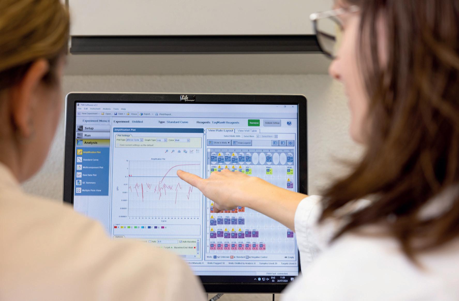
A key part of Van Boeckel’s work involves mapping antibiotic resistance across regions, especially in low- and middle-income countries. This requires assembling data from a wide range of sources. “In many of these countries, there is no systematic surveillance system,” he says. “We often rely on small studies, samples from markets, or data published by local microbiologists. These studies are often scattered across a variety of journals.”
To make sense of this fragmented data, Van Boeckel and his team consolidate it into a central database. “We spend thousands of hours reading and extracting data from these studies,” he notes. “It’s labor-intensive, but it allows us to build statistical models that map resistance patterns and identify geographic hotspots.”
The team is now looking at automating this data collection process. Van Boeckel explains, “We have submitted a grant application to test how accurate our findings based on automated methods are compared to data extracted ‘by hand’. If we can show that the accuracy is high—around 90 percent—we believe it could reliably augment or replace the labor-intensive work currently done by humans. This would allow us to scale our work and conduct it in real time.”
“Our role is to present policymakers with data-driven scenarios, illustrating potential outcomes based on different courses of action.”
Thomas Van Boeckel, One Health Institute, University of Zurich
Real-world impact and policy implications
The work of OHI has significant implications for policy and practice in animal husbandry and public health. Van Boeckel emphasizes the importance of providing evidence to support informed decision-making. “Our role is to present policymakers with data-driven scenarios, illustrating potential outcomes based on different courses of action,” he explains. While making progress can present challenges, there are encouraging examples. Van Boeckel cites Thailand as one such case: “Thailand was early among low and middle income countries to establish a systematic surveillance system for antibiotic use in animals. They recognized early on that improving transparency and reducing overuse would not only benefit public health but also strengthen the reputation of their agricultural exports.”
The OHI, established in May 2023, is a unique institution jointly founded by the Vetsuisse Faculty, the Faculty of Medicine, and the Faculty of Science at the University of Zurich. The institute integrates human, animal, and environmental health in a holistic approach.
The OHI’s organizational structure reflects its interdisciplinary nature, with the institute simultaneously being part of three faculties. This innovative setup provides a solid long-term foundation for the One Health approach at the University of Zurich. While currently focused on research, the institute has plans for expanding its educational offerings too.
“We are actively developing One Health teaching modules,” says Van Boeckel. “I will be leading courses at the Faculty of Science next year, including a dedicated course on superbugs. Our long-term vision includes establishing comprehensive teaching programs, potentially culminating in a master’s degree in One Health here in Zurich.”
The interdisciplinary nature of OHI is central to its success. Van Boeckel emphasizes the importance of diverse
expertise in addressing complex health challenges. “One Health is fundamentally interdisciplinary,” he explains. “My background is in engineering, but I transitioned to epidemiology. Our team included medical doctors, microbiologists, and mathematicians as well as physicists.”
This diverse team reflects the broader mission of the One Health Institute: to address complex health challenges by integrating knowledge from multiple disciplines. Van Boeckel highlights the mutual learning that occurs within the team: “The collaboration between different disciplines enhances everyone’s skills. For instance, veterinarians working alongside mathematicians and ecologists often develop a deeper appreciation for statistical modeling.”
One Health research, while important, operates in a different funding landscape from traditional medical research areas. Van Boeckel describes the current situation. “For diseases like cancer or cardiovascular conditions, there are well-established foundations and dedicated funding programs,” he explains. “One Health, however, does not fit neatly into this structure, as funding is traditionally organized along disciplinary lines.”
The unique nature of One Health research, with its focus on the interconnectedness of human, animal, and environmental health, requires substantial resources for data collection, analysis, and interdisciplinary collaboration. Van Boeckel sees potential for growth in this field as awareness of its importance increases.
“A majority of the classes of antibiotics that we use in animals are the same antibiotics used in humans. ”
Thomas Van Boeckel, One Health Institute, University of Zurich
As the One Health Institute continues to grow, its focus on interdisciplinary collaboration and evidence-based solutions positions it to address some of the most complex global health challenges.
“The health of humans, animals, and the environment is interconnected,” says Van Boeckel. “This means that veterinarians, medical doctors, environmental scientists, and many other experts can benefit from working together to address global health challenges.”
Justus Krueger is an independent journalist based in Hong Kong, who specializes in business, health, and technology. His work has appeared in a variety of publications, including Mare, Internationale Politik, the Berliner Zeitung, the Neue Zürcher Zeitung and Der Spiegel.
Animal health is the mission of the Vetsuisse Faculty’s University Animal Hospital in Zurich. Financial Director Beatrice Gasser and Medical Director Professor Dr. Jean-Michel Hatt are proud to head an internationally renowned institution that combines research, education, and patient care for a broad spectrum of animal species. What are their expectations and plans for 2025? Interview by Philipp Grätzel von Grätz
The University Animal Hospital is among the leading veterinary clinics in Europe. Running such a highly specialized institution in an academic environment is a challenge. But there are also huge gains for animal owners, since the University Animal Hospital can offer access to cutting-edge diagnostics and treatments that are hard to find elsewhere.
Can you briefly introduce the University Animal Hospital?
beatrice gasser: The University Animal Hospital takes care of around 28,000 patients – and 20,000 animal owners – per year. Fifty-nine percent of the animal owners come from the canton of Zurich, the remaining 41 percent from the rest of Switzerland and from abroad. Half of our patients are dogs, about a quarter are cats, 14 percent are horses, eight percent farm animals, and five percent zoo animals, pets, and wildlife.
What are the advantages of an academic animal hospital?
jean-michel hatt: We see ourselves as a specialist hospital with a far broader spectrum of services than other animal hospitals. We are especially proud of our emergency service, which is open 24 hours a day, 365 days a year. Another hallmark is that, as an academic hospital, we combine patient care, teaching, and research. This is appealing not only for students, but also for veterinarians from all over the world who want to specialize. And it is attractive for animal owners, who get access to diagnostic procedures and treatments that are not readily available in other places.
What examples can you give of particularly innovative services or treatments?
hatt: We are one of the main centers for oncological radiation therapy in Europe. There is a broad spectrum of surgical procedures both for small and large animals. For horses, specifically, we are covering cardiology, which is pretty unique. There are also ophthalmological services, some of which are highly specialized.
Can you give an overview of the teaching activities?
hatt: Most important are our veterinary students, obviously. We usually have around 500 at a time. But there is more to it than that. We are also engaged in veterinary nursing education. And we are offering advanced training to veterinarians who want to specialize, for example, in cardiology, in dermatology, or in herd medicine.
“The big challenge is finding the right balance between the basic education of students on the one hand and offering ever more complex care for patients on the other.”
Beatrice Gasser, Financial Director University Animal Hospital Zurich
This usually takes about three to four years, and the graduates get a certificate that is internationally recognized.
What are some current research topics that are being addressed?
hatt: A very active research area is brain tumors, gliomas specifically. We have very precise and minimally invasive technologies available that can be used to diagnose and remove brain tumors. This is also about interdisciplinary collaboration. We are running clinical trials to evaluate the combination of surgery and chemotherapy – always with the goal to minimize the burden on patients as much as possible. Another interesting project is our Growing Dog Project, a long-term clinical trial with a strong focus on prevention. In large animals, I would mention a diagnostic research project in horses with cardiac valve diseases. In this area, we are cooperating with other universities and animal hospitals, for example the University of Ghent in Belgium.
What are the specific challenges and the opportunities for an academic animal hospital?
gasser: The big challenge is finding the right balance between the basic education of students on the one hand and offering ever more complex care for patients on the other. This is not only a question of staffing, but also of infrastructure and, in the end, of money. We need excellent staff, both veterinarians and nurses, and we are lucky that we get these people. The staff shortage is an issue, but being an attractive and internationally renowned hospital, it is easier for us than for many others. When it comes to opportunities, since we are part of an academic faculty, we can draw on non-clinical expertise of all kinds. The University of Zurich’s Institute of Parasitology, to give just one example, has veterinary parasitology as one focus. This is good both for research and for patients.

What goals does the University Animal Hospital have in 2025?
gasser: A high priority project is choosing a new hospital information system. This will be supported by our Information Technology team. It is not only about buying new software, but also about interface management: We have a large number of medical devices, and we have other medical systems like a medical picture archiving and communication system. All this needs to be integrated properly. Another core task in 2025, as in any other year, is financial sustainability. As a university hospital, we have certain duties to fulfil that are not necessarily cost-covering – teaching, for example. To be able to do that, we have to make sure that we offer attractive services for which there is a demand among
“We are especially proud of our emergency service, which is open 24 hours a day, 365 days a year.”
Jean-Michel Hatt, Medical Director University Animal Hospital Zurich
animal owners. This is a dynamic field. We have to think about the right balance and make adjustments every year.
hatt: From a medical perspective, I would add infection prevention as another focus for 2025. This is rapidly evolving, both in medicine and in veterinary medicine. It is closely related to biosecurity, where we have a very
high standard already that we want to develop further. Infection prevention efforts are good for patients, but they have a financial dimension, too – think of outbreaks of notifiable diseases, such as bluetongue disease, that can be very costly. Finally, some reconstruction projects will continue to keep us busy in 2025. Most important are stabilization efforts at the Equine Hospital. There is also construction being done by the faculty at non-clinical departments like the Institute of Virology. All these efforts and projects will make the University Animal Hospital Zurich even more attractive – for animal owners, but also for our employees.
Philipp Grätzel von Grätz, a trained doctor, works as a medical journalist in Berlin.
A 50-Million-Franc Infrastructure Boost for Virology/Parasitology and the Equine Hospital
The comprehensive renewal of the infrastructure of the Vetsuisse Faculty’s Equine Hospital, Institute of Virology, and Institute of Parasitology will ensure our scientists stay at the forefront of their fields – and give students the opportunity to practice in a modern and fit-for-purpose environment. Text by Roman Elsener
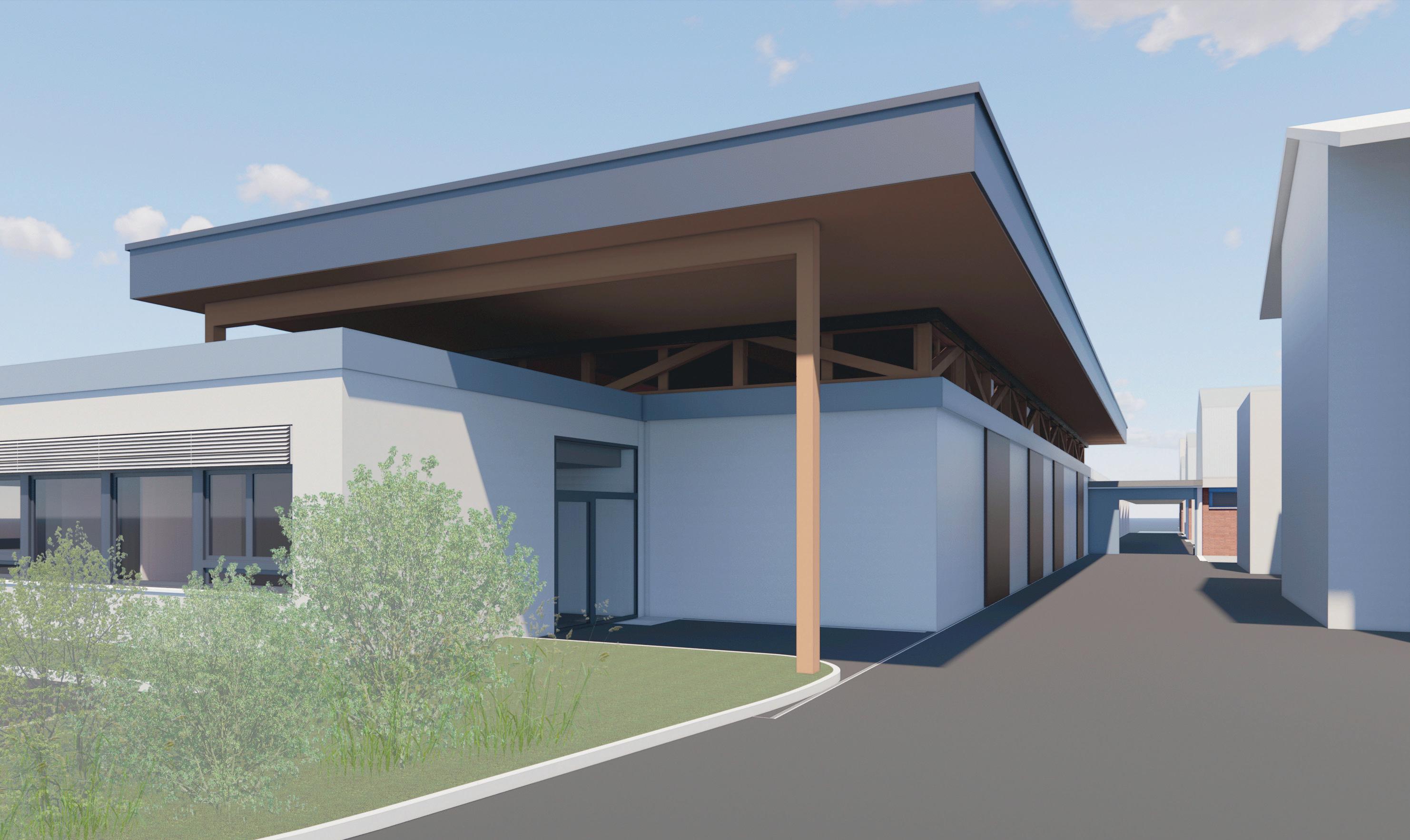
Let’s take a leap into the future: in 30 to 40 years, most of the buildings at the Irchel North Campus of the University of Zurich will have been demolished, replaced by a new, densified L-shaped building hosting the Vetsuisse Faculty. Clinics will occupy the ground floor, with labs and offices above. As one of the leading international centers for veterinary medicine, the faculty’s four clinical departments and numerous institutes are bustling hubs of activity. Roger Stephan, Dean of the Vetsuisse Faculty, describes this scenario, the University of Zurich’s long-term strategy, as a realistic outlook and an important sign that Irchel North will remain the faculty’s main site, strategically connected to nearby campuses.
Aging infrastructure
But 2060 is a long way off, and edifices stemming from the 1960s are reaching the end of their lifespan now. “Some of the buildings belonging to the Equine Hospital are a par-
ticular challenge,” Stephan notes. Treating approximately 3,600 horses annually, the clinic combines medicine, surgery, ophthalmology, and sports medicine under one roof. However, the outdated infrastructure struggles to compete with private practices. Similarly, the Institute of Virology and the Institute of Parasitology face difficulties. One temporary building from 1972, built in a no-construction zone, is still in use. Despite years of planning, no suitable renovation options have been identified.
“The renovation will create a modern space for equine patient care, student training, and clinical research, benefiting both animals and owners.”
Colin Schwarzwald, Director at the Equine Hospital of the University Animal Hospital Zurich

“The University of Zurich’s goal of achieving CO₂ neutrality by 2030 prioritizes sustainable construction. It is integral to the project, encompassing building materials, insulation, and heating and ventilation. ”
Roger Stephan, Dean of the Vetsuisse Faculty, University of Zurich
Three years ago, Stephan got the ball rolling. “I thought it would be better on our campus if the same things were strategically located in the same place, i.e. lab to lab,” says the Dean.
Plans for the Equine Hospital as well as Virology/Parasitology were combined into one project. The University management recognized the strategic benefits and supported the 50-million-Swiss-franc (55.3 million US dollars) investment for the refurbishments.
What makes the renovation project especially challenging is that both the institutes and the Equine Hospital will continue to operate during the renovation. Rotating offices in existing buildings will minimize disruptions. Stephan aims to limit relocations, with the goal of moving groups directly into their new spaces once complete. Professor Dr. med. vet. Colin Schwarzwald, Director of the Equine Hos-
pital of the University Animal Hospital, acknowledges that carrying out renovation work during ongoing operations leads to logistical challenges. But Schwarzwald is convinced, “We are able to overcome these thanks to our highly motivated nursing and veterinary teams.”
“The renovation will improve infrastructure to meet the growing number of patients and students, as well as higher hygiene and safety standards,” he adds. It will create a modern space for patient care, student training, and clinical research, benefiting both animals and owners.
The upgrade is essential for the faculty, as it depends on equine patients for hands-on student training. “We rely on owners bringing their animals so students gain real-world experience,” Stephan explains. “We don’t buy animals so that we can show the students what an animal looks like. They do that in other countries, in so-called teaching hospitals.”

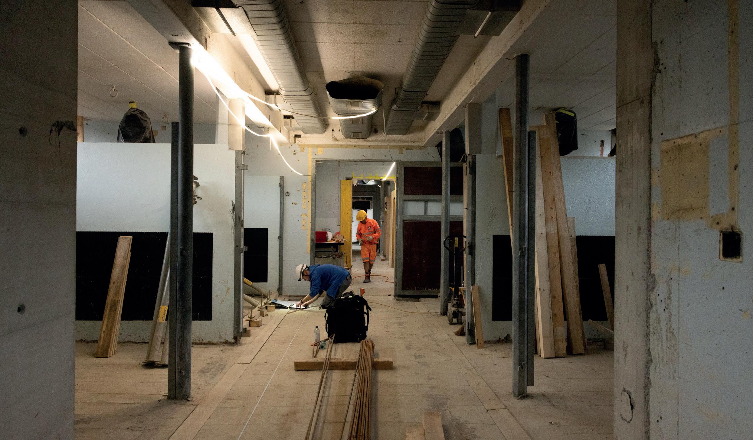
The Virology Institute will remain largely in the same building during renovations. Staff and students are adapting, using noise alerts on the faculty’s website to plan remote work.
“Our building was built as a temporary structure, not just a few years ago, but half a century ago,” says Professor Dr. Cornel Fraefel, Director of the Institute. “Space is very confined and due to the permeability of the building envelope, temperatures in the laboratories in summer often exceed 30 degrees Celsius, meaning that experiments cannot be carried out under normal conditions.”
The construction project gives his team a real perspective for the future. “Previously, we had very little office space available; doctoral students and undergraduates had their desks in the laboratories,” he explains. “After the renovation, the laboratories and offices will be spatially separated in two different parts of the building, allowing optimal and efficient use.”
The renovation ensures that the buildings meet today’s standards and also enables the continuation of the good work that is already being done by the institute. ”The constant climatic conditions in the laboratories will improve the reproducibility of the experiments and thus support our competitiveness in research,” says Fraefel. It will also be easier to implement the necessary safety guidelines such as biosafety measures, fire hazards and controlled access, and ensure proper hygiene to combat hospital-acquired infections.
In addition to research and teaching, the Institute of Virology and the Institute of Parasitology operate service labo -
“The modern infrastructure and constant climate conditions in the laboratories will improve the reproducibility of our experiments and thus support our competitiveness in research. ”
Cornel Fraefel, Head of the Institute of Virology, Vetsuisse Faculty, University of Zurich
ratories. Approximately 2,500 tests for viruses relevant to veterinary medicine in pets, livestock, and zoo animals are carried out per year.
The institutes conduct highly competitive research worldwide in the field of diseases that affect humans, too. “Virology and parasitology are both specialist areas that deal with infectious diseases, including zoonoses from animals to humans, as the world witnessed during COVID-19,” Stephan says. “If you look at the incidence of disease worldwide, the prevalence of infectious diseases is expected to rise in the future. That’s why it’s also very important for these specialist areas to have an appropriate research infrastructure.”
Construction work on the Equine Hospital, including a new building for internal medicine, will be completed by mid-2026. Parasitology too can move into a converted building by mid-2026, and the Institute of Virology will shine in new splendor a year later.
Roman Elsener is an independent journalist who divides his time between Switzerland and New York. He has worked as U.S. Correspondent for various Swiss media such as NZZ and the Swiss news agency Keystone-SDA.
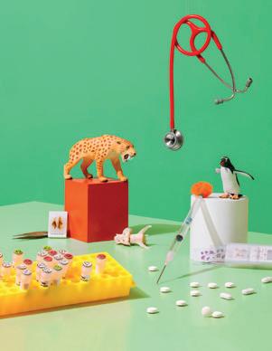
Image concept of the Vetsuisse Faculty Annual Report 2024
Science thrives at the intersection of disciplines — where seemingly unrelated ideas converge into meaningful discoveries.
This image series captures exactly that essence: inspired by a look into labs and clinics, where research takes shape in a full range of bright colors — and not only in the black and white of stated facts.
Each still life photograph represents one of the six key articles in this year's faculty report. Thoughtfully composed, they bring together laboratory instruments, clinical tools, diagnostic elements, and symbolic objects that reflect each research theme.
By playfully mixing small toys with real samples and devices, the series highlights the balance between precision and curiosity that defines cutting-edge scientific discovery — where, much like in art, every element is chosen with both clear intention and intuition to create new enriching insights and spark inspiration.
Michelle Aimée Oesch, science photographer
Vetsuisse Faculty UZH
Imprint
Vetsuisse Faculty UZH Annual Report 2024
Vetsuisse Faculty UZH
Winterthurerstrasse 204
CH-8057 Zurich
Phone: +41 (0)44 635 81 21
vet.uzh.ch
Concept, Design & Layout
Roger Stephan, Dean
Michelle Aimée Oesch, science photographer
Jeanne Peter, graphics
Vetsuisse Faculty UZH
Fabian Elsener, layout
Primafila AG
Photos
Michelle Aimée Oesch, science photographer
Mayra Ibarra, trainee science photographer
Vetsuisse Faculty UZH
Additional images
Pascal Halder (Long COVID, pages 29–31)
Enzo Franchini, Zoo Zurich (Avian malaria, pages 18–19)
Deborah Cooke (Tuberculosis, page 34 bottom, page 37)
Editorial board
Writing: Primafila Correspondents team
Project management (texts): Kim Rupli, Primafila AG
Editing: Morven McLean, Primafila AG
Online issuu.com/uzhch/docs/vetsuisse_report_2024