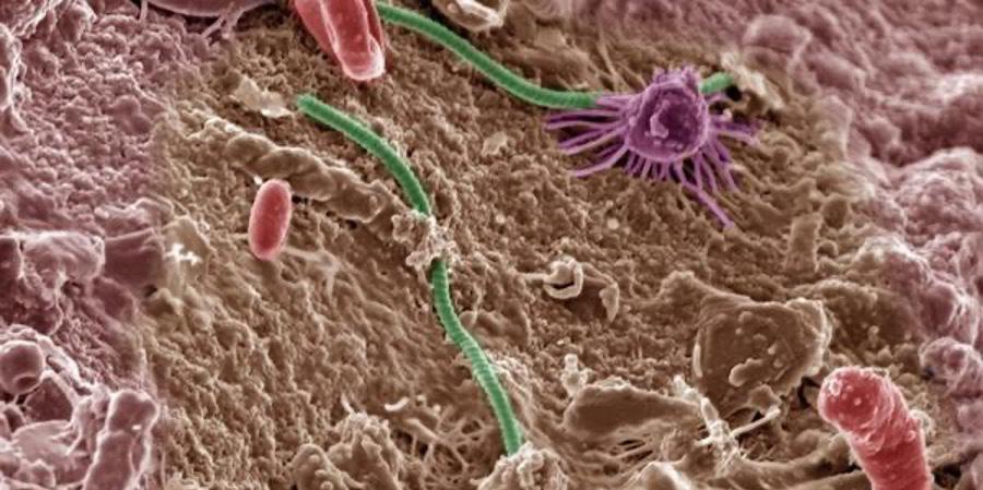
Ultrasound Mediated Delivery of Therapeutics BY SOYEON (SOPHIE) CHO '24 Cover Image: A mouse’s brain after intravenous administration of experimental nanoparticles that can bypass the blood-brain barrier (BBB). Cell nuclei are shown in blue, blood vessels in red, and human cancer cells in green. These experimental nanoparticles are one of the current therapeutic efforts, such as ultrasound, to administer drugs across the BBB. Image Source: Wikimedia Commons
Introduction Ultrasound is defined as sound waves with high frequencies that are not audible by humans. Because sound waves are much safer than other methods used for imaging, due to the lack of radiation, ultrasound has been used for diagnostic imaging of fetuses in pregnant women as well as swelling, infection, or other forms of pain in internal organs. Also, unlike x-rays, ultrasound scans clearly indicate soft tissues, which explains its widespread use. However, applications of ultrasound outside imaging have been explored in the past several years. Nonimaging uses of ultrasound use the frequencies between the audible region (less than 20 kHz) and the diagnostics ultrasound region (more than 10 MHz) (Mitragotri, 2005). This large range of ultrasound frequencies allows for many different uses outside of imaging, including the delivery of therapeutic drugs for patients.
Effects of Ultrasound Cavitation The direct and indirect effects of ultrasound waves allow for a wide range of ultrasound applications in therapeutics. The primary effect is that the ultrasound waves directly interact with mediums like cells and tissues through periodic 47
oscillations of a frequency and amplitude determined by the ultrasound source (Mitragotri, 2005). For the secondary effects, ultrasound increases temperature as the medium absorbs the oscillating sound waves. This impacts tissues with a higher absorption coefficient for ultrasound waves, such as bones rather than muscle tissue (Suslick, 1988). A more significant secondary effect comes from cavitation, which is the cleavage, growth, and oscillation of microbubbles hit by ultrasound waves. Microbubbles have a diameter of around 3 µm and are inserted into the site of interest through intravenous injection. In addition to assisting delivery of drugs and gene products, microbubbles can also act as contrast agents for imaging, since they oscillate in response to ultrasound much more than cells (Blomley et al., 2001). Despite initial concerns about having gaseous spaces within the blood vessels, clinical studies have demonstrated no safety hazards from microbubbles, due to their small size (Nanda et al., 1997). There are two major types of cavitation, dependent on the stability of the oscillatory motions of the microbubbles. Stable cavitation involves stable, DARTMOUTH UNDERGRADUATE JOURNAL OF SCIENCE


