

Molecular Pathology for the FRCPath part 1
Matt Evans


Disclosures
I have received honoraria from
Agilent Technologies
Amgen
AstraZeneca
Boehringer Ingelheim
Bristol-Myers Squibb
Eli Lilly
Incyte
Merck Sharp & Dohme
Pfizer
Roche
Servier
Almost 11 hours of recorded lectures
Document covering molecular pathology matched to the curriculum

Lots of resources!
https://www.molecularpathologyuk.net/

Overview


What are the key types of molecular alteration?
Genomic: small variants, structural variants, copy number variants, epigenetic alterations (methylation)
Non-genomic: IHC surrogates of genomic alterations, MMR IHC, PD-L1 IHC
How can we test for molecular alterations?
Small variants: real-time PCR, NGS
Structural variants: IHC, ISH, RT-PCR, NGS
Copy number variants: IHC, ISH
Non-genomic variants: MMR IHC (and MSI), PD-L1 IHC
What are the key molecular alterations in common cancers?
NTRK and DPYD
Non-small cell lung cancer: EGFR, BRAF, KRAS, ALK, ROS1, RET, METex14, PD-L1
Colon cancer: MMR/MSI, KRAS, NRAS, BRAF
Breast cancer: HER2, Oncotype DX, PD-L1, PIK3CA
High-grade ovarian cancer: BRCA, HRD
Endometrial cancer: MMR, POLE
Melanoma: BRAF, PD-L1

What are they key types of molecular alteration?
What are the key types of molecular alteration?
Genomic alterations
A T A G G C C A>C substitution
A T A G G C A EGFR, KRAS, NRAS, BRAF, KIT, PDGFRA, PIK3CA, POLE, BRCA
Indel: C deletion + AA insertion
CD74
Translocation, rearrangement
Fusion
, ROS1, RET, NTRK, FGFR2, sarcoma, lymphoma
Amplification, gain Deletion, loss
What are the key types of molecular alteration?
Non-genomic alterations
Immunohistochemical surrogates for genomic alterations
Small variants: BRAF V600E
Structural variants: ALK, ROS1, NTRK
Small variants in the MMR genes (usually germline)
Methylation of the MLH1 promoter region (usually somatic)
Reflects the ability of tumour cells to evade the immune system
Copy number variants: HER2

How can we test for molecular alterations?
How does pathological processing affect nucleic acids?
This will continue for many hours in the centre of a resection specimen if it is not opened/sliced/inflated! C>T sequence changes
Why do we need to assess samples?


The higher the neoplastic cell %, the lower the risk of missing variants
Why do we need to assess samples?
Minimum neoplastic cell percentage 20%
Neoplastic cell percentage 50%
There is no evidence of small variants involving the EGFR, BRAF or KRAS genes. There is no evidence of structural variants involving the ALK, ROS1, RET, NTRK1, NTRK2 or NTRK3 genes. There is no evidence of MET exon 14 skipping.
Neoplastic cell percentage 5%
There is no evidence of small variants involving the EGFR, BRAF or KRAS genes. There is no evidence of structural variants involving the ALK, ROS1, RET, NTRK1, NTRK2 or NTRK3 genes. There is no evidence of MET exon 14 skipping.
Can safely say there is nothing here This could be a false negative!


A colorectal cancer sample comprises 10% neoplastic cells. NGS testing is undertaken (minimum neoplastic cell percentage 20%). This reveals a pathogenic KRAS variant.
Which is the appropriate interpretation?
A. This result could represent a false positive in view of the low neoplastic cell percentage.

B. Long duration of formalin fixation would bring this result into doubt.
C. This result is likely to be reliable.
D. It is likely that other clinically relevant variants have been missed in view of the low neoplastic cell percentage.
E. This should be considered a failed test.
How can we test for genomic alterations?
Small variants
Variant detection technologies
Are any of these variants of interest present in this sample?
Structural variants Copy number variants Methylation
Immunohistochemistry
Can I infer a structural variant from protein expression?
Immunohistochemistry
Can I infer a CNV from protein expression?
In situ hybridisation
Real-time PCR
Sequencing
Are any variants present in this sample?
Direct sequencing
Pyrosequencing
Next-generation sequencing
Is the gene in an abnormal location?
Reverse transcription PCR
Are any of these fusions of interest present?
Next-generation sequencing
Are any fusions present?
In situ hybridisation
Is there more of this gene than usual?
Next-generation sequencing
Are there extra copies of this gene?
Small variant detection
Real-time PCR: the theory
Small variant detection
Real-time PCR: the results
Small variant detection
Real-time PCR: the results
Fluorescence
Small variant detection
Real-time PCR: the practical side
Only specific variants detected
Will miss rare variants not covered by the included probes

https://www.biocartis.com/sites/default/files/2021-03/Idylla_KRASIVD_Tech-Sheet.pdf
Multiple variants detected by a single type of fluorescence
Cannot discriminate the exact variant present
Small variant detection
Real-time PCR: overview
Detects specific variants of interest
There is a limit to the number of variants detectable with a single test
Only feasible for genes with limited number of hotspots
Generally need one test per gene
Pros:
Fast
Low neoplastic cell percentage requirement
Small amount of DNA required (per test)
Cons:
Will miss rare variants
If multiple genes needed, will end up consuming lots of DNA
Small variant detection
Next-generation sequencing: library preparation
Small variant detection
Next-generation sequencing: library preparation
Small variant detection
Next-generation sequencing: read alignment
Reference sequence
GAGTGATAGC


Small variant detection
Next-generation sequencing: read alignment
Low depth/coverage (repetitive sequence?)

High depth/coverage


Small variant detection
Next-generation sequencing: overview
Massively parallel sequencing
Can assess multiple samples for multiple genes (up to whole genome)
In theory, will detect any variant in the DNA sequenced
Pros:
Should not miss variants
Can assess multiple genes in parallel
If large numbers of targets need assessing, likely to require less DNA overall
Cons:
Slower
Higher neoplastic cell percentage requirement
Large amount of DNA required if only 1-2 targets needed
Expensive infrastructure, need skilled staff
Small variant detection
Next-generation sequencing: panels
Generally higher failure rates, longer turnaround times


https://www.oncomine.com/hubfs/Downloadable%20PDFs/Genexus_Webinar_Slides.pdf
https://www.e-crt.org/upload/media/crt-2019-305-suppl1.pdf
https://www.illumina.com/content/dam/illumina/gcs/assembled-assets/marketingliterature/trusight-oncology-500-data-sheet-m-gl-00173/trusight-oncology-500-and-ht-datasheet-m-gl-00173.pdf
A 57 year old female non-smoker is diagnosed with lung adenocarcinoma. It is widely metastatic and she is rapidly deteriorating. The oncologist asks for EGFR small variant testing and needs a result as quickly as possible.
Which would be the most appropriate technique?
A. Immunohistochemistry
B. Next-generation sequencing (panels)
C. Next-generation sequencing (whole gene sequencing)
D. Real-time PCR
E. Fluorescence in situ hybridisation (FISH)
How can we test for genomic alterations?
Small variants Structural variants Copy number variants Methylation
Variant detection technologies
Are any of these variants of interest present in this sample?
Real-time PCR
Sequencing
Are any variants present in this sample?
Direct sequencing
Pyrosequencing
Next-generation sequencing
Immunohistochemistry
Can I infer a structural variant from protein expression?
Immunohistochemistry
Can I infer a CNV from protein expression?
In situ hybridisation
Is the gene in an abnormal location?
Reverse transcription PCR
Are any of these fusions of interest present?
Next-generation sequencing
Are any fusions present?
In situ hybridisation
Is there more of this gene than usual?
Next-generation sequencing
Are there extra copies of this gene?
Structural variant detection Immunohistochemistry
Others

https://jcp.bmj.com/content/75/3/145
https://www.researchgate.net/publication/11224272_RET_Expression_i
n_Papillary_Thyroid_Cancer_from_Patients_Irradiated_in_Childhood_for_ Benign_Conditions
Others


Structural variant detection
Immunohistochemistry: overview
Use protein expression to infer the presence of an underlying fusion
More useful for certain genes than others
Pros:
Fast
Very few tumour cells needed
Neoplastic cell percentage irrelevant
Not specific for particular fusions
Fairly robust
Cons:
Only looks at one gene at a time (generally) – inefficient if multiple targets needed
Many markers do not have an objective definition of positivity
Sensitive to under-fixation
Structural variant detection
In situ hybridisation: the theory
Structural variant detection
In situ hybridisation: the detection methods


https://www.wicell.org/home/characterization/cgmp-testing-services/cgmp-fluorescence-insitu-hybridization-fish/gmp-fluorescence-in-situ-hybridization-fish.cmsx
https://www.researchgate.net/figure/Silver-in-situ-hybridization-for-a-case-of-polyCEP17-withHER2-signals-a-and-CEP17_fig1 275046947
https://www.researchgate.net/figure/HPV-ISH-Diffuse-signal-pattern-H-E-400-presents-the-ISHsignals-of-HPV-shown-by_fig1 51488619
https://www.mlo-online.com/home/article/13005291/automated-her2-testing-personalizedhealthcare-for-breast-cancer-patients-enabled-by-novel-molecular-morphology-methods
Structural variant detection FISH: interpretation
Structural variant detection
FISH: interpretation
Structural variant detection FISH: interpretation
Normal
Rearranged
Structural variant detection
FISH: overview
Look at the relative location of the gene of interest to identify rearrangements
Pros:
Can be fast
Neoplastic cell percentage irrelevant
Fairly robust
Cons:
Need a good number of well-preserved tumour cells
Only looks at one gene at a time – inefficient if multiple targets needed
Subjective assessment
Rearrangements at the DNA level do not always result in targetable fusion proteins
Small-scale (e.g. intrachromosomal) rearrangements may be missed
Need fluorescence microscope, need skilled staff
Structural variant detection Reverse transcription PCR: the theory
Structural variant detection
Reverse transcription PCR: the theory
GUGAUAAUAGUGCGCGUGUAAAUAGCU
Gene 1 Gene 2
Structural variant detection Reverse transcription PCR:


https://www.biocartis.com/sites/default/files/202103/Idylla_GeneFusion-RUO_Tech-Sheet.pdf
Structural variant detection
Reverse transcription PCR: overview
Much like real-time PCR
Detects specific fusions of interest
There is a limit to the number of fusions detectable with a single test
Generally need one test per gene
Pros:
Fast
Low neoplastic cell percentage requirement
Small amount of DNA required (per test)
Cons:
Will miss rare fusions
If multiple genes needed, will end up consuming lots of RNA
Less robust than IHC/FISH
Structural variant detection
Next-generation sequencing
Long repetitive sequence
Difficult to sequence Transcription
Structural variant detection
Next-generation sequencing: overview
Massively parallel sequencing
Typically use DNA for small variants and CNVs; RNA for structural variants
Can assess multiple samples for multiple genes (up to whole genome)
In theory, will detect any rearrangement in the RNA sequenced
Pros:
Should not miss fusions
Can assess multiple genes in parallel
If large numbers of targets need assessing, likely to require less RNA overall
Cons:
Slower
Higher neoplastic cell percentage requirement
Large amount of RNA required if only 1-2 targets needed
High failure rate
Expensive infrastructure, need skilled staff
A lung adenocarcinoma is tested for ALK rearrangements using FISH. This demonstrates a rearrangement. Subsequent ALK immunohistochemistry is negative.
Which of the following is most likely to be explanation for this discordance?
A. The rearrangement has not given rise to a functional fusion protein
B. The rearrangement has given rise to a fusion which the immunohistochemical antibody cannot detect
C. Suboptimal tissue processing has given a false positive on FISH
D. The rearrangement has occurred over a short distance, giving rise to false positive result on FISH
E. Non-neoplastic cells have mistakenly been assessed on FISH
How can we test for genomic alterations?
Small variants Structural variants Copy number variants Methylation
Variant detection technologies
Are any of these variants of interest present in this sample?
Real-time PCR
Sequencing
Are any variants present in this sample?
Direct sequencing
Pyrosequencing
Next-generation sequencing
Immunohistochemistry
Can I infer a structural variant from protein expression?
Immunohistochemistry
Can I infer a CNV from protein expression?
In situ hybridisation
Is the gene in an abnormal location?
Reverse transcription PCR
Are any of these fusions of interest present?
Next-generation sequencing
Are any fusions present?
In situ hybridisation
Is there more of this gene than usual?
Next-generation sequencing
Are there extra copies of this gene?
Copy number variant detection Immunohistochemistry
Gene amplification
Overexpression of mRNA
Overexpression of protein
Copy number variant detection Immunohistochemistry


https://diagnostics.roche.com/global/en/products/tests/ventana-her2dual-ish-dna-probe-cocktail-assay.html

Copy number variant detection
Immunohistochemistry:
overview
Use protein expression to infer the presence of an underlying CNV
Only works for certain genes
Often good for ruling out amplification or confirming high-level amplification
Often struggles with grey area in between
Pros:
Fast
Few tumour cells needed
Neoplastic cell percentage irrelevant
Fairly robust
Cons:
Only looks at one gene at a time (generally) – inefficient if multiple targets needed
Subjective – tendency to defer to an alternative technique
Sensitive to under-fixation
Copy number variant detection
In situ hybridisation: normal
Mean copy number 2
1
Copy number variant detection
In situ hybridisation: amplification
Mean copy number 4
Copy number variant detection
In situ hybridisation: polysomy
Mean copy number 4
Copy number variant detection
In situ hybridisation: overview
Look at the number of copies of gene of interest, relative to reference gene
Pros:
Can be fast
Neoplastic cell percentage irrelevant
Fairly robust
Cons:
Need a good number of well-preserved tumour cells
Only looks at one gene at a time – inefficient if multiple targets needed
Subjective assessment
(need fluorescence microscope, need skilled staff)
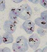
Non-genomic alterations Mismatch
repair
IHC

Non-genomic alterations
Mismatch repair IHC
MMR-proficient (pMMR)

MMR-deficient (dMMR)

Non-genomic alterations
Mismatch repair IHC
BRAF variant testing V600E detected V600E not detected Possibly germline Colon only
Non-genomic alterations
Mismatch repair IHC/microsatellite instability
Maternal copy Paternal copy
An endometrial cancer shows the following result on MMR immunohistochemistry:


Which of the following is true?
A. The patient likely has Lynch syndrome
B. The next step is to organise referral to clinical genetics.
C. The next step is MLH1 promoter methylation testing.
D. The next step is BRAF V600E testing.
E. The next step is MSI testing.

Non-genomic alterations
PD-L1 IHC

Non-genomic alterations
PD-L1 IHC
PD-L1
PD-1
Non-genomic alterations
PD-L1 IHC
Depending on tumour type, response to immune checkpoint inhibitors may be correlated with:
Extent of PD-L1 expression
PD-L1 has:
Multiple assays
Inability to repair DNA damage
e.g. MMR defects
Multiple assessment methods (tumour cells, inflammatory cells, both)
Depends on tumour type and drug to be prescribed
What is the ‘best’ way of identifying small variants?
Oncogenes
EGFR, BRAF, KRAS, NRAS, PIK3CA

Real-time PCR may be viable (except for KIT, PDGFRA)
Tumour suppressors
BRCA

Only sequencing viable
What is the ‘best’ way of identifying small variants?
Real-time PCR
Generally faster
Generally lower neoplastic cell percentage requirement
Generally lower failure rate
Will miss rarer variants
Generally need one test for each gene of interest
Next-generation sequencing
Generally slower
Generally higher neoplastic cell percentage requirement
Generally higher failure rate
Should detect all variants
Can look at dozens/hundreds of genes with no additional tissue
What is the ‘best’ way of identifying structural variants?
DNA-based technologies
FISH DNA NGS
RNA-based technologies IHC
Reverse transcription PCR RNA NGS
Not all rearrangements at the DNA level make it into targetable fusion proteins
A rearrangement detected at the RNA level is more likely to give rise to a targetable fusion protein More false positives
false positives Lower failure rate
failure rate
What is the ‘best’ way of identifying structural variants?
Fast
Few cells needed
Should cover all fusions
Robust
Easy to implement
May not work for some genes
One gene per stain
Questionable definitions of positivity
Can be fast
Works with low neoplastic cell percentage
Needs decent number of cells
One gene at a time
Subjective
May identify non-productive rearrangements
May miss small-scale rearrangements
Fast Works with low neoplastic cell percentage
Low DNA requirement
Should not miss fusions
Can assess multiple genes in parallel
Lower RNA requirements if multiple targets assessed
Misses rare fusions
Generally one gene per reaction
Higher failure rate than IHC/FISH Slower
High neoplastic cell percentage requirement
High failure rate
Expensive

What
are the key molecular alterations in common cancers?
What are the key molecular alterations in common cancers?
Non-small cell lung cancer
Colon cancer
Breast cancer
High-grade ovarian cancer
Endometrial cancer Melanoma
NTRK structural variants
NTRK1, NTRK2, NTRK3
A patient with a solid tumour harbouring an NTRK rearrangement and who has no ‘satisfactory’ treatment options is eligible for targeted therapy, irrespective of histological tumour type
Secretory carcinoma of breast
Secretory carcinoma of salivary gland
Infantile fibrosarcoma
Congenital mesoblastic nephroma
‘Wild-type’ GIST
Spitzoid neoplasms
Papillary thyroid carcinoma (esp. paediatric)
Paediatric gliomas
Common cancers
Molecular testing in non-small cell lung cancer
EGFR small variant
BRAF V600 small variant
PCR or sequencing (10-15%)
Sensitive to TKIs
Sensitive to BRAF/MEK inhibitors PCR or sequencing (<5%)
KRAS G12C small variant
ALK structural variant
Sensitive to TKIs IHC, FISH or sequencing (2-3%)
ROS1 structural variant Sensitive to TKIs
or sequencing, ?IHC screening (1-2%)
NTRK structural variant Sensitive to TKIs
Sensitive to KRAS G12C inhibitors PCR or sequencing (10-15%) RET structural variant Sensitive to TKIs
or sequencing, ?IHC screening (1%)
or sequencing (1-2%)
exon 14 skipping variant Sensitive to TKIs
or sequencing (3-4%)
Sensitive to immune checkpoint inhibitors
EGFR small variants in NSCLC
More common in females, non-/light smokers, especially of (South)
East Asian heritage, with adenocarcinomas
L858R and exon 19 deletions commonest and associated with strong sensitivity to anti-EGFR TKIs
T790M associated with resistance
Patients invariably become resistant after around a year
Depending on the TKI given, may be important to test for acquisition of secondary T790M variant
Repeat biopsy or plasma testing
May prompt change to a different TKI
MET exon 14 skipping variant in NSCLC
PD-L1 expression in NSCLC


A 55 year old man (performance status 1) is diagnosed with widely metastatic lung adenocarcinoma. Molecular testing identifies an ALK rearrangement. No alterations are identified in EGFR, BRAF, KRAS, ROS1, RET, NTRK or MET. PD-L1 TPS is 100%.
Which is the most appropriate first-line treatment?
A. Chemotherapy
B. Immune checkpoint inhibitor therapy
C. Tyrosine kinase inhibitor therapy
D. Combined chemotherapy and immune checkpoint inhibitor therapy
E. Surgery
Molecular testing in colorectal cancer
MMR expression
MSI testing
KRAS variant
NRAS variant
BRAF V600E variant
IHC
MSI testing (15-20%)
PCR or sequencing (30-50%)
PCR or sequencing (5%)
PCR or sequencing (10-15%)
Lynch syndrome, better prognosis, sensitive to immune checkpoint inhibitors
Resistant to anti-EGFR therapy
Resistant to anti-EGFR therapy
Resistant to anti-EGFR therapy, poor prognosis, sensitive to MEK inhibitors
A 64 year old woman (performance status 0) is diagnosed with metastatic colorectal cancer. Molecular testing reveals no alterations in KRAS, NRAS or BRAF. MMR protein expression is preserved.
Which is the most appropriate first-line treatment?
A. Tyrosine kinase inhibitor therapy
B. Chemotherapy
C. Surgery
D. Chemotherapy with anti-EGFR monoclonal antibodies
E. Immune checkpoint inhibitor therapy
Molecular testing in breast cancer
HER2 amplification
Sensitive to anti-HER2 therapy IHC+/-ISH or ISH (20%)
Oncotype DX
(ER+ HER2- only) Predicts recurrence risk Commercial assay
PD-L1 expression (TNBC only)
PIK3CA small variant (ER+, HER2- only)
Sensitive to immune checkpoint inhibitors IHC
Sensitive to PI3K inhibitors PCR or sequencing (30-40%)
HER2 amplification testing in breast cancer
https://www.spandidos-publications.com/ol/14/5/5265?text=fulltext https://diagnostics.roche.com/global/en/products/tests/ventana-her2dual-ish-dna-probe-cocktail-assay.html 0/1+


Oncotype DX testing in breast cancer

https://www.oncotypeiq.com/en-CA/breast-cancer/healthcareprofessionals/oncotype-dx-breast-recurrence-score/interpreting-theresults
Early-stage ER+ HER2-
Intermediate risk of recurrence post-surgery Gene expression profile assay Gives risk of recurrence postsurgery to help establish riskbenefit balance of adjuvant chemotherapy
Molecular testing in high-grade ovarian cancer
Tumour BRCA1/BRCA2 variant
HRD testing
Sequencing (10-25%)
Sensitive to PARP inhibitors
Sensitive to PARP inhibitors + bevacizumab Commercial assay (50%)
Should be done in parallel with germline BRCA1/BRCA2 testing on blood
What is homologous recombination deficiency (HRD)?
How can we test for HRD?
Option 1: Look for the causes
Homologous recombination deficiency (HRD)
Inability to repair double strand DNA breaks
Option 2: Look for the effects
A 71 year old woman (performance status 1) is diagnosed with stage IV primary peritoneal high-grade serous carcinoma. HRD testing on tumour tissue returns the following result:
Genomic instability score: 66 (HRD present)
Tumour BRCA1/BRCA2: No pathogenic variants detected
Which is the correct interpretation?
A. This patient requires further testing to assess for a germline BRCA1/BRCA2 variant
B. This patient is not eligible for PARP inhibitors
C. A tumour BRCA1 or BRCA2 pathogenic variant has been missed
D. HRD testing is not appropriate in primary peritoneal carcinomas
E. A germline BRCA1/BRCA2 variant is exceptionally unlikely in a patient of this age
Molecular testing in endometrial cancer
MMR expression
POLE small variant
More indolent behaviour, lack of benefit from adjuvant therapy Sequencing (10%) All cases at diagnosis
Lynch syndrome, likely better prognosis, sensitive to immune checkpoint inhibitors IHC (15-20%)
A 53 year old woman is diagnosed with poorly differentiated endometrial carcinoma. It shows loss of MSH2 and MSH6 on MMR immunohistochemistry.
Which is the correct interpretation?
A. The MMR result suggests a diagnosis of serous carcinoma
B. MLH1 promoter methylation testing is required
C. This patient has Lynch syndrome
D. This tumour is likely to respond to immune checkpoint inhibitors
E. This tumour is more likely to behave aggressively
Molecular testing in melanoma
BRAF V600 small variant
PD-L1 expression
Sensitive to BRAF/MEK inhibitors
Determines immune checkpoint inhibitor selection PCR or sequencing (50%)
A 67 year old woman is diagnosed with widely metastatic melanoma, with no evidence of a primary cutaneous lesion. Molecular testing reveals a BRAF V600D variant. PD-L1 immunohistochemistry is negative (TPS 0%).
Which is the correct interpretation?
A. This patient’s first treatment option should be chemotherapy
B. This patient should not receive immune checkpoint inhibitors
C. This patient would only be eligible for BRAF/MEK inhibitors if the tumour harboured a V600E variant
D. Detection of a BRAF variant makes a cutaneous origin likely
E. This patient is eligible for monoclonal antibody therapy
Summary
What are the key types of molecular alteration?
Genomic: small variants, structural variants, copy number variants, epigenetic alterations (methylation)
Non-genomic: IHC surrogates of genomic alterations, MMR IHC, PD-L1 IHC
How can we test for molecular alterations?
Small variants: real-time PCR, NGS
Structural variants: IHC, ISH, RT-PCR, NGS
Copy number variants: IHC, ISH
Non-genomic variants: MMR IHC (and MSI), PD-L1 IHC
What are the key molecular alterations in common cancers?
NTRK
Non-small cell lung cancer: EGFR, BRAF, KRAS, ALK, ROS1, RET, METex14, PD-L1
Colon cancer: MMR/MSI, KRAS, NRAS, BRAF
Breast cancer: HER2, Oncotype DX, PD-L1, PIK3CA
High-grade ovarian cancer: BRCA1/2, HRD
Endometrial cancer: MMR, POLE
Melanoma: BRAF, PD-L1
Almost 11 hours of recorded lectures
Document covering molecular pathology matched to the curriculum
Lots of resources!
https://www.molecularpathologyuk.net/

Practice questions
A 79 year old man is diagnosed with metastatic melanoma. Which of the following set of targets is the oncologist most likely to request?
A. BRAF, NRAS and KIT small variant, and PD-L1
immunohistochemistry
B. EGFR, BRAF and KRAS small variant, ALK, ROS1, RET, NTRK small variant, MET exon 14 skipping, and PD-L1 immunohistochemistry
C. KRAS, NRAS and BRAF small variant
D. HRD testing
E. KIT and PDGFRA small variant, and NTRK structural variant
A 65 year old female never-smoker is diagnosed with metastatic lung adenocarcinoma. Performance status is 0 and she is well. Molecular testing reveals an EGFR exon 19 deletion. No clinically relevant variations were detected in the BRAF, KRAS, ALK, ROS1, RET, NTRK or MET genes.
PD-L1 tumour proportion score was 0%.
What is the most appropriate initial treatment?
A. Chemotherapy
B. Surgery
The EGFR variant should be targeted as a priority
Metastatic lung cancer does generally no undergo surgery
C. Radiotherapy
Palliative radiotherapy would only be an option for symptomatic relief
D. Immunotherapy
PD-L1 is low; actionable genomic alterations should be the priority
E. Tyrosine kinase inhibitor therapy
A 76 year old man presents with metastatic melanoma. He has extensive brain metastatic disease and is rapidly deteriorating on ITU. The oncologist tells you that they need to know as quickly as possible whether the patient will be eligible for targeted therapy.
Which would be the most appropriate technique?
A. Fluorescence in situ hybridisation (FISH)
B. Real-time PCR
C. Next-generation sequencing (NGS)
D. RNA microarray
This is used to look for structural or copy number variants
A good option, but would take longer
Used to assess gene expression levels
E. Dual-colour dual-hapten in situ hybridisation (DDISH)
As per FISH
A 65 year old man is diagnosed with caecal adenocarcinoma. MLH1 promoter methylation testing is performed; this demonstrates evidence of MLH1 promoter methylation.
Which is the most likely outcome?
A. Expression of the MLH1 gene is likely to be reduced.
B. MLH1 expression is likely to be preserved on IHC.
C. PMS2 expression is likely to be preserved on IHC.
Expression of both MLH1 and PMS2 is likely to be reduced/lost.
D. There is likely to be a small variant in the MLH1 gene.
E. This patient requires assessment by clinical genetics.
This essentially excludes a germline mechanism.
A 63 year old woman is diagnosed with ductal carcinoma NST. HER2 expression is graded as 3+ on immunohistochemistry.
Identify the best descriptor of HER2.
A. DNA repair protein
B. Immune checkpoint
C. Receptor tyrosine kinase
and BRCA proteins
D. Steroid hormone receptor
E. Transcription factor
A 66 year old man undergoes mediastinal lymph node excision.
Histology demonstrates a lymph node with a tiny focus of subcapsular metastatic adenocarcinoma. Molecular testing needs to be undertaken.
Which of the following is most likely to improve the quality of results obtained.
A. Cut a 10 µm section rather than the usual 5 µm
B. Perform macrodissection
C. Perform microdissection
This will increase the amount of DNA/RNA but still only a tiny amount will be tumour
Would help, but never done in clinical practice
D. Use direct (Sanger) sequencing
Direct sequencing has high neoplastic cell percentage requirements, so this would be a bad option
E. Use a stained section for DNA extraction
Extraction should always be from freshly cut sections wherever possible


Thank you