15 minute read
Department of Bioengineering, Department of Psychiatry, Department of Neurology, Physician Scientist Training Program, University of Pittsburgh School of Medicine
Association between functional dedifferentiation and amyloid in preclinical Alzheimer’s Disease
*Elizabeth J. Mountza , *Jinghang Lia , Akiko Mizuno, PhDb , Ashti M Shah, BSc, Andrea Weinstein, PhDb , Ann D. Cohen, PhDb, William E. Klunk, PhDb, d , Beth E. Snitz, PhDb, d, Howard J. Aizenstein, MD/PhDa, b , Helmet T. Karim, PhDb
aDepartment of Bioengineering, bDepartment of Psychiatry, cPhysician Scientist Training Program, University of Pittsburgh School of Medicine, dDepartment of Neurology * These authors contributed equally to this work.
Jinghang Li Jinghang is a senior Biomedical Engineering student from Wenzhou, China. He has been working in the Geriatric psychiatry neuroimaging lab since May 2020. After graduation he plans on pursuing his PhD in Biomedical Engineering applying deep learning on imaging diagnostics.
Elizabeth J. Mountz Elizabeth Mountz is a Junior studying Bioengineering at the University of Pittsburgh Swanson School of Engineering. She hopes to continue her research efforts in Graduate School.
Howard J. Aizenstein, M.D., Ph.D. Dr. Aizenstein is an expert on the cognitive and affective neuroscience of aging and geriatric brain disorders. He is trained as a geriatric psychiatrist and also a computer scientist. His research program is recognized for expertise in MRI analyses methods, as well as their use for clinical research in aging.
Significance Statement
Preclinical Alzheimer’s disease (AD) is a stage of AD defined by the presence of brain amyloid without overt cognitive impairment, occurring up to two decades prior to diagnosis. Amyloid may deposit asymmetrically, which has been shown to affect neural functional asymmetry in AD but not during the preclinical period. We show that altered functional asymmetry in the hippocampus may appear as early as the preclinical stages of AD and is associated with amyloid deposition.
CATEGORY: Experimental Research
Keywords: preclinical Alzheimer’s Disease,
dedifferentiation, amyloid, memory encoding
Abstract
Preclinical Alzheimer’s disease (AD) is characterized by significant brain amyloid-β (Aβ) pathology without overt cognitive impairment. Aβ has been shown to deposit asymmetrically and has been shown to be associated with asymmetric brain glucose metabolism. In clinical AD, Aβ burden may exceed the compensatory reserve threshold, leading to greater neural functional asymmetry in AD individuals compared to elderly controls, where asymmetry is associated with cognitive function. To better understand AD progression, we investigated the association between markers of asymmetry, AD pathology, and cognitive function in cognitively normal older adults. Using fMRI during a memory encoding task, we calculated functional asymmetry and spread of activation in the hippocampus and dorsolateral prefrontal cortex, which are part of the core and extended memory network. Using positron emission tomography (PET), we measured brain Aβ and global glucose metabolism. We also collected data on APOE allele status and cognitive function. We conducted multivariate linear regression with measures of dedifferentiation (i.e., asymmetry index and spread of activation) as the outcome and markers of AD as independent variables. We additionally investigated their associations with domains of cognitive function. We found that greater global Aβ deposition was associated with greater hippocampal functional asymmetry and lower left hippocampal activation spread during a memory encoding task. Functional asymmetry and spread were not associated with cognitive function. Similar to studies in AD, we found that functional asymmetry was associated with greater Aβ in cognitively normal older individuals. This may hint at the early neurodegeneration in preclinical AD.
1. Introduction
Alzheimer’s disease (AD) is a process of progressive neurodegeneration which leads to severe cognitive dysfunction, including impairments in memory loss and executive control. AD is associated with a buildup of Amyloid-β (Aß) and neurofibrillary tangles in the brain. These cytotoxic proteins especially affect areas associated with memory, such as the hippocampus and dorsolateral prefrontal cortex (DLPFC) [1]. Greater Aß deposition, as measured by positron emission tomography (PET), is associated with functional changes in the brain including hippocampal atrophy, low cerebral glucose metabolism [2] and functional changes in neural activation as measured by functional magnetic resonance imaging (fMRI) [3].
Aß deposition is gradual with a preclinical stage where significant Aß deposition can be detected but no cognitive dysfunction is observed [4]. Aß deposition is often asymmetric, burdening one hemisphere more than the other [5]. One hypothesis for this lack of cognitive dysfunction with significant Aß pathology during the preclinical stage, is that compensatory neural recruitment can help delay cognitive dysfunction. In healthy individuals, neural activation is often lateralized and localized during tasks such as memory encoding due to the highly specialized roles of
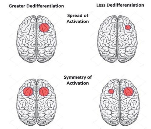
neural networks within the hemispheres [6]. Compared to healthy individuals, greater bilateral neural recruitment as well as greater spatial extent of activation are thought to be two measures of compensatory activation which are termed dedifferentiation, or the general loss of neural specificity (i.e., regions become more domain general in terms of function). Dedifferentiation may allow the pathology-burdened hemisphere to recruit greater neural resources from the other hemisphere in order to maintain cognitive function [7]. Dedifferentiation in cognitively normal individuals is thus measured through both greater symmetry in (bilateral) fMRI activation (i.e., left dominant DLPFC activation spilled over into right DLPFC), as well as greater spread of local activation (i.e., more widespread DLPFC activation within the hemisphere) (Figure 1) [7]. The Hemispheric Asymmetry Reduction in Old Adults (HAROLD) general aging model posits that, dedifferentiation in the frontal regions of older adults may help maintain cognitive function despite the stresses of aging by recruiting greater neural resources, both bilaterally and locally [7]. These compensatory dedifferentiation mechanisms may still be intact in preclinical AD and may help maintain cognitive function during early Aß deposition.
Figure 1. A schematic representation of two forms of dedifferentiation. Greater dedifferentiation may be indicated by greater spread (upper image) and greater symmetry (lower image) of activation due to greater recruitment of neural resource locally and laterally during a given task.
In later stages of AD, increased Aß burden may exceed compensatory capacity, which may result in cognitive impairment [2, 8]. In prodromal [i.e., mild cognitive impairment (MCI)] and mild-to-moderate AD, asymmetric Aß deposition is associated with asymmetric cerebral hypometabolism [5]. Asymmetric (i.e., lateralized to left hemisphere) glucose hypometabolism is associated with Aβ positive (i.e., greater Aß burden than a cut-off) MCI patients [9]. Thus, later stages of AD are associated with functional asymmetry.
In this study, we sought to characterize the relationship between functional asymmetry and Aß deposition in preclinical AD in order to better understand methods of early intervention and diagnosis, as Aß deposition may be undetectable in the earliest stages of AD trajectory. While large bodies of research exist for structural asymmetry in AD progression [10], there are little to no studies on functional asymmetry in preclinical AD. Based on the HAROLD general aging model, we hypothesized that in cognitively normal individuals, greater AD pathology (e.g., Aß deposition and cerebral hypometabolism) would be associated with greater dedifferentiation, measured with greater symmetry and spread of activation. Because our participants were cognitively normal participants, we did not expect to observe an association between cognitive function and activation symmetry or spread. We used the face-name encoding fMRI task because it reliably activates the hippocampus and DLPFC [10]. The hippocampus is critical for memory and learning and shows neuronal degeneration early in the onset of AD [1], while the DLPFC is part of an extended memory encoding network by coordinating the relational information [7].

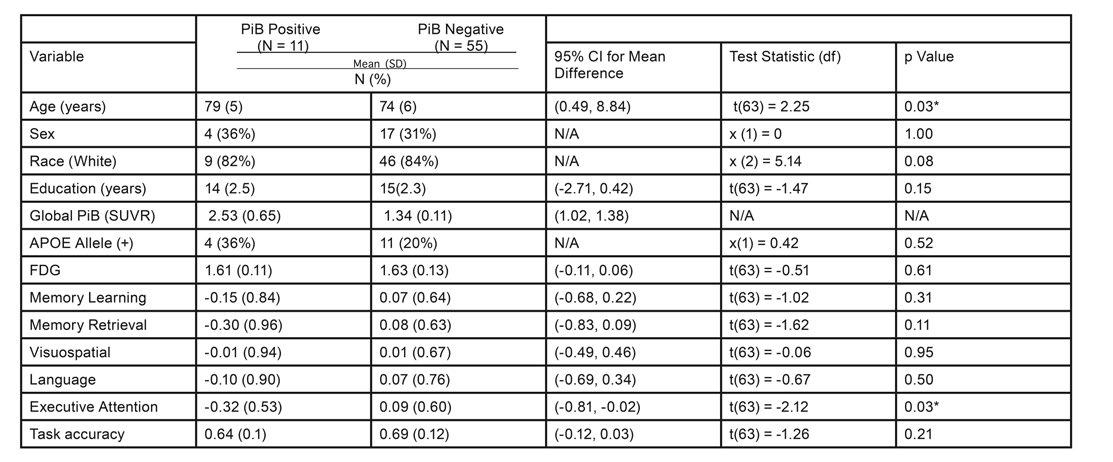
Table 1. Demographics and Summary of Cognitive Domain Scores Note: *p < 0.05. We did not conduct a statistical test on Global PiB (SUVR) since this is how these groups were defined.
2. Methods
Participants and Assessments: We analyzed cross-sectional data from 87 cognitively normal older adults (>65 years). All participants underwent a neuropsychological test battery used by the University of Pittsburgh Alzheimer Disease Research Center (ADRC) to assess cognitive function. Test scores were combined into domain composite z-scores including memory (Logical Memory, Modified Rey-Osterreith Figure, ADRC Word List); visuospatial abilities (Block Design, Modified Rey-Osterreith Figure Copy); language (Animal and Letter Fluency, the 60-Item Boston Naming Test); and executive attention (Trail Making Test A and B, Clock Drawing, Maximum Digit Span Forward and Backward, Stroop Interference Score, and Digit Symbol Substitution) [11] (Table 1). The memory domain was split into learning and retrieval to isolate the role of the DLPFC in delayed memory retrieval. We also collected demographic data and genetic AOPE status.
Neuroimaging Data Acquisition and Preprocessing: PET scans were used to measure participant cerebral Aβ deposition with Pittsburgh Compound B (PiB) and glucose metabolism with fluorodeoxyglucose (FDG) tracers. Participants were classified as PiB positive or negative based on our previous study [12]. We collected whole-brain (excluding cerebellum) fMRI (a 3T Siemens Trio scanner) during a face-name memory encoding task. During the task, participants were shown a face-name pair and were asked to subjectively determine whether each face fit the name. This task consisted of two blocks: unfamiliar facename pairs (novel condition) and familiar (i.e., previously seen) face-name pairs (control condition). This task was administrated over three runs and contained 50 facename pairs. We computed the contrast between novel and control. For our analysis, we extracted functional activation in two regions of interest (ROIs): hippocampus and DLPFC of each hemisphere based on the automated anatomical labeling atlas. After performing the task in the scanner, participants took a post-scan test in which they saw the same faces with two name options. They were asked to select the name they saw in the scanner as an estimate of recognition accuracy.
Computation of Dedifferentiation Measures: We computed the asymmetry index (AI) as the hemispheric laterality of the mean activation in each region. We calculated the asymmetry index using the equation:
where L (left) and R (right) are the mean activation values of a given ROI in the corresponding hemisphere [13]. The equation yields a value between -1 and 1, where 0 represents symmetric activation between the hemispheres, and a negative or positive AI indicates right or left hemispheric dominant activation, respectively. We also computed absolute AI (abs_AI) as a measure of non-directional laterality. Smaller abs_AI indicates greater dedifferentiation (i.e., greater symmetric activation). We also measured dedifferentiation with the spread of activation within each ROI [14]. For each participant, we identified the peak activation voxels, then generated a series of spheres with radii ranging from 1 voxel to 25 voxels centered around peak activation, where we calculated the average activation within each sphere. We then plotted the mean activation with respect to different radii. We used a linear interpolation method to estimate the neural activity decline and took the radius where the activation value was half of the observed peak activation as the spread of the activation [e.g., full width half maximum (FWHM)]. Greater FWHM indicates greater spread (or dedifferentiation).
Statistical Analyses: We conducted eight multivariate linear regressions in R to investigate the associations between measures of dedifferentiation (AI, abs_AI, left
hemispheric FWHM, and right hemispheric FWHM, in both ROIs) and AD related factors (PiB status, global cerebral glucose metabolism, and APOE E4 status). We conducted four linear regressions to investigate the association between left/right hemispheric spread of activation and abs_AI for both ROIs. We also conducted ten multivariate linear regressions to investigate the association between five cognitive domains and both measures of dedifferentiation (abs_AI and FWHM). Other AD related factors were included as predictor variables. All models controlled for demographic factors (age, education, sex, and race), and post-scan recognition accuracy was included as a covariate to predict cognitive function.
3. Results
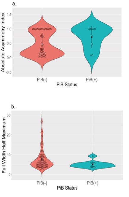
Among our multivariate linear regressions, two models showed marginal significance: absolute hippocampus AI (F(7,53) = 1.95, R2 = 0.205, p = 0.08) and spread of activation in the left hippocampus (F(7,53) = 2.07, R2 = 0.215, p = 0.063). PiB positive individuals (11/66) showed greater asymmetric activation in the hippocampus (ß = 0.33, p = 0.024, Figure 2a). PiB positive individuals showed lower spread of activation in the left hippocampus (ß = -4.18, p = 0.019, Figure 2b). DLPFC dedifferentiation measures were not associated with any AD related factors.

Figure 2. PiB positive group showed significantly greater asymmetric hippocampal activation (a) and less spread of activation in the left hippocampus (indexed by FWHM) (b) during a memory encoding task compared to the PiB negative group.
Individuals with lower activation spread in the left hippocampus had greater absolute asymmetry of hippocampal activation (F(1,82) = 12.14, R2 = 0.129, p = 0.001; ß = -4.33, p = 0.001). Only executive attention domain score was significantly associated with PiB positive status in the two separate models with all AD factors: abs_AI (F(10, 50) = 2.68, R2 = 0.349, p = 0.01; ß = -0.53, p = 0.016) and spread (F(12, 48) = 3.39, R2 = 0.459, p = 0.001; ß = -0.61, p = 0.004). No cognitive domain scores were associated with markers of dedifferentiation.
4. Discussion
Contrary to our hypothesis, we found that PiB positive individuals had greater hippocampal asymmetry and lower spread of activation in the left hippocampus. Unlike the prefrontal dedifferentiation pattern posited in the HAROLD model, there were no associations with DLPFC asymmetry or spread of activation. Greater asymmetry in preclinical AD may be a mechanism of either greater recruitment of activation in the right hippocampus or reduced activation of the left hippocampus. We found that lower left hippocampal spread of activation was associated with greater hippocampal activation asymmetry. PiB positive individuals who are cognitively normal may also have greater cognitive reserve, which enables them to compensate for greater pathology to maintain cognitive function. Therefore, the association between PiB positive individuals and greater asymmetry and less spread may be indicative of the individual differences of greater cognitive reserve, which may help to maintain normal cognitive function despite pathology.
In the clinical stages of AD, functional changes such as asymmetric cerebral hypometabolism are associated with asymmetric Aß deposition [5]. Further, Aβ positive status in AD is associated with leftward lateralization of glucose metabolism decline [9]. Our results indicate that early pathological Aß deposition in the brain is associated with greater asymmetry in preclinical stages of AD, which is in agreement with previous studies that have associated asymmetry with AD, but hints toward an earlier neurodegenerative effect of Aß deposition. We found no associations between dedifferentiation measurements and cognitive function, as these compensatory mechanisms may help to maintain cognitive function. On the other hand, greater Aβ deposition was associated with lower executive attention. These may suggest that Aß deposition is a predictor of the higher order cognitive function regardless of dedifferentiation patterns among cognitively normal individuals or that a functional MRI task that engages executive function may be a better potential functional marker (i.e., face-name task is primarily memory encoding).
There are several limitations to this study – primarily, the associations between dedifferentiation measures and Aß deposition should be validated in other samples and in studies that utilize longitudinal designs to observe changes in activation spread and symmetry in relation to changes in Aß deposition. These results show correlations and do not imply causation of Aß deposition on functional brain activity. This study did utilize a fairly large cross-sectional sample with both functional imaging and intricate PET imaging, which is a strength.
5. Conclusion
We used two measurements (e.g., functional asymmetry and spread of activation) to quantify the extent of neuronal dedifferentiation as a tool to assess the association with Alzheimer’s disease pathology in preclinical AD. We identified significant differences in markers of neural activity associated with the presence of AD risk factors in the hippocampus. We did not find associations between DLPFC and any AD risk factors. We speculate that AD risk factors such as significant Aß deposition may limit the spread of activation in the left hippocampus which affects functional asymmetry. This observation in preclinical AD suggests an early neurodegenerative effect of Aß, before any onset of cognitive symptoms. Future longitudinal studies should identify whether these changes track the progression of Aß or are an indirect consequence of Aß accumulation.
6. Acknowledgements
This study was supported by the SSOE Summer Research Internship and P50 AG005133, P01 AG025204, R37 AG025516 from the National Institute of Health. We would like to acknowledge the GPN Lab for providing the research infrastructure and continuous mentorship.
7. References
[1] Svenningsson, A.L., et al., beta-amyloid pathology and hippocampal atrophy are independently associated with memory function in cognitively healthy elderly. Sci Rep, 2019. 9(1): p. 11180. [2] Cohen, A.D., et al., Basal cerebral metabolism may modulate the cognitive effects of Abeta in mild cognitive impairment: an example of brain reserve. J Neurosci, 2009. 29(47): p. 14770-8. [3] Edelman, K., et al., Amyloid-Beta Deposition is Associated with Increased Medial Temporal Lobe Activation during Memory Encoding in the Cognitively Normal Elderly. Am J Geriatr Psychiatry, 2017. 25(5): p. 551-560.
[4] Dubois, B., et al., Preclinical Alzheimer’s disease: Definition, natural history, and diagnostic criteria. Alzheimers Dement, 2016. 12(3): p. 292-323. [5] Frings, L., et al., Asymmetries of amyloid-beta burden and neuronal dysfunction are positively correlated in Alzheimer’s disease. Brain, 2015. 138(Pt 10): p. 3089-99. [6] Dolcos, F., H.J. Rice, and R. Cabeza, Hemispheric asymmetry and aging: right hemisphere decline or asymmetry reduction. Neuroscience & Biobehavioral Reviews, 2002. 26(7): p. 819-825. [7] Cabeza, R., Hemispheric asymmetry reduction in older adults: the HAROLD model. Psychol Aging, 2002. 17(1): p. 85-100. [8] Jack, C.R., Jr., et al., Tracking pathophysiological processes in Alzheimer’s disease: an updated hypothetical model of dynamic biomarkers. Lancet Neurol, 2013. 12(2): p. 207-16. [9] Weise, C.M., et al., Left lateralized cerebral glucose metabolism declines in amyloid-beta positive persons with mild cognitive impairment. Neuroimage Clin, 2018. 20: p. 286-296. [10] Sperling, R.A., et al., Encoding novel face-name associations: a functional MRI study. Hum Brain Mapp, 2001. 14(3): p. 129-39. [11] Snitz, B.E., et al., Risk of progression from subjective cognitive decline to mild cognitive impairment: The role of study setting. Alzheimer’s & Dementia, 2018. 14(6): p. 734-742. [12] Price, J.C., et al., Kinetic modeling of amyloid binding in humans using PET imaging and Pittsburgh Compound-B. Journal of Cerebral Blood Flow & Metabolism, 2005. 25(11): p. 1528-1547. [13] Seghier, M.L., Laterality index in functional MRI: methodological issues. Magn Reson Imaging, 2008. 26(5): p. 594-601. [14] Gordon, B.A., et al., Spread of activation and deactivation in the brain: does age matter? Front Aging Neurosci, 2014. 6: p. 288.


