Controlling cell sociology with microrobotics The mechanobiology of cells is an important aspect of medicine. We spoke to Mahmut Selman Sakar, Tenure Track Assistant Professor at EPFL in Lausanne, about the work of the ROBOCHIP project in developing microrobotic technologies, research which could open up new perspectives on human physiology and pathology. The human body is made up of trillions of cells, and the ways that these cells are structurally organised inside various tissues exerts a major influence on the designated function of our organs. Cells are exquisitely sensitive and responsive to their dynamic mechanical environment. Both externally applied forces and the mechanical properties of the extracellular matrix (ECM) have been shown to modulate cell behaviour. As the Principal Investigator of the ROBOCHIP project, Professor Sakar aims to develop a robotic toolkit that could facilitate the investigation of how cells communicate with each other and organize through the ECM. This work centres around developing untethered microscopic machines that can be placed inside engineered three-dimensional (3D) microtissues, where they generate spatiotemporally resolved signals. “We can precisely control the magnitude and duration of the deformation applied by the synthetic microactuators. Using this novel technology, we are studying how mechanical signals propagate within the tissues, how they are sensed and processed by the resident cells, and finally how cells collectively respond to mechanical loading,” outlines Professor Sakar.
ROBOCHIP project The results of this work are expected to provide fundamental new information about the mechanobiology of cells, such as the
12
quantification of cell behaviour under welldefined static and dynamic loading conditions. “Our group has developed a number of different techniques for powering microactuators wirelessly. They can be powered by magnetic fields, near infrared light, and ultrasound,” says Professor Sakar. The methodology also involves engineering the ECM from mechanically characterized fibrous materials and time-lapse imaging. “We track where the cells are and
architecture of the ECM in these processes?” asks Professor Sakar. As of yet these questions have not been answered.
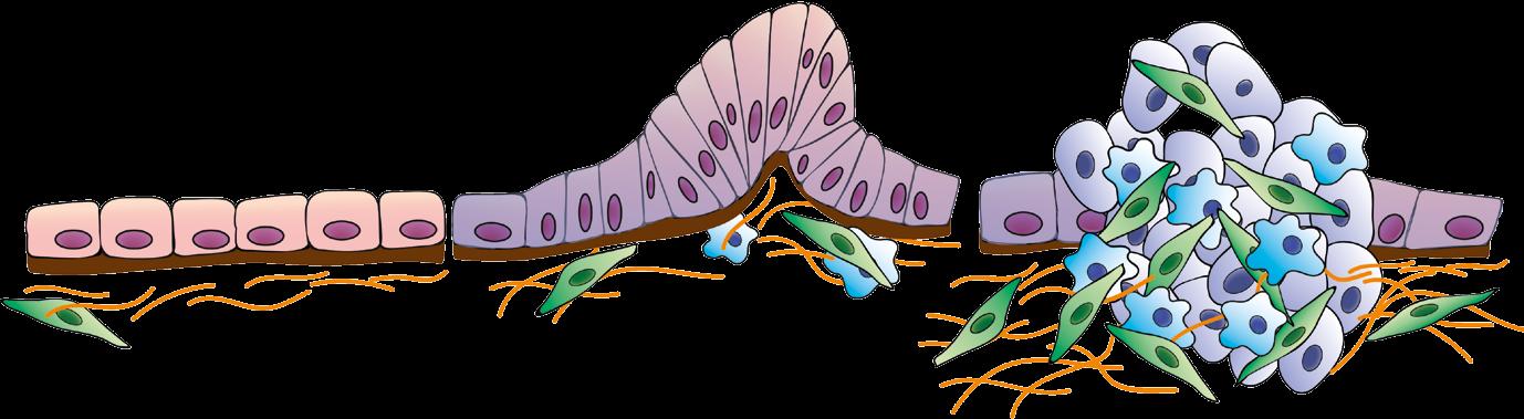
Cellular communication The aim here is to essentially uncover the physical rules behind multicellular organization, research that holds wider relevance to our understanding of disease. For example, certain conditions drive the formation of cysts, tumours,
The microrobotic toolkit that we are developing will perfectly complement modern imaging modalities and biochemical manipulation techniques. Together, they will provide a more complete picture on how the resident cells of a particular tissue interrogate and remodel their microenvironment as a community. what they do in real-time using fluorescent markers so that we can correlate the action of the robotic tools with the biological output,” continues Professor Sakar. The mechanical loading conditions mimic the types of signals that cells are exposed to within the body. The goal then for researchers is to identify what specific factors disrupt homeostasis and what kind of stimulation may facilitate healing. “Do the cells identify the magnitude or the frequency of the mechanical signal? When do they start changing their phenotype and moving beyond physiological limits? What is the contribution of the
or fibrotic scarring in almost every tissue within the body, and the situation may become much more serious if these masses are allowed to grow and spread unchecked. “The abnormal cell clusters are made of our cells. What are the signals that convince healthy cells to abandon their physiological roles and form nonfunctional and even destructive tissues? Recent work has shown that fibroblasts and immune cells residing around a tumour may apply forces, which stimulate the cancer cells to spread. This is only one example of how mechanical factors play a key role in the initiation and progression of certain diseases,” outlines Professor Sakar.
EU Research