


Case Series
1

4
Supraclavicular Brachial Plexus Block for Challenging Anterior Shoulder Dislocations: A Case Series
M Shalaby, O Gregory, C Raciti, M Rosselli, O Mechanic
Serratus Anterior Plane Block for Procedural Anesthesia for Pigtail Tube Thoracostomy: A Case Series
E Lopez, R Sahni, M Cooper, M Shalaby
Case Reports
10
14
17
Analgesia in the Emergency Department for Lower Leg and Knee Injuries: A Case Report
M Shalaby, Y Lee, J McShannic, M Rosselli
Pericapsular Nerve Group Block for Prosthetic Hip Reduction in the Emergency Department: Case Report
R Thomas, M Carr, J Fry, T Hoffman
Takotsubo Syndrome Following Status Epilepticus in a Heart Transplant Recipient: A Case Report
T Shikama, M Shikama, N Hayase, K Doi
21 A Case Report of Status Epilepticus in a Patient with Acute HAE
D Weinberg, S Gayda, K Hultz, H Atia, B Kohen, E Boccio
25 A Tic-ing Time Bomb: Case Report of a Unique Presentation of Sudden-onset Tics
A Neto, V Perez, K Manwaring, L Averill, V Ovalle
28 Case Report of HIV and Neurosyphilis Coinfection in a Recent Migrant: Old Diseases in New Faces
AV Besien, IP Mauricio, R Belfort, J Lykins


Contents continued on page iii


About Us: Penn State Health is a multi-hospital health system serving patients and communities across central Pennsylvania. We are the only medical facility in Pennsylvania to be accredited as a Level I pediatric trauma center and Level I adult trauma center. The system includes Penn State Health Milton S. Hershey Medical Center, Penn State Health Children’s Hospital and Penn State Cancer Institute based in Hershey, Pa.; Penn State Health Hampden Medical Center in Enola, Pa.; Penn State Health Holy Spirit Medical Center in Camp Hill, Pa.; Penn State Health Lancaster Medical Center in Lancaster, Pa.; Penn State Health St. Joseph Medical Center in Reading, Pa.; Pennsylvania Psychiatric Institute, a specialty provider of inpatient and outpatient behavioral health services, in Harrisburg, Pa.; and 2,450+ physicians and direct care providers at 225 outpatient practices. Additionally, the system jointly operates various healthcare providers, including Penn State Health Rehabilitation Hospital, Hershey Outpatient Surgery Center and Hershey Endoscopy Center.
We foster a collaborative environment rich with diversity, share a passion for patient care, and have a space for those who share our spark of innovative research interests. Our health system is expanding and we have opportunities in both academic hospital as well community hospital settings.
Benefit highlights include:
• Competitive salary with sign-on bonus
• Comprehensive benefits and retirement package
• Relocation assistance & CME allowance
• Attractive neighborhoods in scenic central Pennsylvania

FOR MORE INFORMATION PLEASE CONTACT:
Heather Peffley, PHR CPRP
Penn State Health Lead Physician Recruiter hpeffley@pennstatehealth.psu.edu


Indexed in PubMed and full text in PubMed Central
Rick A. McPheeters, DO, Editor-in-Chief Kern Medical/UCLA- Bakersfield, California
R. Gentry Wilkerson, MD, Deputy Editor University of Maryland School of Medicine
Mark I. Langdorf, MD, MHPE, Senior Associate Editor University of California, Irvine School of Medicine- Irvine, California
Shahram Lotfipour, MD, MPH, Senior Associate Editor University of California, Irvine School of Medicine- Irvine, California
Shadi Lahham, MD, MS, Associate Editor Kaiser Permanente- Orange County, California
John Ashurst, DO, Decision Editor/ ACOEP Guest Editor Kingman Regional Health Network, Arizona
Anna McFarlin, MD, Decision Editor Louisiana State University Health Science Center- New Orleans, Louisiana
Lev Libet, MD, Decision Editor Kern Medical/UCLA- Bakersfield, California
Amin A. Kazzi, MD, MAAEM
The American University of Beirut, Beirut, Lebanon
Anwar Al-Awadhi, MD
Mubarak Al-Kabeer Hospital, Jabriya, Kuwait
Arif A. Cevik, MD United Arab Emirates University College of Medicine and Health Sciences, Al Ain, United Arab Emirates
Abhinandan A.Desai, MD University of Bombay Grant Medical College, Bombay, India
Bandr Mzahim, MD
King Fahad Medical City, Riyadh, Saudi Arabia
Barry E. Brenner, MD, MPH Case Western Reserve University
Brent King, MD, MMM University of Texas, Houston
Daniel J. Dire, MD University of Texas Health Sciences Center San Antonio
David F.M. Brown, MD Massachusetts General Hospital/Harvard Medical School
Amal Khalil, MBA
Edward Michelson, MD Texas Tech University
Edward Panacek, MD, MPH University of South Alabama
Erik D. Barton, MD, MBA Icahn School of Medicine, Mount Sinai, New York
Francesco Dellacorte, MD
Azienda Ospedaliera Universitaria “Maggiore della Carità,” Novara, Italy
Francis Counselman, MD Eastern Virginia Medical School
Gayle Galleta, MD
Sørlandet Sykehus HF, Akershus Universitetssykehus, Lorenskog, Norway
Hjalti Björnsson, MD Icelandic Society of Emergency Medicine
Jacob (Kobi) Peleg, PhD, MPH Tel-Aviv University, Tel-Aviv, Israel
Jonathan Olshaker, MD Boston University
Katsuhiro Kanemaru, MD University of Miyazaki Hospital, Miyazaki, Japan
UC Irvine Health School of Medicine
Elena Lopez-Gusman, JD
California ACEP
American College of Emergency Physicians
DeAnna McNett, CAE
American College of Osteopathic Emergency Physicians
John B. Christensen, MD California Chapter Division of AAEM
Randy Young, MD
California ACEP
American College of Emergency Physicians
Mark I. Langdorf, MD, MHPE
UC Irvine Health School of Medicine
Jorge Fernandez, MD
California ACEP
American College of Emergency Physicians University of California, San Diego
Peter A. Bell, DO, MBA
American College of Osteopathic Emergency Physicians
Baptist Health Science University
Robert Suter, DO, MHA
American College of Osteopathic Emergency Physicians UT Southwestern Medical Center
Shahram Lotfipour, MD, MPH UC Irvine Health School of Medicine
Brian Potts, MD, MBA
California Chapter Division of AAEM Alta Bates Summit-Berkeley Campus
Christopher Sampson, MD, Decision Editor University of Missouri- Columbia, Missouri
Joel Moll, MD, Decision Editor
Virginia Commonwealth University School of Medicine- Richmond, Virginia
Steven Walsh, MD, Decision Editor Einstein Medical Center Philadelphia-Philadelphia, Pennsylvania
Melanie Heniff, MD, JD, Decision Editor University of Indiana School of Medicine- Indianapolis, Indiana
Austin Smith, MD, Decision Editor Vanderbilt University Medical Center-Nashville, Tennessee
Rachel A. Lindor, MD, JD, Decision Editor Mayo Clinic College of Medicine and Science
Jacqueline K. Le, MD, Decision Editor Desert Regional Medical Center
Christopher San Miguel, MD, Decision Editor Ohio State Univesity Wexner Medical Center
Khrongwong Musikatavorn, MD King Chulalongkorn Memorial Hospital, Chulalongkorn University, Bangkok, Thailand
Leslie Zun, MD, MBA Chicago Medical School
Linda S. Murphy, MLIS University of California, Irvine School of Medicine Librarian
Nadeem Qureshi, MD
St. Louis University, USA Emirates Society of Emergency Medicine, United Arab Emirates
Niels K. Rathlev, MD Tufts University School of Medicine
Pablo Aguilera Fuenzalida, MD Pontificia Universidad Catolica de Chile, Región Metropolitana, Chile
Peter A. Bell, DO, MBA Baptist Health Science University
Peter Sokolove, MD University of California, San Francisco
Robert M. Rodriguez, MD University of California, San Francisco
Robert Suter, DO, MHA UT Southwestern Medical Center
Robert W. Derlet, MD University of California, Davis
Rosidah Ibrahim, MD
Hospital Serdang, Selangor, Malaysia
Samuel J. Stratton, MD, MPH Orange County, CA, EMS Agency
Scott Rudkin, MD, MBA University of California, Irvine
Scott Zeller, MD University of California, Riverside
Steven Gabaeff, MD Clinical Forensic Medicine
Steven H. Lim, MD Changi General Hospital, Simei, Singapore
Terry Mulligan, DO, MPH, FIFEM ACEP Ambassador to the Netherlands Society of Emergency Physicians
Vijay Gautam, MBBS University of London, London, England
Wirachin Hoonpongsimanont, MD, MSBATS Siriraj Hospital, Mahidol University, Bangkok, Thailand
Isabelle Nepomuceno, BS Executive Editorial Director
Ian Oliffe, BS WestJEM Editorial Director
Emily Kane, MA WestJEM Editorial Director
Tran Nguyen, BS CPC-EM Editorial Director
Stephanie Burmeister, MLIS WestJEM Staff Liaison
Cassandra Saucedo, MS Executive Publishing Director
Nicole Valenzi, BA WestJEM Publishing Director
Alyson Tsai CPC-EM Publishing Director
June Casey, BA Copy Editor
Official Journal of the California Chapter of the American College of Emergency Physicians, the America College of Osteopathic Emergency Physicians, and the California Chapter of the American Academy of Emergency Medicine


Available in MEDLINE, PubMed, PubMed Central, Google Scholar, eScholarship, DOAJ, and OASPA.

Editorial and Publishing Office: WestJEM/Depatment of Emergency Medicine, UC Irvine Health, 3800 W. Chapman Ave. Suite 3200, Orange, CA 92868, USA Office: 1-714-456-6389; Email: Editor@westjem.org
Volume 9, no. 1: January 2025
Clinical Practice and Cases in Emergency
Indexed in PubMed and full text in PubMed Central
This open access publication would not be possible without the generous and continual financial support of our society sponsors, department and chapter subscribers.
Professional Society Sponsors
American College of Osteopathic Emergency Physicians
California ACEP
Academic Department of Emergency Medicine Subscribers
Albany Medical College Albany, NY
American University of Beirut Beirut, Lebanon
Arrowhead Regional Medical Center Colton, CA
Augusta University Augusta GA
Baystate Medical Center Springfield, MA
Beaumont Hospital Royal Oak, MI
Beth Israel Deaconess Medical Center Boston, MA
Boston Medical Center Boston, MA
Brigham and Women’s Hospital Boston, MA
Brown University Providence, RI
Carl R. Darnall Army Medical Center Fort Hood, TX
Conemaugh Memorial Medical Center Johnstown, PA
Desert Regional Medical Center Palm Springs, CA
Doctors Hospital/Ohio Health Columbus, OH
Eastern Virginia Medical School Norfolk, VA
Einstein Healthcare Network Philadelphia, PA
Emory University Atlanta, GA
Genesys Regional Medical Center Grand Blanc, Michigan
Hartford Hospital Hartford, CT
Hennepin County Medical Center Minneapolis, MN
Henry Ford Hospital Detroit, MI
State Chapter Subscribers
Arizona Chapter Division of the American Academy of Emergency Medicine
California Chapter Division of the American Academy of Emergency Medicine
Florida Chapter Division of the American Academy of Emergency Medicine
International Society Partners
INTEGRIS Health
Oklahoma City, OK
Kaweah Delta Health Care District Visalia, CA
Kennedy University Hospitals Turnersville, NJ
Kern Medical Bakersfield, CA
Lakeland HealthCare
St. Joseph, MI
Lehigh Valley Hospital and Health Network Allentown, PA
Loma Linda University Medical Center Loma Linda, CA
Louisiana State University Health Sciences Center New Orleans, LA
Madigan Army Medical Center Tacoma, WA
Maimonides Medical Center Brooklyn, NY
Maricopa Medical Center Phoenix, AZ
Massachusetts General Hospital Boston, MA
Mayo Clinic College of Medicine Rochester, MN
Mt. Sinai Medical Center Miami Beach, FL
North Shore University Hospital Manhasset, NY
Northwestern Medical Group Chicago, IL
Ohio State University Medical Center Columbus, OH
Ohio Valley Medical Center Wheeling, WV
Oregon Health and Science University Portland, OR
Penn State Milton S. Hershey Medical Center Hershey, PA
Presence Resurrection Medical Center Chicago, IL
California Chapter Division of AmericanAcademy of Emergency Medicine
Robert Wood Johnson University Hospital New Brunswick, NJ
Rush University Medical Center Chicago, IL
Southern Illinois University Carbondale, IL
St. Luke’s University Health Network Bethlehem, PA
Stanford/Kaiser Emergency Medicine Residency Program Stanford, CA
Staten Island University Hospital Staten Island, NY
SUNY Upstate Medical University Syracuse, NY
Temple University Philadelphia, PA
Texas Tech University Health Sciences Center El Paso, TX
University of Alabama, Birmingham Birmingham, AL
University of Arkansas for Medical Sciences Little Rock, AR
University of California, Davis Medical Center Sacramento, CA
University of California Irvine Orange, CA
University of California, Los Angeles Los Angeles, CA
University of California, San Diego La Jolla, CA
University of California, San Francisco San Francisco, CA
UCSF Fresno Center Fresno, CA
University of Chicago, Chicago, IL
University of Colorado, Denver Denver, CO
University of Florida Gainesville, FL
University of Florida, Jacksonville Jacksonville, FL
University of Illinois at Chicago Chicago, IL
University of Illinois College of Medicine Peoria, IL
University of Iowa Iowa City, IA
University of Louisville Louisville, KY
University of Maryland Baltimore, MD
University of Michigan Ann Arbor, MI
University of Missouri, Columbia Columbia, MO
University of Nebraska Medical Center Omaha, NE
University of South Alabama Mobile, AL
University of Southern California/Keck School of Medicine Los Angeles, CA
University of Tennessee, Memphis Memphis, TN
University of Texas, Houston Houston, TX
University of Texas Health San Antonio, TX
University of Warwick Library Coventry, United Kingdom
University of Washington Seattle, WA
University of Wisconsin Hospitals and Clinics Madison, WI
Wake Forest University Winston-Salem, NC
Wright State University Dayton, OH
Uniformed Services Chapter Division of the American Academy of Emergency Medicine
Virginia Chapter Division of the American Academy of Emergency Medicine
To become a WestJEM departmental sponsor, waive article processing fee, receive print and copies for all faculty and electronic for faculty/residents, and free CME and faculty/fellow position advertisement space, please go to http://westjem.com/subscribe or contact: Emergency Medicine Association of Turkey Lebanese Academy of Emergency Medicine MediterraneanAcademyofEmergencyMedicine
Stephanie Burmeister
WestJEM Staff Liaison
Phone: 1-800-884-2236
Email: sales@westjem.org
Sociedad Chileno Medicina Urgencia ThaiAssociationforEmergencyMedicine
Indexed in PubMed and full text in PubMed Central
Clinical Practice and Cases in Emergency Medicine (CPC-EM) is a MEDLINE-indexed internationally recognized journal affiliated with the Western Journal of Emergency Medicine (WestJEM). It offers the latest in patient care case reports, images in the field of emergency medicine and state of the art clinicopathological and medicolegal cases. CPC-EM is fully open-access, peer reviewed, well indexed and available anywhere with an internet connection. CPC-EM encourages submissions from junior authors, established faculty, and residents of established and developing emergency medicine programs throughout the world.
33 Cullen Sign Associated with External Iliac Artery Aneurysm Rupture: A Case Report
KM Douglas, WR Davis, JD Raper
37 Challenges in Diagnosis and Management of Altered Mental Status in the Setting of Urosepsis and Hydrocephalus Secondary to an Occlusive Cyst of the Fourth Ventricle: A Case Report
M Van Ligten, M Hudson, JJ Parker, WA Martini
41 Cholecystoduodenal Fistula and Urosepsis in A Febrile Emergency Department Patient: A Case Report
A Nawaz, D Elizondo, B Hussein, R Theophanous
45 Emergence of Invasive Group A Streptococcus Infection in an Infant: A Case Report
AN Roach, R Kwong, S Sylvester
49 A Case Report of Obstructive Shock from an Esophageal Bolus Leading to Left Atrial Compression
S Kalam, Sergio Marquez, EJ Samones, T Phan, VA Dinh
53 Gastroduodenal Obstruction Secondary to Pica-associated Bezoar: A Case Report
M Attia, AA Lavoie-Forrest, P Langius, L Melnitsky, S Lopez, E Boccio
57 Bigeminy with Prolonged QT Interval as an Ominous Sign for Impending Torsades de Pointes: A Case Report
T Vu, J Valentine
61 Drug-induced Leukocytoclastic Vasculitis Secondary to Trimethoprim/Sulfamethoxazole: A Case Report
A Shivarajpur, S Londono, J Shaw, C Boccio, L Melnitsky, J McKay, B Kohen, E Boccio
65 Superficial Dorsal Vein Thrombosis of the Penis and Pulmonary Embolism in a 15-year-old Boy: A Case Report
T Leng, JH Hommem, J Anderson
69 Hydroxyapatite Deposition Disease as Cause of Atraumatic Shoulder Pain: A Case Report
HDTruong, A Gonedes, J Haidar, K Wilson, M Remaly, E Boccio
73 Interfacility Transfer for VA-ECMO in Beta Blocker and Calcium Channel Blocker Overdoses: A Report of Two Cases
R Fisher, SB Minaya, H Brunette, J Nogar, P Sud
Policies for peer review, author instructions, conflicts of interest and human and animal subjects protections can be found online at www.cpcem.org.
Indexed in PubMed and full text in PubMed Central
Clinical Practice and Cases in Emergency Medicine (CPC-EM) is a MEDLINE-indexed internationally recognized journal affiliated with the Western Journal of Emergency Medicine (WestJEM). It offers the latest in patient care case reports, images in the field of emergency medicine and state of the art clinicopathological and medicolegal cases. CPC-EM is fully open-access, peer reviewed, well indexed and available anywhere with an internet connection. CPC-EM encourages submissions from junior authors, established faculty, and residents of established and developing emergency medicine programs throughout the world.
78 Polyarticular Septic Arthritis Caused by Haemophilus Influenzae in an Asplenic Patient: A Case Report
R Desarden, R Caloi
82 “I’m Seeing Dead People”: A Case Report on Salicylate Poisoning in a Patient with Hallucinations
J Meyers, S McCormick, PD Levy, MJ Twine
86 A Case Report of Acute-on-Chronic Methemoglobinemia
G Lathrop, R Fullme
90 Successful Management of Pseudo-Ludwig Angina from Supratherapeutic Warfarin Use: A Case Report
U Ekin, Arham Hazari, N Alyassin, A Alcantara, MH Azzam, M Ismail
95 Case Report: Testicular Pseudoaneurysm Rupture
C Baber, E Daas, M Mouri
98 Active Liver Bleed Caught During FAST Exam from Spontaneous Hemangioma Rupture: A Case Report
R Rodriguez, N Aviles
Images in Emergency Medicine
102 Play Turned Painful: A Teenager’s Tibial Pilon Fracture from A Simple Jump
VM Koniuk, BM Fogleman, L Tjiattas-Saleski
105 Urinary Catheter Causing Paracentesis-induced Circulatory Dysfunction
JG Assis, J Rua
107 Woman with a Painful Rash
L Shalabi, W Eilbert
109 ST-elevation in aVR with Diffuse ST-segment Depression: Need for Urgent Catheterization?
BM Lo, Megyn K. Christensen, KE Schaffer, TJ Tzavaras
111 Persistent Odynophagia 27 Days After Emergent Intubation
R White, K Mander, CA Koziatek, S Mohan
114 Removal of an Aural Foreign Body by Magnetism
Policies for peer review, author instructions, conflicts of interest and human and animal subjects protections can be found online at www.cpcem.org.
Indexed in PubMed and full text in PubMed Central
Clinical Practice and Cases in Emergency Medicine (CPC-EM) is a MEDLINE-indexed internationally recognized journal affiliated with the Western Journal of Emergency Medicine (WestJEM). It offers the latest in patient care case reports, images in the field of emergency medicine and state of the art clinicopathological and medicolegal cases. CPC-EM is fully open-access, peer reviewed, well indexed and available anywhere with an internet connection. CPC-EM encourages submissions from junior authors, established faculty, and residents of established and developing emergency medicine programs throughout the world.
Table of Contents continued
117 Spontaneous Evisceration, or “Burst Abdomen,” in Patient with Prior Flood Syndrome Surgical Repair
M Barden, D Marinelli, K Cable
120 Obstructive Nephropathy from Misplaced Suprapubic Catheter with Antegrade Migration into the Urethra
AW Lipinski, JR Pollock, N Tan, D Rappaport
123 Gastric Outlet Obstruction as a Result of an Inguinal Hernia
L Wohlford, R Bounds
Policies for peer review, author instructions, conflicts of interest and human and animal subjects protections can be found online at www.cpcem.org.

Michael Shalaby, MD*†
Gregory Oliva, MD†
Christopher Raciti, MD†
Michael Rosselli, MD†
Oren Mechanic, MD*†
Section Editor: Steven Walsh, MD
Herbert Wertheim College of Medicine, Department of Emergency Medicine and Critical Care, Miami Beach, Florida
Mount Sinai Medical Center, Department of Emergency Medicine, Miami Beach, Florida
Submission history: Submitted June 12, 2024; Revision received September 3, 2024; Accepted September 10, 2024
Electronically published January 16, 2025
Full text available through open access at http://escholarship.org/uc/uciem_cpcem DOI: 10.5811/cpcem.24850
Introduction: Emergency physicians frequently manage anterior shoulder dislocations (ASD). While there are many effective methods to reduce an ASD, adequate analgesia is imperative.
Case Series: We used the supraclavicular brachial plexus (SBP) block to reduce ASD in three patients.
Conclusion: The SBP block reliably anesthetizes the entire upper extremity, including the shoulder, by targeting all trunks and divisions of the brachial plexus. Complications are rare. Considering its ease of implementation and paucity of complications, the SBP block may be an effective means for reducing ASD. [Clin Pract Cases Emerg Med. 2025;19(1):1-4.]
Keywords: regional anesthesia; supraclavicular brachial plexus; anterior shoulder dislocation; ultrasound.
Anterior shoulder dislocations (ASD) are a common orthopedic emergency, accounting for 45% of all joint dislocations and with a prevalence of 2% in the general population.1 First-time ASDs are typically traumatic in nature and most often occur in young male athletes or in domestic falls.2 Recurrent ASD occurs in up to 95% of patients, especially those who experience dislocation early or patients with associated Hill-Sachs deformity.1 Diagnosis is classically made via radiography. Adequate analgesia is imperative to the successful reduction of ASD. Options for emergency physicians include oral or intravenous analgesics, procedural sedation and anesthesia (PSA), intra-articular injection of anesthetic, and regional anesthesia (RA).
The supraclavicular brachial plexus (SBP) block is one of the oldest techniques for achieving upper extremity anesthesia. The brachial plexus is formed by the fifth cervical to first thoracic (C5-T1) nerve roots, which merge into three trunks (lower, middle, and upper) and descend toward the first rib. The SBP courses through the supraclavicular fossa encased within a sheath
and containing all the nerve roots of the brachial plexus.3 To perform the SBP block, a linear ultrasound (US) probe is placed in the supraclavicular fossa, roughly in the medial to middle third of the clavicle, where the SBP is visible lateral to the subclavian artery and superior to the first rib and pleura (Image).

A needle (usually spinal) is advanced from lateral to medial, and local anesthetic is instilled within the SBP sheath or around it (Video). Injection near the “corner pocket,” a segment within the SBP sheath closest to the subclavian artery, helps to ensure dense anesthesia by ensuring the inferior trunk of the SBP is anesthetized.3 The SBP block anesthetizes the entire upper limb, including the shoulder, by targeting all nerve roots of the brachial plexus. In this case series, we demonstrate the utility of the SBP block for a challenging ASD reduction.
Case 1
An 18-year-old man presented to the emergency department (ED) with pain and a visible deformity of his right shoulder after falling off his skateboard onto an outstretched hand. Radiograph confirmed an ASD. The treating emergency physician administered 30 milligrams (mg) of intravenous (IV) ketorolac and 1 mg of IV hydromorphone, but the reduction was unsuccessful secondary to the patient’s pain. The patient’s care was subsequently handed off to the oncoming emergency team, who offered the PSA or SBP block. The patient chose a SBP block, which was performed with 15 milliliters (mL) 2% lidocaine, and his right shoulder was painlessly reduced on the first attempt. On follow-up the next day, the patient had a full return of strength and sensation and stated that the anesthesia lasted about six hours.
Case 2
A 26-year-old man presented to the ED with pain and a deformity of his left shoulder after a motorcycle collision. A radiograph revealed an ASD. The treating emergency team initially attempted reduction with 30 mg IV ketorolac and 1 mg IV hydromorphone, but the reduction was unsuccessful secondary to the patient’s pain. The patient then consented to PSA, which was performed with 0.5 mg per kilogram each of ketamine and propofol, but this was also ineffective secondary to spasms of the patient’s shoulder girdle muscles. His care was transitioned to the oncoming emergency physician, who consented the patient to a SBP block, which was performed with 10 mL 2% lidocaine. The patient experienced complete anesthesia, and his left shoulder was easily reduced on the first attempt. On follow-up the next day, the patient had regained full strength and sensation in the left upper extremity and stated that the anesthesia lasted about three hours after discharge.
A 32-year-old woman presented to the ED after having awoken that morning with pain and a visible deformity of the left shoulder. The patient had had two previous ASDs, and a radiograph confirmed the diagnosis. The patient was offered a choice between PSA and a SBP block, but she preferred RA. A SBP was performed with 10 mL 2% lidocaine, and
CPC-EM Capsule
What do we already know about this clinical entity?
Anterior shoulder dislocation is common. Reduction can be performed with or without analgesia, with procedural sedation, or with a regional block.
What makes this presentation of disease reportable?
This is the first case series to report using the supraclavicular brachial plexus block to successfully reduce anterior shoulder dislocation.
What is the major learning point?
The supraclavicular brachial plexus block is relatively easy to perform, safe, and useful for managing anterior shoulder dislocation.
How might this improve emergency medicine practice?
Physicians now have another nerve block to help reduce anterior shoulder dislocation.
the patient’s left shoulder was reduced painlessly on the first attempt. On follow-up the next day, the patient regained full strength and sensation in the left upper extremity and stated that the anesthesia had lasted around four hours.
Multiple reduction techniques can be employed to reduce an ASD, primarily based on patient comfort and physician preference,2 but reduction without adequate analgesia portends a high failure rate.4 Attaining adequate analgesia for patients with ASD in the ED can be challenging, and patients who cannot be reduced will ultimately require hospital admission and open reduction. Options for analgesia include parenteral medications, PSA, intra-articular anesthetic, and RA. Parenteral analgesics, most often opioids in the case of joint dislocations, usually only dull pain and do not eliminate the noxious sensation from dislocation. Furthermore, opioids are associated with acute complications during reduction, such as respiratory depression, hypoxia, and nausea,5 and ultimately impart a higher failure rate than PSA.4
Procedural sedation and anesthesia, on the other hand, is effective at sedating patients and providing analgesia during reduction. However, PSA is time- and labor-intensive, requires cardiac and airway monitoring during the procedure and recovery, and mandates the presence of multiple personnel at the bedside. This, in turn, delays care for other patients in the ED. Moreover, PSA may mandate high doses of sedatives,
prolong recovery times, and induce respiratory depression, nausea, vomiting, and hypotension.5 Notably, PSA does not paralyze but only relaxes muscles that actively resist reduction, which may still hinder reduction even in sedated patients. Intra-articular anesthetic can provide significant analgesia and is easy to perform via a landmark-based technique owing to the widened glenohumeral joint space in an ASD. Furthermore, patients managed with intra-articular anesthetic achieve reduction as often as patients who undergo PSA while experiencing fewer complications.6 However, intraarticular anesthetic does not anesthetize or paralyze spastic shoulder girdle muscles, and if it is not performed under sterile technique may result in septic arthritis.
The glenohumeral joint and intrinsic shoulder muscles derive their sensory innervation from the axillary, lateral pectoral, suprascapular, and lower subscapular nerves.7 By targeting these sensory nerves, the SBP block anesthetizes the glenohumeral joint capsule and alleviates pain induced by ASD. Additionally, disruption of the other cords of the brachial plexus paralyzes muscles of the upper extremity that actively resist reduction,5 making the SBP block an effective technique for reducing ASD. There are many favorable qualities of the SBP block that make it useful for the management of ASD as well as for other upper extremity injuries.
To begin, setup for the block is simple, usually requiring only a short linear probe (available on all cart-based systems) owing to the shallow course of the SBP 1-2 cm beneath the skin. Furthermore, patients can remain in a comfortable position with the arm adducted. Although the SBP block is associated with its own risks, adverse events are rare. Pneumothorax, for example, is a known risk of the SBP block, but its incidence has significantly decreased with the use of US. In a pooled analysis of more than 2,500 patients who received an US-guided SBP block, there were no instances of pneumothorax.8-11 The first rib acts as a backstop to the needle’s trajectory, so even if the needle is inadvertently directed past the SBP, a pneumothorax might be averted. Puncture of the subclavian artery is also minimized via US use and by the fact that the artery is distal to the needle’s intended trajectory. The rate of permanent neuropraxia is less than 0.05%.12
Transient hemidiaphragmatic paralysis, due to blockade of the C5 nerve root, which is a component of the phrenic nerve, occurs in up to 70% of patients but is usually well-tolerated in those without chronic cardiac or pulmonary disease.13 This is perhaps due to the compensatory effects of the contralateral hemidiaphragm and the ipsilateral “minor” muscles of respiration (sternocleidomastoid, scalene, and intercostals). Transient Horner syndrome is also a risk of the SBP block. As in any form of RA, local anesthetic systemic toxicity (LAST) is a serious adverse event consisting of central nervous system excitation with or without cardiac instability, culminating in seizures and even possibly cardiac arrest. However, common
to most of these adverse events is a decreasing incidence with increasing operator proficiency. Lastly, physicians who employ RA for ASD should be aware that most patients will experience a return of sensation to the upper extremity hours after discharge from the ED, preventing emergency physicians from assessing for axillary nerve damage. Therefore, all patients with ASD should have close follow-up with an orthopedic surgeon.
Other RA techniques have been described for the management of ASD. The suprascapular nerve block anesthetizes only the posterior glenohumeral joint and paralyzes only the supraspinatus and infraspinatus muscles.14 Therefore, the suprascapular nerve block may not provide adequate analgesia for an ASD (which distends the glenohumeral joint anteriorly and inferiorly) and will not paralyze all the spastic shoulder girdle muscles, which hinders reduction. Yu et al demonstrated that the retroclavicular approach to the infraclavicular region, an infraclavicular brachial plexus block, is a feasible option for ASD.5 However, the needle’s blind trajectory behind the clavicle followed by a narrow path between the pleura and the axillary artery makes less experienced users of RA hesitant to perform this block.
Lastly, the interscalene block, which targets the C5-C7 brachial plexus roots in the middle of the neck, is a more commonly used technique for ASD. However, in our opinion the interscalene block is more difficult to perform than a SBP block. First, the target nerves are smaller and more challenging to localize. Additionally, probe manipulation for an interscalene block requires that the physician maintain a steady grip on the probe farther up the neck while performing the block, as opposed to the SBP, which allows the physician to rest the probe in the supraclavicular fossa. Moreover, the interscalene block has been shown to carry a slightly higher risk of permanent neuropraxia.15
Lastly, all emergency physicians who perform regional anesthesia should be aware of the potential for LAST, a potentially fatal complication in which anesthetic is absorbed into systemic circulation.16 Local anesthetics cause toxicity by blocking sodium, calcium, and potassium channels in cardiovascular and neural tissue, culminating in the most extreme cases in coma, seizures, and cardiovascular collapse. Physicians can prevent LAST by placing all patients on a cardiorespiratory monitor prior to performing a nerve block. Early warning signs may include hypertension, dysrhythmias (brady- or tachycardia), subjective paresthesias, or perioral tingling. Patients who do experience LAST should be treated immediately with intralipid emulsion according to the American Society of Regional Anesthesia guidelines.17
Considering its ease of implementation, low rate of complications, and the dense anesthesia and upper extremity paralysis that it imparts, the SBP block is practical for the reduction of ASD. In each of the patients in this case series,
Supraclavicular Brachial Plexus Block
SBP blockade allowed for quick, painless, and uncomplicated reduction. More significantly, two of the three patients likely would have required surgery had reduction failed despite conservative management.
Video. Performance of a left-sided supraclavicular brachial plexus block. The left side of the screen is medial, the right side is lateral. With a high-frequency linear probe oriented obliquely in the supraclavicular fossa, the supraclavicular brachial plexus is immediately lateral to the subclavian artery.
The authors attest that their institution requires neither Institutional Review Board approval, nor patient consent for publication of this case report. Documentation on file.
Address for Correspondence : Michael Shalaby, Mount Sinai Medical Center, Department of Emergency Medicine, 4300 Alton Road, Miami Beach, Florida 33140. Email: michael.shalaby@msmc.com.
Conflicts of Interest: By the CPC-EM article submission agreement, all authors are required to disclose all affiliations, funding sources and financial or management relationships that could be perceived as potential sources of bias. The authors disclosed none.
Copyright: © 2025 Shalaby et al. This is an open access article distributed in accordance with the terms of the Creative Commons Attribution (CC BY 4.0) License. See: http://creativecommons.org/ licenses/by/4.0/
1. Khiami F, Gérometta A, Loriaut P. Management of recent firsttime anterior shoulder dislocations. Orthop Traumatol Surg Res. 2015;101(1 Suppl):S51-7.
2. Baden DN, Visser MFL, Roetman MH, et al. Effects of reduction technique for acute anterior shoulder dislocation without sedation or intra-articular pain management: a systematic review and metaanalysis. Eur J Trauma Emerg Surg. 2023;49(3):1383-92.
3. Soares LG, Brull R, Lai J, et al. Eight ball, corner pocket: the optimal needle position for ultrasound-guided supraclavicular block. Reg Anesth Pain Med. 2007;32(1):94-5.
4. Furuhata R, Kamata Y, Matsumura N, et al. Risk factors for failure of reduction of anterior glenohumeral dislocation without sedation. J
Shoulder Elbow Surg. 2021;30(2):306-11.
5. Yu M, Shalaby M, Luftig J, et al. Ultrasound-guided retroclavicular approach to the infraclavicular region (Raptir) brachial plexus block for anterior shoulder reduction. J Emerg Med. 2022;63(1):83-7.
6. Wakai A, O’Sullivan R, McCabe A. Intra-articular lignocaine versus intravenous analgesia with or without sedation for manual reduction of acute anterior shoulder dislocation in adults. Cochrane Database Syst Rev. 2011;2011(4):CD004919.
7. Laumonerie P, Dalmas Y, Tibbo ME, et al. Sensory innervation of the human shoulder joint: the three bridges to break. J Shoulder Elbow Surg. 2020;29(12):e499-e507.
8. Perlas A, Lobo G, Lo N, et al. Ultrasound-guided supraclavicular block: outcome of 510 consecutive cases. Reg Anesth Pain Med. 2009;34(2):171-6.
9. Williams SR, Chouinard P, Arcand G, et al. Ultrasound guidance speeds execution and improves the quality of supraclavicular block. Anesth Analg. 2003;97(5):1518-23.
10. Kapral S, Krafft P, Eibenberger K, et al. Ultrasound-guided supraclavicular approach for regional anesthesia of the brachial plexus. Anesth Analg. 1994;78(3):507-13.
11. Franco CD, Gloss FJ, Voronov G, et al. Supraclavicular block in the obese population: an analysis of 2020 blocks. Anesth Analg. 2006;102(4):1252-4.
12. Barrington MJ, Watts SA, Gledhill SR, et al. Preliminary results of the Australasian Regional Anaesthesia Collaboration: a prospective audit of more than 7000 peripheral nerve and plexus blocks for neurologic and other complications. Reg Anesth Pain Med. 2009;34(6):534-41.
13. El-Boghdadly K, Chin KJ, Chan VWS. Phrenic nerve palsy and regional anesthesia for shoulder surgery: anatomical, physiologic, and clinical considerations. Anesthesiology. 2017;127(1):173-91.
14. Chan C, Peng PWH. Suprascapular nerve block: a narrative review. Reg Anesth Pain Med. 2011;36(4):358-73.
15. Hussain N, Goldar G, Ragina N, et al. Suprascapular and interscalene nerve block for shoulder surgery: a systematic review and meta-analysis. Anesthesiology. 2017;127(6):998-1013.
16. Shalaby M, Sahni R, Hamilton R. Local anesthetic systemic toxicity: awareness, recognition, and risk mitigation in the emergency department. Clin Exp Emerg Med. 2024 Jun;11(2):121–6.
17. Neal JM, Barrington MJ, Fettiplace MR, et al. The third American Society of Regional Anesthesia and Pain Medicine practice advisory on local anesthetic systemic toxicity: Executive Summary 2017. Reg Anesth Pain Med. 2018 Feb;43(2):113–23.
Edward Lopez, MD*
Raghav Sahni, DO†
Maxwell Cooper, MD, RDMS‡
Michael Shalaby, MD*§
Mount Sinai Medical Center, Department of Emergency Medicine, Miami Beach, Florida
Drexel University College of Medicine, Department of Emergency Medicine, Philadelphia, Pennsylvania
Drexel University College of Medicine, Department of Emergency Ultrasound, Philadelphia, Pennsylvania
Herbert Wertheim College of Medicine at Florida International University, Department of Emergency Medicine and Critical Care, Miami Beach, Florida
Section Editor: Christopher Sampson, MD
Submission history: Submitted May 25, 2024; Revision received November 27, 2024; Accepted October 14, 2024
Electronically published January 19, 2025
Full text available through open access at http://escholarship.org/uc/uciem_cpcem DOI: 10.5811/cpcem.21251
Introduction: Pneumothoraces are frequently treated by emergency physicians. Tube thoracostomy, the definitive treatment for a spontaneous pneumothorax, is associated with significant pain. Analgesia prior to tube thoracostomy often involves the administration of opioids and local infiltration of anesthetics. Thus far, regional anesthesia prior to pigtail tube thoracostomy in the emergency department (ED) has not been well described; it offers promise in alleviating pain associated with this procedure. Due to its ability to anesthetize all or most of the structures associated with tube thoracostomy—skin, serratus anterior muscles, intercostal muscles, and the parietal pleura—the serratus anterior plane block (SAPB) is a potentially promising fascial plane block prior to pigtail tube thoracostomy.
Case Series: We present three cases of patients in the ED who received a SAPB and had nearly complete or complete anesthesia during pigtail tube thoracostomy.
Conclusion: Pigtail tube thoracostomies are commonly performed in the ED and can be associated with significant pain despite a multimodal approach to pain management. The SAPB offers a safe and effective approach to anesthesia for patients in the ED undergoing a pigtail tube thoracostomy. [Clin Pract Cases Emerg Med. 2025;19(1):5-9.]
Keywords: chest tube; tube thoracostomy; serratus anterior plane block; ultrasound; regional anesthesia.
The pleural space is bordered by the parietal and visceral pleura, which are connective tissues that line the chest wall and the lungs, respectively. Small amounts of physiological fluid within the pleural space aids in lung sliding and expansion. Pneumothorax (PTX), a condition in which air infiltrates the pleural space and causes compression and deflation of the lung, is frequently encountered in the emergency department (ED).1-5
Pneumothoraces manifest in various forms, generally categorized as spontaneous or non-spontaneous, with primary (no known lung pathology) and secondary (underlying lung pathology) subtypes for spontaneous occurrences, and
iatrogenic and traumatic origins for non-spontaneous cases.4 Changes in ambient atmospheric pressure, smoking, and connective tissue disorders, such as Marfan syndrome, are all potential factors that contribute to the creation of a PTX.4,6,7 Regardless of the etiology, tension physiology occurs when sufficient air enters the pleural cavity to cause hemodynamic compromise.4,8 While tension PTX requires immediate intervention to prevent life-threatening decompensation, smaller PTXs may allow emergency physicians time to take a more calculated approach, leading to greater patient comfort.4
Tube thoracostomy is a procedure in which a tube is placed (normally between the fourth and fifth intercostal
spaces) to allow evacuation of air from the pleural space.1,2 En route to the pleural space, a chest tube enters through the skin and traverses the subcutaneous tissue, serratus anterior and intercostal muscles, and finally the parietal pleura. These structures have sensory innervation arising from the intercostal nerve and its lateral cutaneous branches, the long thoracic nerve, and the intercostal nerves.2,9-11 Despite its life-saving potential, tube thoracostomy often induces significant pain to structures innervated by these nerves, necessitating effective pain management strategies.2,9
Currently, emergency physicians often use a combination of local anesthesia and intravenous analgesics such as opioids.9 While thoracic wall regional anesthesia techniques such as the serratus anterior plane block (SAPB) have been extensively used for chest wall analgesia and anesthesia for thoracic surgeries, there is a lack of literature on its use in the ED for pigtail tube thoracostomy.12,13 The SAPB could play a key role in easing discomfort associated with pigtail tube thoracostomy.9 It is performed by placing a patient in the supine position with the ipsilateral arm extended with the chest wall exposed. Using a high-frequency linear probe, the physician visualizes the lateral aspect of the fourth or fifth rib and directs a needle in-plane either between the latissimus dorsi and serratus anterior muscles (superficial technique), or between the serratus anterior muscles and the rib itself (deep technique), and then deposits local anesthetic (Image).9,14
Subsequently, anesthetic spreads to the lateral cutaneous branches of the second through ninth thoracic intercostal nerves along with the intercostal, long thoracic, and thoracodorsal nerves, depending on the volume of anesthetic applied and the area of administration.8,9,14,15 We present a series of three patients with spontaneous PTX for whom a


CPC-EM Capsule
What do we already know about this clinical entity?
The serratus anterior plane block (SAPB) is a well-defined technique in regional anesthesia that has demonstrated effectiveness in anesthesia of the hemithorax.
What makes this presentation of disease reportable?
We present a technique for pre-procedural anesthesia for a common procedure performed in the emergency department.
What is the major learning point?
The SAPB is an effective technique that emergency physicians can use to reduce pain when performing pigtail tube thoracostomy.
How might this improve emergency medicine practice?
The SAPB can improve pain control and reduce the need for opioids in the emergency department.
SAPB provided significant analgesia or complete anesthesia prior to pigtail tube thoracostomy.
Case One
A 32-year-old male without significant past medical history presented to the ED with a chief complaint of chest pain and mild dyspnea. The patient denied any preceding trauma, air travel, or ocean dives. His vital signs were within normal limits and body mass index (BMI) was 19.2 kilograms per meter squared (kg/m2) (normal range: 18.5-24.9 g/m2). A chest radiograph (CXR) revealed a left-sided PTX affecting 20% of the lung. The patient consented to a SAPB, which was performed with 20 milliliters (mL) of bupivacaine 0.5% without epinephrine. The physicians instilled 10 mL of bupivacaine in the superficial plane (between the latissimus dorsi and the serratus anterior muscles) and 10 mL in the deep plane (between the serratus anterior muscles and the rib). The patient was then reassessed after roughly five minutes to assess for anesthesia. He was noted to have reached adequate anesthesia, after which insertion of the pigtail thoracostomy was initiated. Thereafter, the patient experienced no pain with the procedure, which was performed in the fourth intercostal space. A repeat CXR revealed appropriate placement of the
pigtail tube. The patient was admitted and discharged the next day without any complications.
A 45-year-old male presented to the ED with chest pain and dyspnea. Other than daily cigarette smoking, the patient denied any past medical history, preceding trauma, air travel, or ocean dives. The patient’s heart rate was elevated to the low 100s beats per minute, but otherwise his oxygen saturation, respiratory rate, and blood pressure were within normal limits. His BMI was 28.9 kg/m2. A CXR demonstrated a right-sided PTX affecting 25% of the lung. The patient consented to a SAPB, which was performed with 20 mL bupivacaine 0.5% without epinephrine; 10 mL were instilled in the superficial plane and 10 mL in the deep plane. The patient was then reassessed after roughly five minutes to assess for anesthesia. The patient was noted to have reached adequate anesthesia, after which insertion of the pigtail thoracostomy was initiated. The patient complained of no pain during tube thoracostomy up to the intercostal muscles, but when the pigtail traversed the parietal pleura he complained of 2/10 pain. A repeat CXR revealed appropriate placement of the pigtail tube. The patient was admitted to the hospital and discharged the next day without any complications.
A 17-year-old male without significant prior medical history presented to the ED with a chief complaint of chest pain that began the preceding night. The patient endorsed left-sided chest pain that was aggravated by inspiration. On physical exam the patient had stable vital signs and was breathing comfortably with a 95% oxygen saturation on room air; but lung auscultation revealed decreased breath sounds on the left. The patient’s BMI was 19.8 kg/m2. The patient rated his pain as a 4/10, and 15 milligrams of intravenous ketorolac was administered. A subsequent CXR revealed a PTX. The patient and mother then consented to a left-sided SAPB prior to placement of a pigtail tube thoracostomy. A deep SAPB was performed with 15 mL of bupivacaine 0.5% with epinephrine. The patient was then reassessed after roughly five minutes to assess for anesthesia. The patient was noted to have reached adequate anesthesia, after which insertion of the pigtail thoracostomy was initiated. During the tube thoracostomy the patient denied any pain or discomfort. A repeat CXR revealed good tube placement after which the patient was admitted and then discharged on hospital day three without complications.
Pneumothoraces are frequently treated by emergency physicians. Tube thoracostomy, a definitive treatment for a spontaneous PTX, is associated with significant pain.1,2,4,5 Analgesia prior to pigtail tube thoracostomy often involves the administration of systemic opioids and local infiltration of anesthetics.9 However, the rise of the opioid epidemic in the
Western world has also bolstered the use of regional anesthesia in the ED. Thus far, regional anesthesia prior to pigtail tube thoracostomy in the ED has not been well described; it offers promise in alleviating physical suffering associated with this painful procedure.
Due to its ability to anesthetize all or most of the structures associated with pigtail tube thoracostomy—skin, serratus anterior muscles, intercostal muscles, and the parietal pleura— the SAPB is a potentially promising fascial plane block prior to pigtail tube thoracostomy.9,14,15 Furthermore, its shallow depth (even in patients with elevated BMI, as in Case Two) and straightforward technique make the SAPB accessible to emergency physicians familiar with needle-guided procedures and can be performed on both adults and pediatric patients alike, as demonstrated in this case series. Employing shortacting local anesthetics like lidocaine reduces the risk of local anesthetic systemic toxicity compared to bupivacaine, ensuring further safety during SAPB administration.16 Importantly, in cases where there is a pre-existing PTX, the risk of inadvertently advancing the needle into the pleura, a typical concern with SAPB, is no longer a concern, emphasizing a critical safety advantage of this technique.
The difference between analgesia and anesthesia with a SAPB prior to pigtail tube thoracostomy lies in its ability to anesthetize the parietal pleura, which is innervated by the intercostal nerve. The SAPB targets the lateral cutaneous branches of the intercostal nerves (Figure). It could be


Figure. The structures involved in a serratus anterior plane block, including ribs with intercostal muscles, serratus anterior muscle lateral, and the latissimus dorsi muscle most superficial.
presumed that local anesthetic from a SAPB may not reach the intercostal nerve itself, which lies between the innermost and the internal intercostal muscles that traverse the space between ribs deep to the serratus muscles. However, clinically the SAPB may in fact target the intercostal nerve, as is seen in this case series. Herein lies the difference in the anesthesia obtained via SAPB and traditional methods (skin wheel and infiltration of the tube pathway), as anesthetizing these nerves allows for both procedural and post-procedural surgical level anesthesia—taking less than five minutes in our cases.
Additionally, SAPB carries the benefit of providing anesthesia for other pain-causing pathology that may be present, such as rib fractures. Individual patient characteristics such as neural and muscular anatomy or body habitus may determine the difference between complete anesthesia and significant analgesia with a SAPB. For example, in certain patients local anesthetic may diffuse past the serratus anterior muscle to reach between the internal and innermost intercostal muscles; or perhaps in some patients the parietal pleura also receives innervation from the lateral cutaneous branches of the intercostal nerve. All three patients had normal or only slightly elevated BMI; therefore, the patient in Case Two may have had intrinsic anatomical characteristics that prevented him from experiencing complete anesthesia due to the intercostal nerve not being anesthetized by the SAPB. Given the limited size of this case series, the true extent of involvement of the intercostal nerve and, thus, anesthesia of the parietal pleura is yet to be elucidated. However, even mild discomfort with pigtail tube thoracostomy with a low-risk, easily performed regional anesthesia technique marks a significant advancement in emergency medicine.
Additionally, use of the SAPB may allow emergency physicians to avoid the use of opioids, which are associated with nausea, vomiting, respiratory depression, and addiction.9 As with any fascial plane block that involves large amounts of local anesthetic, emergency physicians should safeguard patients against local anesthetic systemic toxicity, which consists of systemic uptake of local anesthetic and is toxic to the central nervous and cardiovascular systems and can result in a cascade of neurological and cardiovascular symptoms, the extremes of which include seizures and cardiac arrest.16 Emergency physicians should always employ ideal body-weight dosing, such as described in Miller’s Basics of Anesthesia, and keep patients on cardiopulmonary monitoring since the first signs of systemic uptake include tachyarrhythmias.17 Lastly, while two patients received both deep and superficial SAPB, studies have demonstrated no difference in pain levels with one or the other.15 Our third patient only received a deep SAPB and experienced complete anesthesia.
Here, we illustrate how emergency physicians trained in ultrasound-guided procedures performed SAPBs, achieving complete or near-complete anesthesia in three patients undergoing pigtail tube thoracostomy for PTX. Importantly, none of the patients experienced adverse events during or after
the procedure. Given the practicality and safety of SAPB, we advocate for its wider adoption among emergency physicians for patients requiring tube thoracostomy due to stable, symptomatic, spontaneous pneumothoraces.
Pigtail tube thoracostomies are commonly performed in the ED and can be associated with significant pain despite a multimodal approach to pain management. The serratus anterior pain block offers a safe and effective approach to anesthesia for patients in the ED undergoing a pigtail tube thoracostomy.
The authors attest that their institution requires neither Institutional Review Board approval, nor patient consent for publication of this case report. Documentation on file.
Address for Correspondence: Michael Shalaby, MD, Mount Sinai Medical Center, Department of Emergency Medicine, 4300 Alton Road, Miami Beach, FL 33140. Email: michael.shalaby@msmc.
Conflicts of Interest: By the CPC-EM article submission agreement, all authors are required to disclose all affiliations, funding sources and financial or management relationships that could be perceived as potential sources of bias. The authors disclosed none.
Copyright: © 2025 Lopez et al. This is an open access article distributed in accordance with the terms of the Creative Commons Attribution (CC BY 4.0) License. See: http://creativecommons.org/ licenses/by/4.0/
1. Dalbec DL and Krome RL. Thoracostomy. Emerg Med Clin North Am. 1986;4(3):441-57.
2. Kwiatt M, Tarbox A, Seamon MJ, et al. Thoracostomy tubes: a comprehensive review of complications and related topics. Int J Crit Illn Inj Sci. 2014;4(2):143-55.
3. Charalampidis C, Youroukou A, Lazaridis G, et al. Pleura space anatomy. J Thorac Dis. 2015;7(Suppl 1):S27-32.
4. Baumann MH and Noppen M. Pneumothorax. Respirology 2004;9(2):157-64.
5. Roberts DJ, Leigh-Smith S, Faris PD, et al. Clinical presentation of patients with tension pneumothorax: a systematic review. Ann Surg. 2015;261(6):1068-78.
6. Scott GC, Berger R, McKean HE. The role of atmospheric pressure variation in the development of spontaneous pneumothoraces. Am Rev Respir Dis. 1989;139(3):659-62.
7. Hall JR, Pyeritz RE, Dudgeon DL, et al. Pneumothorax in the Marfan syndrome: prevalence and therapy. Ann Thorac Surg. 1984;37(6):500-4.
8. Houston MC. Pathophysiology of shock. Crit Care Nurs Clin North Am. 1990;2(2):143-9.
9. Marshall K and McLaughlin K. Pain management in thoracic surgery. Thorac Surg Clin. 2020;30(3):339-346.
10. Patel SJ and Augoustides JGT. Serratus anterior plane block: a
Lopez et al.
SAPB for Procedural Anesthesia for Pigtail Tube Thoracostomy: A Case Series
promising technique for regional anesthesia in minimally invasive cardiac surgery. J Cardiothorac Vasc Anesth. 2020;34(11):2983-5.
11. Davies F, Gladstone RJ, Stibbe EP. The anatomy of the intercostal nerves. J Anat. 1932;66(Pt 3):323-33.
12. Lin J, Hoffman T, Badashova K, et al. Serratus anterior plane block in the emergency department: a case series. Clin Pract Cases Emerg Med. 2020;4(1):21-5.
13. Khalil PA and Becker E. Point-of-care ultrasound-guided serratus anterior plane block for chest tube placement in a spontaneous pneumothorax. Pediatr Emerg Care. 2022;38(8):406-8.
14. Chen JQ, Yang XL, Gu H, et al. The role of serratus anterior plane block during in video-assisted thoracoscopic surgery. Pain Ther. 2021;10(2):1051-66.
15. Amaral S, Medeiros H, Lombardi R. OP047 Going deep or staying superficial – which serratus anterior plane block wins for analgesia: a meta-analysis. Reg Anesth Pain Med 2023;48(Suppl 1):A28-9.
16. Dillane D and Finucane BT. Local anesthetic systemic toxicity. Can J Anaesth. 2010;57(4):368-80.
17. Berde C, Koka A, Drasner K. Local anesthetics. In: Pardo M, ed. Miller’s Basics of Anesthesia. Elsevier; 2018:145.
Michael Shalaby, MD*†
Yonhoon Lee, MD†
Joseph McShannic, MD†
Michael Rosselli, MD†
Section Editor: Austin Smith, MD
Herbert Wertheim College of Medicine at Florida International University, Department of Emergency Medicine, Miami Beach, Florida
Mount Sinai Medical Center Miami Beach, Department of Emergency Medicine, Miami Beach, Florida
Submission history: Submitted January 14, 2024; Revision received September 4, 2024; Accepted September 10, 2024
Electronically published January 16, 2025
Full text available through open access at http://escholarship.org/uc/uciem_cpcem
DOI: 10.5811/cpcem.7201
Introduction: Lower extremity injuries are commonly evaluated and treated in the emergency department (ED). Pain management for these injuries often consists of acetaminophen, nonsteroidal anti-inflammatories, and opioids. Despite this treatment regimen, adequate analgesia is not always achieved.
Case Report: A 38-year-old man presented to the ED with a non-displaced tibia-fibula fracture. The patient did not attain analgesia with intravenous medications but did get complete anesthesia of his lower leg with a combination saphenous and popliteal sciatic nerve block.
Conclusion: Emergency physicians possess the skill set required to effectively perform a saphenous and popliteal sciatic nerve block and should consider adding this procedure to their armamentarium of pain management techniques in treating injuries distal to the knee. [Clin Pract Cases Emerg Med. 2025;19(1):10–13.]
Keywords: saphenous; adductor canal; popliteal sciatic; regional anesthesia; lower limb; fracture.
Lower extremity (LE) injuries account for nearly 15% of emergency department (ED) visits yearly, with trauma to the knee and distal comprising an overwhelming majority (greater than 75%).1 Lower extremity injuries are painful, particularly fracture-dislocations.2 Analgesia for LE injuries is highly variable in time to administration, dosing, and adequacy. For example, patients with LE injuries tend to wait longer than average for analgesics (especially ambulatory patients).3 Moreover, even when treated with opioids, most patients with serious LE injuries do not attain adequate pain control in the ED.4 Opioids also lead to complications such as nausea, vomiting, hypotension, and respiratory depression.5 Elderly patients with LE injuries are especially susceptible to increased mortality and morbidity,6 perhaps partly due to the administration of opioids.
Lower extremity limb injuries requiring inpatient hospitalization can lead to significant financial, psychosocial,
and quality-of-life burdens for patients, which extend far beyond the hospital stay.7 Herein we present the case of a patient with a combined tibia-fibula fracture with intractable pain despite significant amounts of opiate analgesics, but who achieved complete anesthesia with saphenous and popliteal sciatic nerve blocks.
A 38-year-old male presented via emergency medical services (EMS) after sustaining a right lower leg injury from falling off a skateboard. The lower leg had no visible deformity, but the patient was in severe pain, which he described as the worst of his life. He had received 10 milligrams (mg) of intramuscular morphine by EMS without improvement. Given his significant pain level, upon arrival to the ED he was given 1 mg of intravenous (IV) hydromorphone, which was repeated 15 minutes later with minimal improvement. The patient subsequently received two
separate doses of 0.1 mg per kilogram of IV ketamine, after which his pain was minimally relieved. A radiograph was performed and showed a tibia-fibula fracture. The patient had soft LE compartments, full sensation, and 2+ dorsalis pedis and posterior tibial pulses, so there was no concern for acute compartment syndrome.
After minimal relief with opioids and ketamine, the patient consented to an adductor canal and a popliteal sciatic block. The adductor canal block was performed with 15 milliliters (mL) bupivacaine 0.5% without epinephrine, and the popliteal sciatic block was performed with 10 mL bupivacaine 0.5% without epinephrine. Within 10 minutes, the patient noted complete resolution of his pain and ironically opted to leave against medical advice instead of being admitted for future pain control and operative planning. On follow-up with the patient one week later, he noted that the anesthetic lasted about 14 hours and that he had presented to another hospital two days later where he underwent successful and uncomplicated open reduction and internal fixation of his injury.
Anatomy of the Saphenous Nerve
The saphenous nerve is the largest cutaneous branch of the femoral nerve,8 consisting of purely sensory neurons without a motor component.9 The saphenous nerve provides sensation to the patella, the medial femoral and tibial condyles, and the medial malleolus (Figure). The saphenous nerve courses immediately lateral to the femoral artery in the distal thigh between the adductor longus and vastus medialis muscles, a potential space known as the “adductor canal.” Thus, the saphenous nerve block is synonymous with the “adductor canal block.” Although the saphenous nerve is difficult to visualize directly on point-of-care ultrasound (POCUS), it can be presumed to course immediately anterolateral to the femoral artery in the middle to medial lower third of the thigh. This view is already familiar to most emergency physicians who perform POCUS for deep vein thrombosis of the LE. Most commonly, the adductor canal can

CPC-EM Capsule
What do we already know about this clinical entity?
Saphenous and sciatic nerve blocks have been well documented for use in emergency medicine. What makes this presentation of disease reportable?
Used together as a form of dense anesthesia, these nerve blocks proved effective for rapid pain relief in a patient with a non-displaced tibia-fibula fracture.
What is the major learning point?
Saphenous and sciatic nerve blocks are relatively straightforward to perform and effective for pain control.
How might this improve emergency medicine practice?
Lower extremity injuries are painful. These nerve blocks can provide emergency physicians with the tools to alleviate pain from any injury distal to and including the knee.
be visualized anywhere from the middle anterior to the lower medial third of the thigh based on patient anatomy.
Anatomy of the Sciatic Nerve
The sciatic nerve has a unique architecture. It is comprised of the tibial nerve and the common peroneal nerve, each with its own epineurium, surrounded by a paraneural sheath.10 These two nerves diverge from each other in the popliteal fossa, where the popliteal sciatic nerve block is performed. The sciatic nerve provides sensory innervation to the rest of the lower leg not covered by the saphenous

nerve, including the lateral calf and the entire foot (Figure). Unlike the saphenous nerve, the sciatic nerve also has a motor component, which imparts function to all the muscles of the lower leg and the foot. The popliteal, or “distal,” sciatic nerve can be visualized in the popliteal fossa, usually superficial to the popliteal vein (Image 1). The paraneural sheath, which surrounds the sciatic nerve, is visible as a hyperechoic fascial layer separating the nerve from the surrounding musculature. Physicians may also be familiar with POCUS of the sciatic nerve since it is the same view for the popliteal vein component of the deep vein thrombosis exam.
To perform the saphenous nerve block, the patient should be supine (Image 2). The femoral artery should be visualized within the middle of the screen, with the adductor canal lateral to it (Image 3). From anterolateral to posteromedial, a spinal needle is advanced in-plane to the transducer. To ensure that no anesthetic is wasted, the physician should first hydrodissect the adductor canal with normal saline to


visualize the “unzipping” of the fascial plane prior to instilling anesthetic. The procedure may be performed with a variety of anesthetics depending on treatment goals: bupivacaine 0.5% and ropivacaine 0.5% provide anesthesia on the order of hours to days, while anesthesia from lidocaine 1% usually lasts less than three hours.
For physicians performing a popliteal sciatic block, we recommend first blocking the saphenous if the patient is already supine, and then allowing the patient to turn to lateral decubitus with the affected leg up (Image 4). Patients do not have to be prone, which may be difficult with LE injuries. Given its depth in most patients, the popliteal sciatic nerve block should also be performed with a spinal needle in a lateral-to-medial trajectory. The sciatic nerve is usually visualized immediately superficial to or adjacent to the popliteal vein (Image 1). The most crucial aspect of the popliteal sciatic block is to instill anesthetic within the surrounding paraneural sheath, which provides denser and faster blockade.10, 11 As with the saphenous nerve block, bupivacaine and ropivacaine impart longer lasting anesthesia compared to lidocaine. While it is common for physicians to block immediately at the bifurcation of the common peroneal and tibial nerves, blocking proximally to the bifurcation has been

successfully described.12 Blockade proximally may be technically easier and equally effective since it allows for a larger target than at the exact point of bifurcation. Physicians must provide crutches to any ambulatory patient receiving a popliteal sciatic block since the block will result in lower leg paralysis.
Emergency physicians regularly treat patients with LE limb injuries. Frequent opioid analgesic administration for such patients carries high complication rates and does not guarantee adequate analgesia. Lower extremity injuries impose significant costs in both hospital charges and days lost of production, as well as psychosocial burdens.7 The saphenous nerve block combined with the popliteal sciatic block is a powerful tool for physicians to treat and eliminate any pain from the knee down. Both blocks boast relatively straightforward sonoanatomy, with which physicians who are proficient with POCUS may already be familiar. Furthermore, while each block carries intrinsic risks such as nerve damage, vascular puncture, and local anesthetic systemic toxicity (as with all methods of regional anesthesia), these techniques are relatively safe given the lack of risky anatomic structures nearby, such as the lungs or carotid arteries with brachial plexus blocks.
In our experience, both the saphenous nerve block and the popliteal sciatic block are relatively quick procedures that can be
et al. Analgesia for Lower Leg and Knee
performed within a few minutes each. Additionally, if long-acting anesthetics such as bupivacaine or ropivacaine are employed for blockade, patients can experience hours to days of anesthesia. Thus, regional anesthesia in general can reduce patients’ use of opioids. Moreover, time- and labor-intensive procedural sedation and anesthesia, which carries risks of respiratory depression, hypotension, and vomiting, can be avoided for LE fractures requiring reduction.13 Lastly, the use of regional anesthesia for patients with LE injuries and exam findings concerning for compartment syndrome (such as significant edema, tenderness, altered sensation, coolness to touch, or pulselessness) is controversial. While the American Society of Regional Anesthesia does not oppose the use of regional anesthesia in suspected compartment syndrome, citing that compartment pressure measurement is the most accurate method for determining the need for emergent fasciotomy,14 emergency physicians should consult with their surgical team before performing regional anesthesia, as this may disguise worsening compartment syndrome and the need for emergent fasciotomy.
Lower extremity limb injuries are common and can be quite painful. The combined saphenous nerve block and popliteal sciatic blocks can provide dense anesthesia to the lower extremity from the knee down. Emergency physicians who are familiar with in-plane needle-guided procedures (such as ultrasound-guided peripheral IV lines) possess the skill set required to effectively perform a saphenous and popliteal sciatic nerve block and should consider adding this procedure to their multimodal approach to analgesia for injuries distal to the knee.
The authors attest that their institution requires neither Institutional Review Board approval, nor patient consent for publication of this case report. Documentation on file.
Address for Correspondence: Michael Shalaby, MD, Mount Sanai Medical Center, Department of Emergency Medicine, 4300 Alton Road, Miami Beach, Florida 33140. Email: michael.shalaby@msmc.com.
Conflicts of Interest: By the CPC-EM article submission agreement, all authors are required to disclose all affiliations, funding sources and financial or management relationships that could be perceived as potential sources of bias. The authors disclosed none.
Copyright: © 2025 Shalaby et al. This is an open access article distributed in accordance with the terms of the Creative Commons Attribution (CC BY 4.0) License. See: http://creativecommons.org/ licenses/by/4.0/
1. Lambers K, Ootes D, Ring D. Incidence of patients with lower extremity injuries presenting to US emergency departments by anatomic region, disease category, and age. Clin Orthop Relat Res. 2012;470(1):284-90.
2. Clapp ADM, Thull-Freedman J, Mitra T, et al. Patient-reported pain outcomes for children attending an emergency department with limb injury. Pediatr Emerg Care. 2020;36(6):277-82.
3. Abbuhl FB and Reed DB. Time to analgesia for patients with painful extremity injuries transported to the emergency department by ambulance. Prehosp Emerg Care. 2003;7(4):445-7.
4. Neighbor ML, Honner S, Kohn MA. Factors affecting emergency department opioid administration to severely injured patients. Acad Emerg Med. 2004;11(12):1290-6.
5. Ramadan M, Alnashri Y, Ilyas A, et al. Assessment of opioid administration patterns following lower extremity fracture among opioid-naïve inpatients: retrospective multicenter cohort study. Ann Saudi Med. 2022;42(6):366-76.
6. Sharfman ZT, Parsikia A, Rocker TN, et al. Increased morbidity and mortality in elderly patients with lower extremity trauma and associated injuries: s review of 420,066 patients from the National Trauma Database. Injury. 2021;52(4):757-66.
7. Dischinger PC, Read KM, Kufera JA, et al. Consequences and costs of lower extremity injuries. Annu Proc Assoc Adv Automot Med. 2004;48:339-53.
8. Sebastian MP, Bykar H, Sell A. Saphenous nerve and IPACK block. Reg Anesth Pain Med . 2019:rapm-2019-100750.
9. Rasouli MR and Viscusi ER. Adductor canal block for knee surgeries: an emerging analgesic technique. Arch Bone Jt Surg 2017;5(3):131-2.
10. Karmakar MK, Reina MA, Sivakumar RK, et al. Ultrasound-guided subparaneural popliteal sciatic nerve block: there is more to it than meets the eyes. Reg Anesth Pain Med. 2021;46(3):268-75.
11. Perlas A, Wong P, Abdallah F, et al. Ultrasound-guided popliteal block through a common paraneural sheath versus conventional injection: a prospective, randomized, double-blind study. Reg Anesth Pain Med. 2013;38(3):218-25.
12. Tran DQH, González AP, Bernucci F, et al. A randomized comparison between bifurcation and prebifurcation subparaneural popliteal sciatic nerve blocks. Anesth Analg. 2013;116(5):1170-5.
13. Shalaby M, Smith M, Tran L, et al. Utility of supraclavicular brachial plexus block for anterior shoulder dislocation: could it be useful? West J Emerg Med. 2023;24(4):793-7.
14. Lam D, Pierson D, Salaria O, et al. Pain control with regional anesthesia in patients at risk of acute compartment syndrome: review of the literature and editorial view. J Pain Res 2023;16:635-48.
Rebecca Thomas, DO
Matthew Carr, MD
Janae Fry, DO
Taryn Hoffman, MD
Section Editor: Shadi Lahham, MD
Submission history: Submitted May 2, 2024; Revision received June 25, 2024; Accepted June 26, 2024
Electronically published December 17, 2024
Full text available through open access at http://escholarship.org/uc/uciem_cpcem
DOI: 10.5811/cpcem.21137
Introduction: A pericapsular nerve group (PENG) block is unique compared to other regional anesthetic techniques (femoral nerve and fascia iliaca blocks) because it is a motor-sparing block. It also provides anesthesia to more nerves that innervate the anterior capsule of the femoroacetabular joint when compared to the femoral nerve and fascia iliaca blocks. Additionally, regional anesthesia carries fewer risks and requires less resources when compared with procedural sedation, which is the typical method for reducing a dislocated femoroacetabular joint.
Case Report: We describe a novel case in which a PENG block was used in the emergency department (ED) to reduce a prosthetic hip dislocation.
Conclusion: The PENG block is a safe and effective method of achieving sufficient analgesia to reduce prosthetic hips in the ED. [Clin Pract Cases Emerg Med. 2025;19(1):14–16.]
Keywords: PENG block; femoral nerve block; fascia iliaca block; case report.
The incidence of hip dislocation after undergoing total hip arthroplasty (THA) ranges from 1-10%.1,2 The conventional treatment for prosthetic hip dislocation is either reduction under procedural sedation in the emergency department (ED) or general anesthesia in the operating room.3,4 However, procedural sedation is resource-intensive and is associated with multiple complications, including respiratory depression, hypotension, adrenal insufficiency, and immunosuppression.5-7
The pericapsular nerve group (PENG) block was first described in the literature in 2018 by Giron et al, as a means to provide pre- and postoperative analgesia to patients with proximal femur fractures.8 As a relatively new procedure, literature demonstrating its use in the ED is limited and to our knowledge has yet to be reported in the management of acute prosthetic hip dislocation. We describe a case of a patient with recurrent prosthetic hip dislocations who underwent successful reduction in the ED using only the PENG block on three separate visits.
An 81-year-old male with a past medical history significant for hypertension, congestive heart failure, and surgical history of three-vessel coronary artery bypass grafting and left THA in 1996 presented to the ED for evaluation of left hip pain after bending over to tie his shoe. The patient had a history of numerous hip dislocations. Revision THA was attempted in 2021; however, the patient suffered cardiac arrest upon general anesthesia induction, and the procedure was aborted. Hip radiographs obtained in the ED were remarkable for a posterior dislocation of the left prosthetic hip. The patient received acetaminophen by mouth, and a PENG block was performed with sufficient analgesia for the left hip to be reduced by the Allis maneuver without any complications. He was discharged home with a knee immobilizer and standard posterior hip dislocation precautions. The door-to-disposition time for this encounter was 4.5 hours. Subsequently, this patient presented with a left hip dislocation on two additional occasions, and the left hip was successfully reduced during
these two other encounters using the PENG block and either acetaminophen or ketorolac. He provided written authorization for his information to be released in this case report.
The use of regional anesthesia first emerged in the emergency medicine literature in the 1980s with articles demonstrating the use of femoral nerve blocks to provide analgesia for proximal femur fractures.9 Multiple studies have since illustrated the benefits of regional analgesia compared to systemic analgesia, including reduced mortality rate, decreased length of hospital stay, and lower pain scores.10,11 Historically, the most common way to achieve analgesia of the proximal femur was with a femoral nerve or fascia iliaca compartment block.12
A fascia iliaca block involves injecting local anesthetic into the fascia iliaca compartment, which contains both the femoral and lateral femoral cutaneous nerves.13 However, the anterior femoroacetabular joint capsule receives sensory innervation from the femoral, obturator, and accessory obturator nerves.14 So, the fascia iliaca and femoral nerve blocks do not provide complete analgesia when attempting to anesthetize the femoroacetabular joint. Furthermore, these two nerve blocks affect the motor fibers of the femoral nerve, resulting in weakness of knee extension.
The PENG block is a relatively new, regional anesthetic technique developed to provide more complete analgesia of the terminal nerves that innervate the femoroacetabular joint capsule.13 This is an interfascial plane block where local anesthetic is deposited between the psoas tendon and the ileum to achieve analgesia of the terminal branches of the three nerves that provide sensation to the anterior femoroacetabular joint capsule, demonstrated in the Image.13 An additional benefit of the PENG block is that it is motor-sparing and, therefore, ideal for patients who will be discharged from the

CPC-EM Capsule
What do we already know about this clinical entity?
Pericapsular nerve group (PENG) blocks provide local analgesia to the proximal femur and have historically been used for pain control for proximal femur fractures.
What makes this presentation of disease reportable?
We describe a novel case in which a PENG block was used in the emergency department (ED) for local analgesia for prosthetic hip reduction.
What is the major learning point?
A PENG block is a safe and effective method of achieving sufficient analgesia to reduce prosthetic hips in the ED.
How might this improve emergency medicine practice?
Using a PENG block for hip reduction is less resource intensive than moderate sedation and is motor-sparing, which is ideal for patients who will be discharged from the ED.
ED. However, the motor-sparing effects of the PENG block can also be considered a disadvantage as there is more muscle relaxation achieved with the fascia iliaca block. For patients who are experiencing their first prosthetic hip dislocation, more muscle relaxation may be required for hip reduction than for patients who have a history of recurrent dislocations.
The technique for performing a PENG block is as follows:13,15
Using a low-frequency curvilinear probe with the probe indicator facing the patient’s right side, place the probe parallel to and just inferior to the patient’s inguinal ligament. First, identify the head of the femur, and then slide the probe cranially and medially until you obtain a view of both the anterior inferior iliac spine and the iliopubic eminence as seen in the image.
After anatomic landmarks have been identified, clean and drape the region using standard sterile technique, including a sterile probe cover. An 8-10 centimeter 20-22 gauge needle should then be advanced from lateral-to-medial using in-plane needle visualization to reach the target area between the psoas tendon and ilium. After negative aspiration, 15-20 milliliters of local anesthetic (ideally an agent with a longer half-life such as bupivacaine) is deposited below the psoas tendon.
Pericapsular Nerve Group Block
Contraindications to performing a PENG block include patient allergy to local anesthetic, inability to visualize the anatomic landmarks, and infection overlying the injection site. Complications of the PENG block include infection at the injection site, failure of the block to provide sufficient analgesia, damage to local structures, and intravascular injection of local anesthetic resulting in systemic toxicity.
This case demonstrates an expansion to the indications for a pericapsular nerve group block in the ED. The PENG block is a safe and effective method of achieving sufficient analgesia to reduce prosthetic hips in the ED. This method of hip reduction eliminates the adverse effects of procedural sedation and the necessity for resource-intensive sedation in the ED.
The authors attest that their institution does not require Institutional Review Board approval publication of this case report. Patient consent has been obtained. Documentation on file.
Address for Correspondence : Rebecca Thomas, DO, HCA Orange Park Hospital, Emergency Department, 3845 Miruelo Cir. S, Jacksonville, FL 32217. Email: Rebecca.Thomas2@hcahealthcare.com
Conflicts of Interest: By the CPC-EM article submission agreement, all authors are required to disclose all affiliations, funding sources and financial or management relationships that could be perceived as potential sources of bias. This research was supported (in whole or in part) by HCA Healthcare and/or an HCA Healthcare affiliated entity. The views expressed in this publication represent those of the authors and do not necessarily represent the official views of HCA Healthcare or any of its affiliated entities.
Copyright: © 2025 Thomas et al. This is an open access article distributed in accordance with the terms of the Creative Commons Attribution (CC BY 4.0) License. See: http://creativecommons.org/ licenses/by/4.0/
1. Bannister G, Ahmed M, Bannister M, et al. Early complications of total hip and knee replacement: a comparison of outcomes in a regional orthopaedic hospital and two independent treatment centres. Ann R Coll Surg Engl. 2010;92(7):610–4.
2. Gillinov SM, Joo PY, Zhu JR, et al. Incidence, timing, and predictors of hip dislocation after primary total hip arthroplasty for osteoarthritis. J Am Acad Orthop Surg. 2022;30(21):1047-53.
3. Gagg J, Jones L, Shingler G, et al. Door to relocation time for dislocated hip prosthesis: multicentre comparison of emergency department procedural sedation versus theatre-based general anaesthesia. Emerg Med J. 2009;26(1):39–40.
4. Frymann SJ, Cumberbatch GL, Stearman AS. Reduction of dislocated hip prosthesis in the emergency department using conscious sedation: a prospective study. Emerg Med J. 2005;22(11):807–9.
5. Dursteler BB and Wightman JM. Etomidate-facilitated hip reduction in the emergency department. Am J Emerg Med. 2000;18(2):204–8.
6. Mathieu N, Jones L, Harris A, et al. Is propofol a safe and effective sedative for relocating hip prostheses? Emerg Med J. 2009;26(1):37–8.
7. Dela Cruz JE, Sullivan DN, Varboncouer E, et al. Comparison of procedural sedation for the reduction of dislocated total hip arthroplasty. West J Emerg Med. 2014;15(1):76-80.
8. Girón-Arango L, Peng PWH, Chin KJ, et al. Pericapsular nerve group (PENG) block for hip fracture. Reg Anesth Pain Med. 2018;43(8):859-63.
9. McGlone R, Sadhra K, Hamer DW, et al. Femoral nerve block in the initial management of femoral shaft fractures. Arch Emerg Med. 1987;4(3):163–8.
10. Scurrah A, Shiner CT, Stevens JA, et al. Regional nerve blockade for early analgesic management of elderly patients with hip fracture: a narrative review. Anaesthesia. 2018;73(6):769–83
11. Richman JM, Liu SS, Courpas G, et al. Does continuous peripheral nerve block provide superior pain control to opioids? A meta-analysis. Anesth Analg. 2006;102(1):248–57.
12. Kumie FT, Gebremedhn EG, Tawuye HY. Efficacy of fascia iliaca compartment nerve block as part of multimodal analgesia after surgery for femoral bone fracture. World J Emerg Med. 2015;6(2):142-6.
13. Hadzic A. (2022). Fascia iliaca block and PENG (hip) block. In A. Hadzic (Ed.), Nysora Nerve Block Manual (pp. 107-124). New York: New York School of Regional Anesthesia.
14. Short AJ, Barnett JJ, Gofeld M, et al. Anatomic study of innervation of the anterior hip capsule: implication for image-guided intervention. Reg Anesth Pain Med. 2018;43(2):186–92
15. Kolli S, Nimma SR, Kukreja P, et al. How I do it: pericapsular nerve group (PENG) block. ASRA Pain Medicine News2023;48. Available at: https://doi.org/10.52211/asra080123.007.
Takeshi Shikama, MD
Mio Shikama, MD
Naoki Hayase, MD, PhD
Kent Doi, MD, PhD
The University of Tokyo, Department of Emergency and Critical Care Medicine, Tokyo, Japan
Section Editor: Christopher San Miguel, MD
Submission history: Submitted October 31, 2022; Revision received April 20, 2023; Accepted April 20, 2023
Electronically published January 17, 2025
Full text available through open access at http://escholarship.org/uc/uciem_cpcem
DOI: 10.5811/cpcem.41970
Introduction: Takotsubo syndrome (TTS) expresses transient wall motion abnormality of the left ventricle, reportedly induced by sympathetic overstimulation. Takotsubo syndrome is unlikely to be included in the differential diagnosis of heart transplant patients with sudden cardiac dysfunction given the complete denervation occurring during the transplantation.
Case Report: In this case report we describe the case of a female heart transplant recipient who showed apical ballooning on an echocardiogram following status epilepticus. Detailed clinical examinations and her clinical course confirmed the diagnosis of TTS. An iodine-123 meta iodobenzylguanidine myocardial scintigraphy revealed partial cardiac sympathetic reinnervation in the transplanted heart.
Conclusion: This case demonstrates that TTS can manifest itself even in a transplanted heart with partial sympathetic reinnervation. [Clin Pract Cases Emerg Med. 2025;19(1):17-20.]
Key Words: Takotsubo syndrome; heart transplantation; reinnervation; case report
Takotsubo syndrome (TTS) is characterized by a reversible left ventricular dysfunction that commonly represents hypokinesis of the apical segment of the left ventricle (LV) after emotional and physical stress.1 It is believed that sympathetic overstimulation may play an important role in TTS pathogenesis. In theory the autonomic nerve signals in the transplanted heart should be blocked since allografts are entirely denervated during heart transplantation.2 Here, we present a case of a female patient who developed TTS following status epilepticus 15 months after heart transplantation.
A 44-year-old woman, diagnosed at age five with hypertrophic obstructive cardiomyopathy, had undergone heart transplantation for the condition 15 months prior to her transfer to our emergency department (ED) after three witnessed episodes of seizure lasting a few minutes. Altered
mental status on the scene was observed prior to the arrival of paramedics who found her unresponsive with rightward conjugate eye deviation.
Two years before the transplant surgery, a continuousflow left ventricular assist device had been implanted as a bridge to the transplantation because of severely worsening heart failure. She also suffered from a cardiogenic cerebral embolism, infectious intracranial aneurysm, and symptomatic epilepsy secondary to those cerebrovascular complications. After successful heart transplant surgery, she’d had no severe complications since then. Her immunosuppressants contained tacrolimus and mycophenolate mofetil. Additionally, her antiepileptic regimen included levetiracetam, zonisamide, and lacosamide. She took these antiepileptics regularly. However, dose adjustment was frequently required because of several episodes of epileptic seizures in the most recent six months.
On ED arrival, the patient manifested generalized tonic seizures with a Glasgow Coma Scale score of E4V1M1. Her
blood pressure was 140/100 millimeters of mercury, heart rate 135 beats per minute, respiratory rate 15 breaths per minute, and oxygen saturation 99% under oxygen supplementation by mask with reservoir at 15 liters per minute. The convulsion was terminated immediately after 10 milligrams (mg) diazepam was administered intravenously (IV). Additionally, she received 1000 mg IV levetiracetam.
Because we suspected cardiac syncope due to acute coronary syndrome, we measured cardiac enzymes, which revealed the following: high-sensitivity cardiac troponin I, 1,872 picograms per milliter (pg/mL) (reference range: 0-26.2 pg/mL); creatine phosphokinase, 109 units per liter (U/L) (43-165 U/L); creatine phosphokinase-Muscle/Brain, 10 U/L (0-15 U/L). A chest radiograph showed no pulmonary edema. Electrocardiogram revealed a regular sinus rhythm without significant changes in the ST segment. Transthoracic echocardiogram demonstrated hypokinesis of the left ventricle (LV) apical segment, which extended beyond the territory of a single coronary artery (Image 1). Computed tomography and magnetic resonance imaging of the head showed an embolized

CPC-EM Capsule
What do we already know about this clinical entity?
Takotsubo syndrome (TTS) expresses transient wall motion abnormality of the left ventricle. The possible pathogenetic mechanism is sympathetic overstimulation.
What makes this presentation of disease reportable?
In this case, TTS manifested in a transplanted heart with partial sympathetic reinnervation.
What is the major learning point?
TTS can develop in a transplanted heart despite sympathetic reinnervation occurring only partially.
How might this improve emergency medicine practice?
This case highlights the importance of cardiological evaluation for transplant patients with seizure and of close communication with transplant physicians.
aneurysm of the right middle cerebral artery (MCA) and an old infarction in both MCA areas, but no acute lesions.
Based on these findings, systemic tonic convulsions and impaired consciousness were diagnosed as symptomatic epilepsy associated with old cerebral infarction. Because of a prolonged coma, she was admitted to our intensive care unit after endotracheal intubation and sedation with propofol for suspected non-convulsive status epilepticus. Two days later, her consciousness level improved by increasing zonisamide dosage, and she was extubated. T-wave inversion and QTc prolongation were detected on the follow-up electrocardiogram on Day 4 (Image 2). Coronary angiography was electively conducted on Day 13 and showed no findings of obstructive coronary artery disease. Simultaneously, a myocardial biopsy was performed, and no acute cellular or antibody-mediated graft rejection was shown (International Society for Heart and Lung Transplantation Grade 0 and pathologic Antibody-Mediated Rejection Grade 0).3,4 The wall motion in her LV gradually improved and returned to normal on day 19. The coefficient of variation of R-R intervals (CVRR) was examined on day 20 to evaluate parasympathetic function. We discovered that CVRR was decreased (1.08%). Furthermore, an iodine-123 meta iodobenzylguanidine (123I-MIBG) scintigraphy was conducted on day 34. Early and

Image 2. Serial electrocardiogram on day of admission, and Days 4, 10, and 19. Black arrows denote T-wave inversion.
delayed heart-to-mediastinum (H/M) ratios were 1.56 and 1.25, respectively. Global washout rate for 123I-MIBG was 39%. Early images of single-photon emission computed tomography showed decreased MIBG accumulation at the inferior-posterior and lateral walls and apex (Image 3). These nuclear medicine evaluations implied that the heart was largely but not completely denervated. Consequently, she was discharged from our hospital on day 38. Per the diagnostic criteria in the International Expert Consensus Document on Takotsubo Syndrome, the reversible wall motion abnormality at the apex of this patient was finally diagnosed as TTS triggered by status epilepticus.1
Takotsubo syndrome is a transient LV dysfunction, typically involving the apical segment, without evidence of obstructive coronary artery disease. Although the pathogenetic mechanism is still unconfirmed, there has been emerging evidence that sympathetic overstimulation may play a crucial role in the pathogenesis.2 Furthermore, catecholamine-induced toxicity on cardiomyocytes,5 microcirculatory dysfunction,6 and epicardial spasm7 have been identified as potential mechanisms by which excess catecholamine induces myocardial stunning. Recent case reports have shown that TTS occurs in transplanted hearts, although complete allograft denervation commonly occurs during heart transplantation.8-10 A previous study demonstrated that partial sympathetic reinnervation occurred in up to 40% of recipients one year after heart transplant surgery.11 However, some case reports on TTS in transplanted hearts showed no evidence of reinnervation.9,10
In this case, the result of 123I-MIBG scintigraphy presented decreased H/M ratio on both the early and delayed images. Furthermore, MIBG accumulation was reduced at the inferiorposterior and lateral walls and apex. These results indicated that her heart remained largely denervated. However, the anteroseptal area showed myocardial uptake of 123I-MIBG, suggesting partial sympathetic reinnervation in her transplanted heart. There are two explanations as to why this patient developed TTS without global sympathetic reinnervation. First, the transplanted heart may be hypersensitive to catecholamines because of the upregulation of

Image 3. Iodine-123 meta iodobenzylguanidine (MIBG) singlephoton emission computed tomography images on Day 34. Black arrows show decreased MIBG accumulation. ant, anterior; lat, lateral; inf, inferior; sept, septal.
β-adrenergic receptors.12 Second, insufficient parasympathetic reinnervation corroborated by decreased CVRR may make the heart more susceptible to circulating catecholamines. Based on this case, we suggest that emergency physicians consider the following points. First, cardiological evaluation is necessary for the ED patient manifesting seizure. Second, emergency physicians should be in close communication with transplant physicians regarding transplant cases presenting to ED because of their complex anatomy and physiology. We were able to diagnose this case in collaboration with transplant physicians.
We present a case of a female patient who developed TTS following status epilepticus 15 months after undergoing heart transplant surgery. This case shows that TTS could develop in a transplanted heart after status epilepticus despite sympathetic reinnervation occurring only partially.
Patient consent has been obtained and filed for the publication of this case report.
Address for Correspondence: Takeshi Shikama, MD, The University of Tokyo, Department of Emergency and Critical Care Medicine, 7-3-1 Hongo, Bunkyo-ku, Tokyo, Japan, 113-0033. E-mail: shikama.patho@gmail.com.
Conflicts of Interest: By the CPC-EM article submission agreement, all authors are required to disclose all affiliations, funding sources and financial or management relationships that could be perceived as potential sources of bias. The authors disclosed none.
Copyright: © 2025 Shikama et al. This is an open access article distributed in accordance with the terms of the Creative Commons Attribution (CC BY 4.0) License. See: http://creativecommons.org/ licenses/by/4.0/
1. Ghadri JR, Wittstein IS, Prasad A, et al. International Expert Consensus Document on Takotsubo Syndrome (Part I): Clinical Characteristics, Diagnostic Criteria, and Pathophysiology. Eur Heart J. 2018;39(22):2032-46.
2. Paur H, Wright PT, Sikkel MB, et al. High levels of circulating epinephrine trigger apical cardiodepression in a β2-adrenergic receptor/Gi-dependent manner: a new model of Takotsubo cardiomyopathy. Circulation. 2012;126(6):697-706.
3. Stewart S, Winters GL, Fishbein MC, et al. Revision of the 1990 working formulation for the standardization of nomenclature in the diagnosis of heart rejection. J Heart Lung Transplant 2005;24(11):1710-20.
4. Kobashigawa J, Crespo-Leiro MG, Ensminger SM, et al. Report from a consensus conference on antibody-mediated rejection in heart transplantation. J Heart Lung Transplant. 2011;30(3):252-69.
5. Wittstein IS. Stress cardiomyopathy: a syndrome of catecholamine-mediated myocardial stunning? Cell Mol Neurobiol . 2012;32(5):847-57.
6. Galiuto L, De Caterina AR, Porfidia A, et al. Reversible coronary microvascular dysfunction: a common pathogenetic mechanism in apical ballooning or tako-tsubo syndrome. Eur Heart J
2010;31(11):1319-27.
7. Tsuchihashi K, Ueshima K, Uchida T, et al. Transient left ventricular apical ballooning without coronary artery stenosis: a novel heart syndrome mimicking acute myocardial infarction. Angina pectorismyocardial infarction investigations in Japan. J Am Coll Cardiol 2001;38(1):11-8.
8. Gastwirth VG, Yang HS, Steidley DE, et al. Dobutamine stressinduced cardiomyopathy in an orthotopic heart transplant patient. J Heart Lung Transplant. 2009;28(9):968-70.
9. Miyake R, Ohtani K, Hashimoto T, et al. Takotsubo syndrome in a heart transplant recipient with poor cardiac sympathetic reinnervation. ESC Heart Fail. 2020;7(3):1145-1149.
10. Al Humaid M, Ahmed M, Galzerano D, et al. A rare case of takotsubo syndrome within the first day after heart transplantation. Monaldi Arch Chest Dis. 7 2019;89(3):1149.
11. Buendia-Fuentes F, Almenar L, Ruiz C, et al. Sympathetic reinnervation 1 year after heart transplantation, assessed using iodine-123 metaiodobenzylguanidine imaging. Transplant Proc 2011;43(6):2247-8.
12. von Scheidt W, Böhm M, Schneider B, et al. Isolated presynaptic inotropic beta-adrenergic supersensitivity of the transplanted denervated human heart in vivo. Circulation. 1992;85(3):1056-63.
Danielle Weinberg, MD*
Steven Gayda, MD*
Kyle Hultz, PharmD†
Hanan Atia, MD*
Brian Kohen, MD*
Memorial Healthcare System, Department of Pharmacy, Hollywood, Florida * †
Memorial Healthcare System, Department of Emergency Medicine, Hollywood, Florida
Eric Boccio, MD, MPH, FACEP*
Section Editor: Anna McFarlin, MD
Submission history: Submitted October 31, 2023; Revision received April 8, 2024; Accepted May 8, 2024
Electronically published January 12, 2025
Full text available through open access at http://escholarship.org/uc/uciem_cpcem DOI: 10.5811/cpcem.4816
Introduction: Hereditary angioedema (HAE) is a genetic disorder associated with recurrent episodes of angioedema in the absence of urticaria and pruritus. Hereditary angioedema is inherited in an autosomal dominant pattern and results in a quantitative deficiency (HAE type I) or dysfunction (HAE type II) of the C1-esterase inhibitor (C1-INH) protein. A very rare third type of HAE which is associated with normal quantitative and functional levels of C1-INH (HAE-nl-C1-INH) has been described.
Case Report: A 54-year-old female with past medical history significant for HAE-nl-C1-INH presented to the emergency department (ED) for an acute attack of HAE and seizures. The patient arrived postictal after experiencing a total of three witnessed seizures, each lasting approximately 30 seconds. After the initial seizure was witnessed in the ED, the patient received 4200 Units of recombinant C1-INH intravenously. The patient’s mental status did not return to baseline, and she experienced two additional seizures. She was given a dose of the kallikrein inhibitor, ecallantide, as well as standard dosing of lorazepam and levetiracetam. The patient returned to her baseline and had no subsequent seizures while in the ED. Inpatient work-up included continuous video electroencephalography monitoring and magnetic resonance imaging of the brain, both of which were normal. The remainder of the inpatient course was uncomplicated, and the patient was discharged home neurologically intact.
Conclusion: We present a case of status epilepticus in a patient with HAE-nl-C1-INH. The focus of emergent medical management of status epilepticus includes airway protection, respiratory support, and administration of abortive and prophylactic antiepileptic drugs. The emergency medicine physician should also consider and treat possible underlying etiologies. The treatment of an acute attack of HAE should focus on replacing C1-INH and preventing the formation and limiting the action of bradykinin. [Clin Pract Cases Emerg Med. 2025;19(1):21-24.]
Keywords: Hereditary angioedema, hereditary angioedema with normal C1-esterase inhibitor, HAE-nlC1-INH, C1-esterase inhibitor, C1-INH, status epilepticus, case report.
Hereditary angioedema (HAE) has an estimated prevalence of 1 per 60,000 individuals and occurs equally between sexes assigned at birth, with the exception of HAE with normal C1-esterase inhibitor (HAE-nl-C1-INH), which
is more prevalent in females.1,2 Hereditary angioedema types I and II are autosomal dominant diseases in which decreased quantitative or dysfunctional C1-esterase inhibitor (C1-INH) activity results in loss of regulation of the complement system, contact activating system, and the fibrinolytic pathway.
This leads to increased kallikrein-mediated cleavage of high molecular weight kininogen to form bradykinin, leading to vasodilation and swelling. HAE-nl-C1-INH is related to a variety of genetic mutations which can affect factor XII, plasminogen, angiopoietin-1, or high and low molecular weight kininogens, and is difficult to distinguish from types I and II, clinically.3 While the pathophysiology of HAE-nlC1-INH is not completely understood, in many cases acute exacerbations appear to be associated with increased levels of estrogen, oral contraceptive usage, and pregnancy. Bradykinin is believed to be the primary mediator responsible for the development of angioedema during acute attacks.4
Clinically, HAE is characterized by recurrent episodes of angioedema in the absence of urticaria and pruritus. Commonly affected organ systems include the skin, respiratory and circulatory systems, and the gastrointestinal tract. Hereditary angioedema may cause facial and laryngeal swelling with subsequent airway obstruction, respiratory failure, and asphyxiation. Less common are central nervous system (CNS) manifestations. While there exists a paucity of reports in the literature, CNS manifestations of HAE include headaches, weakness and paresthesia, dizziness, visual disturbances, and seizures.5-10 The pathophysiology of HAE and how it affects the CNS is not well understood, although resultant cerebral edema has been posited. We present the first reported case of a patient experiencing an acute attack of HAE-nl-C1-INH and subsequently experiencing status epilepticus. The authors hope that this case report will serve as a general review of HAE-nl-C1-INH, raise awareness of the possibility of CNS manifestations of HAE, and provide an overview of the prophylactic and on-demand pharmacologic management for HAE.
A 54-year-old female with past medical history significant for HAE-nl-C1-INH receiving treatment with lanadelumabflyo, a monoclonal antibody targeting kallikrein, 300 milligrams (mg) subcutaneously (SQ) every two weeks for prophylaxis presented to the emergency department (ED) by ambulance for an acute attack of HAE and seizures. The family reported to emergency medical services (EMS) that the patient was suffering from edema of the face and upper neck and had begun experiencing full body tremors, tongue biting, and extensor posturing. Review of the patient’s electronic health record was significant for previous ED presentations for HAE attacks marked by facial and neck angioedema, syncope, visual disturbances, and generalized tonic-clonic seizures complicated by status epilepticus refractory to antiepileptic drugs and benzodiazepines requiring intubation and admission to the medical intensive care unit. Inpatient and outpatient neurology workups for underlying generalized seizure disorder were all unremarkable. Initial vital signs were remarkable for a blood pressure of 152/82 millimeters of mercury, heart rate of 130 beats per minute, and respiratory
CPC-EM Capsule
What do we already know about this clinical entity?
Albeit rare, hereditary angioedema (HAE) can affect the central nervous system (CNS) manifesting clinically as headache, weakness, paresthesia, encephalopathy, and seizure.
What makes this presentation of disease reportable?
We present the first case of a patient suffering from status epilepticus secondary to HAE with normal C1-esterase inhibitor and outline the recommended therapeutic management options.
What is the major learning point?
Therapeutic management of HAE with CNS involvement includes C1 esterase inhibitors, bradykinin-2 receptor antagonists, kallikrein inhibitors, and fresh frozen plasma.
How might this improve emergency medicine practice?
The initial treatment of HAE with CNS involvement including status epilepticus is more complex than that secondary to generalized seizure disorders.
rate of 26 breaths per minute. The patient’s peripheral oxygen saturation was 100% while wearing a non-rebreather mask with oxygen flow rate at 15 liters/minute, and the patient was afebrile. The airway was protected and free of secretions. The face and upper neck were swollen while the lips, tongue, uvula, and posterior oropharynx were normal in appearance. Breath sounds were clear to auscultation in all lung fields, and there was no stridor. There was no evidence of tongue laceration, urinary incontinence, or trauma, and the skin was free of urticaria. The patient’s past medical history was corroborated by the family at bedside who also presented a letter from the patient’s immunologist describing the typical presentation of symptoms and outlining the recommended therapeutic management of HAE-nl-C1-INH attacks with neurologic involvement. According to these recommendations, in the event of cognitive or motor function decline, loss of consciousness, or laryngeal edema, ecallantide 30 mg SQ should be prioritized as first line treatment. Should symptoms continue 30 minutes after receiving ecallantide, recombinant C1-INH (rC1-INH) 4200 units (U) intravenous (IV) or icatibant 30 mg SQ was recommended. A repeat dose of each
medication could be given 40-60 minutes following the initial dose, not to exceed more than two doses within 24 hours. Emergency medical services administered the patient’s home on-demand abortive medications for HAE which included ecallantide 30 mg SQ and icatibant 30 mg SQ en route. After arrival, the patient had a seizure lasting approximately 30 seconds followed by a postictal period characterized by psychomotor retardation, confusion, and inappropriate word use without a return to baseline mental status. After the initial seizure, rC1-INH 4200 U IV was administered. The patient’s mental status did not return to baseline, and she experienced two additional seizures each lasting approximately 30 seconds. She was given a second dose of ecallantide in addition to what was given by EMS as well as lorazepam 8 mg IV and levetiracetam two grams IV. All seizure activity resolved approximately 10 minutes after administration of the second dose of ecallantide. The patient returned to her baseline and had no subsequent seizures while in the ED. Initial diagnostic testing included a basic metabolic panel and complete blood count which were within normal limits. C1-INH level and factor XII assay were normal. Non-contrast computed tomography (CT) of the brain performed in the ED demonstrated no acute processes. Inpatient work-up included continuous video electroencephalography (EEG) monitoring and a noncontrast magnetic resonance imaging (MRI) of the brain, both of which showed no acute abnormalities. The remainder of the inpatient course was uncomplicated. The patient’s mental status remained at baseline with no recurrence of seizures, the upper neck edema resolved while the facial edema improved, and the patient was discharged home neurologically intact.
Central nervous system involvement in HAE is not well understood nor easily diagnosed. In the absence of advanced neuroimaging, central nervous system symptoms of HAE attacks may mimic those of a generalized seizure disorder. Diagnosis is often clinical and relies on understanding the history, risk factors, and resolution of symptoms following administration of therapeutic medications aimed at treating HAE.11,12 Neuroimaging such as CT brain and MRI brain are not sensitive for HAE.7 Cerebral edema manifests as hypodensities and loss of differentiation between gray and white matter on CT brain and appears as a bright signal on T2weighted imaging and FLAIR pulse series and a low signal on T1-weighted imaging on MRI brain.
According to the United States Hereditary Angioedema Association Medical Advisory Board 2020 Guidelines for the Management of Hereditary Angioedema, medications approved for HAE focus on long and short-term prophylaxis and on-demand treatment for acute attacks.13 United States Food and Drug Administration (FDA)-approved medications for HAE prophylaxis include plasma-derived nanofiltered
C1-INH (pdC1-INH) and plasma kallikrein inhibitors such as lanadelumab-flyo. Attenuated androgens such as danazol and stanozolol are no longer considered first line agents, and antifibrinolytics such as epsilon aminocaproic acid are no longer approved for HAE treatment.14 For ondemand treatment of HAE attacks, ecallantide, icatibant, pdC1-INH, and rC1-INH can be administered. Whereas icatibant, pdC1-INH, and rC1-INH can be self-administered, ecallantide cannot.13 Fresh frozen plasma (FFP) contains C1-INH and may be given if none of the FDA-approved on-demand medications are available, however, it should be noted that FFP also contains factor XII, prekallikrein, and high-molecular-weight kininogen which may lead to the increased production of bradykinin thus worsening symptoms of HAE.15,16 The For Angioedema Subcutaneous Treatment (FAST) 2 trial comparing icatibant to tranexamic acid (TXA) in patients with HAE presenting with cutaneous or abdominal attacks revealed a median time to first improvement of symptoms as assessed by patients and investigators of 12.0 hours for the TXA group suggesting that is has no role in the treatment of acute attacks.17 Additionally, patients on TXA for long-term prophylaxis did not experience a significant decrease in the number or duration of HAE attacks per year compared to controls.18
The use of multiple on-demand agents with varying mechanisms of action and at higher than recommended doses may increase the likelihood of aborting an HAE attack. The patient received ecallantide and icatibant during EMS transport and additional ecallantide and rC1-INH upon arrival to the ED. Ecallantide is relatively fast acting with an initial onset of 10-30 minutes and reaches a peak concentration at two to three hours.19 The CT and MRI brain imaging and EEG were all unremarkable, therefore cerebral edema secondary to an HAE exacerbation cannot be confirmed as the cause of the patient’s seizures; however, given the patient’s history of similar presentation, presence of facial and upper neck edema without urticaria or pruritis, and the observation that the seizures broke approximately 10 minutes following administration of the second dose of ecallantide, status epilepticus secondary to HAE-nl-C1-INH is likely. These observations support the use of fast acting kallikrein inhibitors as first line agents in the management of CNS manifestations of HAE-nl-C1-INH.
Central nervous system symptoms of type I and II HAE have been reported in the allergy and immunology and neurology literature.5-8,10 To the best of our knowledge, this case is the first to describe status epilepticus during an HAEnl-C1-INH attack. Given the diagnostic challenges of HAEnl-C1-INH including the lack of provider awareness, clinical similarities shared with other types of angioedema, and limited availability of necessary diagnostic tests, its incidence is most likely underreported. Further research investigating
the prevalence and pathophysiology of CNS manifestations of HAE across all subtypes is warranted.
Documented patient informed consent and Institutional Review Board approval has been obtained and filed for publication of this case report.
Address for Correspondence: Eric Boccio, MD, MPH, Memorial Healthcare System, Department of Emergency Medicine, 3501 Johnson Street, Hollywood, FL 33021 Email:eboccio@mhs.net.
Conflicts of Interest: By the CPC-EM article submission agreement, all authors are required to disclose all affiliations, funding sources and financial or management relationships that could be perceived as potential sources of bias. The authors disclosed none.
Copyright: © 2025 Weinberg et al. This is an open access article distributed in accordance with the terms of the Creative Commons Attribution (CC BY 4.0) License. See: http://creativecommons.org/ licenses/by/4.0/t
1. Nordenfelt P, Nilsson M, Björkander J, et al. Hereditary Angioedema in Swedish Adults: Report from the National Cohort. Acta Derm Venereol. 2016;96(4):540-5.
2. Schöffl C, Wiednig M, Koch L, et al. Hereditary angioedema in Austria: prevalence and regional peculiarities. J Dtsch Dermatol Ges. 2019;17(4):416-23.
3. Busse PJ and Christiansen SC. Hereditary Angioedema. N Engl J Med. 2020;382(12):1136-48.
4. Bork K, Wulff K, Witzke G, et al. Treatment of patients with hereditary angioedema with the c.9v88A>G (p.Lys330Glu) variant in the plasminogen gene. Orphanet J Rare Dis. 2020;15(1):52.
5. Testori A and Melamed I. Neurologic manifestations of hereditary angioedema. Ann Allergy Asthma Immunol. 2017;118(1):119-120. Erratum in: Ann Allergy Asthma Immunol. 2017;118(4):527.
6. Ando T, Torii R, Kazuta T, et al. Hereditary Angioedema Type 1 with
Recurrent Dizziness. Intern Med. 2019;58(13):1961-3.
7. Lasek-Bal A, Holecki M, Handzlik-Orlik G, et al. Hereditary angioedema with dominant cerebral symptoms finally leading to chronic disability. Clin Neurol Neurosurg. 2015;135:38-40.
8. Kuwahara S, Fukunaga A, Ohata M, et al. High prevalence of epilepsy in HAE with normal C1-INH. Allergol Int. 2020;69(4):630-2.
9. Bonnaud I, Rouaud V, Guyot M, et al. Exceptional stroke-like episodes in a patient with type I autosomal angioedema. Neurology. 2012;78(8):598-9.
10. Molina-Gil J, Azofra J, González-Fernández L. Hereditary angiooedema with C1 inhibitor deficiency type I, an unusual stroke mimic. BMJ Case Rep. 2022;15(4):e249040.
11. Obenaus A and Badaut J. Role of the non-invasive imaging techniques in monitoring and understanding the evolution of brain edema. J Neurosci Res. 2022;100(5):1191-200.
12. Zhang X, Huang P, Zhang R. Evaluation and Prediction of Poststroke Cerebral Edema Based on Neuroimaging. Front Neurol. 2022;12:763018.
13. Busse PJ, Christiansen SC, Riedl MA, et al. US HAEA Medical Advisory Board 2020 Guidelines for the Management of Hereditary Angioedema. J Allergy Clin Immunol Pract. 2021;9(1):132-50.e3.
14. Wilkerson RG and Moellman JJ. Hereditary Angioedema. Emerg Med Clin North Am. 2022;40(1):99-118.
15. Wentzel N, Panieri A, Ayazi M, et al. Fresh frozen plasma for ondemand hereditary angioedema treatment in South Africa and Iran. World Allergy Organ J. 2019;12(9):100049.
16. Adebayo O and Wilkerson RG. Angiotensin-converting enzyme inhibitor-induced angioedema worsened with fresh frozen plasma. Am J Emerg Med. 2017;35(1):192.e1-2.
17. Cicardi M, Banerji A, Bracho F, et al. Icatibant, a new bradykininreceptor antagonist, in hereditary angioedema. N Engl J Med. 2010;363(6):532-41. Erratum in: N Engl J Med. 2010;363(15):1486.
18. Zanichelli A, Vacchini R, Badini M, et al. Standard care impact on angioedema because of hereditary C1 inhibitor deficiency: a 21-month prospective study in a cohort of 103 patients. Allergy 2011;66(2):192-6.
19. Farkas H and Varga L. Ecallantide is a novel treatment for attacks of hereditary angioedema due to C1 inhibitor deficiency. Clin Cosmet Investig Dermatol. 2011;4:61-8.
Arino Neto, MD*†
Vanessa Perez, MD†
Kim Manwaring, MD‡
Lauren Averill, MD§
Victoria Wurster Ovalle, MD, MEd†
Section Editor: Steve Walsh, MD
Ann & Robert H. Lurie Children’s Hospital of Chicago, Division of Emergency Medicine, Chicago, Illinois
Nemours Children’s Hospital, Florida, Division of Emergency Medicine, Orlando, Florida
Nemours Children’s Hospital, Florida, Division of Neurosurgery, Orlando, Florida
Nemours Children’s Hospital, Delaware, Division of Radiology, Wilmington, Delaware
Submission history: Submitted April 18, 2024; Revision received July 26, 2024; Accepted August 13, 2024
Electronically published January 16, 2025
Full text available through open access at http://escholarship.org/uc/uciem_cpcem DOI: 10.5811/cpcem.20915
Introduction: Tics in children are commonly diagnosed and not usually a cause for concern. Rarely, they may present as a symptom of underlying intracranial pathology.
Case Report: We describe an adolescent with sudden-onset tics following a fall who presented to the emergency department and was diagnosed with an arteriovenous malformation with parenchymal hemorrhage. He underwent a successful embolization, after which his tics resolved.
Conclusion: When evaluating a patient with tics, an atypical history or abnormal physical exam findings should raise suspicion for possible secondary etiologies, including arteriovenous malformation and stroke. [Clin Pract Cases Emerg Med. 2025;19(1):25-27.]
Keywords: Tics; arteriovenous malformation; parenchymal hemorrhage; hemorrhagic stroke.
Tics are hyperkinetic movement disorders. They are repetitive, brief, and usually suppressible.1 Tics in children are well-recognized and typically not a cause for concern. When tic disorders arise abruptly or are persistent, secondary causes should be considered. Albeit infrequent, the development of a tic disorder following cranioencephalic trauma has been reported in the literature.2 We present a rare case demonstrating the importance of maintaining a broad differential diagnosis when a patient presents with sudden-onset tics.
A 16-year-old male with autism and attention-deficit/ hyperactivity disorder was referred to the emergency department (ED) for a new onset of tics following a head injury. Before the injury, the patient complained of malaise and diarrhea for several days. Approximately 24 hours before presenting to the ED, he was showering, briefly lost
consciousness and fell, hitting the back of his head on the floor. He immediately had one episode of non-bilious, nonbloody emesis. Afterward, he returned quickly to his neurologic baseline. There was no external bleeding, bruising, abnormal movement, weakness, altered mental status, or fever.
The next day, the patient developed grimacing-type facial movements lasting one to two seconds at a time, occurring several times per hour. The patient was seen by his pediatrician and referred to the ED. Cardiovascular, pulmonary, and abdominal exams were unremarkable. On neurological evaluation, he was alert, oriented, and talking normally. The patient had no weakness or tremors. Cranial nerves II-XII were intact, and his gait was normal. However, he had frequent episodes of facial grimacing, occurring approximately every five minutes and lasting for about one second. Computed tomography (CT) of the head was obtained and showed a heterogeneous area of mixed attenuation in the left frontal lobe in the middle cerebral artery distribution (Image 1).

1. Axial image computed tomography of the head shows a rounded abnormality with mixed attenuation (solid arrow) in the left frontal lobe with surrounding hypodense vasogenic edema (dashed arrows).
Neurology and neurosurgery services were consulted. The initial differential diagnosis included hemorrhagic stroke with underlying arteriovenous malformation (AVM) or tumor. An electroencephalogram was not performed. Nonetheless, the patient received levetiracetam for seizure prophylaxis given the concern for an intracranial process. Magnetic resonance imaging and angiography (MRI/MRA) studies of the brain were obtained, and he was admitted to the pediatric intensive care unit (PICU) for monitoring. The MRI/MRA of the brain showed a left posterior frontal lobe arteriovenous malformation with parenchymal hemorrhage and surrounding vasogenic edema (Image 2).
In the PICU, the patient received neuroprotective measures and remained stable. On day three of hospitalization, he was transferred to another hospital for an angiogram and endovascular neurosurgery assessment. He underwent a successful embolization of the AVM and started an intensive rehabilitation program. At three-month follow-up post procedure, the patient was doing well overall but had residual right-sided facial weakness and word-finding difficulties. He remains tic-free after the procedure.
Tic disorders may manifest in up to 10% of healthy children. They are commonly diagnosed between 3-10 years of age, are suppressible, and often there is a family history. The provisional tic disorder, formerly transient tic
CPC-EM Capsule
What do we already know about this clinical entity?
Tics in children are common and typically do not warrant further workup. Arteriovenous malformations (AVM) are a rare cause of tics.
What makes this presentation of disease reportable?
We report a 16-year-old male who developed sudden-onset tics and was found to have an AVM. Only one other similar case appears in the literature.
What is the major learning point?
Patients with tics who present to the emergency department with an atypical history or abnormal physical exam findings may have an underlying cause (ie, acute intracranial pathology).
How might this improve emergency medicine practice?
Emergency physicians have a crucial role in identifying concerning features in patients with neurological complaints that may warrant further workup.
Image 2. Magnetic resonance image (MRI) and angiogram of the head. (A) Axial T2-weighted MRI of the brain shows intraparenchymal hemorrhage in the left frontal lobe (long, solid arrow) with surrounding vasogenic edema (dashed arrow). Numerous tangled, low-signal vessels are seen posterior and medial to the hemorrhage, including a large draining vein (short arrow). (B) Coronal maximum intensity projection from timeof-flight magnetic resonance angiogram also shows the ovoid, hyperintense intraparenchymal hemorrhage (long arrow) as well as numerous abnormal feeding arteries (short arrow) indicative of an arteriovenous malformation arising from branches of the middle cerebral artery.
disorder, usually lasts for less than one year.3 Our patient was a 16-year-old male with no prior history of tics. Despite his overall well appearance and reassuring examination, the sudden onset of tics following his fall and loss of consciousness was concerning. His MRI/MRA showed a large left frontal AVM with initial concern for hemorrhagic stroke.
Reports in the literature of tic disorders developing after head trauma are rare.2 In a case series with 22 adult and three pediatric patients with emerging tics following traumatic brain injury (TBI), Ricketts et al found that 40% of the patients were diagnosed with damage, including hemorrhaging, to the basal ganglia, ventricular system, and temporal region. The authors concluded that tics emerging after TBI are relatively uncommon in youth and that it is challenging to confirm whether abnormal head imaging findings were pre-existing conditions.4 De Souza reported 13 cases of tics that developed after TBI. All but one patient was an adult, and four patients had positive, nonspecific findings on CT head or MRI brain. No cases of AVM or stroke were reported in this series.5
There are even fewer reports of AVM causing tics as the primary presenting symptom. Yochelson and David reported a 16-year-old male who presented with a complex tic disorder associated with vocalizations following a hemorrhage from a left frontal AVM.6 The patient was initially treated with carbamazepine due to concern for complex partial seizure. However, there were no changes to the movement disorder despite the therapeutic blood levels of antiseizure medication. His medication was transitioned to clonidine, and the frequency and severity of the tics had improved remarkably at six-month follow-up.
Neuroanatomical models propose that the pathophysiology of tics is related to basal ganglia and subthalamic dysfunction.7 There is a report in the literature of two eight-year-old boys presenting with Tourette syndromelike symptoms and hemidystonia following right subcortical strokes involving the basal ganglia.8 A similar finding of vocal tics developing in a 71-year-old man following an acute lacunar infarct is also reported.9
We theorize that our patient had a non-traumatic rupture of his AVM, which led to a new-onset tic disorder. After evaluation by his pediatrician, the patient was referred to our ED, where we obtained a CT head. Our patient did not have classic signs of stroke, basal ganglia, or subthalamic dysfunction on neuroimaging. Nevertheless, we hypothesize that the hemorrhage from his AVM caused corticostriatal tract dysfunction, ultimately leading to basal ganglia activation and a hyperkinetic state.7,8 The patient’s tics improved after the embolization of his AVM, which also corroborates this neuroanatomical hypothesis for secondary tic development.
We present a rare case of a hemorrhagic stroke due to arteriovenous malformation masquerading as a benign entity in
a pediatric patient. Most tic disorders in the pediatric population are benign, transient, and require no treatment. However, when evaluating a patient with tics, certain aspects of the history and physical examination should raise concern for mimickers or underlying pathology, including AVM and stroke.
Publication made possible in part by support from the Nemours Grants for Open Access from Library Services.
The authors attest that their institution requires neither Institutional Review Board approval, nor patient consent for publication of this case report. Documentation on file.
Address for Correspondence: Victoria Wurster Ovalle, MD, MEd, Nemours Children’s Hospital, Florida, Division of Emergency Medicine, 6535 Nemours Pkwy, Orlando, FL 32827. Email: victoria. wursterovalle@nemours.org.
Conflicts of Interest: By the CPC-EM article submission agreement, all authors are required to disclose all affiliations, funding sources and financial or management relationships that could be perceived as potential sources of bias. The authors disclosed none.
Copyright: © 2025 Neto et al. This is an open access article distributed in accordance with the terms of the Creative Commons Attribution (CC BY 4.0) License. See: http://creativecommons.org/ licenses/by/4.0/
1. Sanger TD, Chen D, Fehlings DL, et al. Definition and classification of hyperkinetic movements in childhood. Mov Disord. 2010;25(11):1538-49.
2. Krauss JK and Jankovic J. Tics secondary to craniocerebral trauma. Mov Disord. 1997;12(5):776-82.
3. American Psychiatric Association. (2013). Diagnostic and Statistical Manual of Mental Disorders, Fifth Ed. (DSM-5). Arlington, VA: American Psychiatric Association.
4. Ricketts EJ, Wu MS, Leman T, et al. A review of tics presenting subsequent to traumatic brain injury. Curr Dev Disord Rep 2019;6:145-58.
5. de Souza A. New-onset tic disorder following circumscribed brain injury. J Clin Neurosci. 2020;75:234-9.
6. Yochelson MR and David RG. New-onset tic disorder following acute hemorrhage of an arteriovenous malformation. J Child Neurol 2000;15(11):769-71.
7. Jeffries KJ, Schooler C, Schoenbach C, et al. The functional neuroanatomy of Tourette’s syndrome: an FDG PET study III: functional coupling of regional cerebral metabolic rates. Neuropsychopharmacol 2002;27(1):92-104.
8. Kwak CH and Jankovic J. Tourettism and dystonia after subcortical stroke. Mov Disord. 2002;17(4):821-5.
9. Gomis M, Puente V, Pont-Sunyer C, et al. Adult onset simple phonic tic after caudate stroke. Mov Disord. 2008;23(5):765-6.
Alexa Van Besien, MD*
Ian Paolo Mauricio, MD*
Ronald Belfort†
Joseph Lykins, MD*
Section Editor: R. Gentry Wilkerson, MD
Chobanian & Avedisian School of Medicine, Department of Emergency Medicine, Boston University, Boston, Massachusetts
Boston Medical Center, Interpretive Services, Boston, Massachusetts
Submission history: Submitted April 29, 2024; Revision received July 19, 2024; Accepted July 26, 2024
Electronically published November 25, 2024
Full text available through open access at http://escholarship.org/uc/uciem_cpcem
DOI: 10.5811/cpcem.20943
Introduction: Coinfection with human immunodeficiency virus (HIV) and Treponema pallidum represents a unique challenge in management, with increased risk of neurological complications. Haiti is well-known for being disproportionately impacted by the HIV epidemic, with rates of infection ~6 times higher than in the United States (US). Rates of coinfection in Haiti are incompletely characterized but likely high. The US has seen a marked increase in migration from Haiti, with implications for public health and migrant health management.
Case Report: A 69-year-old male, recent Haitian migrant presented for subacute altered mental status and visual and auditory hallucinations for approximately four weeks. The patient’s neurological exam was non-focal, but laboratory evaluation showed an elevated paraprotein gap (6.7 grams per deciliter). This prompted concern for infectious etiology. The patient was diagnosed with HIV/AIDS with a CD4+ count of 154 cells per cubic millimeter and a positive rapid plasma reagin test (titer 1:128), with cerebrospinal fluid demonstrating elevated white blood cell count and protein concentration, consistent with neurosyphilis. The patient completed 14 days of intravenous benzathine penicillin G, with hospitalization complicated by hyponatremia and vomiting, which resolved after antibiotics.
Conclusion: This case highlights the risk of coinfection with HIV and neurosyphilis in the Haitian migrant population and suggests possible benefit in routine screening for HIV and syphilis in the emergency department, particularly for at-risk populations. Neurosyphilis can be difficult to diagnose, requiring a high index of suspicion. Migrant patients can have difficulty accessing healthcare services, and the emergency department may have a role in screening and initiation of treatment in this population. [Clin Pract Cases Emerg Med. 2025;19(1):28-32.]
Keywords: neurosyphilis; Treponema pallidum; HIV; coinfection; Haiti; case report.
Treponema pallidum, the spirochete causing syphilis, can invade the central nervous system (CNS), affecting brain, eyes, spinal cord, and meninges. Presence of T pallidum in cerebrospinal fluid can occur at any stage of infection, whether symptomatic or not. Neurosyphilis within the first year of infection typically presents with meningitis, but it has a more varied presentation as the organism persists in the CNS, including meningovascular and parenchymatous lesions, CNS
gummata, and ocular syphilis.1 The presentation of neurosyphilis is often non-specific and can mimic microvascular and neurodegenerative disorders, infectious encephalitides, complications of substance use, and primary HIV-induced neurocognitive decline. These conditions are frequently seen in geriatric patients and potentially present with neuropsychiatric symptoms.
Rates of primary and secondary syphilis have risen steadily, and the United States Centers for Disease Control and
Prevention (CDC) estimates that from 2020 to 2021 rates of syphilis cases in the United States (US) increased 32%.2 Neurosyphilis is particularly prevalent among HIV-positive individuals with untreated syphilis, as high as 40%.3 The burden of this syndemic is distributed unequally across the globe, with Haiti being disproportionately impacted. Many Haitians face severe stigma, limiting access to already limited care.4 Certain populations within Haiti are particularly affected, including sex workers and sexual and gender minorities.5 Data is sparse on rates of syphilis in Haiti. Increased Haitian immigration to the US makes these questions relevant to US clinicians, particularly those in the emergency department (ED), where many recently arrived migrants experience their first contact with the US healthcare system.6
Our case (see Figure for hospital course) highlights the need for a high index of suspicion for coinfection of HIV and syphilis, particularly in geriatric patients and recently arrived migrants who might be at elevated risk and whose healthcare access can be limited. It impresses a call to action for emergency physicians to take a more public health approach to our work, addressing challenges through novel approaches such as routine disease screening from the ED for vulnerable populations.
A 69-year-old Haitian immigrant male with hypertension presented to the ED with his daughter for altered mental status, neck pain, and hallucinations. He had not seen a physician for over 20 years. The patient was alert and oriented to person and place but not to time. He complained of three days of paraspinal neck pain but denied recent trauma, photophobia, new incontinence, back pain, skin changes, or focal neurological changes. The patient’s daughter reported a fourweek decline in his mental status, including increased confusion, disorientation to time, and visual and auditory hallucinations of deceased family members.
His ED vital signs were heart rate 61 beats per minute, blood pressure 200/94 millimeters of mercury, respiratory rate 17 breaths per minute, oxygen saturation 98% on room air, and temperature 98.4° Fahrenheit. He had full extraocular motility without anisocoria, afferent pupillary defect, or Argyll Robertson pupils. The patient’s neck was supple without meningismus. Cranial nerves were intact, strength was 5/5, and sensation was intact in the bilateral upper and lower extremities. Cerebellar testing and gait were normal. Given the patient’s non-specific presentation with recent arrival to the US four weeks prior, a broad diagnostic evaluation was initiated.
Initial labs showed mild normocytic anemia (hemoglobin 12.8 grams per deciliter [g/dL]) (13.5-17.5 g/dL) without leukocytosis. Comprehensive metabolic panel was unremarkable with the exception of mild hyponatremia (132 millimoles per liter (mmol/L), mild hyperglycemia (133 milligrams per deciliter [mg/ dL]), and elevated (normal < 4 g/dL) serum protein (10.3 g/dL) (6.8-8.6 g/dL). With a normal serum albumin (3.6 g/dL) (3.5-5.0 g/dL), the patient had a calculated paraprotein gap of 6.7 g/dL,
CPC-EM Capsule
What do we already know about this clinical entity?
Coinfection between neurosyphilis and HIV can have profound impact on health, presents heterogeneously, and diagnosis is often delayed.
What makes this presentation of disease reportable?
This case presents in a recent migrant to the US from Haiti, likely with significantly delayed diagnosis and possible long-term neurocognitive implications.
What is the major learning point?
Recently arrived migrants experience barriers to care. Neurosyphilis and HIV, which can present with altered mental status, can be screened for in the ED.
How might this improve emergency medicine practice?
In expanding emergency physicians’ role to include bridging care for migrants, we promote health equity and diagnose conditions early, preventing long-term sequelae.
which was elevated. Thyroid stimulating hormone, and vitamin B12 levels were normal. Serum toxicology screen for tricyclic antidepressants, salicylate, ethanol, and acetaminophen was negative. Hepatitis C antibody testing was negative. Computed tomography angiogram of the head and neck showed no acute abnormalities. Hypertensive encephalopathy was also included within the differential diagnosis but was deemed less likely in the absence of any other signs of end-organ injury. Consent was obtained for HIV and syphilis testing. Results of the rapid plasma reagin were positive at 1:128 titer, and HIV-1 antibody testing was positive. The absolute CD4+ count was measured at 154 cells per cubic millimeter, and the viral load was 184,000 copies per milliliter. Lumbar puncture was unsuccessful in the ED.
During the patient’s hospitalization, both infectious diseases and neurology were consulted. Initial treatment for neurosyphilis was delayed secondary to difficulty obtaining cerebrospinal fluid (CSF). The patient reported possible sexual exposures but denied history of intravenous (IV) drug use or sexual contact with men. He was started on trimethoprimsulfamethoxazole (TMP-SMX) for Pneumocystis jirovecii pneumonia (PJP) prophylaxis. Antiretroviral therapy (ART) was deferred initially due to risk of immune reconstitution inflammatory syndrome. The CSF was ultimately obtained by

Figure. Hospital course of 69-year-old Haitian immigrant coinfected with HIV and neurosyphilis ART, anti-retroviral therapy; ED, emergency department; ID, infectious diseases; SIADH, syndrome of inappropriate antidiuretic hormone.
interventional radiology, revealing elevated white blood cell count (115 per microliter (µL) with polymorphonuclear predominance, an elevated total protein (85 mg/dL), and a normal glucose (45 mg/dL). Venereal Disease Research Laboratory testing of the CSF was unavailable due to lab transporting error. Mycobacterium tuberculosis polymerase chain reaction of CSF was negative, as was the bioMérieux BioFire meningitis/encephalitis CSF panel.
Magnetic resonance imaging (MRI) of the brain demonstrated global volume loss. The patient was prescribed vitamin B12 and a 14-day course of IV penicillin for treatment of neurosyphilis. At this point ART was again deferred due to vomiting and worsening hyponatremia (128-131 mmol/L). Nephrology was consulted, attributing the hyponatremia to medication-induced syndrome of inappropriate antidiuretic hormone from TMP-SMX. The PJP prophylaxis was transitioned to atovaquone with improvement in serum sodium. The patient was subsequently discharged on appropriate ART and prophylaxis, having completed neurosyphilis treatment. On follow-up two months after hospitalization, the patient’s daughter reported he was functioning well at home, eating and ambulating independently, but he continued to have intermittent confusion and memory loss.
Altered mental status always necessitates a broad differential diagnosis. In this patient, it included neurosyphilis, syphilitic gummata, HIV-induced
encephalopathy, herpes simplex virus encephalitis, a primary neurocognitive disorder like dementia, neoplasms, cryptococcal meningitis, and progressive multifocal leukoencephalopathy among other processes. Suspicion for HIV infection was increased due to elevated paraprotein gap noted on initial labs, as well as the prevalence of HIV in the patient’s country of origin. The paraprotein gap is calculated by the difference between total protein and serum albumin. These values are available on a hepatic panel or comprehensive metabolic panel, which are regularly ordered ED tests. A difference >4 g/dL, an elevated paraprotein gap, suggests multiple possible causes, including an underlying chronic infection such as HIV or hepatitis C virus.7
Syphilis is known classically as “the great imitator” with William Osler credited as saying, “He who knows syphilis knows medicine.” The patient’s subacute, rather than gradual, onset of neuropsychiatric symptoms prompted consideration of an infectious process like neurosyphilis. It should be noted that definitive diagnosis of neurosyphilis could not be made with CSF testing, although review of the literature suggests that no single test can definitively diagnose this infection in all patients. In the migrant population, this is particularly salient. One study estimated rates of syphilis among refugees to the US to be 373 per 100,000, though this may be an underestimate.8 Syphilis might not have been considered, however, given the patient’s age. This case provides an opportunity to discuss the “desexualization” of geriatric patients, making assumptions about their sexual behavior and
neglecting the sexual history.9 In a sample of older US adults, only 17% discussed sexual issues with their doctors, with 61% of these discussions being patient-initiated.10
Previous studies note higher syphilis prevalence among refugees and migrants in certain South American communities. This is perhaps unsurprising given limited access to sexually transmitted infection (STI) screening, comprehensive sex education, and contraceptive practices, as well as the risk of sexual violence.11,12 Furthermore, it highlights how migration contributes to acquisition of HIV and other STIs, with increased proportions of infections in high-income countries stemming, at least in part, from migrants from lower or middle-income countries.13 This case emphasizes the vigilance that emergency physicians must bring to care of migrant and refugee patients, with high indexes of suspicion for STIs especially for individuals who have barriers to access. It highlights the need for increased sexual health services for migrants, as well as commitment at physician, community, and policy levels to provide culturally appropriate prevention and healthcare to these often-marginalized communities.
Routinely implemented screening for STIs and other conditions in the ED could contribute to public health, particularly for migrant patients. The CDC has recommended ED-based opt-out HIV screening since 2006, with academic medical centers leading implementation.14 The American College of Emergency Physicians has issued a policy statement encouraging universal opt-out HIV screening.15 Academic EDs comprise 3% of total EDs but account for the majority of sites providing HIV screening, with roughly 1 in 4 providing targeted screening and almost 1 in 5 providing nontargeted screening in a national survey performed in 2009.16,17 Benefits of screening include identifying patients with acute HIV at higher risk for viral transmission, indicating added public health benefit.18 This study included sites in Illinois, Texas, California, Louisiana, and Pennsylvania, indicating the practice has spread across the US.
Screening for syphilis in the ED, a more novel proposition, is a natural outgrowth of HIV testing. Previous data suggests syphilis is often ignored at visits testing for gonorrhea and Chlamydia 19 One study found routine syphilis screening feasible and noted high rates of infection even in groups typically considered “low risk.”20 Implementing and improving screening programs in community EDs will be vital for equitable care, meeting patients where they are and addressing their medical needs when other parts of the healthcare system cannot or will not.
Notably, our patient presented with hallucinations. Our literature review found that <20% of neurosyphilis cases present with psychiatric symptoms, including paranoia, behavioral changes, hallucinations, mania, and cognitive impairment.21 A case series revealed that multiple patients were admitted to psychiatric units before diagnosis of neurosyphilis was established.22 This illustrates that, especially in older patients without pre-existing psychiatric history, new-onset
neuropsychiatric symptoms require a broader differential, and in the appropriate clinical context CSF examination must be considered.23 This is especially true for recent migrants from high-risk areas and patients with known comorbid HIV infection. This case highlights the impact of shared population experience on ED clinical decision-making. The patient’s status as a recent migrant from a high-prevalence area with a comparatively fragmented healthcare system prompted consideration of HIV and neurosyphilis as potential explanations for his subacute presentation. Best practices from this case include early involvement of interpreter services to interpret not simply the words being communicated but the cultural context in which such communication occurs. Our own interpreter team suggests the importance of coordination with interpretive services prior to meeting the patient, a “pre-brief” in which goals of the encounter, clinician concerns about cognition, and other factors might be communicated, such that the interpreter can assess for culturally relevant factors and medically relevant details.
We add to the literature a case of neurosyphilis—novel in highlighting social determinants of health for immigrant populations with high HIV prevalence. In keeping with its status as “the great imitator,” syphilis should remain high on the differential, particularly in undifferentiated altered mental status and new-onset neuropsychiatric abnormalities in individuals from higher prevalence areas or with risk factors. High index of suspicion for syphilis and HIV coinfection must be maintained, particularly in EDs that serve migrant populations and are often the first point of contact with the US healthcare system. This case further highlights the importance of taking a more focused public health lens to care in the ED, potentially leading to the conclusion that screening for HIV and syphilis, as is part of routine care in multiple EDs across the country, might better be viewed as standard of care, and would identify patients before they develop irreversible complications, reducing consequent morbidity and mortality.
Patient consent has been obtained and filed for the publication of this case report.
Address for Correspondence: Joseph Lykins, MD, Boston University, Chobanian & Avedisian School of Medicine, Department of Emergency Medicine, 725 Albany St, Boston, MA 02118. Email: joseph.lykins@bmc.org.
Conflicts of Interest: By the CPC-EM article submission agreement, all authors are required to disclose all affiliations, funding sources and financial or management relationships that could be perceived as potential sources of bias. The authors disclosed none.
Copyright: © 2025 Van Besien et al. This is an open access article distributed in accordance with the terms of the Creative Commons Attribution (CC BY 4.0) License. See: http://creativecommons.org/ licenses/by/4.0/
1. Hamill MM, Ghanem KG, Tuddenham S. State-of-the-art review: neurosyphilis. Clin Infect Dis. 2024;78(5):e57-68.
2. Centers for Disease Control and Prevention. Sexually Transmitted Disease Surveillance, 2021. Available at: https://www.cdc.gov/std/ statistics/2022/2021-STD-Surveillance-Report-PDF_ ARCHIVED-2-16-24.pdf. Accessed January 2, 2024.
3. Pialoux G, Vimont S, Moulignier A, et al. Effect of HIV infection on the course of syphilis. AIDS Rev. 2008;10(2):85-92.
4. Human Immunodeficiency Virus in Haiti -September 2022 | Fact Sheet | Haiti. U.S. Agency for International Development. Available at: https:// www.usaid.gov/sites/default/files/2023-05/Health%20HIV%20 %28Level%203%29%20Public%20Fact%20Sheet%20-%20Sept%20 2022%20-%20FINAL_1.pdf. Accessed January 2, 2024.
5. Zalla LC, Herce ME, Edwards JK, et al. The burden of HIV among female sex workers, men who have sex with men and transgender women in Haiti: results from the 2016 Priorities for Local AIDS Control Efforts (PLACE) study. J Int AIDS Soc. 2019;22(7):e25281.
6. Dain B and Batalova J. Haitian Immigrants in the United States. Available at: https://www.migrationpolicy.org/article/haitian-immigrantsunited-states. Accessed January 2, 2024.
7. Liu GY, Tang O, Brotman DJ, et al. Gamma gap thresholds and HIV, hepatitis C, and monoclonal gammopathy. PloS One 2020;15(1):e0224977.
8. Mohareb AM and Hyle EP. HIV and other sexually transmitted infections. In: Annamalai A, ed. Refugee Health Care: An Essential Medical Guide (p. 127-39). 2nd Ed. Midtown Manhattan, NY: Springer International Publishing, 2020.
9. Steckenrider J. Sexual activity of older adults: Let’s talk about it. Lancet Healthy Longev. 2023;4(3):e96-7.
10. Agochukwu-Mmonu N, Malani PN, Wittmann D, et al. Interest in sex and conversations about sexual health with health care providers among older U.S. adults. Clin Gerontol. 2021;44(3):299-306.
11. Du H and Li X. Acculturation and HIV-related sexual behaviours among international migrants: a systematic review and meta-analysis. Health Psychol Rev. 2015;9(1):103-22.
12. Guerra-Ordoñez JA, Benavides-Torres RA, Zapata-Garibay R, et al. Facilitating and inhibiting factors of sexual behavior among migrants in transition from Mexico to the United States. Front Public Health
2017;5:149.
13. Ghimire S, Hallett J, Gray C, et al. What works? Prevention and control of sexually transmitted infections and blood-borne viruses in migrants from Sub-Saharan Africa, Northeast Asia and Southeast Asia Living in high-income countries: a systematic review. Int J Environ Res Public Health. 2019;16(7):1287.
14. Branson BM, Handsfield HH, Lampe MA, et al. Revised recommendations for HIV testing of adults, adolescents, and pregnant women in health-care settings. MMWR Recomm Rep. 2006;55(RR14):1-17; quiz CE1-4.
15. Bloodborne Pathogens in Emergency Medicine. American College of Emergency Physicians. Available at https://www.acep.org/patient-care/ policy-statements/bloodborne-pathogens-in-:emergency-medicine. Last Revised 2023. Accessed January 2, 2024.
16. Rothman RE, Hsieh Y-H, Harvey L, et al. 2009 US emergency department HIV testing practices. Ann Emerg Med. 2011;58(1, Supplement):S3-9.e4.
17. Haukoos JS, Hopkins E, Hull A, et al. HIV testing in emergency departments in the United States: a national survey. Ann Emerg Med. 2011;58(1 Suppl 1):S10-6.e68.
18. White DAE, Giordano TP, Pasalar S, et al. Acute HIV discovered during routine HIV screening with HIV antigen-antibody combination tests in 9 US emergency departments. Ann Emerg Med 2018;72(1):29-40.e2.
19. White DAE, Alter HJ, Irvin NA, et al. Low rate of syphilis screening among high-risk emergency department patients tested for gonorrhea and chlamydia infections. Sex Transm Dis. 2012;39(4):286.
20. Stanford KA, Hazra A, Friedman E, et al. Opt-out, routine emergency department syphilis screening as a novel intervention in at-risk populations. Sex Transm Dis. 2021;48(5):347.
21. Taki El-Din Z, Iqbal H, Sharma A. Neurosyphilis-induced psychosis: a unique presentation of syphilis with a primary psychiatric manifestation. Cureus. 2023;15(3):e36080.
22. Saik, S, Kraus, J, McDonald, A, et al. Neurosyphilis in newly admitted psychiatric patients: three case reports. J Clin Psych 2004;65(7):919-21.
23. Drago F, Merlo G, Ciccarese G, et al. Changes in neurosyphilis presentation: a survey on 286 patients. J Euro Acad Derm Vener 2016;30(11):1886-900.
Kendra M. Douglas, DO
William R. Davis, MD
Jaron D. Raper, MD
Section Editor: Joel Moll, MD
University of Alabama at Birmingham, Department of Emergency Medicine, Birmingham, Alabama
Submission history: Submitted June 28, 2024; Revision received August 13, 2024; Accepted August 26, 2024
Electronically published January 12, 2025
Full text available through open access at http://escholarship.org/uc/uciem_cpcem DOI: 10.5811/cpcem.24997
Introduction: Cullen sign is an area of periumbilical ecchymosis that results from blood tracking along the round ligament. Any source of retroperitoneal or abdominal hemorrhage can cause Cullen sign, but it is often described in association with acute pancreatitis.
Case Report: Here we report a case of a chronically ill male who presented with a bulging sensation in his lower abdomen and lower abdominal pain. On physical examination this patient was noted to have a large area of periumbilical ecchymosis predominantly on the left aspect of the umbilicus, consistent with Cullen sign. Computed tomography abdomen and pelvis were remarkable for an enlarging left external iliac artery aneurysm with adjacent hematoma and multifocal intraperitoneal hematoma tracking into the right side of the abdomen, concerning for aneurysmal rupture. The patient was taken to the operating room for a left iliac artery arteriogram and stent placement.
Conclusion: Isolated iliac artery aneurysms are rare and represent less than 2% of all abdominal aneurysmal disease; furthermore, external iliac artery aneurysms are exceedingly rare and account for the least common abdominal aneurysmal pathology. This case demonstrates the importance of considering other etiologies of Cullen sign beyond pancreatitis, including aneurysmal ruptures. [Clin Pract Cases Emerg Med. 2025;19(1):33-36.]
Keywords: Cullen sign; aneurysm; iliac artery.
Cullen sign is an often taught but rarely observed complication of multiple intra-abdominal and retroperitoneal processes. First described in 1918 in association with a ruptured ectopic pregnancy, it is defined as subcutaneous edema and bruising around the umbilicus. Cullen sign has more recently been described in association with acute pancreatitis, where it is observed in 1-3% of patients.1,2 However, the etiologies causing Cullen sign include a variety of other sources of retroperitoneal and abdominal hemorrhage, among them rectal sheath hematomas, splenic rupture, and duodenal ulcer perforation.3
In this case we describe a patient who was noted to have Cullen sign caused by a large, ruptured, left external iliac artery pseudoaneurysm. While there are several case reports
that attribute Cullen sign to aortic and internal iliac artery pathology, we are unaware of any report of Cullen sign related to a ruptured external iliac artery aneurysm.4
A 75-year-old male with a past medical history of chronic obstructive pulmonary disease (COPD), peripheral artery disease, hypertension, hyperlipidemia, abdominal aortic aneurysm (previously repaired in 2015), and sarcoidosis presented to the emergency department for evaluation of lower abdominal pain and a bulging sensation in his lower abdomen. The patient reported he had been hospitalized two weeks prior to this presentation secondary to a COPD exacerbation, but he reported his dyspnea to be improving. Review of systems was otherwise negative.
On the initial physical examination, the patient was in no acute distress and hemodynamically stable. His abdominal exam was remarkable for a large area of periumbilical ecchymosis predominantly on the left aspect of the umbilicus, consistent with Cullen sign (Image 1). The patient’s basic metabolic panel and liver enzymes were unremarkable. Hemoglobin was 10.9 grams per deciliter (g/dL), down from 14.2 g/dL (obtained two months prior) (reference range: 11.3 gm/dL-15.2 gm/dL). Computed tomography (CT) abdomen and pelvis with contrast demonstrated an enlarging left external iliac artery aneurysm with adjacent hematoma and multifocal intraperitoneal hematoma tracking into the right side of the abdomen, concerning for an interval aneurysmal rupture (Image 2 and Image 3). The patient’s hemoperitoneum and hematomas were entirely new since a previous positron emission tomography-CT obtained two months prior. Also reported was a stable 2.7-centimeter (cm) right common femoral artery pseudoaneurysm and patent infrarenal abdominal aorta iliac bypass grafts.
The patient was subsequently transferred to a regional tertiary care center for vascular surgery consultation. He was taken to the operating room for left iliac artery arteriogram with stent placement. Initially, the vascular surgery team exposed the left femoral artery and then performed an aortoiliac angiogram via the left iliac artery. Subsequently, vascular surgery placed a Viabahn 11 millimeter x 10 cm stent (WL Gore & Associates, Inc, Newark, DE) in the left external iliac artery. Left dorsalis pedis and posterior tibial pulses were confirmed via Doppler upon case completion.
Cullen sign is an area of ecchymosis around the periumbilical region that results from blood tracking along the round ligament. This can occur with any hemorrhagic fluid

CPC-EM Capsule
What do we already know about this clinical entity?
Cullen sign and iliac artery aneurysmal disease are well described clinical findings without known association.
What makes this presentation of disease reportable?
This case report demonstrates a relationship between Cullen sign and a ruptured iliac artery aneurysm, which has not been previously described.
What is the major learning point?
It is vital is to broaden the existing clinical differential for Cullen sign to include the emergent vascular pathology of iliac artery aneurysms.
How might this improve emergency medicine practice?
A heightened awareness of this association will help clinicians form their differential for patients presenting with Cullen sign.
from the retroperitoneal compartments when the fluid extends along the gastrohepatic and falciform ligaments to the umbilicus, allowing entry into the abdominal wall muscles. Usually, the abdominal wall musculature is isolated by various fascia and sheaths; however, when these protective structures are damaged or missing, blood may track into the subcutaneous region of the muscle.3
Isolated iliac artery aneurysms are rare, representing less than


2% of all abdominal aneurysmal disease; furthermore, external iliac artery aneurysms are exceedingly rare and account for the least common abdominal aneurysmal pathology.5 External iliac artery aneurysms can be isolated but can also be associated with more proximal aneurysms that have undergone extension. Traumatic or iatrogenic injury to the arterial wall can also lead to aneurysms but are also associated with pseudoaneurysm. The clinical presentation of an external iliac artery aneurysm can vary widely from being asymptomatic to symptoms of mass effect with venous occlusion and lower extremity edema to hemorrhage from rupture.6 In this case, the left external iliac artery aneurysm ruptured, causing intra-abdominal hemorrhage, and resulted in periumbilical ecchymosis (Cullen sign).
An additional physical exam characteristic often associated with pancreatitis is the Grey-Turner sign, which is seen as ecchymosis in the subcutaneous tissue of the flanks.1,7 Like Cullen sign it is a rare finding seen in severe intra-abdominal pathology including splenic rupture, retroperitoneal hemorrhage, or severe liver pathology. 8 The mechanism is like Cullen sign in which blood can traverse the typical intraabdominal and abdominal wall fascia to become deposited within the subcutaneous tissue on the flank. The blood can take several days to accumulate and degrade to a point it can be appreciated on exam.7 Unfortunately, when disease processes have been able to develop long enough to manifest as either Grey-Turner or Cullen sign, they are often more severe.
The patient presented in this case was overall well appearing and did not have significant tenderness on abdominal exam. It was not until the skin on his abdomen was examined that the large areas of ecchymosis were revealed. Cullen sign was well demonstrated on exam, and it was the only exam characteristic that signaled how critically ill the patient could become. The rupture of his arterial aneurysm
was relatively controlled at that time but could easily have worsened if proper treatment had been delayed. This case helps reiterate the importance of thorough physical exams and the role they can play in targeting or broadening a workup when a history or initial labs alone are reassuring.
While Cullen sign has been widely reported in the setting of pancreatitis, there are limited reports of other causes of this exam finding. When present, Cullen sign should be seen and interpreted as an indication of some type of either retroperitoneal or intra-abdominal hemorrhage, rather than simply a sign associated with pancreatitis, as previously thought. We are unaware of any other reports of Cullen sign associated with ruptured external iliac pseudoaneurysms.
Cullen sign is an important clinical exam finding that indicates retroperitoneal or intraperitoneal hemorrhage, which can be caused by a variety of etiologies. We describe a previously unrecorded pathologic cause of Cullen sign. It is important for emergency physicians to maintain a broad differential when Cullen sign is present, as it may be indicative of any hemorrhagic source in the abdomen.
The authors attest that their institution requires neither Institutional Review Board approval, nor patient consent for publication of this case report. Documentation on file.
Address for Correspondence: William R. Davis, MD, University of Alabama at Birmingham, Department of Emergency Medicine, 521 19th Street South, Suite 203, Birmingham, AL 35233. Email: wdavis@uabmc.edu.
Conflicts of Interest: By the CPC-EM article submission agreement, all authors are required to disclose all affiliations, funding sources and financial or management relationships that could be perceived as potential sources of bias. The authors disclosed none.
Copyright: © 2025 Davis et al. This is an open access article distributed in accordance with the terms of the Creative Commons Attribution (CC BY 4.0) License. See: http://creativecommons.org/ licenses/by/4.0/
1. Bosmann M, Schreiner O, Galle PR. Coexistence of Cullen’s and Grey Turner’s signs in acute pancreatitis. Am J Med. 2009 Apr;122(4):333-4.
2. Rahbour G, Ullah MR, Yassin N, et al. Cullen’s sign - Case report with a review of the literature. Int J Surg Case Rep. 2012;3(5):143-6.
3. Harris S and Naina HV. Cullen’s sign revisited. Am J Med. 2008 Aug;121(8):682-3.
4. Su CY, Lee CW, Huang CY. Ruptured abdominal aortic aneurysm presented as Cullen’s sign and Grey Turner’s sign. Anatol J Cardiol 2020 Feb;23(3):E7-E8.
5. Uberoi R, Tsetis D, Shrivastava V, et al. Standard of practice for the interventional management of isolated iliac artery aneurysms. Cardiovasc Intervent Radiol. 2011 Feb;34(1):3-13.
6. van de Luijtgaarden KM and Brehm V. External iliac artery aneurysm causing severe venous obstruction. Eur J Vasc Endovasc Surg. 2019 May;57(5):739.
7. Guldner GT and Magee EM. (2022). Grey-Turner sign. In: StatPearls [Internet]. Treasure Island (FL): StatPearls Publishing. Available at: https://www.ncbi.nlm.nih.gov/books/NBK534296/. Accessed October 11, 2023.
8. Álvarez-García M, Otegui Martínez-Peñuela L, Calvo Hernández R, et al. Uncommon etiology of Cullen´s sign and Grey Turner sign. Rev Esp Enferm Dig. 2024;116(5):280.
Matthew Van Ligten, MS*
Miles
Hudson, MD†
Jonathon
J. Parker, MD, PHD*
Wayne A. Martini, MD†
Section Editor: Steven Walsh, MD
Mayo Clinic, Arizona Department of Emergency Medicine, Phoenix, Arizona Mayo Clinic, Arizona Department of Neurosurgery, Phoenix, Arizona
Submission history: Submitted July 17, 2024; Revision received September 10, 2024; Accepted September 26, 2024
Electronically published January 1, 2025
Full text available through open access at http://escholarship.org/uc/uciem_cpcem DOI: 10.5811/cpcem.29335
Introduction: Hydrocephalus presents a diagnostic and therapeutic challenge due to its diverse clinical manifestations and underlying causes. Symptoms can vary from feelings of unsteadiness to focal symptoms such as weakness, difficulty ambulating, or urinary incontinence. Due to the wide variety of symptoms, hydrocephalus can present a difficult diagnosis for any physician and may require different interventions depending on the underlying cause.
Case Report: This case report highlights a 69-year-old female with altered mental status, initially diagnosed with communicating hydrocephalus and sepsis. The patient’s symptoms, including confusion, urinary dysfunction, and gait ataxia, initially masked the hydrocephalus, emphasizing the importance of considering this condition in patients with prolonged progression of neurological deficits. Brain imaging, including magnetic resonance imaging (MRI) and computed tomography (CT), facilitated the diagnosis, suggesting hydrocephalus with downward tonsillar herniation. The acute management involved empirical antibiotic therapy for associated sepsis, followed by the placement of an external ventricular drain for cerebrospinal fluid diversion and sampling, including cytology and cell counts, given the concern for tonsillar herniation with a lumbar puncture. Cine MRI and CT cisternogram demonstrated a cyst filling the volume of the fourth ventricle. Subsequent surgical fenestration of the cyst using a suboccipital craniotomy for cyst resection alleviated symptoms and stabilized ventricular size.
Conclusion: Hydrocephalus can present with unique and varying symptoms, and it can have a variety of underlying causes. This case underscores the necessity for individualized treatment approaches tailored to the underlying etiology of hydrocephalus, including temporizing measures and more aggressive approaches once infection has improved. [Clin Pract Cases Emerg Med. 2025;19(1):37-40.]
Keywords: hydrocephalus; trauma; case report; altered mental status; external ventricular drain.
Hydrocephalus is defined as the abnormal accumulation of cerebrospinal fluid (CSF) in the cerebral ventricles. Symptoms are widely variable and can occur from a variety of causes ranging from trauma and genetic diseases to infection and
autoimmune processes. In the United States, hydrocephalus represents a significant burden to our healthcare system with the average cost of surgical treatments well over $35,000 per intervention. Here, we present a unique case of hydrocephalus in the setting of recent trauma, altered mental
status, and sepsis that required advanced imaging to correctly diagnose and treat the hydrocephalus. This case highlights the diagnostic challenge that hydrocephalus can present when an affected patient presents to the emergency department (ED).
A 69-year-old female with a medical history of breast cancer status post mastectomy and hysterectomy presented to the ED for altered mental status after an episode of severe confusion. Upon presentation, the patient stated that she had been feeling progressively dizzy and weak, causing her to fall and strike her head with no loss of consciousness. She also reported difficulty emptying her bladder, dysuria, and two episodes of non-bloody emesis two days prior.
Vitals were notable for tachycardia and low-grade fever. The physical exam was benign except for mild dysmetria and gait ataxia. Laboratory workup was notable for leukocytosis, a urinalysis consistent with a UTI, normal troponins with no notable change over two hours, and a normal lactate at 1.3 millimoles per liter (mmol/L) (0.5 - 2.2 mmol/L). Basic Metabolic Panel (BMP) showed an elevated serum creatinine at 1.63 mg/dL (0.59 - 1.04 mg/dL) significantly changed from her reported baseline of 0.88 mg/dL. Basic metabolic panel showed signs consistent with dehydration. Chest radiograph revealed no signs of pneumonia, but a computed tomography (CT) head and neck angiogram demonstrated communicating hydrocephalus pattern with dilation of the lateral, third, and fourth ventricles with no signs of intracranial bleeding or skull fracture (Figure 1). The initial differential diagnoses included meningitis, normal-pressure hydrocephalus (NPH), and leptomeningeal tumor spread. Due to the uncertainty, neurology was consulted and recommended a broad infectious workup and magnetic resonance imaging (MRI). Since the patient met criteria for systemic inflammatory response syndrome, she was admitted for further management of her sepsis and hydrocephalus. She was started on vancomycin, ampicillin, and ceftriaxone for empiric meningitis coverage

initially, but due to blood cultures growing Escherichia coli and the lack of findings concerning meningitis, she was later
CPC-EM Capsule
What do we already know about this clinical entity?
Hydrocephalus can present with diverse symptoms, often requiring advanced imaging and individualized treatment.
What makes this presentation of disease reportable?
An occlusive fourth ventricle arachnoid cyst, initially occult on imaging, required advanced techniques for diagnosis in the setting of urosepsis and hydrocephalus.
What is the major learning point?
Hydrocephalus requires high suspicion in altered mental status cases; advanced imaging may be necessary to diagnose underlying causes that might not be visualized on CT.
How might this improve emergency medicine practice?
Recognizing that hydrocephalus may be contributing to altered mental status in patients with sepsis, and using advanced imaging, can prevent misdiagnosis.
de-escalated to oral ciprofloxacin.
Later imaging via MRI demonstrated slight contrast enhancement of the dura, with the differential including meningitis given her sepsis or leptomeningeal spread of her breast cancer. The MRI also demonstrated periventricular edema with concerns for transependymal flow. These findings were concerning for meningitis vs leptomeningeal tumor spread and communicating hydrocephalus with tonsillar herniation. Given the concern for exacerbation of her downward herniation and crowding of the foramen magnum, a lumbar puncture was deferred, and neurosurgery was consulted. An external ventricular drain (EVD) was placed by the neurosurgical team for CSF diversion and sampling. A full CSF workup was sent, including cultures, cell count, protein, glucose, and cytology. The CSF studies showed a normal opening pressure and no signs of infection, malignancy, or inflammatory process.
Given the negative CSF studies, an MRI with cine protocol was performed to visualize the flow throughout her intracranial ventricular system. The study demonstrated an obstruction at the exit of the aqueduct into the fourth ventricle (Figure 2). A CT cisternogram was performed through the patient’s EVD to further assess the CSF flow dynamics. The
study revealed a fourth ventricular cyst obstructing the fourth ventricle, only allowing a small amount of CSF to flow along the right ventrolateral aspect of the cyst (Figure 3). After extensive discussion with the patient, the neurosurgical team recommended a suboccipital craniotomy for fenestration and resection of the cyst to remove the obstruction and decrease the risk of the patient needing placement of a CSF shunt. The procedure was completed without complication, and nine days after admission, her only remaining neurological


Figure 3. Computed tomography cisternogram. A: 10 minutes after contrast injection demonstrating obstructed flow into the 4th ventricle with some ventral spread of contrast. B: 30 minutes after contrast injection demonstrating further ventral descent of contrast. C: 105 minutes after contrast injection demonstrating ventrolateral spread of contrast to the obex of the 4th ventricle. finding was bilateral nystagmus. An EVD clamp trial was performed, which showed stable ventricular size. The drain was subsequently removed, and she was discharged to a rehab facility for further outpatient management without the need for ventriculoperitoneal (VP) shunting.
Hydrocephalus is a process whereby CSF accumulates inside the cerebral ventricles, causing neurological symptoms
Urosepsis and Hydrocephalus
that overlap with those of increased intracranial pressure. In adults, there are four distinct types of hydrocephalus: communicating, non-communicating, hypersecretory, and NPH. Common etiologies of communicating hydrocephalus include post-hemorrhagic or post-inflammatory changes, while obstructive hydrocephalus may be secondary to massoccupying lesions such as tumors or cysts. Normal pressure hydrocephalus is classically defined by the “wet, wobbly, and wacky” triad of urinary incontinence, gait ataxia, and altered mental status. Hypersecretory hydrocephalus is more common in children and due to CSF-producing papillomas or carcinomas.1 Regardless of the type, the diagnosis and treatment of hydrocephalus present a challenge to any physician, as the severity and symptoms can range widely and may require advanced imaging, such as the studies used in our case, to successfully treat the underlying reason for the fluid accumulation. This case presented a unique diagnostic challenge as the pathophysiology was obstructive secondary to a large cyst (non-communicating) but presented with a typical communicating hydrocephalus imaging pattern on standard CT and MRI acquisitions.
Given the wide variety of symptoms, hydrocephalus can be found incidentally when investigating a headache, urinary incontinence, or altered mental status in the setting of trauma, as in our patient. Hydrocephalus is often initially diagnosed on CT, but the gold standard is MRI.2 Cerebrospinal fluid studies should also be conducted to investigate underlying etiologies for obstruction and hydrocephalus. Lumbar puncture should not be pursued if there is an obstructive hydrocephalus pattern or evidence of downward tonsillar herniation. If initial testing is negative, further imaging, such as an MRI cine, may be useful for diagnosing the underlying cause of the hydrocephalus. For example, MRI cine can be used to differentiate between communicating and noncommunicating arachnoid cysts by flow pattern and can be used to gauge response to and monitor for complications of surgical interventions such as VP shunts.3 Another useful imaging modality is CT cisternogram, which can be used to accurately diagnose NPH and predict treatment response.4 The “tap test” is also an option if imaging is consistent with NPH, where one removes CSF via high-volume lumbar puncture to determine whether a patient’s symptoms improve.5
Due to the broad range of possible causes for hydrocephalus, treatment varies widely, with some cases requiring surgical evacuation, VP shunt placement for longterm diversion of CSF, or being treated with resection of the space-occupying lesions. Given this, it is crucial that a physician be aware of advanced imaging techniques such as MRI cine and CT cisternography, which can help elucidate the underlying cause of hydrocephalus and ensure proper treatment. If not identified, temporizing measures such as the EVD that was placed in our case can be done to relieve pressure while investigating possible underlying causes of the CSF buildup.2 Outcomes for other interventions, such
Urosepsis and Hydrocephalus Ligten
as ventricular shunts, have also been studied, showing improvement in symptoms in 59% of cases and a 6% severe complication rate.6 Today, definitive treatment for hydrocephalus is almost entirely surgical via placement of a shunt, a tappable reservoir to help with serial lumbar punctures, or removal of the obstructing lesion.7 Thus, hydrocephalus represents a complex, often puzzling, set of diagnoses that may require further advanced imaging for accurate diagnosis to properly treat the underlying etiology for the accumulation.
Our case demonstrates a unique presentation of a patient with a slow neurological decline from hydrocephalus complicated by an acute worsening due to urosepsis. Further complicating the presentation, the patient’s initial imaging was consistent with a communicating hydrocephalus pattern, which was highly suggestive of meningitis or leptomeningeal tumor spread. As a lumbar puncture was deemed unsafe by neurosurgery, an external ventricular drain was placed for diagnostic and therapeutic purposes to obtain CSF for culture and cytology and to drain CSF. Advanced brain imaging with a cine flow MRI and CT cisternogram confirmed the unique diagnosis of an occlusive fourth ventricular cyst that resulted in a communicating hydrocephalus initial CT pattern in the setting of an obstructive hydrocephalus physiology. This case highlights the need for the emergency physician to look
1. Koleva M. Hydrocephalus. StatPearls [Internet]. August 23, 2023. Available at: https://www.ncbi.nlm.nih.gov/books/NBK560875/.
Accessed May 26, 2024.
2. Shprecher D, Schwalb J, Kurlan R. Normal pressure hydrocephalus: diagnosis and treatment. Curr Neurol Neurosci Rep. 2008;8(5):371-376.
for symptoms of hydrocephalus in patients where lumbar puncture may be considered. The authors attest that their institution does not require Institutional Review Board approval for publication of this case report. Documentation on file. Patient consent has been obtained and filed for the publication of this case report.
Address for Correspondence: Wayne Martini, MD, Mayo Clinic, Department of Emergency Medicine, 5777 E. Mayo Blvd, Phoenix, Arizona 85054. Email: Martini.Wayne@Mayo.edu.
Conflicts of Interest: By the CPC-EM article submission agreement, all authors are required to disclose all affiliations, funding sources and financial or management relationships that could be perceived as potential sources of bias. The authors disclosed none.
Copyright: © 2025 Ligten et al. This is an open access article distributed in accordance with the terms of the Creative Commons Attribution (CC BY 4.0) License. See: http://creativecommons.org/ licenses/by/4.0/
3. Battal B, Kocaoglu M, Bulakbasi N, et al. Cerebrospinal fluid flow imaging by using phase-contrast MR technique. Br J Radiol. 2011;84(1004):758-765.
4. Kartal MG and Algin O. Evaluation of hydrocephalus and other cerebrospinal fluid disorders with MRI: an update. Insights Imaging. 2014;5(4):531-541.
5. Mihalj M, Dolić K, Kolić K, et al. CSF tap test — obsolete or appropriate test for predicting shunt responsiveness? A systemic review. J Neurol Sci 2016;362:78-84.
6. Hebb AO and Cusimano MD. Idiopathic normal pressure hydrocephalus: a systematic review of diagnosis and outcome. Neurosurgery. 2001;49(5):1166-1186.
7. Hochstetler A, Raskin J, Blazer-Yost BL. Hydrocephalus: historical analysis and considerations for treatment. Eur J Med Res 2022;27(1):168.
Amna Nawaz, MD*
Denise Elizondo, MD*
Bushra Hussein, MD*
Rebecca Theophanous, MD, MHSc*†
Section Editor: Jacqueline Le, MD
Duke University School of Medicine, Department of Emergency Medicine, Durham, North Carolina
Durham Veterans Affairs Healthcare System, Durham, North Carolina
Submission history: Submitted June 1, 2024; Revision received August 1, 2024; Accepted July 25, 2024
Electronically published January 16, 2025
Full text available through open access at http://escholarship.org/uc/uciem_cpcem DOI: 10.5811/cpcem.21291
Introduction: Point-of-care ultrasound (POCUS) is a rapid bedside tool, particularly in undifferentiated emergency department patients. Point-of-care ultrasound can investigate potential intra-abdominal infections in febrile patients, especially in the elderly, who often present atypically without abdominal pain or localizing symptoms.
Case Report: We highlight the important POCUS findings of cholecystoduodenal fistula and staghorn calculus in a febrile, elderly patient with dementia.
Conclusion: Early recognition of cholecystoduodenal fistula and staghorn calculus using POCUS can expedite appropriate antibiotic and interventional treatment for improved patient outcomes. [Clin Pract Cases Emerg Med. 2025;19(1):41-44.]
Keywords: Fever; staghorn calculus; cholecystoduodenal fistula; point-of-care ultrasound; case report.
Point-of-care ultrasound (POCUS) is a useful clinical tool in guiding diagnostic and management plans, particularly in undifferentiated emergency department (ED) patients.1,2 The benefits of ultrasound include the ability to perform the exam bedside in unstable patients, reduced ED and hospital costs, decreased lengths of stay, and improved patient safety avoiding ionizing radiation.2 Recognition of intra-abdominal POCUS findings is important in guiding appropriate treatment in patients requiring early surgical or interventional procedures, for example in acute cholecystitis, choledocholithiasis, nephrolithiasis, and urinary retention.3-11
Cholecystoduonedal fistula and staghorn calculi can lead to serious complications if not recognized and treated early. These unusual findings can be visualized bedside with POCUS.6-11 Aside from a few older case reports, literature on this topic is sparse. We describe the characteristic findings that are critical for emergency physicians to recognize in order to expedite treatment.6-11 We also describe how POCUS can be used in undifferentiated febrile patients to expedite or narrow
the diagnosis, including in older adults who often have insidious etiologies and atypical presentations without abdominal pain or localizing symptoms.12-14
We present a case report of a febrile, elderly patient with dementia. We describe the important POCUS findings of cholecystoduodenal fistula causing Clostridium perfringens bacteremia and staghorn calculus causing urosepsis, and we compare these findings to computed tomography (CT) imaging.
An 89-year-old elderly man with hypertension, diabetes mellitus, benign prostatic hypertrophy, seizure disorder, and prior subdural hemorrhage presented to the ED via ambulance after two unwitnessed falls at his skilled nursing facility. Paramedics placed him in a cervical collar, and he received acetaminophen for fever. On ED arrival, the patient was confused and not oriented to self, place, or time. Vital signs included blood pressure 92/49 millimeters of mercury, heart rate 100 beats per minute, oxygen saturation 98% on room air, respiratory rate 16 breaths per minute, and temperature of 39.1° degrees Celsius. On
Cholecystoduodenal Fistula and Urosepsis in a Febrile Patient
physical exam, the patient was confused, with a Glasgow Coma Scale of 14. His lungs were clear, and abdomen was soft, non-tender, and non-distended with normal bowel sounds. He had a right periorbital contusion. His neurological exam was limited due to his underlying cognitive deficits and confusion, but he moved all extremities with full strength and sensation.
On traumatic workup, chest and pelvis radiographs were normal. Non-contrast computed tomography of his head and cervical spine revealed a non-operative, right nondisplaced zygomatic arch fracture and chronic right subdural hematoma. Laboratory tests revealed a leukocytosis of 18.2x10⁹ per liter (L) (reference range 3.2-9.8 x10⁹/L), hemoglobin of 9 grams per deciliter (g/dL) (13.7-17.3 g/dL), hematocrit of 28.9% (39-49%), normal electrolytes, elevated blood urea nitrogen of 29 milligrams (mg) per dL (mg/dL) (7-20 mg/dL), creatinine of 1.1 mg/dL (0.6-1.3 mg/dL), glucose of 128 mg/dL (70-140 mg/dL), aspartate aminotransferase of 51 units (U)/L (15-41 U/L), alanine aminotransferase of 26 U/L) (15-50 U/L), total bilirubin of 0.7 mg/dL (0.4-1.5 mg/dL), conjugated bilirubin of 0.3 mg/dL (0.1-0.6 mg/dL), alkaline phosphatase of 76 U/L (24-110 U/L), lactate of 1.4 millimoles (mmol)/L (0.6-2.2 mmol/L), and pH 7.41. His coronavirus disease test was negative.
Due to the patient’s fever and altered mental status, while awaiting CT imaging transport and performance, emergency physicians performed a biliary POCUS exam that showed a gallbladder full of air and sludge with shadowing and a duodenal fistula at the medial aspect of the gallbladder view (Supplemental Video 1). Gallbladder thickness was normal (0.27 centimeters); the common bile duct was not visualized due to shadow artifact. Renal POCUS exam also revealed a left intrarenal staghorn calculus (hyperechoic stone with posterior shadowing) and moderate hydronephrosis (dilation of renal pelvis and calyces) (Supplemental Video 2).
An abdominal/pelvic CT confirmed 1) a gas-containing gallbladder with fistulous connection to the proximal duodenum (Image 1), and 2) left staghorn calculus with significant hydroureteronephrosis and urothelial thickening (Image 2). He also had partial small and large bowel obstructions due to a new colorectal mass concerning for primary malignancy, with rectosigmoid inflammation and an adjacent contained perforation, and liver metastases.
Urology was consulted since the patient could not urinate. A urethral stricture was dilated, and a Foley catheter was placed. His urinalysis had greater than 182 white and red blood cells per high power field (hpf) (reference range <5/hpf and <3/hpf), 2+ protein, 3+ blood, 3+ leukocytes, (reference range negative), negative nitrite, 5 squamous cells, and gross blood from traumatic Foley placement. The patient was treated for urosepsis with intravenous (IV) fluids and IV piperacillintazobactam and vancomycin, and he was admitted to the internal medicine service.
Two days later, both blood cultures grew C perfringens (presumed from biliary/perforated gastrointestinal source), and
CPC-EM Capsule
What do we already know about this clinical entity?
Point-of-care ultrasound (POCUS) as a bedside tool can investigate potential intra-abdominal infections in febrile patients.
What makes this presentation of disease reportable?
We highlight the important POCUS findings for cholecystoduodenal fistula and staghorn calculus in a febrile, elderly patient.
What is the major learning point?
Cholecystoduodenal fistula is an abnormal gallbladder and duodenal connection with air. Staghorn calculus is a hyperechoic structure with shadowing in the kidney.
How might this improve emergency medicine practice?
Early recognition of intra-abdominal disease using POCUS can expedite appropriate antibiotic and interventional treatment for improved patient outcomes.
the urine culture grew Enterococcus faecalis. After goals of care discussions with the patient’s family, due to his frail condition, severe dementia, and likely new diagnosis of


metastatic colon cancer with intestinal obstruction and contained bowel perforation, invasive therapeutic intervention with endoscopic retrograde cholangiopancreatography and surgical intervention of the cholecystoduodenal fistula and staghorn calculi were deferred. The patient was treated with IV antibiotics for seven days and discharged back to his facility with goals to prioritize comfort and avoid further hospitalizations.
Point-of-care ultrasound can expedite diagnosis and guide treatment decisions, especially in unstable patients with hypotension or sepsis.1,2 Because intra-abdominal infections can present atypically in older adults, as was evident in our patient, POCUS can help evaluate potential sources including biliary or renal pathology.1,2,12,13 Very few cases of spontaneous gallbladder-duodenal fistula resulting from stone erosion through the gallbladder wall into the duodenum have been described.6-10 Gallbladder-duodenal fistula is important to diagnose because serious complications and illness from bacterial invasion and leakage into the biliary system and intra-abdominal cavity can occur if not treated quickly with antibiotics and surgery. The most common bilioenteric fistula is cholecystoduodenal, occurring in 40% of cases.6-10
Risk factors include female gender, old age, large stones, and recurrent cholangitis.6-10 Malignancy, peptic ulcer disease, hydatid disease, and iatrogenic injury can also form aberrant connections between the gallbladder and enteric tract.8,9 Point-of-care ultrasound and CT findings may reveal air and mixing of biliary/intestinal contents, with an abnormal fistula between the gallbladder wall and duodenum, and visualization of air bubbles traveling between the gallbladder and
Cholecystoduodenal Fistula and Urosepsis in a Febrile Patient
duodenum, as seen in our patient.6,7,10 Point-of-care ultrasound can also demonstrate a finding called “dirty shadowing,” which is caused by intra-abdominal gas or air that reflects the sound waves and appears as reverberations against a solid organ, which is abnormal.1,10
Regarding our patient’s infected staghorn calculus, POCUS was used to diagnose intrarenal nephrolithiasis and hydronephrosis.11 On POCUS, staghorn calculi are large hyperechoic or bright white structures with posterior acoustic shadowing (vertical line of darkness inferior to the object on the screen) that conform to the shape of the renal pelvis. These stones can cause hydroureteronephrosis, which is seen as dilation of the renal pelvis and the ureter.1 Again, recognizing these POCUS findings can yield improved patient outcomes with early antibiotics and surgical removal.1,2 Stone analysis can guide treatment.
Struvite or calcium carbonate apatite staghorn calculi are associated with urease-producing bacteria, requiring antibiotics and expulsion therapy.14 In contrast, metabolic stones composed of calcium phosphate, uric acid, calcium oxalate, or cystine require changes in urine alkalinization and fluid rehydration to promote expulsion and prevent reformation.14 Staghorn calculi can cause pyelonephritis and sepsis, especially in dehydration or decreased renal flow. Urologic interventions such as stone removal, intraureteral stenting, or percutaneous nephrostomy tube placement are critical and time-sensitive treatments in septic patients and can be expedited with renal POCUS at the bedside.11,14
This case also highlights the importance of a thorough workup and evaluation of fever in the undifferentiated ED patient without early anchoring, especially in those with cognitive deficit.12,13,15,16 Because older adults have diminished physiologic reserve, it is imperative to minimize diagnostic delays, as delayed or incorrect treatment can cause permanent morbidity including end-organ damage and death, particularly in sepsis.12,13,15 In addition to infection, the differential for fever in older adults should include drug reactions, polypharmacy, malignancy, autoimmune disorders, and hematologic pathology.15,16 Workup includes a thorough history and physical exam, with assistance from family members or facility staff.13,14 Diagnostics include blood tests with a blood gas, lactate, and blood and urine cultures if there is concern for sepsis.15,16 Diagnostic imaging includes chest radiograph and bedside abdominal ultrasound to expedite diagnosis and treatment, with a low threshold for CT imaging. Lastly, physicians should halt non-essential home medications to avoid symptom-masking and initiate fluid resuscitation, antibiotics, and antipyretics as indicated.15,16
Emergency physicians should incorporate point-of-care ultrasound in undifferentiated febrile patients to optimize correct treatment and expedite patient care, particularly in older adults who present atypically. We highlight the unique
Cholecystoduodenal Fistula and Urosepsis in a Febrile Patient Nawaz et al.
POCUS findings for cholecystoduodenal fistula and intrarenal staghorn calculus. Both findings typically require surgical or interventional treatments in addition to IV antibiotics and, thus, should be identified early to prevent serious complications and mortality.
Supplemental Video 1. Biliary point-of-care ultrasound longitudinal view of the gallbladder full of air and sludge with shadowing and duodenal fistula at the medial aspect of the gallbladder. Unable to visualize the common bile duct.
Supplemental Video 2. Renal point-of-care ultrasound longitudinal view of a left intrarenal staghorn calculus with shadowing and moderate hydronephrosis.
The authors attest that their institution requires neither Institutional Review Board approval, nor patient consent forpublication of this case report. Documentation on file.
Address for Correspondence: Rebecca G. Theophanous, MD, MHSc, Duke University School of Medicine, Department of Emergency Medicine, 2301 Erwin Rd, Durham, NC, 27710, USA. Email: rebecca.theophanous@duke.edu.
Conflicts of Interest: By the CPC-EM article submission agreement, all authors are required to disclose all affiliations, funding sources and financial or management relationships that could be perceived as potential sources of bias. The authors disclosed none.
Copyright: © 2025 Nawaz et al. This is an open access article distributed in accordance with the terms of the Creative Commons Attribution (CC BY 4.0) License. See: http://creativecommons.org/ licenses/by/4.0/
1. Soni NJ, Arntfield R, Kory P, et al. (2019). Point-of-Care Ultrasound, 2nd Ed. Amsterdam, The Netherlands: Elsevier Inc.
2. Hashim A, Tahir MJ, Ullah I, et al. The utility of point of care
ultrasonography (POCUS). Ann Med Surg (Lond). 2021;71:102982.
3. Yarmish GM, Smith MP, Rosen MP, et al. ACR appropriateness criteria right upper quadrant pain. J Am Coll Radiol. 2014;11(3):316-22.
4. Adhikari S, Morrison D, Lyon M. Utility of point-of-care biliary ultrasound in the evaluation of emergency patients with isolated acute non-traumatic epigastric pain. Intern Emerg Med 2014;9(5):583-7.
5. Bennett GL and Balthazar EJ. Ultrasound and CT evaluation of emergent gallbladder pathology. Radiol Clin North Am 2003;41(6):1203-1216.
6. Paul LW. Cholecysto-duodenal fistula with gallstone obstruction of the small intestine: a report of two cases. Radiology. 1934;23(3):363-5.
7. Jaguś D, Karthik A, Tan ZY, et al. Gallbladder-duodenal fistula detected by ultrasound - a case report. J Ultrason 2020;20(82):e214-7.
8. Huang S, Han Y, Chen J. Surgical management of cholecystoenteric fistula in patients with and without gallstone ileus: an experience of 29 cases. Front Surg Sec Visceral Surgery. 2022;9:950292.
9. Crespi M, Montecamozzo G, Foschi D. Diagnosis and treatment of biliary fistulas in the laparoscopic era. Gastroenterol Res Pract 2016;2016:6293538.
10. Kumar PS, Harikrishnan S. Cholecystoduodenal fistula: a case series of an unusual complication of gallstone diseases. Cureus 2022;14(11):e31651.
11. Sibley S, Roth N, Scott C, et al. Point-of-care ultrasound for the detection of hydronephrosis in emergency department patients with suspected renal colic. Ultrasound J. 2020;12(1):31.
12. Anderson RA, Magidson PH, Koujah D. Emergencies in the older adult, an issue of Emerg Med Clin N Am. Elsevier. 2021;39(2):243-52.
13. Lyon C and Clark DC. Diagnosis of acute abdominal pain in older patients. Am Fam Physician. 2006;74(9):1537-44.
14. Terry RS and Preminger GM. Metabolic evaluation and medical management of staghorn calculi. Asian J Urol. 2020;7(2):122-9.
15. Norman DC. Fever in the elderly. Clin Infect Diseases 2000;31(1):148–51.
16. Tal S, Guller V, Gurevich A. Fever of unknown origin in older adults. Clin Geriatr Med. 2007;23(3):649-68, viii.
Alexis N. Roach, DO
Ryan Kwong, MD
Sarah Sylvester, MD
Section Editor: Melanie Heniff, MD, JD
Submission history: Submitted May 10, 2024; Revision received September 7, 2024; Accepted October 6, 2024
Electronically published December 6, 2024
Full text available through open access at http://escholarship.org/uc/uciem_cpcem DOI: 10.5811/cpcem.21172
Introduction: Group A streptococcus (GAS) manifests as a spectrum of illnesses, ranging from mild to life-threatening. While relatively rare in infants, GAS infections can present with grave consequences.
Case Report: An eight-month-old infant was found to have GAS bacteremia complicated by sepsis and disseminated intravascular coagulation, resulting in lower extremity myositis and tissue ischemia. Tissue ischemia progressed to dry gangrene requiring below-knee amputation followed by six weeks of antibiotics.
Conclusion: This case serves as a reminder of the critical importance of vigilance, prompt recognition, and aggressive intervention in the management of invasive GAS infections in infants. [Clin Pract Cases Emerg Med. 2025;19(1):45-48.]
Keywords: Group A streptococcus; Streptococcus pyogenes; osteomyelitis; necrotizing fasciitis.
Group A streptococcus (GAS), also known as Streptococcus pyogenes, is a widely recognized pathogen responsible for a broad spectrum of illnesses, from pharyngitis and impetigo to life-threatening invasive conditions such as necrotizing fasciitis and streptococcal toxic shock syndrome.1-3 Prevalence of GAS infections tends to increase with age, although GAS poses a significant public health concern in the pediatric population.3,4 However, it is uncommon for infants to develop disseminated GAS infection, which can lead to difficulties in diagnosis and treatment.5 This case report focuses on the clinical symptoms, treatment, and complications experienced by an eight-month-old male diagnosed with disseminated GAS bacteremia.
University of Arkansas for Medical Sciences, Arkansas Children’s Hospital, Department of Pediatrics, Section of Emergency Medicine, Little Rock, Arkansas days prior to presentation. Two weeks prior to presentation, the infant experienced multiple sores over his nose that bled intermittently with congestion. At that time, his pediatrician prescribed a five-day course of cefdinir, which he completed. The infant’s two older siblings had recent diagnoses of influenza, and his parents had been diagnosed with GAS pharyngitis 2-3 weeks prior to presentation. The infant then began to experience right upper extremity edema one day prior to presentation, which prompted his parents to return to his pediatrician’s office for further evaluation.
An eight-month-old, previously healthy male presented to the emergency department (ED) due to concern for extremity edema. His parents reported initial symptoms of fever, congestion, diarrhea, and increased fussiness that began five
In his pediatrician’s office, the infant was diagnosed with GAS pharyngitis, using a rapid GAS test, and prescribed oral amoxicillin-clavulanic acid. At that time, the right upper extremity edema was believed to be secondary to insect bites. That evening, the infant began to experience bilateral lower extremity edema with skin discoloration, decreased oral intake, and increased irritability. Due to continued concern, the infant’s parents brought him to an outside ED where laboratory findings revealed hypoalbuminemia (albumin 2.2 grams per deciliter [g/dL] [2.6- 3.6 g/dL]), elevated liver
function tests (aspartate aminotransferase (AST) 128 units per liter [U/L] [20-60 U/L]), and leukopenia (3.3 x 10³ per μL [6-17.5 x 10³ per μL]), therefore, he was transferred to our facility for a higher level of care.
On presentation to our ED, the infant was alert, interactive, non-toxic appearing, tachycardic (heart rate 184 beats per minute), and febrile (38.4 °Celsius). Initial exam revealed edematous right upper arm and forearm, bilateral lower extremity edema, and palpable pulses in all four extremities. He was also noted to have congestion and rhinorrhea. He was hypoglycemic (glucose 50 milligrams [mg]/dL, reference range 70-105 mg/dL) and required a 5 milliliter per kilogram (mL/kg) dextrose 10% bolus. Additional laboratory evaluation revealed metabolic acidosis (pH 7.07, serum bicarbonate 11 millimoles [mmol]/L [18-27 mmol/L]); lactic acid 8.6 mmol/L (0.5- 2.2 mmol/L); elevated inflammatory markers (C-reactive protein 245 mg/L [0-9.9 mg/L]); procalcitonin 16.58 nanograms [ng]/mL [0-2 ng/ mL])15; elevated AST 128 U/L (20-60 U/L); normal alanine aminotransferase, and mild hypoalbuminemia (albumin 2.5 g/dL [2.6-3.6 g/dL]). Intravenous (IV) vancomycin and ceftriaxone were initiated, and a 20 mL/kg normal saline bolus was administered due to persistent tachycardia, fever, and laboratory findings consistent with sepsis.
Radiographs of the right upper extremity were unremarkable. Surgery was consulted due to concern for limb ischemia. Computed tomography of the right humerus with contrast was concerning for myositis and cellulitis without drainable fluid collection, and an ultrasound of the right upper extremity was without thrombus. However, lower extremity perfusion and edema acutely worsened with absence of Doppler signal in the left lower extremity distal to the popliteal fossa. A second 20 mL/kg normal saline bolus was administered due to overt sepsis. Concern for disseminated intravascular coagulation (DIC) was heightened as additional laboratory findings revealed prolonged prothrombin time (15.7 seconds [9.8-13.3 seconds], leukopenia (4.39 x 10³ per μL [6-17.5 x 10³ per μL]; bandemia 25% (5-11%), anemia (hemoglobin 8.9 g/dL [10.5-13.5 g/dL], hematocrit 28% [3339%]); and thrombocytopenia (34 x 103 per μL [150-400 x 103 per μL]).
The infant required admission to the pediatric intensive care unit due the critical nature of his illness. In the setting of DIC with significant thrombocytopenia and anemia, fresh frozen plasma, packed red blood cells, platelets, and cryoprecipitate were given. Antimicrobial coverage was broadened, with continued IV vancomycin and the addition of clindamycin and cefepime. Ultrasound of the left lower extremity revealed venous and arterial thrombi with femoral vein and popliteal artery thrombus. Vascular surgery was consulted, but no revascularization surgical options were identified; therefore, unfractionated heparin was initiated for anticoagulation. Echocardiogram revealed a structurally normal heart with no evidence of intracardiac vegetation.
CPC-EM Capsule
What do we already know about this clinical entity?
Group A streptococcus (GAS) infection results in a spectrum of illnesses, ranging from pharyngitis and impetigo to necrotizing fasciitis and toxic shock syndrome.
What makes this presentation of disease reportable?
A rare example of invasive GAS in an infant, this case highlights the formidable and rapidly progressive nature of systemic disease.
What is the major learning point?
Group A streptococcus infections pose diagnostic challenges in infants, and prompt intervention can attenuate the morbidity associated with systemic complications.
How might this improve emergency medicine practice?
Early recognition and aggressive intervention in the management of GAS infections in infants can mitigate progression to invasive infection.
Blood culture obtained in the ED grew Gram-positive cocci in chains at approximately 12 hours, which was noted to be S pyogenes on polymerase chain reaction, confirmed on culture speciation. Upon availability of bacterial susceptibilities, antimicrobial coverage was narrowed to ampicillin.
The infant required intubation and mechanical ventilation on day 2 of hospitalization due to concern for airway edema in the setting of worsening facial and neck edema with

Roach et al. Invasive Group A Streptococcus Infection
desaturations. He became hypotensive following intubation requiring pressor support and stress-dose hydrocortisone. Due to worsening clinical status, a continuous penicillin infusion was initiated. The infant remained intubated for seven days, and he was quickly weaned to room air following extubation. Magnetic resonance imaging of the right upper extremity revealed three rim-enhancing, soft-tissue collections concerning for abscesses with extensive cellulitis, myositis, and fasciitis surrounding the entire right upper extremity (Image 1). A small elbow joint effusion with synovitis was also noted. The infant underwent irrigation and debridement of the right upper extremity with elbow arthrotomy on day 6 of hospitalization. Orthopedic surgery noted purulence and
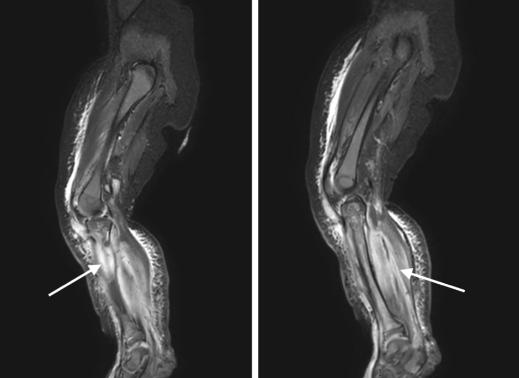
Image 2. Magnetic resonance imaging of the left lower extremity demonstrating osteomyelitis involving the entire left tibia, the mid/ distal left fibula, and the left ilium adjacent to the sacroiliac joint (arrow in left photo). Diffuse muscular edema is visualized in the left gluteal musculature, quadriceps musculature, and extensively involving both the anterior and posterior compartments of the lower leg (arrow in right photo).
necrotic tissue during this procedure and collected cultures. On day 9 of hospitalization, he underwent a second irrigation and debridement of the right upper extremity. His left lower extremity ischemia progressed to dry gangrene requiring left below-knee amputation on day 20 of hospitalization. Magnetic resonance imaging of the left lower extremity directly prior to scheduled, below-knee amputation revealed osteomyelitis involving the entire left tibia, mid/distal left fibula, and left ilium adjacent to the sacroiliac joint (Image 2). Below-knee amputation cultures grew Candida parapsilosis, Enterococcus faecalis, and possible anaerobes. Oral fluconazole was initiated for candida coverage, IV penicillin G provided adequate coverage for E faecalis, and oral metronidazole was initiated for anaerobic coverage. The infant completed six weeks of IV penicillin G, oral fluconazole, and oral metronidazole from source control with amputation, and was safely discharged home with continued outpatient therapies.
Group A streptococcus often infiltrates deep tissues through superficial skin lesions, such as an insect bite in the infant described in this case, with inflammation quickly becoming extensive.3,5,6 Bacteremia frequently occurs concurrently leading to rapid decompensation.1-3,5,6 Invasive streptococcal pharyngitis is a less common source of infection, but it was also relevant to the infant described in this case.2 Appropriate management of GAS complications, including myositis and myonecrosis, osteomyelitis, toxic shock syndrome, and necrotizing fasciitis, requires a multidisciplinary approach to management
Collaboration with medical and surgical subspecialties, including infectious diseases, hematology, vascular surgery, and orthopedic surgery, was necessary to coordinate and enhance inpatient management. Ancillary therapy with physical therapy, occupational therapy, and speech therapy assisted the infant in wound care and continued therapeutic mobility measures. Integrated patient-centered care largely contributed to mitigation of disease progression and the ability to successfully discharge home with continued outpatient management.
This case highlights the formidable nature of invasive group A streptococcus infections in infants, illustrated by the rapid progression to life-threatening sepsis, DIC, and limb ischemia necessitating amputation. Despite advances in medical care, GAS infections continue to pose significant challenges, often presenting with rapid progression and severe systemic manifestations, as demonstrated in this case. Early recognition and initiation of appropriate antimicrobial therapy are paramount to mitigate the progression of disease and improve outcomes. Additionally, vigilance for complications such as DIC and limb ischemia is crucial, as prompt intervention can attenuate morbidity and mortality associated with these sequelae. By sharing our clinical experience and insights, we hope to contribute to the collective knowledge base and ultimately improve clinical outcomes for infants afflicted by this life-threatening condition.
Address for Correspondence: Alexis N. Roach, DO, University of Arkansas for Medical Sciences, Arkansas Children’s Hospital, Department of Pediatrics, Section of Emergency Medicine, 1 Children’s Way, Slot 512-19A, Little Rock, AR 72202. Email: aroach@uams.edu.
Conflicts of Interest: By the CPC-EM article submission agreement, all authors are required to disclose all affiliations, funding sources and financial or management relationships that could be perceived as potential sources of bias. The authors disclosed none.
Copyright: © 2025 Roach et al. This is an open access article distributed in accordance with the terms of the Creative Commons Attribution (CC BY 4.0) License. See: http://creativecommons.org/ licenses/by/4.0/
The authors attest that their institution requires neither Institutional Review Board approval, nor patient consent for publication of this case report. Documentation on file.
1. Thompson KM, Sterkel AK, McBride JA, et al. The shock of strep: rapid deaths due to group A Streptococcus. Acad Forensic Pathol. 2018;8(1):136-49.
2. Vallalta Morales M, Soriano Navarro CJ, Salavert Lletí M, et al. Group A streptococcal bacteremia: outcome and prognostic factors. Rev Esp Quimioter. 2006;19(4):367-75.
3. Waddington CS, Snelling TL, Carapetis JR. Management of invasive group A streptococcal infections. J Infect. 2014;69 Suppl 1:S63-9.
4. Kaul R, McGeer A, Low DE, et al. Population-based surveillance for group A streptococcal necrotizing fasciitis: Clinical features, prognostic indicators, and microbiologic analysis of seventyseven cases. Ontario Group A Streptococcal Study. Am J Med 1997;103(1):18-24.
5. Stevens DL and Bryant AE. Severe group A streptococcal infections. In: Ferretti JJ, Stevens DL, Fischetti VA, eds. Streptococcus pyogenes: Basic Biology to Clinical Manifestations [Internet]. Oklahoma City (OK): University of Oklahoma Health Sciences Center; 2016. Available from: https://www.ncbi.nlm.nih.gov/books/ NBK333425/. Accessed April 10, 2024.
6. Stevens DL and Bryant AE. Necrotizing soft-tissue infections. N Engl J Med. 2017;377(23):2253-65.
Sharmin Kalam, MD
Sergio Marquez, MD
Emmelyn
J. Samones, CCRP
Tammy Phan, BS,
CCRP
Vi Am Dinh, MD
Section Editor: Lev Libet, MD
Loma Linda University Medical Center, Department of Emergency Medicine, Loma Linda, California
Submission history: Submitted May 21, 2024; Revision received September 6, 2024; Accepted October 3, 2024
Electronically published December 12, 2024
Full text available through open access at http://escholarship.org/uc/uciem_cpcem
DOI: 10.5811/cpcem.21220
Introduction: Obstructive shock results from reduced cardiac output due to physical blockage of blood flow, such as cardiac tamponade. Cardiac tamponade compresses cardiac chambers, particularly the left atrium, causing decreased end-diastolic volume and cardiac output. Rapid fluid accumulation within the pericardial sac is the usual cause. Transesophageal echocardiography provides clearer visualization of these structures than transthoracic ultrasound. This case underlines the impact of esophageal pathology on cardiac output and highlights ultrasound’s dynamic diagnostic utility alongside computed tomography.
Case Report: A 64-year-old female with a history of colon cancer and peritoneal metastases status post colostomy presented with altered mental status and urinary symptoms. Laboratory evaluation was notable for leukopenia, hypoglycemia, elevated ammonia, and an abnormal urinalysis that was positive for urinary tract infection. She was initially admitted to the internal medicine service for sepsis secondary to urine as the source of infection. During her hospital stay, she developed hypotension, tachypnea, tachycardia, and complained of chest pressure. Point-ofcare echocardiogram revealed compression of the left atrium by distended gastric and esophageal contents. A nasogastric tube was placed and suctioned partially digested food and liquid with improvement of her condition. Follow-up ultrasound showed improvement of compression and cardiac function.
Conclusion: In evaluation of acute shock, multiple etiologies must be considered. In this case, the cause of reduced cardiac output was direct compression of the left atrium from an adjacent structure. Even with direct visualization and imaging, immediate history and patient-centered approach are still useful to complete the clinical picture and treat the reversible cause of undifferentiated shock. [Clin Pract Cases Emerg Med. 2025;19(1):49-52.]
Keywords: left atrial compression; esophageal mass; extracardiac compression; obstructive shock; case report.
Obstructive shock occurs when there is a physical blockage of blood flow that results in reduced cardiac output and hypoperfusion.1 Although the focus of this report is on pericardial tamponade, other etiologies of
obstructive shock include massive pulmonary embolism, tension pneumothorax, vena cava compression, pulmonary compression, and aortic dissection.1 Cardiac tamponade is a life-threatening condition with a well-understood pathophysiology, necessitating prompt intervention. Clinical
signs include hypotension, evidence of pulmonary edema, chest pain, dyspnea, tachypnea, muffled heart sounds, and jugular venous distention.
The hemodynamic compromise in cardiac tamponade is caused by compression of the cardiac chambers, particularly the left atrium and left ventricle, directly reducing enddiastolic volume and cardiac output.2 The etiology is often rapid accumulation of fluid within the pericardial sac, stemming from trauma, infection, malignancy, or other non-infectious causes.1-3 The close anatomical proximity of the distal esophagus to the left atrium is exploited by transesophageal echocardiography to provide direct visualization of the cardiac structures. This case reemphasizes the direct physical relationship and potential impact of esophageal pathology reducing cardiac output. It also highlights the utility of ultrasound in providing dynamic diagnostic utility to supplement static computed tomography (CT).
A 64-year-old female with a past medical history of colon cancer with peritoneal metastases and prior colostomy with both external and internal metastasis near her ostomy site was brought in by family for altered mental status. She had also been complaining of urinary symptoms, including frequency and urgency. On presentation, vital signs were temperature 98.2 °Fahrenheit, pulse 118 beats per minute, blood pressure 150/92 millimeters of mercury (mm Hg), respiratory rate 22 breaths per minute, and oxygen saturation 100% on room air. On physical examination, the patient was in mild distress and cachectic. She was unable to follow command. Her pupils were equal, round, and reactive to light with no apparent focal deficits. Her abdomen was soft and non-tender with an ostomy bag in place showing brown stool. Heart rate was tachycardic without murmurs and lungs were clear bilaterally.
Laboratory values revealed a white blood count of 4.74 x 10⁹ cells per liter (L) (reference range 4.8-11.8 x 10⁹ cells/L), a blood glucose of 53 milligrams per deciliter (mg/ dL) (70-185 mg/dL) and an elevated ammonia level of 207 micromoles/L (μmol/L) (11-48 μmol /L). Additionally, her urinalysis showed moderate leukocytes, 13 white blood cells per high-powered field (HPF) (0-5 HPF), and bacteriuria. Two ampules of 50% dextrose were subsequently given with mild improvement of her mental status. Clinical suspicion of sepsis was made due to presence of altered mental status despite treatment of hypoglycemia. Infectious workup was obtained with the following results: wwhite blood cells 3.48 x 10⁹ per L, aspartate aminotransferase 36 units/L (U/L) (0-30 U/L), alanine aminotransferase 41 U/L (7-37 U/L), anion gap 14 (716), lactate 3.5 millimole per L (mmol/L) (0.5-20 mmol/L), no growth on blood cultures, and urine culture positive for Enterococcus faecalis. Intravenous ceftriaxone was ordered to treat urinary as source of infection.
CPC-EM Capsule
What do we already know about this clinical entity?
Obstructive shock occurs when blood flow to the heart is obstructed, preventing it from pumping enough blood.
What makes this presentation of disease reportable?
Esophageal tamponade leading to obstructive shock is rare, with few reported cases of this association.
What is the major learning point?
Clinicians should have a broad differential diagnosis for acute presentations of shock, specifically for cardiac tamponade caused by a food bolus.
How might this improve emergency medicine practice?
Point-of-care ultrasound is a rapid and valuable tool to differentiate etiologies of shock, helping assess and guide appropriate treatment.
The following day, the patient required vasopressor support and was transferred to the intensive care unit (ICU). She was found to have a urine culture positive for Enterococcus as well as blood cultures positive for Klebsiella pneumoniae. Her ICU course was complicated by respiratory decompensation due to suspected fluid resuscitation and concurrent hypoalbuminemia resulting in large pleural effusions requiring a thoracentesis and high-flow nasal cannula (HFNC). She was weaned to two liters nasal cannula with aggressive diuresis and fluid restriction and subsequently downgraded. The following morning, shortly after breakfast, she developed tachypnea in the 40 breaths per minute range and was found to be hypotensive with systolic blood pressure in the 70 mm Hg range. She was placed on a nonrebreather mask at 15 L per minute (L/min), and 500 milliliters (mL) normal saline bolus was given with improvement of her systolic blood pressure to 105 mm Hg. She was started on HFNC at 30 L/min and 100% oxygen. In the ICU, she remained hypotensive with systolic blood pressures ranging from 90-100 mm Hg and tachycardia in the low 100s beats per minute but was otherwise alert and oriented with no concern for impending airway compromise. When asked what happened to her, she replied, “It was that shake I drank.” Point-of-care echocardiography was performed and revealed that the left atrium could not be clearly visualized secondary to anatomical compression by distended gastric as well as esophageal contents (Image 1).

Image 1. Apical 4-chamber cardiac view from point-of-care ultrasound showing a food bolus causing gastric outlet obstruction and external compression of the left atrium (arrow). RA, right atrium; RV, right ventricle; LV, left ventricle; LA, left atrium.
As a result of the ultrasound, a nasogastric (NG) tube was placed and immediately suctioned, yielding approximately 600 mL of partially digested food and liquid with subjective relief voiced by the patient, as well as clinical improvement (Image 2A). Her systolic blood pressure improved from 90 to low 100s mm Hg, and her HFNC was weaned to 14 L/min at 45% oxygen. A repeat ultrasound demonstrated visualization of the left atrium and improved cardiac function. Given the patient’s comorbidities, a CT angiogram of the chest was ordered to evaluate for possible pulmonary embolism (PE). This took place following point-of-care ultrasound (POCUS) and suctioning. No PE was found; however, a severely distended esophagus persisted with a large amount of intraluminal fluid (Image 3). The patient was made nil per os. The NG tube was also advanced to continuous suction overnight. A follow-up POCUS the next morning showed that the left atrium was no longer compressed (Image 2B). This correlated clinically with stable hemodynamic status as monitored with an arterial line.

2. (2A)
4-chamber cardiac view from point-of-care ultrasound of the left atrium immediately after nasogastric tube (NG) suction of approximately 600 milliliters of content. (2B) The same view of the left atrium after continued NG tube suctioning the following morning.
RA, right atrium; RV, right ventricle; LV, left ventricle; LA, left atrium.

Obstructive shock is defined as physical obstruction of blood flow resulting in decreased cardiac output ultimately leading to tissue hypoperfusion. Prompt intervention is required for cardiac tamponade due to its life-threatening condition. It is believed that this patient experienced an accumulation of digestive material within the esophagus due to delayed gastric emptying and possibly a gastric outlet obstruction, leading to external compression of the left atrium. The relief of the left atrial obstruction with the NG tube suctioning seems to have reversed the patient’s shock. An esophagram performed three days later showed evidence of residual contrast within the distal esophagus and minimal entry into the stomach. The cause of the obstruction was believed to be related to her progressive metastatic disease. The patient was ultimately discharged home under hospice care.
This case highlights the importance of performing a rapid ultrasound for shock and hypotension exam in undifferentiated shock. The inferior vena cava was plethoric, which is consistent with an obstructive process. The rapid identification of left atrial compression allowed for immediate intervention with marked clinical improvement. Gastrointestinal pathology leading to obstructive shock is rare but has been shown in the setting of compartment syndrome or hepatic pathology from compression of central veins.4 Cases of similar presentations as the one mentioned above have been reported, and esophageal tamponade is an uncommon, yet known, phenomenon.5,6
A recent published report details a case of progressive achalasia leading to heart failure, successfully treated with esophageal dilation, underscoring the anatomic relationship between the mid esophagus and the left atrium.7 In the majority of the cases reviewed, there was a known history of progressive achalasia without any association with metastatic disease or outlet obstruction. One case report did describe
Obstructive Shock from an Esophageal Bolus Kalam et al.
acute heart failure with clinical evidence of hypoperfusion but with preserved hemodynamic stability.8 Additionally, while heart failure and pulmonary edema are mentioned, these cases did not describe acute shock in an ICU setting. Their lower acuity allowed for slower treatment options.9-15 This case represents an example of rapid diagnosis with POCUS allowing for a rapid intervention and reversal of a life-threatening condition.
In evaluation of the patient with acute shock, multiple etiologies must be considered. In this case the cause of reduced cardiac output was direct compression of the left atrium by an adjacent distended esophagus. POCUS was important in diagnosing the condition, and the patient’s hemodynamic status improved with nasogastric suction of the esophageal contents.
The authors attest that their institution does not require Institutional Review Board approval for publication of this case report. Patient consent has been obtained and filed for the publication of this case report. Documentation on file.
1. Pérez C, Diaz-Caicedo D, Almanza Hernández DF, et al. Critical care ultrasound in shock: a comprehensive review of ultrasound protocol for hemodynamic assessment in the intensive care unit. J Clin Med. 2024;13(18):5344.
2. Spodick DH. Acute cardiac tamponade. N Engl J Med. 2003;349(7):684-90.
3. Beck CS. Two cardiac compression triads. JAMA. 1935;104(9):714-6.
4. Murphy MP, Warren HR, Surmaitis RM, et al. An unusual etiology of obstructive shock in the emergency department. Am J Emerg Med. 2022;55:228.e5.
5. Van Rooijen JM and Van den Merkhof LF. Left atrial impression: a sign of extra-cardiac pathology. Eur J Echocardiogr. 2008;9:661-4.
6. De Cock D, Monballyu P, Voigt JU, et al. Extra-cardiac compression and left ventricular inflow obstruction as a complication of a Sengstaken–Blakemore tube. Eur J Echocardiogr. 2011;12(12):973.
Address for Correspondence: Emmelyn J. Samones, CCRP, Loma Linda University Medical Center, Department of Emergency Medicine, 11234 Anderson St., Room A890A, Loma Linda, CA 92354. Email: Esamones@llu.edu.
Conflicts of Interest: By the CPC-EM article submission agreement, all authors are required to disclose all affiliations, funding sources and financial or management relationships that could be perceived as potential sources of bias. The authors disclosed none.
Copyright: © 2025 Kalam et al. This is an open access article distributed in accordance with the terms of the Creative Commons Attribution (CC BY 4.0) License. See: http://creativecommons.org/ licenses/by/4.0/
7. Felix ADS, Quintes AC, Andrade de Sá G, et al. Left atrial compression secondary to massive esophageal dilatation in a patient with idiopathic achalasia. CASE (Phila). 2018;2(6):285-92.
8. Maffè S, Paino AM, Paffoni P, et al. Left atrial compression with acute heart failure caused by esophageal bolus. J Clin Ultrasound. 2018;46(4):259-61.
9. Lee H, Lee SH, Kim JH, et al. A case of esophageal achalasia compressing left atrium diagnosed by echocardiography in patient with acute chest pain. J Cardiovasc Ultrasound. 2012;20(4):218–9.
10. Nakabayashi K, Hirata T, Oka T. Left atrium compression due to oesophageal dilation can induce acute heart failure. BMJ Case Rep. 2015;2015:bcr2014209233.
11. Yilmaz MB, Arat N, Biyikoglu SF, et al. Extrinsic left atrial compression in a patient with achalasia. Int J Cardiol. 2002;85(2-3):301-3.
12. Cacciapuoti F, Paoli VD, Scognamiglio A, et al. Three-Dimensional Trans- Thoracic Echocardiography of Esophageal Achalasia: Description of a Case. J Cardiovasc Echogr. 2014;24(2):57-9.
13. Stoupakis G, Fuhrman MA, Dabu L, et al. The use of contrast echocardiography in the diagnosis of an unusual cause of congestive heart failure: achalasia. Echocardiography. 2004;21(2):149-52.
14. Chen SY, Toh HS, Chang WT, et al. A rare cause of left atrium compression. Int Heart J. 2021;62(4):944-8.
15. Polina A, Dawdy J, Afonso L. A Case of Severe Left Atrial Compression by a Mid- Esophageal Metastatic Mass. Authorea. 2023:1-6.
Mariam Attia, DO*
Ashley A. Lavoie-Forrest, BS, MS†
Phoebe Langius, BS, BA‡
Leon Melnitsky, DO*
Sandra Lopez, MD*
Eric Boccio, MD, MPH, FACEP*
Section Editor: Austin Smith, MD
Memorial Healthcare System, Department of Emergency Medicine, Hollywood, Florida
Nova Southeastern University, Dr. Kiran C. Patel College of Osteopathic Medicine, Fort Lauderdale, Florida
Florida International University, Herbert Wertheim College of Medicine, Miami, Florida
Submission history: Submitted June 6, 2024; Revision received September 14, 2024; Accepted September 29, 2024
Electronically published January 6, 2025
Full text available through open access at http://escholarship.org/uc/uciem_cpcem DOI: 10.5811/cpcem.21300
Introduction: While mild or moderate iron-deficiency anemia may not cause any symptoms, more severe deficiencies may present clinically as fatigue, shortness of breath, exertional dyspnea, lightheadedness, tachycardia, and presyncope or syncope, and, in rare instances, pica. Pica is defined as the developmentally inappropriate ingestion of non-nutritive, non-food substances for more than one month. We present the case of a duodenal obstruction secondary to a picaassociated bezoar in a patient with iron-deficiency anemia who presented to the emergency department (ED) with abdominal pain.
Case Report: A 40-year-old female with past medical history of iron-deficiency anemia, asthma, and Von Willebrand disease and allergies to both oral and intravenous (IV) iron presented to the ED with one day of acute and severe abdominal pain associated with nausea and vomiting. The patient’s last bowel movement was one day prior to presentation. The abdominal exam revealed mild distention and generalized tenderness with no evidence of rebound or guarding. Computed tomography of the abdomen and pelvis with IV and oral contrast demonstrated gastric distention and a fecalized distal duodenum with wall thickening concerning for a duodenal obstruction. Given the patient’s known history of iron-deficiency anemia, the emergency physician inquired about ingestion of non-nutritive substances to which the patient replied that she had been consuming cotton foam. The patient was admitted to the hospital for gastroenterology consultation and esophagogastroduodenoscopy.
Conclusion: Pica-associated gastrointestinal bezoars are a rare complication with a variety of reported substances being consumed. Patients presenting with small gastroduodenal bezoars may benefit from endoscopic removal, but large non-fragmentable bezoars can only be removed through surgical intervention. [Clin Pract Cases Emerg Med. 2025;19(1):53-56.]
Keywords: gastroduodenal obstruction; gastroduodenal bezoar; pica; iron-deficiency anemia; case report.
While mild or moderate iron-deficiency anemia may not cause any symptoms, more severe deficiencies may present clinically as fatigue, shortness of breath, exertional dyspnea, lightheadedness, tachycardia, and presyncope or
syncope. In rare instances, pica, or the developmentally inappropriate ingestion of non-food substances for more than one month, may be observed. Features of pica include the compulsory consumption of a variety of both organic and inorganic, digestible and indigestible, non-nutritive substances
including ice, soil, clay, paper, hair, and cotton foam. While evidence suggests a strong association between pica and irondeficiency anemia which is strengthened by the observation that pica improves or resolves following iron transfusions, the pathophysiology of pica is currently unknown. We present the case of a duodenal obstruction secondary to a pica-associated bezoar in a patient with iron-deficiency anemia who presented to the emergency department (ED) with abdominal pain.
A 40-year-old female with past medical history of irondeficiency anemia, asthma, and Von Willebrand disease, past surgical history of Cesarean section, and abdominal gunshot wound status post laparotomy (2019), and allergies to ferrous sulfate, iron dextran, and iron sucrose presented to the ED with one month of intermittent abdominal pain. The patient reported one day of acutely worsened and severe abdominal pain associated with nausea, non-bloody and nonbilious vomiting, and inability to pass flatus. The patient’s last bowel movement was one day prior to presentation and was described as grossly normal. The patient denied urinary complaints, fever, shaking, and chills. Initial vital signs including blood pressure (110/71 millimeters of mercury), heart rate (67 beats per minute), respiratory rate (18 breaths per minute), peripheral capillary oxygen saturation (100%, room air), and temperature (36.5 °Celsius, oral) were within normal limits.
The abdominal exam revealed mild distention and generalized tenderness with no evidence of rebound or guarding. The points of maximal abdominal tenderness were the left upper quadrant and middle of the epigastric region. Murphy’s sign was not present. There were no visible or palpable abdominal masses. The complete blood count was significant for a microcytic anemia (hemoglobin 8.9 grams per deciliter (g/dL) [reference range 12.0-15.5 g/dL], hematocrit 32.1% [36-44%], and mean corpuscular volume 62.9 femtoliters (fL) [80-95 fL]). The comprehensive metabolic panel demonstrated hypokalemia (potassium 3.4 millimoles per liter (mmol/L) [3.5-5.5 mmol/L]). Iron level and transferrin saturation were low (25.0 micrograms per deciliter (mcg/dL) [60-170 mcg/dL] and 6% [20-50%], respectively). Zinc and calcium levels were not measured. Computed tomography of the abdomen and pelvis with intravenous (IV) and oral contrast demonstrated gastric distention and a distended fecalized distal duodenum with wall thickening, adjacent fat stranding, and prominence of vasa recta concerning for a duodenal obstruction; duodenal bezoar could not be excluded (Image 1).
The patient received 6 milligrams (mg) of morphine IV, 4 mg ondansetron IV, 40 milliequivalents of potassium chloride IV over four hours, and 1 L of sodium chloride IV over one hour. The patient could not receive iron supplementation due to documented allergies. Given the patient’s known history of iron-deficiency anemia, the emergency physician inquired about ingestion of non-nutritive substances to which the
CPC-EM Capsule
What do we already know about this clinical entity?
Severe deficiencies in iron may present clinically as fatigue, shortness of breath, tachycardia and, in rare instances, pica.
What makes this presentation of disease reportable?
Pica-associated gastrointestinal bezoars are a rare complication with a variety of non-nutritive consumed substances having been reported.
What is the major learning point?
It is important to inquire about dietary behaviors and frequency of non-nutritive substance ingestion when clinical suspicion for pica is high.
How might this improve emergency medicine practice?
When diagnosing a gastrointestinal bezoar in the severely iron-deficient patient, consider picarelated behaviors.

patient replied that she had been consuming cotton foam since 2012 with last ingestion two days earlier. The patient appeared to have insight that eating cotton foam was illogical but could not verbalize the rationale behind her indulgence. The patient presented a large piece of cotton foam from her purse to show the clinical team (Image 2).
The cotton foam was confiscated, and the patient was made nil per os and admitted to the hospital for gastroenterology consultation and esophagogastroduodenoscopy for duodenal obstruction secondary to pica-associated bezoar, presumably cotton foam.

Image 2. Piece of cotton foam (9.5 x 5.1 x 5.1 centimeter) presented to clinical team by patient after being asked about ingestion of non-nutritive substances. The patient stated that she had last ingested cotton foam two days prior to her presentation to the emergency department.
Anemia is a common clinical disorder affecting 1.92 billion people globally as of 2021.1 According to the National Hospital Ambulatory Medical Care Survey reported by the US Centers for Disease Control and Prevention, 800,000 cases of anemia were diagnosed in EDs across the United States in 2021, and iron-deficiency was the leading cause.2 While mild or moderate iron-deficiency anemia may not cause any symptoms, more severe deficiencies may present clinically as fatigue, shortness of breath, exertional dyspnea, lightheadedness, tachycardia, presyncope or syncope, and, in rare instances, pica. Pica is defined by the Diagnostic and Statistical Manual of Mental Disorders, 5th edition, as developmentally inappropriate ingestion of non-nutritive, nonfood substances for more than one month.3,4 It is theorized that pica is a compulsory and compensatory behavior caused by low nutritional status, specifically in iron, zinc, and calcium.5 Pica can be further categorized by the type of non-nutritive non-food substance being ingested. In the US, pagophagia, the ingestion of ice, is most common among patients with irondeficiency. In other parts of the world and especially in Africa, geophagia, the ingestion of earth materials such as soil, dirt, or clay, is both common and socially accepted.6 The ingestion of hair is referred to as trichophagia.
Studies have shown an increased prevalence of pica among children and pregnant women. It is speculated that pica behaviors may develop more frequently in these subgroups due to a negative nutrient state exacerbated by high energy expenditures and increased nutritional needs.7 One study found that over half of pregnant adolescents (<18 years) reported pica behaviors, with pagophagia being the most common. These subjects had a significantly lower iron status than those who did not engage in pica behaviors.5 A study of 262 non-pregnant adults with iron-deficiency anemia found that 87.3% reported pagophagia.8 A meta-analysis of 43 studies involving 6,407 subjects found that individuals with pica were 2.35 times more likely to be anemic and have lower than normal zinc levels.9
Adults with avoidant or restrictive food intake disorder, poor body image, and depression are more likely to exhibit pica than the general population.10 A study of blood donors found an association between pica and low ferritin levels, non-Asian race,
younger age, and restless leg syndrome with no statistically significant difference between the sexes.11 A recent case-control study found an increased prevalence of pica in individuals with autism spectrum disorder, intellectual disability, and developmental disabilities.12 Individuals demonstrated behaviors of chewing on inedible materials without swallowing prior to a formal diagnosis of pica being made.
Pica is an important disorder to identify, however, it is commonly overlooked due to patients’ unwillingness to speak about unusual eating behaviors and physicians’ failure to ask targeted questions. When clinical suspicion is high, the medical history should include direct questions about dietary behaviors, cravings, and frequency of non-nutritive substance ingestion. On physical examination, signs of nutritional deficiencies such as pallor and glossitis may be noted. While there are no specific screening or diagnostic tests for pica, serum laboratory tests including a complete blood count, serum iron level, iron saturation, total iron-binding capacity, unsaturated iron-binding capacity, and ferritin level should be considered. Results will most likely demonstrate a microcytic anemia with elevated, unsaturated iron-binding capacity and total iron-binding capacity and decreased transferrin levels.13 While serum ferritin is typically low, normal levels have been documented in pica cases.11
Therapeutic management of pica involves addressing the underlying cause of anemia and repleting iron and electrolytes when deficient. Treatments include oral and IV iron supplementation and packed red blood cell transfusions. In patients with comorbid iron-deficiency anemia, pica symptoms have been shown to resolve within three weeks following IV iron infusion.6 Additionally, prognosis has been shown to improve when psychiatric care, which includes cognitive behavioral therapy focused on controlling urges and discouraging compulsory ingestion of non-nutritive substances, is maintained.14 Documented allergies to oral and IV forms of iron made this case particularly interesting and the therapeutic management of the iron-deficiency anemia particularly challenging. Long-term management goals would need to focus on improving hemoglobin levels and reducing anemia-associated symptoms. Several treatment options include transfusion of packed red blood cells, administration of epoetin alfa, and desensitization treatment to oral and IV iron.15
Early recognition of pica can lead to better prevention of life-threatening complications such as infection, lead poisoning, gastrointestinal (GI) inflammation secondary to the ingestion of caustic chemicals, and GI obstruction secondary to bezoars.16 Bezoar refers to a mass of indigestible material that forms in the GI tract and may consist of organic or inorganic materials. Often, bezoars may cause GI obstruction, which presents clinically as severe abdominal pain, abdominal distention, nausea, vomiting, constipation, and inability to pass flatus. The workup of patients presenting with signs or symptoms of GI obstruction may include diagnostic imaging studies such as abdominal radiographs or
Gastroduodenal Obstruction
computed tomography. Patients presenting with small gastric bezoars may benefit from endoscopic removal, but large nonfragmentable bezoars can only be removed through surgical intervention via laparotomy or laparoscopic extraction.14
We present the case of a 40-year-old woman with past medical history of iron-deficiency anemia, and with allergies to both oral and IV iron, suffering from abdominal pain secondary to a duodenal obstruction caused by a picaassociated bezoar, presumably cotton foam. Pica-associated gastrointestinal bezoars are a rare complication with a variety of reported substances being consumed, including cotton foam, dirt, paper, and hair. Patients presenting with small gastroduodenal bezoars may benefit from endoscopic removal, but large non-fragmentable bezoars can only be removed through surgical intervention. The clinical workup for pica should include an assessment for iron-deficiency anemia and calcium and zinc levels. Therapeutic management is mostly supportive and should focus on iron repletion, correction of electrolyte abnormalities, and outpatient referral for scheduled iron transfusions and cognitive behavioral therapy.
The authors attest that their institution requires neither Institutional Review Board approval, nor patient consent for publication of this case report. Documentation on file.
Address for Correspondence: Eric Boccio, MD, MPH, FACEP, Memorial Healthcare System, Department of Emergency Medicine, 3501 Johnson Street, Hollywood, FL 33021. Email: eboccio@mhs.net.
Conflicts of Interest: By the CPC-EM article submission agreement, all authors are required to disclose all affiliations, funding sources and financial or management relationships that could be perceived as potential sources of bias. The authors disclosed none.
Copyright: © 2025 Attia et al. This is an open access article distributed in accordance with the terms of the Creative Commons Attribution (CC BY 4.0) License. See: http://creativecommons.org/ licenses/by/4.0/
REFERENCES
1. GBD 2021 Anaemia Collaborators. Prevalence, years lived with disability, and trends in anaemia burden by severity and cause, 19902021: findings from the Global Burden of Disease Study 2021. Lancet
Haematol. 2023;10(9):e713-34.
2. Cairns C and Kang K. National Hospital Ambulatory Medical Care Survey: 2021 emergency department summary tables. Available from: https://ftp.cdc.gov/pub/Health_Statistics/NCHS/Dataset_ Documentation/NHAMCS/doc21-ed-508.pdf. Accessed June 3, 2024.
3. Singh I, Singh P, Patkar P, et al. Geophagia: a case series. Ind Psychiatry J. 202;30(Suppl 1):S240-2.
4. Ganesan PR and Vasauskas AA. The association between pica and iron-deficiency anemia: a scoping review. Cureus 2023;15(4):e37904.
5. Lumish RA, Young SL, Lee S, et al. Gestational iron deficiency is associated with pica behaviors in adolescents. J Nutr. 2014;144(10):1533-9.
6. Goldman RH and Weissmann L. A diagnosis to chew on. N Engl J Med. 2019;381(5):466-73.
7. Papini NM, Bulik CM, Chawner SJRA, et al. Prevalence and recurrence of pica behaviors in early childhood within the ALSPAC birth cohort. Int J Eat Disord. 2024;57(2):400-9.
8. Barton JC, Barton JC, Bertoli LF. Pica associated with iron deficiency or depletion: clinical and laboratory correlates in 262 non-pregnant adult outpatients. BMC Blood Disord. 2010;10:9.
9. Miao D, Young SL, Golden CD. A meta-analysis of pica and micronutrient status. Am J Hum Biol. 2015;27(1):84-93.
10. Hartmann AS, Zenger M, Glaesmer H, et al. Prevalence of pica and rumination behaviours in adults and associations with eating disorder and general psychopathology: findings form a population-based study. Epidemiol Psychiatr Sci. 2022;31:e40.
11. Liu H, Burns RT, Spencer BR, et al. NHLBI Recipient Epidemiology Donor Evaluation Study (REDS)-III. Demographic, clinical, and biochemical predictors of pica in a large cohort of blood donors. Transfusion. 2021;61(7):2090-8.
12. Fields VL, Soke GN, Reynolds A, et al. Pica, autism, and other disabilities. Pediatrics. 2021;147(2):e20200462.
13. Ozbilen M, Yesil S, Demir E. Relationship between pica and laboratory values in patients with iron deficiency. Van Med J. 2023; 30(4):314-20.
14. Cannalire G, Conti L, Celoni M, et al. Rapunzel syndrome: an infrequent cause of severe iron deficiency anemia and abdominal pain presenting to the pediatric emergency department. BMC Pediatr. 2018;18(1):125.
15. Fishbane S. Refractory anemia in a patient with allergy to intravenous iron drugs. Kidney Int. 2006;69(10):1910-3.
16. Juwairiyyah Fatima S, Nepal N, Ochieta D. Surviving critical low hemoglobin levels and pica. Cureus. 2023;15(5):e38812.
Thuc Vu, DO*
Jake Valentine, MD†‡
Kansas City University College of Osteopathic Medicine, Kansas City, Missouri University of Houston Tilman J. Fertitta Family College of Medicine, Houston, Texas HCA Houston Healthcare Kingwood, Kingwood, Texas
Section Editor: Christopher San Miguel, MD
Submission history: Submitted February 22, 2024; Revision received May 30, 2024; Accepted June 6, 2024
Electronically published November 18, 2024 Full text available through open access at http://escholarship.org/uc/uciem_cpcem DOI: 10.5811/cpcem.19484
Introduction: Ventricular ectopic beats and corrected QT interval (QTc) prolongation are both relatively common entities that are typically benign. It is difficult to predict subsequent dysrhythmias from either electrocardiogram (ECG) feature. The combination of both features may better predict the risk of torsades de pointes. We highlight a case of torsades preceded by a bizarre bigeminal rhythm with QTc prolongation likely caused by memantine use and hypokalemia.
Case Report: An 84-year-old female presented to the emergency department with a fall. A syncope workup revealed an ECG demonstrating bigeminy with a prolonged QTc interval. Several minutes after obtaining the ECG, the patient went into torsades. She had multiple subsequent cardiac arrests during the rest of her hospital stay. This case report details the importance of recognizing ventricular bigeminy in the context of QTc prolongation as a harbinger of torsades.
Conclusion: While premature ventricular contractions including bigeminy may be a benign finding, when accompanied by prolonged QTc intervals, they warrant immediate investigation and treatment of potential underlying pathology to prevent torsades and subsequent cardiac arrest. [Clin Pract Cases Emerg Med. 2025;19(1):57-60.]
Keywords: torsades de pointes; long QT; case report; bigeminy.
Ventricular ectopic beats and corrected QT interval (QTc) prolongation are both relatively common entities that are typically benign. It is difficult to predict subsequent dysrhythmias from either electrocardiogram (ECG) feature. The combination of both features may better predict risk of torsades de pointes.1 We highlight a case of torsades preceded by a bizarre bigeminal rhythm with QTc prolongation likely caused by memantine use and hypokalemia. The report includes teaching points on the pathophysiology of torsades and the clinical correlation to typical ECG changes predicting development of torsades beyond the traditional prolonged QTc and “R on T” phenomenon frequently cited in emergency medicine textbooks.
An 84-year-old female presented to the emergency
department (ED) due to a laceration to her forehead. She had a history of dementia, chronic kidney disease, and hypertension, for which she was prescribed memantine and amlodipine. She had been in her usual state of health the night prior but walked downstairs the morning of her ED presentation due to blood trickling down her face. She could not recall whether she had fallen or provide any meaningful details surrounding the event. She denied any other acute medical complaint. The patient’s family had not noticed any outward evidence of illness prior to discovering the wound and reported that the patient was at her baseline mental state. The patient was alert, with a non-focal neurologic exam, and had an isolated twocentimeter laceration to the middle of her forehead without active bleeding. She had a regular rate and rhythm but bizarreappearing morphology on her telemetry monitoring. The emergency physician relayed the plan for trauma and syncope
workups to include computed tomography (CT) of the head and cervical spine, tetanus vaccination, wound irrigation and preparation for closure, along with basic labs and an ECG, the result of which is below (Image 1).

Image 1. Electrocardiogram revealing sinus rhythm with bizarreappearing bigeminy. The subtle low-voltage QRS complexes best seen in leads II and V5 (arrows) represent premature ectopic beats. Heart rate is 85 beats per minute, and the computer reported a QTc of 602 milliseconds. It is difficult to delineate the end of the T-wave in the sinus beat, but the QT interval for the ectopic beat is larger than half the RR interval (brackets), supporting the diagnosis of prolonged QTc.
After reviewing the ECG, the physician ordered two grams of magnesium based on the “bizarre repolarization pattern with QTc of 602 milliseconds.” Intravenous access was obtained, magnesium was started, and the patient was brought expeditiously to CT at which point she went into cardiac arrest, with the bedside nurse reporting ventricular fibrillation as the initial rhythm. The patient achieved return of spontaneous circulation (ROSC) after a single defibrillation and two-minute round of compressions per Advanced Life Support (ALS) protocol. The physician also requested the rest of the magnesium to be delivered at a bolus rate. The patient maintained her baseline neurologic exam after the cardiac arrest. Her post-arrest ECG revealed sinus rhythm without ectopy at a rate of 107 beats per minute with a partial right bundle-branch block, a QTc of 451 that was difficult to confirm by visual inspection due to diffuse T-wave flattening, and no specific evidence of ischemia.
Notable lab results included a potassium of 3.1 millimoles per liter (mmol/L) (reference range 3.5-5.1 mmol/L), magnesium of 1.9 milligrams per deciliter (mg/dL) (1.8-2.4 mg/dL), and minimally elevated troponin of 171 picograms per milliliter (pg/mL) (0-53 pg/mL). Amiodarone had initially been held in order not to propagate QTc prolongation but was initiated at time of lab result along with potassium repletion, given the unexpectedly mild electrolyte derangements and the computer-determined correction of the QTc interval. Several hours later, while awaiting an intensive care unit (ICU) bed, the patient suffered another cardiac arrest, this time captured on telemetry monitoring, which showed torsades (Image 2).
CPC-EM Capsule
What do we already know about this clinical entity?
A prolonged QT interval is described as a classic risk factor for torsades de pointes.
What makes this presentation of disease reportable?
This case highlights useful predictors of torsades de pointes that align with the proposed pathophysiology of this rare disease process.
What is the major learning point?
The combination of frequent ectopy and prolonged QT interval multiplies the risk of torsades de pointes.
How might this improve emergency medicine practice?
Sharing this case will raise awareness of the risk factors and emergency management of torsades de pointes.

Image 2. 12-lead electrocardiogram (ECG) captured during development of torsades. The ECG displays the classic “shortlong-short” sequence prior to development of the dysrhythmia, with “S” representing the short RR interval and “L” representing the long RR interval.
The patient again achieved ROSC with return of baseline neurological status after a single defibrillation and round of compressions. A 300 mg bolus of amiodarone was given at this time at the direction of the ICU physician, and she was promptly transferred to the ICU. Her hospital stay was complicated by at least four more episodes of documented torsades. Her repeat troponin several hours later remained adynamic at 180 pg/mL. She suffered progression of her chronic kidney disease to oliguric renal failure, requested a “do not resuscitate” order, and was discharged to hospice care several days later.
This case underscores the critical importance of recognizing the association of bigeminy with a markedly prolonged QTc interval as a sign of impending torsades. Ventricular ectopy is perhaps the best predictive marker for torsades in the setting of a prolonged QT interval.1 In a retrospective review of Holter-monitor-captured torsades, 103 of 105 episodes were caused by a “short-long-short” RR interval, which is elaborated upon below (Image 3).2
There appear to be at least two theories as to why the appearance of bigeminy with a long QT interval is an ominous sign for torsades. The “early after-depolarizations” explanation attributes bigeminy to a profound repolarization disturbance in which electrical instability across the repolarizing cardiac membrane triggers another depolarization, creating the bigeminal beat.3
An alternate theory directly implicates bigeminy as the cause of torsades.3 In this theory, bigeminy is caused by a competing parasystolic focus. The independent depolarizations create alternating RR intervals, which predispose the patient to the “R on T” phenomenon. This phenomenon is the pathophysiologic underpinning of the

Image 3. (Adapted, with permission, from Locati et al, 19952): Examples of “short-long-short” RR intervals preceding torsades. Brackets denote the RR intervals of interest, with “S” signifying a short interval and “L” signifying a long interval. In example A, there are alternating short and long intervals owing to bigeminy. The premature ventricular contraction (PVC) initiating torsades strikes imperceptibly earlier (arrow), fulfilling the “short-long-short” pattern. In example B, the second of a two-run beat of PVCs (arrow) creates the first short interval. A compensatory pause until the next sinus beat creates the long interval, followed by the initiating PVC (arrowhead) completing another “short-long-short” sequence. Example C is started by a premature atrial contraction (arrow), creating the first short interval. Again, a compensatory pause follows, with another sinus beat and subsequent PVC (arrowhead) initiating torsades.
“short-long-short” sequences detailed above (Image 3). In simpler terms, the alternating “short-long” sequence entailed by bigeminy creates opportunity for a “short-long-short” sequence to trigger torsades. A prolonged QTc creates an opportunity during the “long” sequence for the early arrival of the subsequent depolarization to fall during the critical period of repolarization. This might explain why only the combination, and neither bigeminy nor QTc prolongation alone, markedly increases the risk of torsades.
Our patient’s QTc prolongation may have been medication-induced and exacerbated by hypokalemia. Memantine has been implicated in QTc prolongation in several case reports.4,5 In the case of bigeminy, the QTc interval associated with the longer RR interval ought to be measured, as this is the interval that will provoke torsades if a subsequent depolarization occurs during the “long” cycle repolarization. When in doubt, use the longest QTc measurement from the lead with the most clearly identified T-wave. Physicians must be careful of the case with indiscernible T-waves due to diffuse flattening or other morphologic abnormalities as QT measurements will be unreliable. In cases in which the QT interval appears longer than half the RR interval, a normal computer-derived QTc measurement should be checked manually using Bazett’s formula.6
Critical actions for the presenting ECG would include magnesium, potassium repletion, and avoidance of QTc-prolonging agents. Torsades management includes standard ALS with the exception of amiodarone, and the additional level 1-B recommendation for overdrive pacing or isoproterenol administration in cases refractory to magnesium.7 In hindsight, we would have delayed CT in this patient without a focal neurologic deficit until the electrolytes had resulted and magnesium and potassium had finished infusing. We would have withheld amiodarone after ROSC, recognizing that the QTc on the post-ROSC ECG was indeterminate due to diffuse T-wave flattening. We would have advocated harder against the additional amiodarone ordered by the ICU team and recommended instead to consider overdrive pacing or isoproterenol administration. We hope that the lessons learned from this case can help our readers identify and manage this uncommon ECG pattern in the future.
While premature ventricular contractions including bigeminy may be a benign finding, when accompanied by prolonged QTc intervals they warrant immediate investigation and treatment of potential underlying pathology to prevent torsades and subsequent cardiac arrest.
The authors attest that their institution does not require Institutional Review Board approval for publication of this case report. Patient consent has been obtained and filed for the publication of this case report. Documentation on file.
Address for Correspondence: Jake Valentine, MD, HCA Houston Healthcare Kingwood, 22999 US-59N, Kingwood, TX, 77339. Email: jvalentine@uh.edu
Conflicts of Interest: By the CPC-EM article submission agreement, all authors are required to disclose all affiliations, funding sources and financial or management relationships that could be perceived as potential sources of bias. The authors disclosed none.
Copyright: © 2025 Vu et al. This is an open access article distributed in accordance with the terms of the Creative Commons Attribution (CC BY 4.0) License. See: http://creativecommons.org/ licenses/by/4.0/
1. Marill KA, Lopez S, Hark D, et al. Increased ventricular ectopy precedes torsades de pointes in patients with prolonged QT. J Electrocardiol. 2023;80:17–23.
2. Locati EH, Maison-Blanche P, Dejode P, et al. Spontaneous sequences of onset of torsade de pointes in patients with acquired
prolonged repolarization: quantitative analysis of Holter recordings. J Am Coll Cardiol. 1995;25(7):1564–75.
3. Lerma C, Lee CF, Glass L, et al. The rule of bigeminy revisited: analysis in sudden cardiac death syndrome. J Electrocardiol. 2007;40(1):78–88.
4. Kajitani K, Yanagimoto K, Monji A, et al. Memantine exacerbates corrected QT interval prolongation in Alzheimer’s disease: a case report from an unintentional rechallenge. J Am Geriatr Soc. 2016;64(1):232–3.
5. Takehara H, Suzuki Y, Someya T. QT prolongation associated with memantine in Alzheimer’s disease. Psychiatry Clin Neurosci. 2015;69(4):239–40.
6. Vandenberk B, Vandael E, Robyns T, et al. Which QT correction formulae to use for QT monitoring? J Am Heart Assoc. 2016;5(6):e003264.
7. Al-Khatib SM, Stevenson WG, Ackerman MJ, et al. 2017 AHA/ ACC/HRS guideline for management of patients with ventricular arrhythmias and the prevention of sudden cardiac death: a report of the American College of Cardiology/American Heart Association Task Force on Clinical Practice Guidelines and the Heart Rhythm Society. Circulation. 2018;138(13):e272–391.
Ambika Shivarajpur, DO*
Simon Londono, MD*
Justin Shaw, BS†
Christopher Boccio, PharmD‡
Leonid Melnitsky, DO*
Jheanelle McKay, MD§
Brian Kohen, MD*
Eric Boccio, MD, MPH, FACEP*
Section Editor: Steven Walsh, MD
Memorial Healthcare System, Department of Emergency Medicine, Hollywood, Florida
Florida International University, Herbert Wertheim College of Medicine, Miami, Florida
NYC Health + Hospitals/Bellevue New York, Department of Pharmacy, New York, New York
Joe DiMaggio Children’s Hospital, Department of Pediatric Emergency Medicine, Hollywood, Florida
Submission history: Submitted July 18, 2024; Revision received September 28, 2024; Accepted September 29, 2024
Electronically published January 6, 2025
Full text available through open access at http://escholarship.org/uc/uciem_cpcem DOI: 10.5811/cpcem.31056
Introduction: Leukocytoclastic vasculitis (LCV) is a small vessel vasculitis typically affecting dermal capillaries and venules. The condition is often idiopathic but can be associated with infections, neoplasms, autoimmune disorders, and certain drugs.
Case Report: A 91-year-old female with past medical history of Alzheimer dementia and hypertension, being treated for lower extremity cellulitis, presented to the emergency department for an allergic reaction. Trimethoprim/sulfamethoxazole (TMP/SMX) had been initiated six days earlier. The patient was noted to have normal vital signs. Palpable purpura was discovered on the lower back, buttocks, lower extremities, ankles, and feet. Laboratory studies were within normal limits. Given the clinical presentation, physical exam findings, and normal eosinophil count, the diagnosis of LCV secondary to TMP/SMX was made.
Conclusion: Most cases of LCV are limited to cutaneous symptoms and self-resolve with supportive care. [Clin Pract Cases Emerg Med. 2025;19(1):61-64.]
Key Words: leukocytoclastic vasculitis, Immunoglobulin A vasculitis, trimethoprim/sulfamethoxazole, drug reaction.
Trimethoprim/sulfamethoxazole (TMP/SMX) is commonly prescribed for the treatment of various commonly encountered infections for numerous clinical indications. It is generally safe for immunocompetent patients; however, rare but potentially serious dose-unrelated adverse effects have been linked to both the TMP and SMX components. Gastrointestinal side effects including nausea, vomiting, and diarrhea are commonly reported in 3-8% of patients, with glossitis, stomatitis, and hepatotoxicity occurring more rarely. Cutaneous involvement ranging from maculopapular rashes to urticaria affect 3-4% of users; more severe reactions such
as Stevens-Johnson syndrome are rare but may still occur. Trimethoprim/sulfamethoxazole has been associated with acute renal injury and hyperkalemia with higher rates of occurrence observed when administered at higher doses and in patients with renal dysfunction. Rare reports of hematologic conditions such as anemia and thrombocytopenia occur at rates similar to other sulfonamides. Psychiatric adverse effects including delirium and psychosis are observed at higher incidences in geriatric patients.1
Leukocytoclastic vasculitis (LCV) is a small-sized vasculitis characterized by inflammation and necrosis of arterioles, venules, and capillaries. The condition often
presents as palpable purpura, which is typically confined to the lower extremities, although it may occasionally involve other areas. The etiology of LCV is diverse and has been associated with drugs, infections, autoimmune disorders, and malignancies. In a significant number of cases the cause of LCV remained unidentified. Leukocytoclastic vasculitis poses a diagnostic and therapeutic challenge, requiring careful consideration of potential triggers and management of possible complications. We present a case of drug-induced LCV secondary to TMP/SMX in an elderly female.
A 91-year-old female with a past medical history of Alzheimer dementia and hypertension, undergoing treatment for bilateral lower extremity methicillin-resistant Staphylococcus aureus cellulitis, on day 6 of therapy with TMP 800 milligrams (mg)/SMX 160 mg orally twice a day, presented to the emergency department (ED) with a chief complaint of an allergic reaction. Per the skilled nursing facility, a rash was first noted on the bilateral lower extremities by the wound care nurse earlier that morning during routine dressing changes. The rash did not appear to be pruritic. Medication reconciliation revealed that the patient had also been taking carbidopa-levodopa 25-100 mg orally three times a day, digoxin 125 micrograms orally once daily, aspirin 81 mg orally once daily, and bisacodyl 5 mg orally once daily. The patient had no documented allergies, and no new known possible exposures to environmental allergens were reported. She was reported as being bedbound. The patient received 50 mg diphenhydramine intravenous (IV) from emergency medical services while en route to the ED.
Upon arrival, the patient was unable to provide further history due to advanced Alzheimer dementia. Initial vital signs were remarkable for mild hypertension (133/60 millimeters of mercury) but were otherwise normal (heart rate 67 beats per minute; respiratory rate 17 breaths per minute; peripheral capillary oxygen saturation 97%, room air), and the patient was afebrile (temperature: 36.5 °Celsius, oral). Physical examination revealed palpable purpura on dependent portions of the body, specifically involving the lower back, right upper extremity, bilateral buttocks, lower extremities, ankles, and feet (Image).
Routine laboratory studies were performed. The complete blood count was remarkable for erythrocytosis (hemoglobin 15.8 grams per deciliter [g/dL] [reference range 12.1-15.1 g/dL]), leukocytosis (white blood cell count 13,200 cells per microliter (µL) [4,500-11,000/µL]), thrombocytosis (platelet count 530,000 platelets per µL, [150,000-450,000/µL]), and normal eosinophil count (absolute eosinophil count 31 cells per µL [30-350/ µL]). The basic metabolic panel revealed an elevated serum creatinine compared to that of the last presentation two years prior (creatinine 1.10 mg/dL, increased from 0.51 mg/dL [0.6-1.1 mg/dL]). Given the clinical presentation, physical exam findings and normal eosinophil count, the diagnosis of drug-induced LCV
CPC-EM Capsule
What do we already know about this clinical entity?
Leukocytoclastic vasculitis (LCV) is a small vessel vasculitis that typically affects dermal capillaries and venules.
What makes this presentation of disease reportable?
While most cases of LCV are idiopathic, it may present as an adverse drug reaction to trimethoprim/ sulfamethoxazole.
What is the major learning point?
Failure to recognize the potential causes and manifestations of LCV often leads to missed or delayed diagnosis and unnecessary testing and treatment.
How might this improve emergency medicine practice?
Most cases of LCV are limited to cutaneous symptoms and self-resolve with avoidance of triggers and supportive care.

Image. Typical cutaneous manifestations of leukocytoclastic vasculitis as evidenced by palpable purpura on dependent portions of the body including (A) bilateral ankles and feet, (B) right lateral upper extremity, and (C) sacrum, bilateral buttocks, and lower back (arrows).
secondary to TMP/SMX was made.
The patient was admitted to the hospitalist service for further monitoring, medical management, and non-emergent dermatology consultation. Trimethoprim/sulfamethoxazole was discontinued, and the patient was placed on minocycline 100 mg orally twice a day for seven days. Additional therapeutic treatment included methylprednisolone 40 mg IV twice daily, famotidine 20 mg orally once daily, and diphenhydramine 25 mg IV every eight hours. Dermatology was never consulted, and a skin biopsy was not performed. Per review of inpatient progress notes, the rash completely resolved on hospital day 6, and the patient was discharged on hospital day 7 on prednisone 10 mg orally once daily for five days.Outpatient follow-up at 96 hours revealed no sequelae, rash recurrence, or return visit to the ED.
Vasculitis encompasses a spectrum of inflammatory conditions affecting blood vessels and adjacent tissues and is categorized by the types of vessels affected and the extent of localized or systemic involvement. With an incidence of 30 cases per million people per year, small vessel vasculitis is relatively rare and affects small blood vessels such as arterioles, venules, and capillaries.2 It can develop in association with factors such as infection, malignancy, autoimmune disorders, or drugs. Studies indicate that drugs account for approximately one-third of all cases of LCV, with non-steroidal anti-inflammatory drugs and certain antibiotics such as TMP/ SMX frequently implicated. Antihypertensives, antiepileptics, immunosuppressive agents, hydralazine, and cocaine have also been reported to induce LCV, albeit less frequently.3
Leukocytoclastic vasculitis is characterized by inflammation of small blood vessels due to neutrophilic degranulation (leukocytoclasia) and subsequent infiltration, leading to hemorrhage, fibrinoid necrosis, and endothelial damage.4 The exact mechanism by which TMP/SMX induces LCV remains unclear, but current theories suggest a type 3 hypersensitivity reaction mediated by immune complexes. This process involves antibody-antigen complex deposition in small blood vessels, activating the complement cascade and recruitment of neutrophils, thereby inducing inflammation and vessel damage.5 Trimethoprim/sulfamethoxazole’s sulfurcontaining nature has been theorized as the major contributor of its association with hypersensitivity reactions and has been linked to cases of Stevens-Johnson syndrome.6 Patient genetic factors, such as variations in the N-acetyltransferase 2 enzyme, may also influence susceptibility to TMP/SMXinduced LCV, necessitating further research.7
Most commonly, LCV presents clinically as cutaneous manifestations described characteristically as maculopapular and palpable purpura, often observed in gravity-dependent areas such as the distal extremities. Fever and generalized myalgias may also be present. More severe cases involve inflammation and restriction of blood flow to vital organs and
Drug-induced Leukocytoclastic Vasculitis
tissues, resulting in systemic complications such as kidney failure and gastrointestinal and vascular hemorrhage. The diagnostic approach should involve a careful review of the medical history, a reconciliation of up-to-date medications, and a thorough physical examination. The differential diagnosis may include drug reaction with eosinophilia and systemic symptoms (DRESS syndrome), thrombocytopenic purpura, benign pigmented purpura, and Schamberg disease.8
Per the American College of Rheumatology, criteria for the diagnosis of LCV include age 16 years or older, recent initiation (1-3 weeks) of a new medication, palpable exanthem, and histopathology biopsy results demonstrating neutrophil infiltration of small vessel walls.5
The prognosis of LCV is favorable and related to disease severity as characterized by strict cutaneous involvement versus systemic involvement. The overall mortality rate of LCV is about 2%, and approximately 90% of patients experience resolution of skin lesions within weeks to months, while the remaining 10% average 2-4 years.7 The six-year survival rate is greater than 75% among all subsets of LCV. A large retrospective review of 112 patients revealed that 18% experienced relapse within 1-40 months following resolution of initial symptoms across all LCV subtypes.9
Several case reports have previously described TMP/ SMX as a probable cause of drug-induced LCV. An 83-year-old female who developed purpura two days after starting TMP/SMX was found to have elevated cytoplasmic antineutrophil cytoplasmic antibodies titers, suggestive of small vessel vasculitis. A punch biopsy showed neutrophil and eosinophil infiltration of vessel walls as well as red cell extravasation consistent with LCV.6 Similarly, a 23-year-old male developed a rash on his foot three days after initiating TMP/SMX, which progressed to lower extremity purpura; histopathology was suggestive of small-vessel vasculitis.5 A 14-year-old female who was being treated for skin abscesses and oral ulcers developed purpura four days into her course of TMP/SMX with histopathology revealing perivascular inflammatory infiltrate with eosinophils and extravasation of red blood cells.10 Antibiotics other than TMP/SMX have been associated with drug-induced LCV. A 49-year-old female was administered ceftriaxone IV, which resulted in a violaceous rash on the lower extremities, and skin biopsy findings were consistent with LCV.11 All these cases are representative of uncomplicated LCV, involved only cutaneous manifestations, and were successfully managed with discontinuation of the antibiotic and initiation of steroid treatment.6,7,10,11 A single case report described probable TMP/SMX-induced LCV in a patient on chronic steroids, which was successfully treated with a burst of high-dose steroids.5 This case report suggests a possible dose-dependent relationship of steroids on LCV, which warrants further research.
Clinical manifestations of LCV are typically cutaneous
Drug-induced Leukocytoclastic Vasculitis Shivarajpur et al.
and are characteristically described as palpable purpura localized to dependent areas of the body. Most cases of LCV are idiopathic, present within 1-3 weeks of exposure, are limited to cutaneous symptoms, and self-resolve with supportive care. If an offending drug is suspected, discontinuation and avoidance are mainstays of treatment.
The authors attest that their institution does not require Institutional Review Board approval for publication of this case report. Patient consent has been obtained and filed for the publication of this case report. Documentation on file.
Address for Correspondence: Eric Boccio, MD, MPH, Memorial Healthcare System, Department of Emergency Medicine, 3501 Johnson Street, Hollywood, FL 33021. Email:eboccio@mhs.net.
Conflicts of Interest: By the CPC-EM article submission agreement, all authors are required to disclose all affiliations, funding sources and financial or management relationships that could be perceived as potential sources of bias. The authors disclosed none.
Copyright: © 2025 Shivarajpur et al. This is an open access article distributed in accordance with the terms of the Creative Commons Attribution (CC BY 4.0) License. See: http://creativecommons.org/
1. Masters PA, O’Bryan TA, Zurlo J, et al. Trimethoprimsulfamethoxazole revisited. Arch Intern Med. 2003;163(4):402-10.
2. Perinkulam Sathyanarayanan S, Hamid K, Narayana Gowda S, et al. Trimethoprim-sulfamethoxazole-induced type 2 cryoglobulinemia and leukocytoclastic vasculitis. Am J Ther. 2022;29(5):e592-4.
3. Misra DP, Patro P, Sharma A. Drug-induced vasculitis. Indian J Rheumatol. 2019;14(Suppl 1):S3-9.
4. Sams WM Jr, Thorne EG, Small P, et al. Leukocytoclastic vasculitis. Arch Dermatol. 1976;112(2):219-26.
5. Bhesania S, Raol K, Medina C, et al. Leukocytoclastic vasculitis: depiction of the diagnostic dilemma. Cureus. 2021;13(8):e17462.
6. Bhatt P, Montreuil N, Olayiwola A, et al. Leukocytoclastic vasculitis caused by sulfamethoxazole-trimethoprim. Clin Case Rep. 2024;12(2):e8437.
7. Woodring T, Abraham R, Frisch S. A case of probable trimethoprimsulfamethoxazole induced circulating antineutrophil cytoplasmic antibody-positive small vessel vasculitis. Dermatol Online J. 2017;23(8):13030/qt3j9537pg.
8. Baigrie D, Goyal A, Crane JS. Leukocytoclastic vasculitis. [Updated 2023 Aug 8]. In: StatPearls [Internet]. Treasure Island, FL: StatPearls Publishing; 2024 Jan-. Available at: https://www.ncbi.nlm.nih.gov/ books/NBK482159/. Accessed June 3, 2024.
9. Bouiller K, Audia S, Devilliers H, et al. Etiologies and prognostic factors of leukocytoclastic vasculitis with skin involvement: a retrospective study in 112 patients. Medicine. 2016;95(28):e4238.
10. Majluta Yeb A, Dedic E, Olsen B, et al. Leukocytoclastic vasculitis in an adolescent with new-onset Crohn’s disease. JPGN Rep 2023;4(4):e359.
11. Almasoudi AA, Bablghaith ES, Alaauldeen SI, et al. Ceftriaxoneinduced leukocytoclastic vasculitis: a case report and literature review of antibiotic-induced leukocytoclastic vasculitis. J Int Med Res. 2022;50(5):3000605221097768.
Tomas Leng, MD*‡
Jason H. Homme, MD*
Jana Anderson, MD†
Section Editor: Lev Libet, MD
Mayo Clinic, Department of Pediatric and Adolescent Medicine, Rochester, Minnesota Mayo Clinic, Department of Emergency Medicine, Rochester, Minnesota
ORCID: 0000-0001-8069-3393
Submission history: Submitted May 7, 2024; Revision received July 10, 2024; Accepted July 29, 2024
Electronically published December 6, 2024
Full text available through open access at http://escholarship.org/uc/uciem_cpcem DOI: 10.5811/cpcem.21138
Introduction: Penile pain in children and adolescents is an uncommon presenting symptom in the emergency department (ED). The differential diagnosis includes trauma, priapism, urethral stone, infection, Mondor disease, Peyronie disease, and thrombosis.
Case Report: A 15-year-old male with a high-risk, B-cell acute lymphocytic leukemia and recent pegaspargase administration presented to the ED with new-onset penile pain. After the administration of opioid analgesics, he developed hypoxia prompting an urgent computed tomography pulmonary angiogram that revealed bilateral segmental acute pulmonary embolism (PE). Ultrasound of the penis revealed findings consistent with superficial dorsal vein thrombosis of the penis.
Conclusion: To our knowledge, this is the first case report of an adolescent with superficial dorsal vein thrombosis of the penis and a coexisting PE. Doppler ultrasound can provide a prompt assessment of penile induration and differentiate venous thrombosis from other causes. [Clin Pract Cases Emerg Med. 2025;19(1):65-68.]
Keywords: penile pain; thrombosis; pediatrics; pulmonary embolism.
Penile pain in children and adolescents is an uncommon presenting symptom in the emergency department (ED). The differential diagnosis of acute penile pain in the ED should include trauma, priapism, urethral stone, infection, Mondor disease, and Peyronie disease.1 A growing body of evidence suggests that Doppler ultrasound (US) is the imaging method of choice for the investigation of vascular causes of penile pain.2 For patients with acute severe pain, parenteral opioids are commonly first-line therapy.3 Although a decrease in oxygen saturation is a well-described effect of acute opioid administration, it is usually transient and should not distract from the evaluation for pulmonary embolism (PE) in a patient with prothrombotic risk factors.4 We present a case of an adolescent male with penile pain with superficial dorsal vein thrombosis of the penis who was found to have a coexisting PE.
A 15-year-old male with a medical history significant for high-risk B-cell acute lymphoblastic leukemia (ALL)
and on maintenance chemotherapy presented to clinic for his scheduled chemotherapy, which was deferred due to his report of new-onset, intense penile pain. A limited physical exam revealed an indurated shaft of the penis that was tender to palpation. A complete blood cell count revealed a hemoglobin of 8.7 grams/deciliter (g/dL) (reference range for age: 13.316.9 g/dL) and a platelet count of 73 x 109/liter (L) (139-320 x 109/L). The white cell count was 2.9 x 10⁹/L (3.8-10.4 x 10⁹/L) with absolute neutrophil count of 0.58 x 10⁹/L (1.40-6.10 x 10⁹/L). A swab performed for SARS-CoV-2 was negative. Urinalysis was within normal limits, and a urine culture was sent. Due to his excruciating penile pain, the patient was referred to the ED for further evaluation.
Upon arrival to the ED, the patient was in acute distress due to the severity of his pain, rated as 9/10. He was alert and speaking in complete sentences without respiratory distress. He confirmed acute onset of penile pain lasting for the prior 18 hours with progressive worsening. Initial vital signs were recorded as temperature 36.9° Celsius, heart rate 89 beats per minute (BPM), blood pressure 123/75 millimeters of mercury,
respiratory rate 22 breaths per minute, and oxygen saturation 95% on room air. He received fentanyl (1 microgram per kilogram [kg]) soon after arrival, which decreased his pain level to 2/10. The patient denied any recent trauma, risk factors or concern for sexually transmitted infections or abuse. He reported decreased urinary output and urinary leakage, with darker appearing urine. The pain was described as constant and exacerbated by urination or movement of the penile shaft. He reported decreased physical activity and mobility in the setting of his high-risk B-cell ALL and chemotherapy. He had a history of COVID-19 infection seven months prior but no other significant recent infections.
On genital examination, his circumcised penis was noted to have mild erythema and marked swelling and induration of the shaft most prominent in the dorsal region of the penis as well as tenderness to palpation. The glans of the penis appeared normal. Testicular and scrotal examination were normal. He had an occasional wet cough, but his lungs were clear to auscultation. There was no splenomegaly, and all other routine exam findings were normal. Due to worsening penile pain, the patient received phenazopyridine and morphine 4 milligrams (mg). Pediatric urology was consulted and recommended an evaluation for urolithiasis with a kidney, ureter, and bladder radiograph and kidney US, which was negative.
Approximately 2½ hours after morphine administration, the patient reported improved pain; however, his oxygen saturation decreased intermittently to 75% and improved to 95% with 1 L per minute of oxygen via nasal cannula. Upon reexamination by a second clinician, the patient’s heart rate was noted to have increased to 100 BPM at rest despite improved pain control. A chest radiograph demonstrated mild perihilar interstitial opacities and central pulmonary vascular congestion. Due to continued intermittent desaturations to 75%, tachycardia, and prothrombotic risk factors of ALL and immobility, a computed tomography

CPC-EM Capsule
What do we already know about this clinical entity?
Penile Mondor disease (PMD) is a self-limiting thrombophlebitis of the superficial dorsal penile vein that should be considered in the differential diagnosis of penile pain.
What makes this presentation of disease reportable?
To our knowledge, this is the first reported case of PMD and co-occurring pulmonary embolism.
What is the major learning point?
Doppler ultrasound can provide a prompt assessment of penile induration and differentiate PMD from other causes.
How might this improve emergency medicine practice?
Early identification of PMD may allow for the rapid, noninvasive diagnosis of penile thrombosis and guide disposition.
pulmonary angiogram (CTPA) was obtained. It showed bilateral segmental acute PE involving multiple segmental right and left pulmonary arteries (Images 1, 2) and left lower lobe atelectasis. Point-of-care ultrasound of the penis performed by an experienced emergency physician revealed a noncompressible dorsal vein and internal heterogenous

Leng et al.
echogenicity in the superficial dorsal vein of the penis suggestive of a thrombus. Color Doppler US revealed reduced venous flow signals in this region. Bilateral dorsal arterial flow signals were normal. An electrocardiogram was normal. The CBC revealed thrombocytopenia with a platelet count of 54 x 109/L. Prothrombin time was 17.5 seconds (9.4-12.5 seconds) with international normalized ratio of 1.6 (0.9-1.1). N-terminal pro B-type natriuretic peptide was 120 picograms/milliliter (pg/mL) (≤158 pg/mL); and troponin T was 9 nanograms (ng)/L (≤15 ng/L).
The patient was admitted to the pediatric hematologyoncology service and started on unfractionated heparin. An echocardiogram was performed and did not show any evidence of heart strain. Ultrasound of the upper and lower extremities was also performed without evidence of thrombi. The patient’s pain improved after initiation of heparin infusion and completely resolved after 48 hours. Repeat US of the penis demonstrated resolution of the thrombosis consistent with the observed clinical improvement. After three days of a heparin infusion, the patient was discharged home on subcutaneous enoxaparin 1 mg/kg/day divided twice a day with close follow-up. He reported no recurrence of additional concerns at a followup visit three days after discharge.
This case of superficial dorsal vein thrombosis of the penis and concomitant PE highlights the importance of considering vascular etiologies such as thrombosis in patients with prothrombotic risk factors who present with penile pain. Penile venous thrombosis typically presents with episodic or continuous throbbing pain, and erythema and edema may also be present as was the case for our patient.5 Generally, a superficial dorsal vein thrombosis of the penis has been described in the literature as penile Mondor disease (PMD).6 It is a rare and self-limiting superficial dorsal vein thrombophlebitis that has been described in young and middle-aged, sexually active men as well as in recent SARSCoV-2 positive patients with cardiovascular disease.7 It typically manifests as a visible painful cord located along the dorsal surface of the penis. This contrasts with the findings in our patient who had more generalized shaft induration and urinary dribbling, which would be atypical for a case of PMD. Unlike our patient who presented with acute pain, most cases of PMD described in the literature typically presented with progressive pain on the dorsum of the penis lasting for a few days, which was exacerbated by erection.8
While penile venous thrombosis can be suspected based on a medical history and physical examination, color Doppler US plays an important role in differentiating it from other conditions. Non-compressibility of the dorsal vein with no flow inside the vein on color or spectral Doppler studies should raise concern for thrombosis. Although US of the lower extremities is the first-line modality to evaluate for
Superficial Dorsal Vein Thrombosis of the Penis
thrombosis in major veins, it often cannot effectively visualize venous drainage above the inguinal ligament, which might have been an originating site of the penile thrombus in our case. In such cases, considering a computed tomography of the abdomen and pelvis (CTPA) may reveal the site of deep venous thrombosis that can potentially be responsible for venous thromboembolism at other sites. Segmental PEs alone identified on the CTPA would probably not account for the hypoxia seen in our patient. However, the finding of the left lower lobe atelectasis on the CTPA, along with a suspected viral infection and the administration of opioids, likely contributed to the patient’s hypoxia. Selection of additional appropriate imaging modalities should be guided by the severity and complexity of a patient’s clinical condition.
Management in the ED should focus on pain control and identifying an underlying cause. In most instances of Mondor disease, supportive care and expectant management are usually adequate. Initial treatment typically includes applying warm compresses, nonsteroidal anti-inflammatory drugs, and avoiding irritating clothing or activities. Although the use of anticoagulation for PMD is controversial, some studies have reported the effectiveness of anticoagulation in the acute phase using prophylactic or intermediate doses of low-molecular-weight heparin and subcutaneous administration of fondaparinux.9 The main factors prompting heparin administration and admission in our patient were his risk factors and the co-existing PE. Ultimately, the decision to anticoagulate and admit the patient should be made on a case-by-case basis. Most patients with isolated PMD can be discharged from the ED safely and should be advised to abstain from sexual activity until thrombus resolution. In most previously reported cases, the resolution of thrombosis varied from two to eight weeks.10 Topical heparin-containing creams, thrombectomy, and superficial penile vein resection have been reported as treatment modalities for subacute and chronic cases of PMD.
Venous thromboses in unusual sites in hypercoagulable patients are rare and may involve any portion of the venous system. It should be noted that the PEs in our patient were not from the dorsal vein of the penis but rather from the hypercoagulable state. The prothrombotic state of malignancy and the addition of pegaspargase likely predisposed our patient to thromboemboli. Pegaspargase is known to decrease the levels of anticoagulant proteins C, S and antithrombin III, increasing the risk of thrombosis.4 The differential diagnosis of acute penile pain in the ED includes trauma, priapism, urethral stone, infection, and acute worsening of chronic conditions such as sclerosing lymphangitis and Peyronie disease. In all cases, a detailed physical examination and history should provide diagnostic cues.
Superficial dorsal vein thrombosis of the penis is a rare cause of acute penile pain in the ED that poses a
Superficial Dorsal Vein Thrombosis of the Penis Leng et
diagnostic challenge. Vascular etiologies of penile pain in the ED should always be considered, and risk factors may point toward underlying etiology. Doppler ultrasound is the first-line imaging modality in evaluation of penile induration and can differentiate venous thrombosis from other etiologies.
The authors attest that their institution requires neither Institutional Review Board approval. Patient consent has been obtained and filed for the publication of this case report.
Address for Correspondence: Tomas Leng MD, Mayo Clinic Children’s Center, Department of Pediatric and Adolescent Medicine, Mayo Clinic, 200 First Street SW, Rochester MN, 55902. Email: leng.tomas@mayo.edu.
Conflicts of Interest: By the CPC-EM article submission agreement, all authors are required to disclose all affiliations, funding sources and financial or management relationships that could be perceived as potential sources of bias. The authors disclosed none.
Copyright: © 2025 Leng et al. This is an open access article distributed in accordance with the terms of the Creative Commons Attribution (CC BY 4.0) License. See: http://creativecommons.org/ licenses/by/4.0/
1. Öztürk H. Penile Mondor’s disease. Basic Clin Androl. 2014;24:5.
2. Halls JE, Patel DV, Walkden M, et al. Priapism: pathophysiology and the role of the radiologist. Br J Radiol. 2012;85 Spec No 1(Spec Iss 1):S79-85.
3. Anele UA, Le BV, Resar LM, et al. How I treat priapism. Blood. 2015;125(23):3551-8.
4. Agha BS, Sturm JJ, Simon HK, et al. Pulmonary embolism in the pediatric emergency department. Pediatrics. 2013;132(4):663-7.
5. Nazir SS and Khan M. Thrombosis of the dorsal vein of the penis (Mondor’s disease): a case report and review of the literature. Indian J Urol. 2010;26(3):431-3.
6. Amano M and Shimizu T. Mondor’s disease: a review of the literature. Intern Med. 2018;57(18):2607-12.
7. Balawender K, Pliszka A, Surowiec A, et al. COVID-19 infection as a new risk factor for penile Mondor disease. BMC Urol. 2022;22(1):57.
8. Wild J, Wilson L, Bajaj M. Penile Mondor’s disease - an understated entity. Urol Case Rep. 2020;31:101176.
9. Talhari C, Mang R, Megahed M, et al. Mondor disease associated with physical strain: report of 2 cases. Arch Dermatol. 2005;141(6):800-1.
10. Ouattara A, Paré AK, Kaboré AF, et al. Subcutaneous dorsal penile vein thrombosis or penile Mondor’s disease: a case report and literature review. Case Rep Urol. 2019;2019:1297048.
11. Donadini MP and Ageno W. Unusual site thrombosis. Semin Hematol. 2011;48(4):264-70.
Hong Diem Truong, DO*
Andrew Gonedes, DO*
Jason Haidar, BS†
Kevin Wilson, DO*
Michael Remaly, DO*
Eric Boccio, MD, MPH, FACEP*
Section Editor: Melanie Heniff, MD
Memorial Healthcare System, Department of Emergency Medicine, Hollywood, Florida Florida International University, Herbert Wertheim College of Medicine, Miami, Florida
Submission history: Submitted August 29, 2024; Revision received November 16, 2024; Accepted November 18, 2024
Electronically published January 12, 2025
Full text available through open access at http://escholarship.org/uc/uciem_cpcem DOI: 10.5811/cpcem.35244
Introduction: Hydroxyapatite deposition disease (HADD) is caused by the presence of hydroxyapatite crystals in periarticular spaces oftentimes leading to inflammation, pain, and decreased range of motion.
Case Report: A 40-year-old right hand dominant female presented with three days of atraumatic right shoulder pain. Radiographs of the right shoulder were negative. Computed tomography revealed a hydroxyapatite deposit adjacent to the acromioclavicular joint. The patient was managed with nonsteroidal anti-inflammatory drugs and a referral to orthopedic surgery.
Conclusion: Many instances of HADD will not be diagnosed on plain radiographs, and heightened awareness will provide confidence when ordering confirmatory imaging. Management is typically conservative, however, referral to orthopedic surgery is recommended to ensure improvement and to assess the need for more invasive procedures. [Clin Pract Cases Emerg Med. 2025;19(1):69-72.]
Keywords: Hydroxyapatite deposition disease; HADD; Milwaukee shoulder; shoulder pain; musculoskeletal pain.
Hydroxyapatite deposition disease (HADD) is a systemic condition characterized by the deposition of hydroxyapatite crystals in periarticular tissues, such as tendons, bursae, and joint capsules, leading to localized pain, swelling, and restricted joint movement. Hydroxyapatite deposition syndrome commonly presents in the shoulder, typically involves the supraspinatus tendon, and can often be confused with other shoulder pathologies, such as gout or pseudogout, arthritis, malignancy, or rotator cuff injury. Diagnostic testing for HADD typically begins with plain radiography, which can reveal calcifications of varying sizes and shapes in affected areas. In more complex cases or to better assess soft tissue involvement, advanced imaging techniques such as computed
tomography (CT) or magnetic resonance imaging (MRI) may be employed. These imaging modalities are particularly useful in identifying specific characteristics of calcifications and differentiating HADD from other conditions. Therapeutic management of HADD varies depending on the severity and symptoms. Initial treatment often includes analgesics, cryotherapy, physical therapy, and steroid injections. In more severe cases, or when conservative management fails, surgical intervention may be necessary to remove the deposits. The disease can progress through several stages, each with varying degrees of inflammation and severity of pain. Treatment is tailored based on the stage and progression of disease as well as the severity of associated symptoms. We present a case of HADD as a cause of atraumatic shoulder pain presenting in
the emergency department (ED) and discuss the etiology, clinical presentation, diagnostic workup, and standard therapeutic management options including both conservative and interventional approaches.
A 40-year-old right hand dominant female with a past medical history of anemia, anxiety, depression, uterine fibroids, idiopathic intracranial hypertension, migraine headaches, and supraventricular tachycardia status post cardiac ablation and past surgical history of breast reduction surgery, four Cesarean sections, robotic laparoscopic hysterectomy, bilateral salpingectomy, bilateral tubal ligation, and right ovarian cystectomy presented to the ED with right shoulder pain. The patient stated that the shoulder pain began three days earlier and was present upon awakening from sleep. The patient denied trauma, overuse, fall, and seizure. The patient reported reduced range of motion with inability to abduct, externally rotate, and flex the shoulder due to pain. The patient denied any history of shoulder surgery, previous injury, and past dislocation. The patient denied neck pain and numbness, paresthesia, and weakness of the right upper extremity.
The patient’s vital signs at triage were within normal limits for age (blood pressure, 126/83 millimeters (mm) of mercury; pulse, 83 beats per minute; respiratory rate, 18 breaths per minute; and temperature, 36.3 °Celsius, oral). Physical examination was remarkable for a deformity of the right shoulder with swelling and tenderness over the acromioclavicular joint and proximal third of the humerus. There was no evidence of hyperemia or erythema. The patient experienced pain with active and passive range of motion of the right shoulder. The radial pulse was strong and regular when palpated, and capillary refill was less than three seconds. Two-point discrimination in the right fingertips was intact, and there was no evidence of decreased grip strength. The neck was supple and exhibited full range of motion; there was no midline cervical tenderness, and Spurling test was negative. There was no tenderness with palpation of the midshaft and distal humerus, radius, and ulna. The elbow and wrist joints exhibited full range of motion without resistance or pain. There were no gross signs of trauma on visual inspection of the right upper extremity.
The patient was given morphine 4 milligrams (mg) intravenous (IV) twice for a total of 8 mg and ketorolac 15 mg IV. The differential diagnosis included rotator cuff tendonitis and complete or partial tear, bursitis, osteoarthritis, adhesive capsulitis, and labrum tear. Three-view radiographs of the right shoulder showed no fracture or dislocation (Image 1).
Given the patient’s physical examination findings, unremarkable radiography, and continued complaints of pain and inability to range the shoulder despite analgesics, contiguous 1.25 mm thick transaxial CT imaging of the right shoulder without prior administration of IV contrast material was obtained. Computer generated coronal and
CPC-EM Capsule
What do we already know about this clinical entity?
Hydroxyapatite deposition syndrome (HADD) involves musculoskeletal pain due to inflammation caused by crystals in tendons, bursae, and joint capsules.
What makes this presentation of disease reportable?
Atraumatic shoulder pain and swelling is a common reason for presentation to the emergency department and has a broad differential.
What is the major learning point?
Hydroxyapatite deposition syndrome is a relatively uncommon cause of atraumatic shoulder pain that may not be distinguishable on plain radiographs.
How might this improve emergency medicine practice?
Diagnosis of HADD may require ordering of computed tomography and is treated with antiinflammatories, physical therapy, extracorporeal shock wave therapy, and barbotage.


Image 1. Three-view radiographs of the right shoulder demonstrating no fracture or dislocation in the A) anteroposterior externally rotated, B) anteroposterior internally rotated, and C) scapular Y projections.
sagittal reformations were performed. A 13x3x5 mm calcific deposit was identified along the anterior aspect of the acromioclavicular joint adjacent to the acromion suggestive of HADD. The acromioclavicular joint was intact and overlying subcutaneous edema and heightened fat stranding was visualized without a discrete fluid collection. No fracture or dislocation was identified (Image 2).

Image 2. Computed tomography imaging without intravenous contrast of the right shoulder demonstrating A) axial, B) coronal, and C) sagittal series. A 13x3x5 millimeter calcific deposit overlying the anterior aspect of the acromioclavicular joint adjacent to the acromion consistent with hydroxyapatite deposition disease (dotted lines) is seen. No fracture or dislocation was visualized.
A shoulder sling was applied, and the patient was discharged with an ambulatory referral to orthopedic surgery and prescriptions for naproxen, orphenadrine, and hydrocodone-acetaminophen.
The etiology of HADD involves the deposition of hydroxyapatite crystals in tendons, bursae, and joint capsules, leading to inflammation that manifests as pain, swelling, and reduced mobility. Hydroxyapatite deposition syndrome is estimated to affect 2.7% of adults, is more common in females, and has a peak incidence in the fourth to sixth decades of life.1 The shoulder is the most commonly affected site, and within the shoulder, the supraspinatus is often involved due to its anatomical position and susceptibility to microtrauma and ischemia; periarticular tissues, especially tendons, may also be involved.2 The hip, elbow, wrist, and knee are the subsequent most common sites, and HADD rarely involves the ankle, foot, and fingers.1
Hydroxyapatite deposition disease develops over four stages: the pre-calcific stage marked by fibrocartilaginous transformation due to vascular or mechanical injury, the formative stage during which the transformed tissue is replaced with calcium deposition, the resting stage where additional fibrocartilaginous tissue borders the calcifications, and finally, the resorptive stage which involves the extravasation of the calcifications into adjacent tissues. Hydroxyapatite deposition disease is frequently an incidental finding in its early stages as it is often asymptomatic. As HADD progresses, pain develops, and, therefore, HADD typically presents to the ED and is diagnosed in its advanced stages.3
Typically, patients suffering from HADD present clinically with atraumatic monoarticular pain, although some patients may have a history of prior injury to the joint. Swelling and erythema may be present. If the crystal size becomes large enough or is positioned within the joint space, articulation will be painful and range of motion will be limited. Over time, HADD may result in joint destruction.4 The diagnosis is often established via high clinical suspicion and confirmatory diagnostic imaging. Radiographs are highly sensitive but can miss subtle amorphous calcifications, especially when there are
overlapping structures. Ultrasonography will reveal echogenic foci with posterior acoustic shadowing but utility may be limited in certain locations due to technical difficulties associated with manipulating and positioning the probe. Computed tomography and MRI are highly sensitive for detecting calcification and pericalcific inflammation and, hence, they are most useful in confirming the diagnosis of HADD.5 However, these imaging modalities are infrequently used in acute care settings due to the nonemergent nature of the presentation.6
Therapeutic management strategies vary based on disease stage and severity of symptoms. A conservative approach involving nonsteroidal anti-inflammatory drugs (NSAIDs), cryotherapy, rest, and physical therapy has been shown to provide clinically significant improvement in 72% of patients with calcific tendinitis of the shoulder.7 Lack of resorption of the hydroxyapatite deposits or penetration of liquified crystals into surrounding soft tissue may result in a local inflammatory reaction that is amenable to needling and corticosteroid injections.8 For refractory cases lasting longer than six months or those involving functional impairment, barbotage, ultrasound-guided percutaneous needle aspiration and lavage, and extracorporeal shock wave therapy have been shown to reduce the size of the calcium deposits.9 Ultrasound-guided barbotage which involves the needling down of crystal deposits and lavage to remove the resultant fragments has been shown to effectively remove calcium deposits in up to 91% of patients.10 Lastly, surgical excision should be considered if minimally invasive procedures prove ineffective.7 A cost-effectiveness analysis demonstrated that ultrasound-guided barbotage was the most cost-effective strategy when compared to low-energy extracorporeal shock wave therapy and surgery, and conservative management was considered reasonable in appropriate clinical settings.11
This case describes an atypical presentation of HADD as radiographs failed to capture the hydroxyapatite crystal deposition and CT imaging was required. The calcific deposits were found to be adjacent to the acromion just anterior to the acromioclavicular joint. While the shoulder was the only joint affected, the supraspinatus was spared. Our patient was female and in her fifth decade of life, consistent with common demographic risk factors for HADD. The patient presented with
Hydroxyapatite Deposition Syndrome as Cause of Atraumatic Shoulder Pain
atraumatic monoarticular joint pain and an inability to range the shoulder due to pain likely secondary to the size, location, and associated inflammation and swelling caused by the relatively large 13x3x5 mm hydroxyapatite deposit. Our patient was managed with analgesics and NSAIDs and provided with an ambulatory referral to orthopedic surgery. Per review of the patient’s medical record, there were no future minimally invasive procedures or surgery scheduled to date.
This case report emphasizes the importance of distinguishing HADD from other causes of shoulder pain and highlights the role of imaging in establishing a diagnosis and guiding management. While the most severe stages of HADD may be detected on radiographs, visualization of hydroxyapatite crystals within or adjacent to periarticular structures typically requires CT or MRI. While conservative management including NSAIDs and physical therapy is appropriate in most clinical scenarios, minimally invasive procedures such as needling, barbotage, and extracorporeal shock wave therapy may be necessary for moderate and severe cases or in refractory cases. Surgical intervention is considered for the most severe cases that result in significant disability or when other methods fail.
The authors attest that their institution requires neither Institutional Review Board approval, nor patient consent for publication of this case report. Documentation on file.
1. Lawande MA and Daftary AR. MRI findings in calcific deposits in and around shoulder: atypical locations beyond supraspinatus. Clin Radiol 2020;75(8):579-85.
2. Patel J, Tai R, Sereni C, et al. Hydroxyapatite deposition disease, an overlooked differential diagnosis in the emergency department: a case series and review of literature. Emerg Radiol. 2024;31(2):229-38.
3. Hegazi T. Hydroxyapatite deposition disease: A comprehensive review of pathogenesis, radiological findings, and treatment strategies Diagnostics (Basel). 2023;13(16):2678.
4. Darras K, Andrews G, Forster BB. Shouldering the burden: a case of pain in the middle-aged female athlete. Diagnosis: Hydroxyapatite deposition disease (HADD). Br J Sports Med. 2013;47(17):1079. Erratum in: Br J Sports Med. 2016;50(2):131.
5. Hongsmatip P, Cheng KY, Kim C, et al. Calcium hydroxyapatite deposition disease: Imaging features and presentations mimicking other pathologies. Eur J Radiol. 2019;120:108653.
6. Garcia GM, McCord GC, Kumar R. Hydroxyapatite crystal deposition disease. Semin Musculoskelet Radiol. 2003;7(3):187-93.
7. Cho NS, Lee BG, Rhee YG. Radiologic course of the calcific deposits in calcific tendinitis of the shoulder: does the initial radiologic aspect affect the final results? J Shoulder Elbow Surg. 2010;19(2):267-72.
Address for Correspondence: Eric Boccio, MD, MPH, FACEP Memorial Healthcare System, Department of Emergency Medicine, 3501 Johnson Street, Hollywood, FL 33021. Email: eboccio@mhs.net.
Conflicts of Interest: By the CPC-EM article submission agreement, all authors are required to disclose all affiliations, funding sources and financial or management relationships that could be perceived as potential sources of bias. The authors disclosed none.
Copyright: © 2025 Truong et al. This is an open access article distributed in accordance with the terms of the Creative Commons Attribution (CC BY 4.0) License. See: http://creativecommons.org/ licenses/by/4.0/
8. Goller SS, Hesse N, Dürr HR, et al. Hydroxyapatite deposition disease of the wrist with intraosseous migration to the lunate bone. Skeletal Radiol. 2021;50(9):1909-13.
9. Bannuru RR, Flavin NE, Vaysbrot E, et al. High-energy extracorporeal shock-wave therapy for treating chronic calcific tendinitis of the shoulder: a systematic review. Ann Intern Med. 2014;160(8):542-9.
10. del Cura JL, Torre I, Zabala R, et al. Sonographically guided percutaneous needle lavage in calcific tendinitis of the shoulder: short- and long-term results. AJR Am J Roentgenol 2007;189(3):W128-34.
11. Alaia EF, Subhas N, Da Silva Cardoso M, et al. Common treatment strategies for calcium hydroxyapatite deposition disease: a costeffectiveness analysis. Skeletal Radiol. 2024;53(3):437-44.
Richard Fisher, MD*
Santiago Batista Minaya, MD†
Heather Brunette, MD*
Joshua Nogar, MD†
Payal Sud, MD†
Section Editor: Steven Walsh, MD
Northwell Health, Department of Emergency Medicine, New Hyde Park, New York Northwell Health, Department of Emergency Medicine, Division of Medical Toxicology, New Hyde Park, New York
Submission history: Submitted August 27, 2024; Revision received November 27, 2024; Accepted December 19, 2024
Electronically published January 12, 2025
Full text available through open access at http://escholarship.org/uc/uciem_cpcem DOI: 10.5811/cpcem.34869
Introduction: Calcium channel blocker (CCB) and beta blocker (BB) overdoses are life-threatening conditions that can lead to vasoplegic and cardiogenic shock. Treatment involves a combination of vasopressors, calcium, glucagon, and/or high-dose insulin euglycemia therapy. The most severe overdoses may require venoarterial extracorporeal membrane oxygenation (VA-ECMO), which often results in interfacility transfers. This report describes two successful VA-ECMO transfers for refractory CCB/BB overdose.
Case Reports: Case 1: A 56-year-old male developed severe hypotension after ingesting 40-45 tablets of 10 milligram (mg) amlodipine tablets. After initial treatment approaches were unsuccessful, an early interdisciplinary discussion facilitated timely cannulation at the initial facility and quick transfer for VA-ECMO initiation. The patient was discharged at his neurological baseline after 60 days. Case 2: A 19-year-old female presented to the emergency department after a polypharmacy ingestion including 60 tablets of 20 mg propranolol. An early interdisciplinary discussion between the medical intensive care unit, medical toxicology, and the ECMO team allowed for prompt transfer directly to the receiving hospital catheterization lab for VA-ECMO within three hours of the initial presentation. The patient was discharged to an inpatient psychiatric facility after nine days.
Conclusion: Venoarterial extracorporeal membrane oxygenation for refractory shock due to CCB and BB overdoses can be a life-saving intervention. Interfacility transfer of poisoned patients for VAECMO is logistically challenging, which can delay the appropriate care for patients with an otherwise morbid prognosis. A streamlined interfacility transfer protocol with multidisciplinary collaboration can help optimize outcomes. [Clin Pract Cases Emerg Med. 2025;19(1):73-77.]
Keywords: case series; VA-ECMO; toxicology; beta blocker; calcium channel blocker.
Calcium channel blocker (CCB) and beta blocker (BB) poisonings are associated with significant morbidity and mortality. According to the 2022 Annual Report of the National Poison Data System, CCBs and BBs were ranked sixth and seventh, respectively, for all-cause poisoning
mortality, with a combined total of 323 deaths.1 Even massive overdoses can be asymptomatic early in the course of a poisoning, but the patient can rapidly develop vasoplegia, followed by cardiogenic shock and dysrhythmias. While both CCBs and BBs can cause cardiotoxicity, their mechanism of action differs. Calcium channel blockers can be divided into
nondihydropyridines (diltiazem and verapamil) and dihydropyridines (amlodipine, nifedipine, etc).
Nondihydyropyridines act on L-type calcium channels reducing both myocardial chronotropic and ionotropic activity. Dihydropyridines preferentially act on the peripheral vasculature and are potent vasodilators at therapeutic concentrations. In large overdoses, specificity is lost and all CCBs can impact both the vasculature and myocardium. Beta blockers exert a negative chronotropic and ionotropic effect but less of a direct impact on the peripheral vasculature compared to CCBs.
The current treatment approach involves a combination of vasopressors/inotropes, calcium, glucagon, and high-dose insulin euglycemia therapy (HIET). In refractory cases, methylene blue, lipid emulsion therapy, and venoarterial extracorporeal membrane oxygenation (VA-ECMO) have also been used. The use of VA-ECMO for cardiogenic shock secondary to drug poisonings has increased over the past decade.2 In its 2023 guidelines, the American Heart Association recommends the use of VA-ECMO for cardiogenic shock and cardiac dysrhythmia.3 Frequently, the decision to perform VA-ECMO is a last resort but has been shown to improve acidemia, hemodynamics and, in some studies, mortality.2,4-7 However, a major barrier to the initiation of VA-ECMO is the need for an interfacility transfer, which requires the activation of emergency medical services and multiple specialized medical teams. Within an ECMO-capable health system, a protocol for real-time interdisciplinary discussions between medical toxicology, cardiothoracic and medical intensive care units (ICU), emergency departments (ED), and a transfer center has streamlined the process to determine which poisoned patients may benefit from ECMO therapy. We present two cases of severe cardiogenic shock secondary to CCB and BB blocker toxicity that were transferred successfully for VA-ECMO therapy.
Case One
A 56-year-old male with a past medical history of hypertension and hyperlipidemia presented to the ED with a syncopal episode approximately 17 hours after ingesting 40-45 tablets of 10 milligram (mg) amlodipine tablets in a suicide attempt. He denied any coingestants. Initial vitals revealed an oral temperature of 36.3° Celsius, blood pressure of 89/45 millimeters of mercury (mm Hg), heart rate of 83 beats per minute (bpm), a respiratory rate of 18 breaths per minute, and an oxygen saturation of 100% on room air. In the ED, the patient’s blood pressure initially improved to 93/64 mm Hg with norepinephrine at 0.17 micrograms/kilogram/minute (mcg/ kg/min). However, his blood pressure began to decline over the next several hours prompting aggressive resuscitation with norepinephrine up to 1 mcg/kg/min, vasopressin at 0.04 units/ minute, phenylephrine up to 3 mcg/kg/min, epinephrine up to 0.05 mcg/kg/min, and HIET up to 10 units/kg/hour in
CPC-EM Capsule
What do we already know about this clinical entity?
Venoarterial extracorporeal membrane oxygenation (VA-ECMO) is an effective intervention for refractory beta blocker and calcium channel blocker toxicity.
What makes this presentation of disease reportable?
Two patients successfully transferred to an ECMO-capable site highlights the importance of the interfacility transfer process.
What is the major learning point?
A multidisciplinary approach facilitates quick and effective transport to an ECMO center.
How might this improve emergency medicine practice?
Emergency physicians should be cognizant of ECMO’s role in certain overdoses, and streamlined interfacility transfer protocols could improve patient outcomes.
conjunction with the medical toxicology team. At this time his blood pressure was 84/50 mm Hg. A multidisciplinary conversation between the presenting hospital’s intensivist, the medical toxicology team, the centralized transfer center, the receiving hospital’s intensivist, and the receiving hospital’s ECMO team determined that the patient would benefit from transfer to an ECMO-capable facility in anticipation of further decline. The patient was then cannulated for VA-ECMO at the presenting hospital and subsequently transferred directly to the cardiothoracic ICU at a VA-ECMO-capable site. Upon arrival, VA-ECMO was promptly initiated.
Throughout his hospital course, the patient received HIET (up to 16 units/kg/hr) and VA-ECMO for eight days as well as a combination of vasopressor support (epinephrine up to 0.07 mcg/kg/min, norepinephrine up to 2.10 mcg/kg/ min, vasopressin at 0.04 units/min, and angiotensin II at 30 nanograms/kg/min) for a total of 12 days. However, at the time of ECMO decannulation, he required only vasopressin at 0.04 units/min and norepinephrine at 0.04 mcg/kg/min. He was successfully extubated after 14 days of mechanical ventilation but required several days of hemodialysis secondary to acute kidney injury. His medical course was complicated by acute acalculous cholecystitis, lower extremity weakness, and tachycardia. In total, the patient
was hospitalized for 60 days but had a complete return to baseline neurologic status at discharge.
A 19-year-old female with a history of major depressive disorder presented to the ED 60-90 minutes after ingestion of 60 tablets of 20 mg propranolol, 30 tablets of 50 mg hydroxyzine, and 90 tablets of 60 mg fluoxetine in a suicide attempt. Initial vitals revealed an oral temperature of 36.5°C, blood pressure of 87/57 mm Hg, heart rate of 82 bpm, a respiratory rate of 16 breaths per minute, and an oxygen saturation of 96% on room air. The patient was somnolent and confused and ultimately developed a generalized tonic-clonic seizure that resolved after 4 mg of lorazepam. She was intubated and orogastric lavage was performed, followed by administration of 50 grams (g) of activated charcoal. The patient became bradycardic to 50 bpm and remained hypotensive, with a minimum blood pressure of 67/37 mm Hg. Norepinephrine at 0.05 mcg/kg/min was started as well as HIET at 1 unit/kg/hr in consultation with medical toxicology. She ultimately required norepinephrine up to 5 mcg/ kg/min, epinephrine up to 2 mcg/kg/min, phenylephrine up to 6 mcg/kg/min, 5 mg of glucagon, 3 g of calcium gluconate, and HIET at 1 unit/kg/hr.
An early discussion with the medical toxicology team and a VA-ECMO-capable facility took place following HIET initiation, and the patient was transferred from the presenting ED directly to the catheterization lab at the receiving facility for VA-ECMO within three hours of initial presentation. The patient was successfully decannulated after three days on VA-ECMO. She was extubated on hospital day five and transferred to an inpatient psychiatric facility on day nine. Her course was complicated by a left upper extremity deep vein thrombosis, pneumonia, and heart failure with an ejection fraction of 15% that improved to 55-60% at discharge.
The use of ECMO initially gained recognition in the 1980s as a therapy for neonatal respiratory failure and was quickly recognized as an intervention that could be applied beyond the scope of pediatric cardiac surgery.8 The role of VA-ECMO in the context of a poisoning or overdose was first described in 1997 in a 16-month-old with quinidine toxicity. The patient developed refractory bradycardia and hypotension, required 11 days of VA-ECMO, and was discharged neurologically intact.9 This case suggested that VA-ECMO could be a life-saving intervention in the treatment of drug-induced shock. Compared to data for cardiac arrest, however, the role of VA-ECMO in drug-induced shock is limited to observational studies and case reports. A review of the National Poison Data System database from 2000-2018 showed that the utilization of VA-ECMO for the treatment of poisonings has increased, but without a clear mortality benefit.10 Complications such as bleeding, limb ischemia, and circuit-clotting from thrombi or lipid emulsion are not infrequent.
The role of VA-ECMO in the treatment algorithm of refractory CCB and BB toxicity is of great interest to medical toxicology, poison control centers, and VA-ECMO teams. Our 21-hospital medical toxicology consult service has seen an increasing number of dihydropyridine CCB and BB overdoses of varying severity, morbidity, and mortality. Currently, there are no national treatment guidelines for the timing and utilization of VA-ECMO in the setting of CCB and BB toxicity. One study found that a higher vasoactive-inotropic score (VIS), a scoring system based on the dosages of vasopressors and inotropes used in resuscitation, prior to ECMO, was associated with greater in-hospital mortality in patients with cardiogenic shock.11 However, the study included both ischemic and non-ischemic etiologies for cardiogenic shock, did not consider HIET, and could not be extrapolated to vasoplegic shock.
While our approach does not currently include a specific VIS value for ECMO initiation, it can be used as a generalized framework to ensure that patients with severe hemodynamic instability, or the potential to exhibit it, be expeditiously transferred to facilities where a multidisciplinary bedside evaluation can be performed. For example, in Case Two the patient’s VIS score was initially 5 but rapidly increased to 700 over the span of a few hours prior to transport.
The figure below describes a suggested generalized flowsheet for patients who present with a CCB/BB overdose at a hospital without ECMO capabilities. The ED team consults the medical toxicology team early in the patient’s presentation, regardless of the patient’s hemodynamic status. The medical toxicology team will determine whether the patient could benefit from transfer to an ECMO-capable center for either further monitoring or VA-ECMO initiation. The medical toxicology team initiates a multidisciplinary conversation involving the ED team, ECMO team, ICU, and transfer center. Ultimately, the ECMO team will make the final decision for cannulation if no exclusion criteria are met, with considerable input from medical toxicology.
Our centralized transfer center is then able to direct the transfer of the patient to the facility within our system that can most appropriately care for the patient, based on the current resources available. While no specific hemodynamic triggers exist to discuss initiating VA-ECMO, the conversation will typically occur with increasing vasopressor and HIET support. The ability for this process to occur is in part due to our hospital system’s well-established medical toxicology consult service, as well as a VA-ECMO system in place for cardiac arrests.
According to the Extracorporeal Life Support Organization, there are currently 14 ECMO-capable hospitals in the state of New York. In certain states, this number drops to one or even zero.12 Resources are often limited at the initial facility, making cannulation prior to transfer a challenge. Together, these factors delay care and increase the risk of complications from ECMO. Nevertheless, interfacility transfer is still shown to be a feasible approach, with no difference in complications if cannulated at the initial facility vs the receiving facility.13,14 In Case One, the initial

Figure. Flowchart for venoarterial extracorporeal membrane oxygenation (VA-ECMO) initiation. CCB, calcium channel blocker; BB, beta blocker; HIET, high-dose insulin euglycemia therapy; VIS, vasoactive inotropic score; CICU, cardiac intensive care unit; DNR, do not resuscitate.
hospital had the capability to cannulate before transfer, preventing a delay in care. However, in Case Two, this was not possible, and the patient was quickly transferred directly to the receiving hospital catheterization lab for cannulation and initiation of VA-ECMO. In both scenarios the patients had good neurological outcomes. The hospital system’s pre-existing transfer policy pertaining to the management of shock due to CCB/BB toxicity likely played a role in these results. A VA-ECMO protocol has been described previously in other hospital systems.15
These two cases suggest that VA-ECMO therapy can be used in the setting of severe dihydropyridine-induced vasoplegic shock refractory to aggressive vasopressor support and high-dose insulin euglycemia therapy. This report also emphasizes the effectiveness and importance of establishing an interfacility transfer protocol to VA-ECMO-capable centers. Given the inherent logistical constraints with transferring critically ill patients between facilities, it is important to consider an early transfer of a patient to an
ECMO-capable center before life-threatening CCB/BB toxicity has begun to manifest. Successful coordination of care for patients with life-threatening overdoses may require a multidisciplinary conversation involving services unfamiliar with one another (toxicology, cardiothoracic surgery, medical ICU, shock teams, etc) and is best achieved with pre-planning for such an event.
The authors attest that their institution requires neither Institutional Review Board approval, nor patient consent for publication of this case report. Documentation on file.
Address for Correspondence: Richard Fisher, MD, Northwell Health, Department of Emergency Medicine, 300 Community Dr, Manhasset, NY 11030. Email: rfisher7@northwell.edu.
Conflicts of Interest: By the CPC-EM article submission agreement, all authors are required to disclose all affiliations, funding sources and financial or management relationships that could be perceived as potential sources of bias. The authors disclosed none.
Copyright: © 2025 Fisher et al. This is an open access article distributed in accordance with the terms of the Creative Commons Attribution (CC BY 4.0) License. See: http://creativecommons.org/ licenses/by/4.0/
1. Gummin DD, Mowry JB, Beuhler MC, et al. 2022 Annual Report of the National Poison Data System® (NPDS) from America’s Poison Centers®: 40th Annual Report. Clin Toxicol (Phila) 2023;61(10):717-939.
2. Weiner L, Mazzeffi MA, Hines EQ, et al. Clinical utility of venoarterial-extracorporeal membrane oxygenation (VA-ECMO) in patients with drug-induced cardiogenic shock: a retrospective study of the Extracorporeal Life Support Organization’s ECMO case registry. Clin Toxicol (Phila). 2020;58(7):705-10.
3. Lavonas EJ, Akpunonu PD, Arens AM, et al. 2023 American Heart Association Focused Update on the Management of Patients with Cardiac Arrest or Life-Threatening Toxicity Due to Poisoning: An Update to the American Heart Association Guidelines for Cardiopulmonary Resuscitation and Emergency Cardiovascular Care. Circulation. 2023;148(16):e149-84.
4. Daubin C, Lehoux P, Ivascau C, et al. Extracorporeal life support in severe drug intoxication: a retrospective cohort study of seventeen cases. Crit Care. 2009;13(4):R138.
5. Finn D, Stevens J, Tolkacz M, et al. Calcium channel blocker overdose: What role does extracorporeal membrane oxygenation have in support? A systematic review of the literature. ASAIO J 2024;70(5):404-8.
6. Sorabella RA, Gray WH, Dabal RJ, et al. Central extracorporeal membrane oxygenation support following calcium channel blocker
Fisher et al.
Interfacility Transfer for VA-ECMO in BB and CCB Overdoses overdose in children. ASAIO J. 2024;70(7):e92-6.
7. Masson R, Colas V, Parienti JJ, et al. A comparison of survival with and without extracorporeal life support treatment for severe poisoning due to drug intoxication. Resuscitation 2012;83(11):1413-7.
8. Bartlett RH, Roloff DW, Cornell RG, et al. Extracorporeal circulation in neonatal respiratory failure: a prospective randomized study. Pediatrics. 1985;76(4):479-87.
9. Tecklenburg FW, Thomas NJ, Webb SA, et al. Pediatric ECMO for severe quinidine cardiotoxicity. Pediatr Emerg Care 1997;13(2):111-3.
10. Cole JB, Olives TD, Ulici A, et al. Extracorporeal membrane oxygenation for poisonings reported to U.S. Poison Centers from 2000 to 2018:an analysis of the National Poison Data System. Crit Care Med. 2020;48(8):1111-9.
11. Hyun J, Kim AR, Lee SE, et al. Vasoactive-Inotropic score as a
determinant of timely initiation of venoarterial extracorporeal membrane oxygenation in patients with cardiogenic shock. Circ J 2022;86(4):687-94.
12. Extracorporeal Life Support Organization. Map of ELSO centers with ECMO availability. Available at: https://www.elso.org/membership/ ecmoavailabilitymap.aspx. Accessed March 2024.
13. Wothe JK, Bergman ZR, Kalland KR, et al. Outcomes of patients undergoing interfacility extracorporeal membrane oxygenation transfer based on cannulation location and mode of transport. Crit Care Explor. 2022;4(4):e0664.
14. Broman LM, Holzgraefe B, Palmér K, et al. The Stockholm experience: interhospital transports on extracorporeal membrane oxygenation. Crit Care. 2015;19(1):278t
15. Bonadonna D, Barac YD, Ranney DN, et al. Interhospital ECMO transport: regional focus. Semin Thorac Cardiovasc Surg 2019;31(3):327-34.
Roberto Desarden, MD Roya Caloia, DO
Section Editor: Lev Libet,
MD
Henry Ford Genesys Hospital, Department of Emergency Medicine, Grand Blanc, Michigan
Submission history: Submitted June 5, 2024; Revision received October 23, 2024; Accepted November 4, 2024
Electronically published January 19, 2025
Full text available through open access at http://escholarship.org/uc/uciem_cpcem DOI: 10.5811/cpcem.20761
Introduction: Prevalence of serious infections from Haemophilus influenzae has diminished over the last few decades because of immunizations against the most virulent serotype. However, over the last few years a handful of septic arthritis cases secondary to H influenzae have been documented. Most of the cases documented are in the pediatric and unimmunized population. This is a case of polyarticular septic arthritis in a 69-year-old male who presented with syncope and ankle pain.
Case report: A 69-year-old male presented to the emergency department after a syncopal event at home and complaining of right ankle pain. He was tachycardic and tachypneic on presentation and had an erythematous painful right ankle and right elbow. Aspiration of both joints produced purulent aspirate that grew H influenzae. Antibiotics were started, and the patient was taken to the operating room for emergent joint lavage. The patient made a full recovery and was discharged home with a peripherally inserted central catheter line for continued intravenous (IV) antibiotics.
Conclusion: Our case highlights an atypical presentation for a case of polyarticular septic arthritis caused by H influenzae. We were unable to rule out endocarditis as a source of the bacterial seeding, and the patient improved with IV antibiotics and surgical lavage of the affected joints. [Clin Pract Cases Emerg Med. 2025;19(1):78-81.]
Keywords: polyarticular septic arthritis; sepsis; Haemophilus influenzae.
Haemophilus influenzae is a Gram-negative coccobacillus that is notorious for causing serious infections such as meningitis, endocarditis, and epiglottitis.1 This bacteria has been identified with an encapsulated (typeable) and unencapsulated (untypeable) form.2 Haemophilus influenzae serotype b is known to be the most virulent. Vaccination against this strain has helped prevent lifethreatening illness.3 Even with the increasing prevalence of non-typeable H influenzae, there have been occasional accounts of H influenzae causing polyarticular septic arthritis.4 In this report, we discuss the presentation of a 69-year-old male with polyarticular septic arthritis secondary to H influenzae
A 69-year-old male with a past medical history of hypertension, gastroesophageal reflux disease, and prior splenectomy presented to the emergency department (ED) via private vehicle for evaluation of syncope and right ankle pain. He stated that he was at home walking down the hallway when he started to feel lightheaded and had a syncopal event. He was down for a short amount of time since his wife was present during the event and helped him to his feet. He denied any head trauma but complained of right ankle pain, right elbow pain, myalgias, and rigors after the syncopal episode. The pain in his ankle and elbow was exacerbated with any movement. He believed he strained his joints during the syncopal event. Because of his rigors, he performed a home
COVID-19 test, which was negative. He denied any fever, rhinorrhea, sore throat, cough, shortness of breath, chest pain, nausea, vomiting, diarrhea, recent travel, or any sick contacts.
The patient appeared uncomfortable but not in any distress. His temperature was 98.3° Fahrenheit; heart rate, 104 beats per minute (min); blood pressure, 111/71 millimeters of mercury; respiratory rate, 20 breaths per min; and oxygen saturation, 96% on room air. On exam, his right ankle was erythematous, warm to the touch, and edematous over the lateral malleolus (Image 1).
There was point tenderness over the lateral malleolus without any crepitus. Plantar flexion of the right foot significantly exacerbated the pain in the ankle. The right elbow had no overlying erythema or edema but was tender to palpation over the olecranon process. Extension of the elbow exacerbated the pain. Cardiopulmonary auscultation demonstrated regular tachycardia without any murmurs, wheezes, rhonchi, or rales. The remainder of the exam was normal.
The patient was given a 30 milliliters per kilogram bolus of normal saline, and blood cultures were obtained. Orthopedic surgery was consulted, and arthrocentesis of both

CPC-EM Capsule
What do we already know about this clinical entity?
Haemophilus influenzae septic polyarthritis cases are rare and have mostly been documented in pediatric and unimmunized patients.
What makes this presentation of disease reportable?
In this case the joint infection was caused by an uncommon organism with no confirmed source of infection in an immunized but asplenic patient.
What is the major learning point?
It’s important to consider transesophageal echocardiogram to rule out endocarditis if transthoracic echocardiogram is negative when there is high clinical suspicion
How might this improve emergency medicine practice?
We highlight the importance of the physical and differential while providing insight on testing considerations in an atypical presentation of an orthopedic emergency.
joints was performed. Purulent material was aspirated from the right ankle and right elbow (Image 2).


Vancomycin and ceftriaxone were then started empirically to treat septic arthritis. A complete blood count showed a leukocytosis of 39.1x10³ per mm3 (reference range 4.5-11.0 x10³/mm3) and absolute neutrophil count of 34.6 x10³/mm3 (1.0-8.0 x 10³/mm3). Basic metabolic panel showed an anion gap of 15 millimoles per liter (mmol/L) (8-16 mmol/L); bicarbonate, 24 mmol/L (20-30 mmol/L); creatinine, 1.8 milligrams per deciliter (mg/dL) (0.61-1.24 mg/dL); and blood urea nitrogen, 45 mg/dL (8-26 mg/dL). Lactic acid was 3.2 mmol/L (0.50-2.20 mmol/L). Erythrocyte sedimentation rate was 65 mm per hour (mm/hr) (2-10 mm/hr) with C-reactive protein, 375 mg/L (0.00-5.00 mg/L). Cell count of the right elbow aspirate showed 205,300 nucleated cells/microliter (NUC/µL) (0-200 NUC/µL). Cell count of the right ankle was not performed as the sample was too turbid and viscous. Gram stain of the joint aspirate revealed Gram-negative rods in the aerobic and anaerobic bottle. Plain film radiography of the right ankle and elbow showed soft-tissue swelling, without evidence of gas or fracture.
The patient was taken to the operating room where


arthrotomy and irrigation of both joints was performed. A large amount of purulent material was removed during the procedure, and blunt debridement of the joints was performed. Blood cultures and both joint aspirate samples grew H influenzae that was sensitive to beta-lactam antibiotics. Unfortunately, our institution was unable to determine whether the isolate was encapsulated or unencapsulated. Transthoracic echocardiogram (TTE) was negative for any valvular vegetations. He was discharged on hospital day six with a peripherally inserted central line in place for outpatient antibiotic treatment. The patient’s childhood immunizations were up to date, and he was scheduled for follow-up to see an infectious disease specialist to update his vaccines against encapsulated organisms.
Septic arthritis is usually caused by hematogenous spread or by traumatic inoculation.5 Treatment involves arthrotomy and irrigation of the affected large joint spaces in combination with the appropriate antibiotic regimen.6 Haemophilus influenzae septic arthritis can be treated with 14-21 days of intravenous (IV) ceftriaxone.7 We believe the route of entry for the bacteria in our patient was most likely oral, given that H influenzae is primarily an upper respiratory organism in
humans.8 The patient’s history of a splenectomy likely predisposed him to a more serious infection. A TTE was negative for evidence of endocarditis in this case, but it is not the gold standard as its sensitivity is only 40-63%. In comparison, a transesophageal echocardiogram (TEE) has a sensitivity of 90-100%.9 No justification was made to explain why a TEE wasn’t obtained, but it would have been an important diagnostic test to rule out endocarditis as the source of the patient’s bacteremia. It is possible that the reading cardiologist may have felt comfortable ruling out endocarditis with the TTE, but that would be speculation. A TEE is a more comprehensive, time- and personnel-intensive test and what we would recommend being done as a next step. Unfortunately, in our patient we cannot definitively say we ruled out endocarditis and, therefore, he was treated presumptively for endocarditis with four weeks of IV ceftriaxone.
This case highlights an atypical presentation of polyarticular septic arthritis caused by an uncommon organism in an uncommon demographic. The patient initially believed his elbow and ankle pain were secondary to a strain from a syncopal episode. In addition to highlighting the importance of keeping polyarticular septic arthritis in the differential, this case demonstrates the need to not anchor on presumptive diagnoses provided by patients and to perform a thorough history and physical exam to come up with a differential diagnosis. Polyarticular septic arthritis was diagnosed, and our patient made a full recovery after joint lavage and antibiotic therapy.
The authors attest that their institution requires neither Institutional Review Board approval, nor patient consent for publication of this case report. Documentation on file.
Address for Correspondence: Roberto Desarden, MD, Ascension Genesys, Department of Emergency Medicine, Hospital 1 Genesys Pkwy, Grand Blanc, MI 48439. Email: Roberto. desarden@ascension.org.
Conflicts of Interest: By the CPC-EM article submission agreement, all authors are required to disclose all affiliations, funding sources and financial or management relationships that could be perceived as potential sources of bias. The authors disclosed none.
Copyright: © 2025 Desarden et al. This is an open access article distributed in accordance with the terms of the Creative Commons Attribution (CC BY 4.0) License. See: http://creativecommons.org/ licenses/by/4.0/
1. American Academy of Pediatrics. (2021). Haemophilus influenzae infections. In D.W. Kimberlin, E.D. Barnett, R. Lynfield, & M.H. Sawyer (Eds.), Red Book: 2021-2024 Report of the Committee on
Desarden et al. Polyarticular Septic Arthritis Caused by Haemophilus Influenzae in an Asplenic Patient
Infectious Diseases (p. 345). Itasca, IL: American Academy of Pediatrics.
2. St Geme JW III, Takala A, Esko E, et al. Evidence for capsule gene sequences among pharyngeal isolates of nontypeable Haemophilus influenzae. Infect Dis. 1994;169(2):337.
3. Watt JP, Wolfson LJ, O’Brien KL, et al. Burden of disease caused by Haemophilus influenzae type b in children younger than 5 years: global estimates. Lancet. 2009;374(9693):903-11.
4. Soeters HM, Blain A, Pondo T, et al. Current epidemiology and trends in invasive Haemophilus influenzae disease-United States, 20092015. Clin Infect Dis. 2018;67(6):881.
5. Goldenberg DL. Septic arthritis and other infections of rheumatologic
significance. Rheum Dis Clin North Am. 1991;17(1):149.
6. Stutz G, Kuster MS, Kleinstück F, et al. Arthroscopic management of septic arthritis: stages of infection and results. Knee Surg Sports Traumatol Arthrosc. 2000;8(5):270.
7. Mathews CJ and Coakley G. Septic arthritis: current diagnostic and therapeutic algorithm. Curr Opin Rheumatol. 2008;20(4):457.
8. Thors V, Christensen H, Morales-Aza B, et al. High-density bacterial nasal carriage in children is transient and associated with respiratory viral infections-implications for transmission dynamics. Pediatr Infect Dis J. 2019;38(5):533.
9. Haq IU, Haq I, Griffin B, et al. Imaging to evaluate suspected infective endocarditis. Cleve Clin J Med. 2021;88(3):163-72.
Jessica Meyers, MD*
Sean McCormick, MD*
Phillip D. Levy, MD, MPH*†
Michael J. Twiner, MD, PhD*†
Wayne State University, Department of Emergency Medicine, Detroit, Michigan Wayne State University, Integrative Biosciences Center, Detroit, Michigan
Section Editor: Christopher San Miguel, MD
Submission history: Submitted August 13, 2024; Revision received December 9, 2024; Accepted November 19, 2024
Electronically published January 19, 2025
Full text available through open access at http://escholarship.org/uc/uciem_cpcem
DOI: 10.5811/cpcem.33609
Introduction: Salicylate poisoning remains one of the most common global accidental overdoses and poses a considerable health threat. Typical presentations for salicylate overdoses include nausea, vomiting, and abdominal pain as well as tinnitus, tachypnea, fever, and dehydration resulting in a concomitant metabolic acidosis and respiratory alkalosis. This may progress to a predominance of neurological symptoms such as mental status changes, confusion, delirium, and hallucinations.
Case Report: We describe the case of an accidental, sub-chronic overdose (up to 7.5 grams/day for multiple weeks; ~75 milligrams/kilogram/day) that resulted in predominantly neurological symptoms (ie, tinnitus and hallucinations, including the patient reporting “seeing dead people”) but without the more typical findings classically associated with salicylate toxicity. The patient was started on a sodium bicarbonate drip; after two days, symptoms completely resolved, and she was safely discharged home.
Conclusion: This case serves as a reminder for physicians to have a high index of suspicion for chronic toxicities including salicylates in patients who present as acute psychosis or altered mental status of unknown etiology. [Clin Pract Cases Emerg Med. 2025;19(1):82-85.]
Keywords: aspirin toxicity; case report; hallucinations; overdose; salicylate poisoning.
Acetylsalicylic acid, or aspirin, is a commonly available over-the-counter nonsteroidal anti-inflammatory drug used to reduce pain, fever, and inflammation. It also inhibits aggregation of platelets, making it cardioprotective. Because it is so readily available and inexpensive, it is often taken at inappropriate doses, which may lead to potentially devastating outcomes, particularly in the elderly. Acetylsalicylic acid continues to rank among the most frequently reported drugs associated with accidental poisonings.1 At one point it was estimated that 26% of women aged 65-74 years old were regularly taking aspirin.2 Although toxicity and severity of symptoms is related to dose (generally exceeding ingestion of 150 milligrams per kilogram (mg/kg) or serum concentrations of >100 mg/deciliter [dL]), acute poisonings usually manifest as acute nausea, vomiting, and abdominal pain as well as
tinnitus, tachypnea, fever, and dizziness.3
Subacute and chronic poisonings tend to be quite nonspecific and include similar but milder symptoms than acute cases, although progressive confusion, mental status changes, dehydration, and hypotension may be more likely to develop.3 Due to non-specific symptoms, chronic toxicity is often misdiagnosed or results in a delayed diagnosis. Furthermore, blood salicylate concentrations in chronic toxicity may be misleadingly lower due to tissue distribution and accumulation in the central nervous system (CNS). This is believed to account for the increased number of neurologic symptoms compared to acute toxicity.4 As CNS salicylate concentrations increase, neurologic manifestations such as delirium and hallucinations tend to be more common and profound.1 However, there is limited literature describing the neurologic manifestations seen in chronic aspirin toxicity.
In this case report, we present a patient with sub-chronic salicylate toxicity who presented solely with tinnitus and visual hallucinations that resolved once identified and treated.
A 65-year-old female with a medical history significant for hypertension, diabetes, heart failure, and chronic back pain presented to the emergency department by ambulance for visual hallucinations. Without a prior history of any psychiatric disorders or any other complaints, she requested to see a psychiatrist for her visual hallucinations of “I’m seeing dead people.” She even went as far as to discharge a firearm at these “dead people.” Other than elevated blood pressure, her vital signs were within normal limits: blood pressure 148/84 millimeters of mercury; heart rate 87 beats per minute (min); respiratory rate 16 breaths/min; temperature 37.1 degrees Celsius and pulse oximeter 97% on room air. Weight was 100 kg. Physical exam revealed a calm, cooperative patient who was alert and oriented to person, place, and time, and in no acute distress. Physical exam including neurological exam was normal. The patient denied suicidal or homicidal ideations.
Given that the patient had no prior psychiatry history, it was determined she required medical assessment before consulting psychiatry. Initial laboratory studies demonstrated a normal point-of-care glucose at 119 mg/dL. Serum creatinine was slightly elevated at 1.33 mg/dL (reference range 0.6-1.2 mg/dL). She had a microcytic anemia with a hemoglobin of 8.3 grams (g)/dL (11.5-15.1 g/dL) (baseline ≈7-10 g/dL) and mean corpuscular volume of 74 femtoliters (fL) (82-97 fL), white cell count of 9,500 per microliter (μL) (3,500-10,600/ μL) and platelet count of 430,000/μL (150,000-450,000/μL) (Table). There were no electrolyte abnormalities, with bicarbonate of 22 millimoles (mmol)/liter (L) (21-31 mmol/L) and anion gap of 10 mmol/L (5-15 mmol/L). Creatine phosphokinase was slightly elevated at 261 micrograms (μg)/L (30-223 μg/L) . Urinalysis was negative for an acute urinary tract infection, with a urine pH of 5.5 (5.0-9.0). Urine drug screen was negative.
Influenza, COVID-19, and respiratory syncytial virus swabs were negative. A venous blood gas revealed a mild metabolic acidosis with a pH of 7.33 and a bicarbonate level of 21.6 mmol/L. The treating clinicians also included toxicology screening as part of a broad-spectrum workup, which found acetaminophen, alcohol, and tricyclic levels all below the limit of detection; however, salicylate level was elevated at 28.8 mg/dL. Chest radiograph showed no acute cardiopulmonary process, and non-contrast computed tomography of head showed no acute intracranial process.
At this time, more history was elicited regarding medications with a focus on salicylate use. The patient revealed that she had been taking Bayer® Body and Back pain medication, which contains 500 mg of aspirin and 32.5 mg of caffeine. She admitted to taking upward of 15 pills (7.5 g) a day for multiple weeks, which approximates to 75 mg/kg/day.
CPC-EM Capsule
What do we already know about this clinical entity?
Salicylate poisoning remains one of the most common global accidental overdoses and poses a considerable health threat.
What makes this presentation of disease reportable?
This is a case of a sub-chronic overdose of salicylates that presented with hallucinations and tinnitus but no other findings consistent with typical presentations.
What is the major learning point?
Physicians must maintain a high index of suspicion for chronic toxicities including salicylates when patients present as acute psychosis of unknown etiology.
How might this improve emergency medicine practice?
Physicians should consider a broader toxicological workup when assessing elderly patients with new psychiatric or neurological presentations.
When explicitly asked, she did endorse tinnitus as well. The patient’s case was discussed with the Michigan Poison and Drug Information Center, which recommended initiation of a sodium bicarbonate drip at 120 mL/hour. This was started, and the patient was admitted to the medicine floor after clearance by the intensivist service. Upon repeat testing 11 hours later, the patient’s salicylate level had decreased to 18.3 mg/dL. Within 48 hours, her visual hallucinations resolved, the bicarbonate drip was discontinued, and she was discharged home on admission day two. Due to resolution of her hallucinations and no concern for self-harm, psychiatry was not consulted.
Aspirin and other salicylate-containing products are over-the-counter medications commonly used and easily available, but when taken inappropriately they can cause toxicity that may be difficult to clinically diagnose. Acute salicylate toxicity classically causes both metabolic acidosis and respiratory alkalosis, tinnitus, and gastrointestinal distress. On the other hand, symptoms and clinical presentations for subacute and chronic poisoning are more subtle with variable manifestations involving multiple organ systems, including a preponderance of neurological symptoms. This case serves as
Table. Case patient’s laboratory values.
Toxicology screens
Blood
Nasal
Viral swabs (COVID-19/influenza/RSV) negative negative
Urine Ketones
Blood negative negative
pH 5.5 5.0-9.0
Nitrite negative negative
Leukocyte esterase negative negative
Urine drug screen negative negative
dL, deciliter; fL, femtoliter; g, gram; mm Hg, millimeters of mercury; mol, millimole; mEq, milliequivalents; mg, milligram; mL, milliliter; µIU, micro international units; µg, micrograms; µL, microliters; RSV, respiratory syncytial virus.
a reminder to emergency physicians that the inappropriate use of aspirin or other salicylate products for multiple weeks (subacute/chronic) can result in potentially subtle (ie, tinnitus) and atypical presentations focused solely on neurological symptoms (ie, visual hallucinations) so extreme that the patient in this case found herself to be psychotic after she used a firearm to shoot at “dead people.” Interestingly, these symptoms were in the absence of the other classical findings of acute or chronic salicylate poisoning.
Salicylates are known to irreversibly inhibit platelet cyclooxygenases, which may be cardioprotective but also increases the risk of bleeding.5 In acute poisonings, there is often a rapid loss of potassium through symptomatic vomiting, increased renal excretion of potassium (compensatory response of the initial respiratory alkalosis with increased permeability of the renal tubules), and inhibition of the active transport system.6 Severe, acute salicylate poisoning can progress to hyperthermia, coagulopathy, and pulmonary and cerebral edema. Hypoglycemia may develop due to simulation of glucagon release, increased energy demand, depletion of glycogen stores, and decreased gluconeogenesis.1 Tinnitus may develop due to activation of cochlear N-methyl-D-aspartate receptor activity, leading to an increase in receptor currents.7 Resolution of tinnitus after the exposure generally takes several days.
Subacute and chronic ingestions tend to have more subtle laboratory abnormalities with a predominance of more neurological symptoms. Although the mechanism is poorly understood, longer term ingestions tend to saturate serum albumin, thus allowing free salicylate to more easily cross the blood-brain barrier and potentially contribute to cerebral edema8 and other neurological complications. This is particularly more pronounced in elderly patients as they are predisposed to becoming more ill at a lower serum concentration due to decreased hepatic transformation and renal dysfunction, causing reduced elimination/clearance.8 Thus, early diagnosis through obtaining a sufficient history with a high clinical suspicion is important, as salicylate poisoned patients may mimic other presentations such as delirium, stroke, sepsis, and psychosis.
Although there is no specific antidote for a salicylate overdose, the mainstay treatment for chronic toxicity is intravenous sodium bicarbonate (as given to our patient), which induces blood and urine alkalinization allowing ionized salicylic acid to more readily exit the body through enhanced renal elimination.9,10 Upon presentation, our patient’s serum salicylate level was 28.8 mg/dL; after 11 hours on a sodium bicarbonate drip her level had dropped to 18.3 mg/dL. Within 48 hours, the patient was psychosis-free with resolving tinnitus and discharged home.
Patients with chronic salicylate ingestions may present with non-specific neurological complaints (in this case just visual hallucinations and tinnitus) in the absence of the other
more-classic symptoms and laboratory abnormalities often seen with salicylates. This case serves as a reminder to clinicians that they should have a high index of suspicion for chronic salicylate toxicity, especially in elderly patients with neurologic or psychiatric presentations.
The authors attest that their institution requires neither Institutional Review Board approval, nor patient consent for publication of this case report. Documentation on file.
Address for Correspondence: Michael Twiner, MD, PhD, Wayne State University, Department of Emergency Medicine, 4201 St Antoine, UHC-6G, Detroit, MI 48201. Email: mtwiner@med. wayne.edu.
Conflicts of Interest: By the CPC-EM article submission agreement, all authors are required to disclose all affiliations, funding sources and financial or management relationships that could be perceived as potential sources of bias. The authors disclosed none.
Copyright: © 2025 Meyers et al. This is an open access article distributed in accordance with the terms of the Creative Commons Attribution (CC BY 4.0) License. See: http://creativecommons.org/ licenses/by/4.0/
1. Runde TJ and Nappe TM. Salicylates toxicity. In: StatPearls. Treasure Island, FL, 2024. Available at: https://www.ncbi.nlm.nih.gov/ books/NBK499879/. Accessed on August 2, 2024.
2. Qato DM, Alexander GC, Conti RM, et al. Use of prescription and over-the-counter medications and dietary supplements among older adults in the United States. JAMA. 2008;300(24):2867-78.
3. Temple AR. Acute and chronic effects of aspirin toxicity and their treatment. Arch Intern Med. 1981;141(3 Spec No):364-9.
4. Halani S and Wu PE. Salicylate toxicity from chronic bismuth subsalicylate use. BMJ Case Rep. 2020;13(11):e236929.
5. Baigent C and Patrono C. Selective cyclooxygenase 2 inhibitors, aspirin, and cardiovascular disease: a reappraisal. Arthritis Rheum. 2003;48(1):12-20.
6. Temple AR. Pathophysiology of aspirin overdosage toxicity, with implications for management. Pediatrics. 1978;62(5 Pt 2 Suppl):873-6.
7. Guitton MJ, Caston J, Ruel J, et al. Salicylate induces tinnitus through activation of cochlear NMDA receptors. J Neurosci. 2003;23(9):3944-52.
8. Hatten B. Aspirin and nonsteroidal agents. In: Walls RM, Hockberger RS, Gausche-Hill M, eds. Rosen’s Emergency Medicine: Concepts and Clinical Practice. 10th ed. New York, NY: Elsevier, 2023.
9. Proudfoot AT, Krenzelok EP, Vale JA. Position paper on urine alkalinization. J Toxicol Clin Toxicol. 2004;42(1):1-26.
10. American College of Medical Toxicology. Guidance document: management priorities in salicylate toxicity. J Med Toxicol. 2015;11(1):149-52.
Gabriel Lathrop, DO, MS
Rodney Fullmer, DO, MBS
Section Editor: Melanie Heniff, MD, JD
Endeavor Health Swedish Hospital, Department of Graduate Medical Education, Chicago, Illinois
Submission history: Submitted July 19, 2024; Revision received September 19, 2024; Accepted October 18, 2024
Electronically published January 1, 2025
Full text available through open access at http://escholarship.org/uc/uciem_cpcem DOI: 10.5811/cpcem.31025
Introduction: Methemoglobinemia is a rare hematologic disorder of hemoglobin, in which iron contained within the heme moiety becomes oxidized from ferrous iron to ferric iron at a concentration greater than 1% in the blood. This biochemical change reduces binding affinity for oxygen, leading to impaired oxygen deposition in tissues and subsequent hypoxia and hypoxemia. The etiology of methemoglobinemia is often acquired from exposure to oxidizing agents, commonly antibiotics such as dapsone or local anesthetics such as benzocaine. A rare cause results from congenital deficiency of cytochrome b5 reductase, a nicotinamide adenine dinucleotide dependent enzyme within red blood cells that donates electrons to reduce ferric to ferrous iron.
Case Report: A 22-year-old previously healthy female was referred to the emergency department (ED) by her dentist one week after a dental procedure where she was noted to have low oxygen saturation and dark blood upon reported exposure to benzocaine. Upon arrival to the ED one week after exposure, her vitals were notable for oxygen saturation of 89% on room air. She was placed on 6 liters supplemental nasal cannula oxygen with subsequent improvement of oxygen saturation to 92%. Her exam was concerning with pale appearance, perioral cyanosis, and dusky fingertips. Her laboratory studies were most notable for serum methemoglobin level critically elevated to 31.6% one week after exposure, and she received 1 milligram per kilogram methylene blue in the ED with subsequent reduction of methemoglobin to 0.7%. The patient’s inpatient workup revealed a congenital deficiency in cytochrome b5 reductase.
Conclusion: Methemoglobinemia arises when there is a mismatch between the formation of oxidized ferric iron and the subsequent reduction to ferrous iron. Classically, methemoglobinemia is an acquired pathologic process from acute exposure to any number of oxidative stressors; in rare cases, methemoglobinemia is caused by congenital deficiency in red blood cell-reducing enzymes. We report a case of an acquired methemoglobinemia with prolonged methemoglobinemia in a patient with undiagnosed congenital methemoglobinemia from cytochrome b5 reductase deficiency. [Clin Pract Cases Emerg Med. 2025;19(1):86-89.]
Keywords: acquired methemoglobinemia; congenital methemoglobinemia; cytochrome b5 reductase deficiency; toxicology emergency.
INTRODUCTION
Methemoglobinemia is a rare hematologic disorder of hemoglobin in which levels of methemoglobin (MetHb) are greater than 1% concentration in the blood.1 This occurs when iron within the heme moiety becomes oxidized, from Fe2+ (ferrous iron) to Fe3+ (ferric iron), thereby decreasing
hemoglobin’s affinity for oxygen. Red blood cells inherently possess the ability to reduce ferric iron in MetHb to the ferrous iron found in native hemoglobin. Cytochrome b5 reductase is the enzyme responsible for restoring ferric iron and the heme’s natural oxygen-binding affinity.2,3 Methemoglobinemia develops when the rate of oxidation exceeds the rate of reduction.3,4
The majority of cases of methemoglobinemia are acquired, that is, they are due to an environmental exposure to oxidizing compounds. The list of oxidizing compounds includes many iatrogenic drugs, such as topical anesthetics (tetracaine, benzocaine), urinary analgesic (phenazopyridine), antibiotics (dapsone, nitrofurantoin), and antimalarials (chloroquine, hydroxychloroquine).3,4 In rare cases, methemoglobinemia can result from recessively inherited cytochrome b5 reductase deficiency, rendering the body unable to reduce MetHb to native hemoglobin after exposure to environmental oxidative stressors.3-7 This ultimately leads to a leftward shift of the oxygen-hemoglobin dissociation curve, decreased oxygen deposition in tissues, and subsequent hypoxemia.2
Clinically, a patient with methemoglobinemia will be hypoxic and cyanotic. The patient may present with a variety of cyanotic signs reflective of hypoxemia, such as ashen skin appearance and blue-tinged mucosal surfaces. Objectively, they will develop compensatory vital sign changes with tachycardia and tachypnea.2-7 They will have a low peripheral oxygen saturation, when compared to oxygen saturation by arterial blood gas, that does not respond to 100% fraction of inspired oxygen supplemental oxygen.4,7 The diagnosis is confirmed with a blood MetHb level greater than 1%.
Treatment for most cases of methemoglobinemia is focused on supportive care, with 1-2 milligrams per kilogram (mg/kg) methylene blue indicated for patients with critically elevated concentration of MetHb (usually greater than 30% in asymptomatic individuals, or greater than 20% in symptomatic patients, or for patients at high risk for decompensation if left untreated).1,3,7,9 Methylene blue is contraindicated in patients with glucose-6-phosphate dehydrogenase deficiency due to risk of hemolytic anemia and, thus, a thorough family history is important when interviewing a possible candidate.1 Alternative treatment options for patients in which first-line therapy is not indicated, or is otherwise ineffective consist of hyperbaric oxygen, exchange transfusion, and supplementation with ascorbic acid (vitamin C).1,8,9 Intravenous dextrose can be used as an adjunct therapy that is administered to generate nicotinamide-adenine dinucleotide (via glycolysis) and nicotinamide adenine dinucleotide phosphate (NADPH) (via pentose phosphate pathway).10 These electron donors serve as important cofactors for reduction of MetHb via the cytochrome b5 reductase, and NADPH-MetHb reductase, respectively.1,10
A previously healthy 22-year-old female presented to the emergency department (ED) by referral from her dentist one week after wisdom tooth extraction for chief complaint of “abnormal vital sign.” She was informed by her dentist that her oxygen saturation was low during her procedure and that her blood had a very dark appearance. The patient was unsure of what type(s) of local anesthesia was used during the procedure. Topical benzocaine gel was applied to the extracted tooth socket postoperatively. Upon arrival
CPC-EM Capsule
What do we already know about this clinical entity?
Methemoglobinemia is a hematologic state in which the rate of iron oxidation exceeds iron reduction, leading to decreased oxygen-binding affinity.
What makes this presentation of disease reportable?
The patient’s undiagnosed congenital methemoglobinemia did not permit natural reduction to hemoglobin.
What is the major learning point?
Patients with congenital methemoglobinemia may develop methemoglobinemia after exposure to non-toxic levels of reducing agents and present with a prolonged methemoglobin state.
How might this improve emergency medicine practice?
Congenital methemoglobinemia patients may have chronically elevated levels and become acutely symptomatic upon exposure to reducing agents.
to the ED, the patient’s vital signs showed a blood pressure 127/74 millimeters of mercury, heart rate 77 beats per minute, respiratory rate 20 breaths per minute, temperature 36.9º Celsius, and oxygen saturation of 89% on room air, which improved marginally to 92% with six liters supplemental nasal cannula oxygen. Physical exam was notable for paleappearing female without respiratory distress who displayed cyanosis at the lips and distal extremities.
On review, the patient endorsed a long history of dyspnea on exertion (DOE), poor exercise tolerance, discolored blue lips, and dusky fingertips that were worsened by exposure to cold temperature. She had previously presented to the same ED with similar symptoms of cyanosis and DOE with pleuritic chest pain approximately three years prior. Documentation from that visit showed she underwent a negative cardiopulmonary workup, including D-dimer. At that time, she initially presented with oxygen saturation of 91% and was started on supplemental oxygen and given albuterolipratropium nebulizer treatment. The oxygen saturation sensor was then moved to the patient’s ear, which resulted in an improvement to 98%, and the patient was discharged. There was a family history of monoclonal gammopathy of undetermined significance in her grandmother, but otherwise she denied any known family history of hematologic
Acute-on-Chronic Methemoglobinemia
or rheumatic diseases. Her medication list consisted of fluoxetine, tramadol, acetaminophen-codeine No. 3, and amoxicillin. She was coronavirus disease 2019-vaccinated and denied any recent illness or sick contacts.
Laboratory workup was remarkable for critically elevated MetHb level of 31.6% (reference range: 0.4-1.5%). Arterial blood gas showed oxygen saturation of 67.3% (94-97%). The patient was given 1 mg/kg methylene blue, with subsequent reduction of MetHb to 0.7%. She was admitted to the hospital for observation and further hematologic workup, which revealed reduction in activity of cytochrome b5 reductase to 2.1 units per gram (U/g) hemoglobin (Hb) (7.8-13.1 U/g Hb). Iron studies notable for an iron level of 34 micrograms per deciliter (mg/dL) (42-135 mg/dL), iron saturation of 10.8% (15-50%), and ferritin of 7.0 nanograms per milliliter (ng/mL) (10-291 ng/mL). Glucose-6-phosphate dehydrogenase enzyme activity was normal. The patient was started on supplemental 1,000 mg ascorbic acid three times daily and discharged from the hospital in stable condition. Outpatient follow-up showed resolution of her microcytic anemia with normalization of iron level and iron saturation. The patient continues to be on vitamin C maintenance therapy and receives methylene blue infusions monthly. She had one subsequent hospital admission due to maintenance therapy noncompliance and was admitted for methylene blue infusion with MetHb level of 26.8%.
Methemoglobinemia is an extremely rare and potentially life-threatening emergency if unrecognized. It is widely accepted that most cases arise from exposure to oxidizing agents such as dapsone, hydroxychloroquine, benzocaine, and nitrate/nitrite substances.1,4,6,7 Healthy individuals with intact cytochrome b5 reductase activity can reduce MetHb upon its formation. Methemoglobinemia may arise when the formation of MetHb exceeds the enzymatic rate of reduction, building up to serum levels greater than 1.5%.1 Without reduction ability, methemoglobinemia can arise from otherwise innocuous exposure to an oxidizing compound and stay elevated until there is an appropriate medical intervention, or death— whichever comes first.
We report the first documented case of a patient with an undiagnosed, congenital methemoglobinemia and superimposed acquired methemoglobinemia who presented with prolonged methemoglobinemia one week following benzocaine exposure. The exposure one week prior triggered hemoglobin iron oxidation that could not be reduced to the ferrous state because of a congenital deficiency of cytochrome b5 reductase. With her rare enzymatic deficiency unknown, this patient maintained critically elevated MetHb levels for a week prior to her presentation in the ED.
In the majority of acute methemoglobinemia cases, there will be history of an oxidative exposure from
the prior 24-48 hours. This can be iatrogenic—from a prescribed antibiotic or antimalarial, over-the-counter pharmaceuticals such as phenazopyridine and benzocaine gel—or it may be occupational exposure from aniline dye used in varnish, or nitrate/nitrites found in the meatpacking industry. The constellation of chocolate-brown blood, refractory hypoxia, and saturation-cyanosis gap are clinical clues that may assist emergency physicians in early recognition of methemoglobinemia. The details of this unique case emphasize that populations with congenital methemoglobinemia may sustain prolonged methemoglobin levels for several days, if not weeks, after an inciting event.
The Institutional Review Board approval has been documented and filed for publication of this case report. Patient consent has been obtained and filed for the publication of this case report.
Address for Correspondence: Gabriel Lathrop, DO, MS, Endeavor Health Swedish Hospital, Department of Graduate Medical Education, 5145 N California Ave, Chicago, IL 60625. Email: glathrop@schosp.org.
Conflicts of Interest: By the CPC-EM article submission agreement, all authors are required to disclose all affiliations, funding sources and financial or management relationships that could be perceived as potential sources of bias. The authors disclosed none.
Copyright: © 2025 Lathrop et al. This is an open access article distributed in accordance with the terms of the Creative Commons Attribution (CC BY 4.0) License. See: http://creativecommons.org/ licenses/by/4.0/
1. Rehman HU. Methemoglobinemia. West J Med. 2001;175(3):193-6.
2. Wright RO, Lewander WJ, Woolf AD. Methemoglobinemia: etiology, pharmacology, and clinical management. Ann Emerg Med. 1999;34(5):646-56.
3. Tintinalli JE, Stapczynski J, Ma O, et al. (2020). Tintinalli’s Emergency Medicine: A Comprehensive Study Guide, 9th ed. (pp. 1329-32). New York, NY: McGraw Hill. Cline DM. eds.
4. Skold A, Cosco DL, Klein R. Methemoglobinemia: pathogenesis, diagnosis, and management. South Med J. 2011;104(11):757-61
5. Hegesh E, Hegesh J, Kaftory A. Congenital methemoglobinemia with a deficiency of cytochrome b5. N Engl J Med. 1986;314(12):757-61.
6. Mansouri A and Lurie AA. Concise review: methemoglobinemia. Am J Hematol. 1993;42(1):7-12.
7. Haymond S, Cariappa R, Eby CS, et al. Laboratory assessment of oxygenation in methemoglobinemia. Clin Chem. 2005;51(2):434-44.
8. Cortazzo JA and Lichtman AD. Methemoglobinemia: a review and recommendations for management. J Cardiothorac Vasc Anesth. 2014;28(4):1043-7.
9. Goldfrank LR, Flomenbaum NE, Lewin NA, et al. (1998). Price D.
Lathrop
Methemoglobinemia. In: Goldfrank’s Toxicologic Emergencies. 6th ed. (p 1507-23). Old Tappan, NJ: Appleton & Lange.
10. Zuschlag ZD, Warren MW, K Schultz S. Serotonin toxicity and
urinary analgesics: a case report and systematic literature review of methylene blue-induced serotonin syndrome. Psychosomatics. 2018;59(6):539-46.
Utku Ekin, MD, MPH, MS*
Arham Hazari, DO*
Nizar Alyassin, MD†
Alan Alcantara, MD†
Moh’d Hazem Azzam, MD*
Mourad Ismail, MD*
Section Editor: Steven Walsh, MD
St. Joseph’s University Medical Center, Department of Pulmonary and Critical Care Medicine, Paterson, New Jersey
†
St. Joseph’s University Medical Center, Department of Internal Medicine, Paterson, New Jersey
Submission history: Submitted August 9, 2024; Revision received December 9, 2024; Accepted December 10, 2024
Electronically published January 19, 2025
Full text available through open access at http://escholarship.org/uc/uciem_cpcem DOI: 10.5811/cpcem.20386
Introduction: Ludwig angina is a potentially fatal condition characterized by soft tissue infection of the submandibular, sublingual, and submental compartments. Pseudo-Ludwig angina is a rare condition characterized by sublingual swelling of non-infectious etiology, typically in the setting of supratherapeutic anticoagulation. However, other etiologies, such as angioedema and trauma, have been described.
Case Report: We present the case of a 59-year-old female with pseudo-Ludwig angina that developed in the setting of warfarin therapy and supratherapeutic international normalized ratio. She presented with sublingual swelling and dysphagia. She was successfully treated with dexamethasone, vitamin K, and fresh frozen plasma. The most appropriate imaging modalities in these cases are contrast-enhanced computed tomography (CT) and CT angiogram. If a hematoma is present, antibiotics for anaerobic coverage are also appropriate.
Conclusion: We hope this case sheds light upon this rare pathology and ultimately hastens recognition and proper intervention. [Clin Pract Cases Emerg Med. 2025;19(1):90-94.]
Keywords: pseudo-Ludwig angina; warfarin; anticoagulation; sublingual hematoma; case report.
Pseudo-Ludwig angina (also known as spontaneous sublingual hematoma) is an uncommon condition in which hematoma or soft-tissue swelling arises in the sublingual compartments and may provoke significant airway obstruction.1 In contrast to true Ludwig angina, which is secondary to severe infection, pseudo-Ludwig angina results from non-infectious etiologies.2-5 There have been cases describing this phenomenon due to ill-fitting dentures, trauma, and even possibly uncontrolled hypertension.6 In 1976 Kiviranta published the first reported case of sublingual hematoma due to coagulopathy; since then the number of reported cases have been scarce.7 Anticoagulation with warfarin was reported in the majority of cases, although there
have been cases associated with liver disease coagulopathy and direct oral anticoagulant (DOAC) therapy.1,8,9 It is important to note that this phenomenon has been reported for patients on anticoagulants within therapeutic range and even in patients who are not on any anticoagulants.4 Herein, we present a case of pseudo-Ludwig angina, which was identified and treated swiftly with conservative management, thereby preventing further airway compromise.
A 59-year-old female presented to the emergency department (ED) with complaints of tongue swelling and difficulty swallowing, which she noticed when she woke up from sleep in the morning. She was in her usual state of health
prior to going to sleep the night before and could not recall any inciting events or trauma to the area. She had a history of hypertension, severe mitral regurgitation, and mitral valve replacement several years prior and was taking warfarin daily. She denied any recent changes to her diet or taking any new medications. She denied taking any herbal supplements. Her warfarin dose had been recently increased from 8 milligrams (mg) daily to 11 mg daily and she had been unable to get her weekly bloodwork done in the prior two weeks. On presentation, her vitals demonstrated a blood pressure of 138/80 millimeters of mercury (mmHg), heart rate of 76 beats per minute, respiratory rate of 18 breaths per minute, and temperature of 37.1°C. She was placed on 2 liters/minute nasal cannula for comfort in the ED as she reported mild difficulty breathing. Her tongue and sublingual area appeared swollen with signs of ecchymosis. She additionally was noted to have some drooling from the right side of her mouth (Image 1).
She was initially given dexamethasone 10 mg intravenous (IV) due to the swelling, and the critical care team was consulted for concern of airway compromise. Computed tomography (CT) was obtained with contrast and did not reveal any active IV contrast extravasation suggestive of active bleeding; however, there was significant swelling in the sublingual space (Image 2).
Labs revealed supratherapeutic international normalized ratio (INR) of 9.0 (reference range 0.8-1.1) as well as hemoglobin of 8.3 grams per deciliter (g/dL) (13.5-17.5 g/dL), which was low compared to her baseline hemoglobin of 11.2 g/dL. She denied any melena or hematochezia and denied any gross hematuria. As the patient was protecting her airway and reported mild improvement in symptoms, the decision was made to monitor her in the medical intensive care unit.
We administered phytonadione (vitamin K1) 10 mg IV as


CPC-EM Capsule
What do we already know about this clinical entity?
Pseudo-Ludwig angina (also known as spontaneous sublingual hematoma) is a rare condition that is often mistaken for Ludwig angina or angioedema.
What makes this presentation of disease reportable?
The patient had significant ecchymosis of the sublingual space in the setting of supratherapeutic anticoagulation.
What is the major learning point?
Treatment approaches to Ludwig and pseudoLudwig angina are different. Proper history and examination of the sublingual space is important when considering these diagnoses.
How might this improve emergency medicine practice?
When a patient presents with complaints of sublingual/submental swelling, keeping pseudoLudwig angina on the list of differentials will speed up diagnosis and treatment.
well as 3 units of fresh frozen plasma to the patient to bring INR to therapeutic levels. The next morning, her INR level decreased to 1.2. She reported improvement in her breathing as well as her ability to swallow her secretions (Image 3). The


sublingual swelling improved gradually, and she was discharged several days later with outpatient follow-up.
Ludwig angina is a well-known and frequently managed diagnosis. Ludwig angina pathology includes infectious etiology that leads to cellulitis of the sublingual, submental, and submandibular compartments.10,11 Pseudo-Ludwig angina, on the other hand, is described as soft-tissue swelling in the same sublingual, submental, and/or submandibular compartments, although it is due to non-infectious causes. Non-infectious causes have been described in the literature as trauma, ill-fitted dentures, uncontrolled hypertension, supratherapeutic anticoagulation, and even angioedema.2-6,8,9 Pseudo-Ludwig angina has been reported less frequently compared to Ludwig angina and often presents in patients on anticoagulation such as warfarin or DOACs.1 As INR levels increase, the risk of bleeding increases, and often the suggested etiology is bleeding in the sublingual compartments that progresses to a hematoma.8,12
While Ludwig angina and pseudo-Ludwig angina presents very similarly they have different management pathways. Both entities present with swelling under the tongue that can lead to complaints of difficulty breathing and difficulty swallowing.12 Furthermore, on physical exam, there may be signs of sublingual ecchymosis (Image 1). Unlike Ludwig angina, pseudo-Ludwig angina has no infectious etiology, and patients do not present with fever or chills and lack leukocytosis.2
In our case the patient, who was taking warfarin for mechanical heart valve, presented with difficulty swallowing and sublingual ecchymosis. Although imaging did not reveal a definitive hematoma in the sublingual compartment, there was significant soft-tissue swelling. As has been suggested in previous literature, this could be due to angioedema from the anticoagulant medications as opposed to frank bleeding.2 In these cases steroids can help reduce swelling and inflammation. Additionally, an argument can be made to use steroid therapy even in cases of hematoma presence, as blood in tissues is known to cause local inflammation. It is, therefore, essential for clinicians to recognize these signs and
symptoms due to the potential risk of misclassification and mistreatment. If imaging does not demonstrate a hematoma in this sublingual space, angioedema should be considered as a strong possibility for etiology.
The recommended imaging modality is contrast-enhanced CT. In addition, obtaining a CT angiogram may demonstrate active extravasation of contrast suggestive of active bleeding, in which case the airway should be secured with prompt endotracheal intubation. In the case of this patient, imaging demonstrated soft-tissue swelling without active contrast extravasation, as was also reported by the radiologist. No comments were made regarding the presence of a hematoma in the sublingual space. However, the CT angiogram report did describe this soft-tissue swelling as a “mass.” Previously, the presence of hematoma on CT imaging with contrast has been reported in the literature as a hyperdense lesion/mass.3,4
Many patients with mechanical heart valves are prescribed warfarin with INR goals of 2.5-3.5.13 Close monitoring of patients is always of great importance due to changes in diet, medication dosages, other prescribed medications, and metabolism rate leading to drastic elevation or decline of INR. 14 In patients whose INR levels exceed 4.5 the risk of bleeding and further complications increases exponentially, and some studies have shown that the increased risk of bleeding is also noted with INR levels greater than 3.5.15,16 In this case, with an increased INR >9, the only apparent physical exam presentation was the sublingual ecchymosis and swelling. The patient was then noted at admission to have pseudo-Ludwig angina, and prompt treatment was started.
When managing both Ludwig angina and pseudo-Ludwig angina, the priority is airway management, as the enlarging sublingual compartment due to infection or hematoma can lead to airway compromise.12,17 If not recognized early, patients can deteriorate quickly with sequelae of complications such as anoxic brain injury or death. In one retrospective study, awake tracheostomy was found to be superior in safety to endotracheal intubation.10 Other managements include steroids to aid in inflammation and avoid the necessity for intubation or tracheostomy.10 Ludwig and pseudo-Ludwig angina share this management; however, pseudo-Ludwig angina does not have an infectious cause and would generally not benefit from antibiotic treatment.2 Although arguments have been made in cases with a hematoma, the blood collection may serve as a nidus for infection and, thus, anaerobic coverage should be initiated.2,3,6
Correcting the INR can be achieved by holding anticoagulant medication. Vitamin K can also be administered by IV or oral route based on the degree of INR elevation.18 In the retrospective Supra-War-K study, it was noted that administering vitamin K at any dose while also holding warfarin dose showed a more significant decline in INR compared to holding warfarin alone.19 Additionally we administered 3 units of fresh frozen plasma (FFP), and the patient’s INR the following day decreased to 1.2. She was then
started on continuous heparin infusion and a low dose of warfarin 48 hours after admission to bring INR back to therapeutic levels. To reverse coagulopathy, in addition to FFP, prothrombin concentrate complex (PCC) and tranexamic acid can also be administered as initial treatment.2,4,9
While no clear guidelines exist for the management of pseudo-Ludwig angina, prompt recognition and treatment is of utmost importance. This can be achieved by thorough history and physical exam, looking for sublingual compartment swelling and potential signs of ecchymosis. Imaging with contrast-enhanced CT and CT angiogram is also crucial as it can reveal presence (or absence) of hematoma and whether there is active extravasation of contrast suggestive of active bleeding. In cases of coagulopathy, reversal with FFP, tranexamic acid, and PCC is appropriate. Steroids can also help control inflammation and further swelling, especially in cases of possible angioedema, although they are not frequently used in cases of obvious presence of hematoma. Antibiotics have also been used with focus on anaerobic coverage as collection of blood can serve as a nidus of infection.
We present this case in the hope of increasing awareness of pseudo-Ludwig angina in patients on oral anticoagulation. Given that many patients are now placed on anticoagulation for a variety of pathologies, recognizing pseudo-Ludwig angina will improve patient care, limit the administration of unnecessary antibiotics, and facilitate the consultation of appropriate services such as critical care and oral maxillofacial surgery.
The management of pseudo-Ludwig angina differs from its infectious counterpart. Swift recognition and correction of the supratherapeutic INR are paramount in preventing further complications, as demonstrated in our case. Administration of phytonadione (vitamin K1) and fresh frozen plasma efficiently restored therapeutic INR levels, leading to a remarkable improvement in the patient’s symptoms with conservative management. As many patients are routinely placed on oral anticoagulation, a heightened awareness among clinicians regarding this unique presentation is essential to avoid misclassification and mistreatment. The potential misdiagnosis of Ludwig angina in such cases could lead to unnecessary antibiotic use and subsequent delay in appropriate interventions. Fluctuations in INR levels, whether due to changes in diet, medication dosages, or other factors, can significantly impact the risk of bleeding complications. In raising awareness of pseudo-Ludwig angina, we aim to improve patient care, minimize the overuse of antibiotics, and facilitate the timely involvement of appropriate medical services. This case highlights the significance of clinical acumen, thorough examination, and prompt intervention in managing pseudo-Ludwig angina, ultimately enhancing patient outcomes in this distinctive clinical scenario.
Patient consent has been obtained and filed for the publication of this case report.
Address for Correspondence: Utku Ekin, MD, MPH, MS, St. Joseph’s University Medical Center, Department of Pulmonary and Critical Care Medicine, 703 Main Street, Paterson, NJ USA 07503. Email: r_ekinu@sjhmc.org.
Conflicts of Interest: By the CPC-EM article submission agreement, all authors are required to disclose all affiliations, funding sources and financial or management relationships that could be perceived as potential sources of bias. The authors disclosed none.
Copyright: © 2025 Ekin et al. This is an open access article distributed in accordance with the terms of the Creative Commons Attribution (CC BY 4.0) License. See: http://creativecommons.org/ licenses/by/4.0/
1. Figaro N, Figaro K, Seecheran R, et al. Apixaban-induced pseudoLudwig’s angina. Cureus. 2022;14(7):e26740
2. Lovallo E, Patterson S, Erickson M, et al. When is “pseudo-Ludwig’s angina” associated with coagulopathy also a “pseudo” hemorrhage? J Investig Med High Impact Case Rep 2013;1(2):2324709613492503.
3. Alamoudi U, Alsallumi Y, Rigby MH, et al. Spontaneous submental hematoma, a pseudo-Ludwig’s phenomenon in 101-year-old patient: case report and literature review. Int J Surg Case Rep. 2017;36:98-102
4. Naser A, Uzun Y, Karsli C, et al. Warfarin-induced sublingual swelling: a case of pseudo-Ludwig angina. Eurasian J Crit Care 2023;5(3):113-4.
5. Bitar MA, Kamal MA, Mahfoud L. Spontaneous sublingual haematoma: a rare entity. BMJ Case Rep 2012;2012:bcr1220115367.
6. Satpathy S, Guha R, Satpathy A, et al. Spontaneous sublingual space hematoma secondary to hypertension: a case report and review of literature. Natl J Maxillofac Surg. 2015;6(1):96.
7. Kiviranta UK. Laryngeal haematomas due to coagulopathies. J Laryngol Otol. 1967;81(5):503-13.
8. Zacharia GS, Kandiyil S, Thomas V. Pseudo-Ludwig’s phenomenon: a rare clinical manifestation in liver cirrhosis. ACG Case Rep J 2014;2(1):53–4.
9. Cnossen JD, Schoonderbeek JF, Muller M. Spontaneous sublingual haematoma in a 90-year-old patient: a complication of direct oral anticoagulants. J Crit Care Med (Targu Mures). 2020;6(2):111-4.
10. Pak S, Cha D, Meyer C, et al. Ludwig’s angina. Cureus 2017;9(8):e1588.
11. An J, Madeo J, Singhal M. Ludwig angina. [Internet]. 2023. StatPearls. Available at: https://www.ncbi.nlm.nih.gov/books/ NBK482354. Accessed July 12, 2024.
12. Tiu A and Lapumnuaypol K. Pseudo-Ludwig angina. Cleve Clin J
Med. 2019;86(9):577–8.
13. Ryu R and Tran R. DOACs in mechanical and bioprosthetic heart valves: a narrative review of emerging data and future directions. Clin Appl Thromb Hemost, 2022:10760296221103578.
14. Wells PS, Holbrook AM, Crowther NR, et al. Interactions of warfarin with drugs and food. Ann Intern Med. 1994;121(9):676–83.
15. Bapat VN, Brown K, Nakas A, et al. Retropharyngeal hematoma--a rare complication of anticoagulant therapy. Eur J Cardiothorac Surg 2002;21(1):117–8.
16. Afzal A, Gage BF, Suhong L, et al. Different risks of hemorrhage in
patients with elevated international normalized ratio from chronic liver disease versus warfarin therapy, a population-based retrospective cohort study. J Thromb Haemost. 2022;20(7):1610–7.
17. Lin HC and Chen HC. Spontaneous upper airway hematoma. Ear Nose Throat J. 2020;99(5):337-8.
18. Hanley JP. Warfarin reversal. J Clin Pathol. 2004;57(11):1132–9.
19. Tai C, Wu H, San C, et al. Management of supratherapeutic international normalized ratio without bleeding after warfarin use: an evaluation of vitamin K administration (SUPRA-WAR-K Study). Can J Hosp Pharm. 2017;70(3):207–14.
Caroline Baber, DO
Eshaan Daas, MD
Michelle Mouri, DO
Editor: R. Gentry Wilkerson, MD
Desert Regional Medical Center, Department of Emergency Medicine, Palm Springs, California
Submission history: Submitted September 12, 2024; Revision received November 23, 2024; Accepted December 5, 2024
Electronically published January 19, 2025
Full text available through open access at http://escholarship.org/uc/uciem_cpcem DOI: 10.5811/cpcem.35400
Introduction: Gastroduodenal artery embolization is an increasingly common treatment method in patients with upper gastrointestinal (GI) bleeding who fail endoscopy or as a prophylactic procedure to help prevent further episodes. However, this new technique includes new risks including GI tract ischemia and risks associated with endovascular access such as hematoma formation, pseudoaneurysm development, and arterial dissection.
Case Report: We discuss a case of 51-year-old male with recurrent upper GI bleeding who presented to the emergency department for scrotal swelling following the prophylactic embolization of his gastroduodenal artery. He was subsequently found to have a ruptured testicular artery pseudoaneurysm resulting in hemorrhagic shock, which required massive transfusion protocol and vascular repair.
Conclusion: While endovascular access is relatively safe, patients can develop severe complications such as pseudoaneurysm development and subsequent rupture that may not be obviously apparent on physical exam. Because of this, clinicians must have a high index of suspicion for arterial injury, and risk stratification should be used when selecting appropriate candidates for prophylactic procedures. [Clin Pract Cases Emerg Med. 2025;19(1):95-97.]
Keywords: case report; testicular pseudoaneurysm; GI bleed; prophylactic embolization.
Prophylactic embolization of the gastroduodenal artery (GDA) is an increasingly common procedure used in patients with recurrent upper gastrointestinal (GI) bleeding who fail endoscopy.1,2 This has replaced surgical repair as the next-line treatment.1 Long-term success is seen in 50-80% of cases2,3 with a complication rate of 7-16%.1,2 The most common complication of this procedure is GI tract ischemia with rare reports of ischemic pancreatitis and multiorgan failure.1 Endovascular access can result in additional complications such as hematoma formation, pseudoaneurysm development, and arterial dissection, which occur in up to 8% of cases.3 We discuss a case of testicular artery pseudoaneurysm rupture and subsequent hemorrhagic shock as a complication of prophylactic GDA embolization.
A 51-year-old male with a history of hemochromatosis, alcoholic liver cirrhosis, and recurrent upper GI bleeding presented with significant testicular swelling after prophylactic embolization of the GDA via the superior mesenteric artery two days prior. The patient had been home when he heard a “pop” in his abdomen and subsequently developed testicular swelling. His initial vital signs were significant for mild hypotension with a blood pressure of 107/44 millimeters of mercury (mm Hg). Physical exam showed significant swelling and ecchymosis to the scrotum (Image 1). Dorsalis pedis and posterior tibial artery pulses were detected with Doppler ultrasound. The right groin vascular access site was without erythema, induration, or active hemorrhage. During


Image 1. Significant swelling and ecchymosis to the scrotum with recent right femoral artery access site. Left side shows initial presentation, while right side shows progression with extension up the penile shaft two hours later.
evaluation, the patient became severely hypotensive with a blood pressure of 58/25 mm Hg, and mass transfusion protocol was initiated. Hemostasis was attempted with a 10-pound sandbag and direct pressure over the access site.
The patient’s hemoglobin dropped to 5 milligrams per deciliter (mg/dL) from his baseline of 7 mg/dL (reference range 12-15 mg/dL). Computed tomography angiography (CTA) showed a 15-centimeter (cm) x 3 cm multilocular partially ruptured pseudoaneurysm of the testicular branches of the right external iliac artery involving the right scrotum with adjacent large hematoma and active extravasation of contrast (Image 2). Vascular surgery was consulted, and the patient was taken to the operating room for endovascular repair. He was admitted to the intensive care unit and initially was doing well. Five days later, while still hospitalized, the patient was ambulating when he felt a subsequent pop in his right groin and again developed hemorrhagic shock requiring multiple blood transfusions and vasopressor support. A second repair by vascular surgery was planned; however, the patient transitioned to comfort measure and ultimately died two days later.


Image 2. Computed topography angiogram abdomen/pelvis showing 15-centimeter x 3-cm multilocular partially ruptured pseudoaneurysm of the testicular branches of the right external iliac artery involving the right scrotum with adjacent large hematoma and active extravagation of contrast as highlighted by the white arrows.
CPC-EM Capsule
What do we already know about this clinical entity?
Complications of gastroduodenal artery embolization include gastrointestinal ischemia, while complications of vascular access include hematomas and pseudoaneurysms.
What makes this presentation of disease reportable?
This was a unique presentation of testicular pseudoaneurysm formation and subsequent rupture resulting in severe hemorrhagic shock.
What is the major learning point?
Early recognition of pseudoaneurysm development and prompt vascular repair can prevent mortality and morbidity.
How might this improve emergency medicine practice?
This case highlights the potential complications of this procedure and helps physicians consider pseudoaneurysm development/rupture in postoperative patients.
Embolization of the GDA is generally regarded to be a widely accepted and safe treatment option for patients with recurrent upper GI bleeding or GI bleeding that persists despite endoscopic interventions.4 This has widely replaced surgical intervention. Technical success rates of this procedure are 95-100% while long-term success rates, defined as no rebleeding, occurs 50-80% of the time.1 Additionally, patients with significant co-morbidities such as liver cirrhosis or cardiovascular disease may be poor surgical candidates and better suited for a minimally invasive embolization.
The most common complication of this procedure is GI ischemia, which occurs in 7-16% of cases and can progress to multiorgan failure and sepsis.1 Other case reports highlight rare complications such as endovascular coil migration into the GI lumen.4 However, we must also consider the risks associated with endovascular access. The common femoral artery is the access site most frequently used for various endovascular procedures including arterial embolization and cardiac catheterization.5 Complications include hematoma formation, uncontrolled bleeding, pseudoaneurysm development, arteriovenous fistula development, and arterial dissection.5
Pseudoaneurysm formation occurs in up to 8% of vascular interventional procedures when the arterial puncture site fails to clot, allowing blood to escape from the vascular lumen.3 Commonly, pseudoaneurysms require a high clinical suspicion for diagnosis and may not be apparent if small. The characteristic finding of a pulsatile mass, palpable thrill, and audible murmur is commonly seen,6 with the presence of a pulsatile groin mass having the highest positive predictive value for pseudoaneurysm diagnosis.7 The risk of pseudoaneurysm development increases significantly when platelet counts are fewer than 200,000 per microliter.7 It is essential that pseudoaneurysms be recognized early and treated as they can rupture causing exsanguination and death.6
Femoral pseudoaneurysms commonly occur at the bifurcation of the common femoral artery6 and rarely within the testicular branches of the external iliac artery as seen this case. Because of the deep location, it is less likely that the common characteristic findings of a pulsatile mass would have been noticeable in our patient. This likely allowed the pseudoaneurysms to grow post-procedurally and symptoms to present only after rupture. Given our patient’s underlying liver disease and coagulopathy, the risk of pseudoaneurysm development was significantly increased. Additionally, the deep location of this artery adds additional challenges with manual compression when compared to pseudoaneurysms at the common femoral artery. Thus, our patient did not present to the emergency department until a large amount of exsanguination had occurred, further increasing the risk of mortality.
While prophylactic embolization of the gastroduodenal artery is relatively safe, it is important for clinicians to be aware of possible life-threatening complications such as pseudoaneurysm development and subsequent rupture. These complications may not be apparent in the immediate postprocedural period and often present vaguely; thus, clinicians must maintain a high index of suspicion for vascular injury following endovascular access. Risk stratification should be used when determining whether a patient is a candidate for such procedures, and emergent vascular surgery intervention should be performed in cases of pseudoaneurysm rupture.
The authors attest that their institution requires neither Institutional Review Board approval, nor patient consent for publication of this case report. Documentation on file.
Address for Correspondence: Caroline Baber, DO, Desert Regional Medical Center, Department of Emergency Medicine, 1150 N Indian Canyon Rd Palm Springs, CA 92262. Email: caroline.baber@tenethealth.net.
Conflicts of Interest: By the CPC-EM article submission agreement, all authors are required to disclose all affiliations, funding sources and financial or management relationships that could be perceived as potential sources of bias. The authors disclosed none.
Copyright: © 2025 Baber et al. This is an open access article distributed in accordance with the terms of the Creative Commons Attribution (CC BY 4.0) License. See: http://creativecommons.org/ licenses/by/4.0/
1. Kuyumcu G, Latich I, Fine G, et al. Gastrodoudenal embolization: indications, technical pearls, and outcomes. J Clin Med. 2018;7(5):101.
2. Loffroy R, Rao P, Ota S, et al. Embolization of acute nonvariceal upper gastrointestinal hemorrhage resistant to endoscopic treatment: results and predictors of recurrent bleeding. Cardiovasc Intervent Radiol 2010;33(6):1088-100.
3. Tisi PV and Callam MJ. Treatment for femoral pseudoaneurysms. Cochrane Database Syst Rev. 2009;(2):CD004981.
4. Chua WM, Venkatanarasimha N, Damodharan K. Acute ischemic pancreatitis: a rare complication of empirical gastroduodenal artery embolization. Indian J Radiol Imaging. 2017;27(3):338-41.
5. Tsetis D. Endovascular treatment of complications of femoral arterial access. Cardiovasc Intervent Radiol. 2010;33(3):457-68.
6. Kronzon I. Diagnosis and treatment of iatrogenic femoral artery pseudoaneurysm: a review. J Am Soc Echocardiogr. 1997;10(3):236-45.
7. Mlekusch W, Haumer M, Mlekusch I, et al. Prediction of iatrogenic pseudoaneurysm after percutaneous endovascular procedures. Radiology. 2006;240(2):597-602.
Raul Rodriguez, DO Nicole Aviles, MD
Section
Editor: John Ashurst, DO
Submission history: Submitted June 27, 2024; Revision received November 26, 2024; Accepted November 27, 2024
Electronically published January 28, 2025
Full text available through open access at http://escholarship.org/uc/uciem_cpcem DOI: 10.5811/cpcem.21315
Introduction: This case highlights the advances that have been made when skilled sonographers using point-of-care ultrasound (POCUS) are able to evaluate for more than free fluid on the focused assessment with sonography in trauma (FAST) exam. Specific solid organ injury including an active liver bleed can also be detected during FAST exam, as seen in this case of a unstable hypotensive patient.
Case Report: A 55-year-old male who had recently been admitted to trauma service due to multiple rib fractures presented back to the emergency department (ED) due to an episode of syncope and was found to have an acute, left segmental pulmonary embolism. The patient was started on anticoagulation, and the following day was found to be hypotensive, encephalopathic, and minimally responsive to verbal stimuli. During the resuscitative efforts, a FAST exam performed by the emergency physician showed grossly positive free fluid in various quadrants and active flow around the liver concerning for active bleeding. Computed tomography subsequently confirmed an active subcapsular bleed of the liver, and patient was taken emergently to surgery for hemostasis from a ruptured liver hemangioma. This was then followed by a right hepatic arterial embolization.
Conclusion: While the FAST exam is well established in the setting of trauma, this case further highlights the use of POCUS in a patient with undifferentiated hypotension and shock. It serves as a reminder of how imperative it is to not anchor on the primary diagnosis and reinforces the importance of ultrasonographic competency in physicians of all specialties and not just those in the realm of emergency medicine and critical care. [Clin Pract Cases Emerg Med. 2025;19(1):98-101.]
Keywords: case report; FAST; liver; hemangioma; rupture.
The focused assessment with sonography in trauma (FAST) exam was incorporated into Advanced Trauma Life Support in the 1980s and is still a vital tool in the process of assessing for free fluid specifically in patients who present with traumatic injuries to the region of thorax and abdomen.1 Although we are aware of the importance of the FAST exam is critical in traumatic patients presenting with shock and hypotension, it plays a significant role in non-traumatic patients as well.
Medical training in the United States over the last decade has incorporated more point-of-care ultrasound (POCUS) as part of residents’ daily practice. In emergency medicine, critical care, anesthesia, and across most specialties there is a role for POCUS in the right clinical setting for certain patients, and it can lead to a more expedited diagnosis, management, and definitive care.
The utility of the FAST exam has always been an adjunct to quickly and reliably help physicians to assess for hemoperitoneum and hemopericardium.2 While evaluation of
specific solid organ injury is a known limitation of ultrasound, skilled operators can detect more than just free fluid during the FAST exam. In this unique case our emergency physicians was able to catch, in real time, active color flow noted in the right upper quadrant consistent with what was later confirmed by computed tomography (CT) as an active liver bleed.
Our patient was a 55-year-old male who had been admitted to our trauma service a day prior for pain control and observation after a motor vehicle crash. He had suffered a left seventh and eighth anterior rib fracture and presented on the same day of discharge back to the emergency department (ED) due to an episode of syncope. The patient denied any new symptoms, new trauma, or any changes to his rib pain where the fractures had occurred. Workup in the ED found the patient to have an acute segmental non-occlusive left pulmonary embolism (PE). The patient was subsequently started on anticoagulation with a heparin drip and readmitted to inpatient service for monitoring in the setting of this new acute PE along with recent history of syncope.
The following day a rapid response was called on the patient’s floor by a nurse as the patient was found to be hypotensive, encephalopathic, and minimally responsive to verbal stimuli. The physical exam revealed a tender abdomen and very cool extremities. He was emergently transported for computed tomography (CT) but in the process of transport became increasingly more hypotensive and unstable. At that point the patient was diverted to a resuscitative bay in the ED, and a FAST exam was performed by an emergency physician. The exam was significant for grossly positive free fluid in various quadrants of the abdomen. Furthermore, the emergency physician noted an irregular pattern in the motion of free fluid along with varying echogenicity seen by the liver parenchyma. This promptly the physician to add color doppler to that area of the liver, where an active flow was visualized (as seen in our video--supplemental material), concerning for potential active liver bleed.
The patient was stabilized, given two liters of crystalloids along with blood products, and intubated for airway protection. Radiology and CT of abdomen and pelvis exam confirmed a subscapular hyperdense blush along the right lower hepatic lobe. This was favored to be an active venous subscapular bleed of the liver with new hemoperitoneum from intraparenchymal hematoma given timing of contrast phase (Image). These findings were consistent with what the emergency physician was able to visualize with POCUS in the resuscitation bay.
New laboratory testing on the patient showed a steep drop in hemoglobin from 12.3 grams per deciliter (g/dL) (reference range 13.8-17.2 g/dL) on arrival to 9.0 g/dL along with a new elevated lactic acid level of 7.7 millimoles per liter (mmol/L) (0-2 mmol/L).
The patient was emergently taken to the operative suite where he underwent exploratory laparotomy for evacuation
CPC-EM Capsule
What do we already know about this clinical entity?
Liver hemangioma rupture and bleeding is more prominent in tumors larger than 4 centimeters and can quickly lead to hemorrhagic shock and death.
What is the major impact of the image(s)?
These images provide evidence that specific organ injury and active bleeding could be visualized during a focused assessment with sonography in trauma exam if there is clinical suspicion for it.
How might this improve emergency medicine practice?
This case reinforces the usefulness of point-of care-ultrasound in undifferentiated shock and the need to keep a broad differential for all crashing patients.

Image. Computed tomography of abdomen and pelvis with contrast showing subscapular hyperdense blush along the right lower hepatic lobe. Favored active venous subscapular bleeding of liver with new hemoperitoneum from intraparenchymal hematoma given timing of contrast phase. Subcapsular hematoma is visible around the liver (arrows).
of intrabdominal hematoma, hepatorrhaphy, electrocautery of liver for hemostasis, intrabdominal packing of liver, and temporary close with negative pressure dressing. Surgical notes described a “raw appearance of liver capsule and bleeding from area of ruptured hemangioma.” The patient was then taken to interventional radiology where he had an hepatic angiogram
and gel-foam embolization of the right hepatic artery. He was then extubated after stabilization with great recovery and subsequently downgraded from the intensive care unit.
Hepatic hemangiomas, the most common benign tumors of the liver, are typically small structures less than 4 centimeters (cm) in diameter and found incidentally on imaging. These tumors are composed of atypical masses of blood vessels, and most are asymptomatic.3 Spontaneous rupture is an extremely rare complication with most cases being secondary to traumatic events, although very rarely it can be associated with anticoagulation administration. Rupture often presents with a combination of abdominal pain and hemorrhagic shock.3 Hemangiomas larger than 4 cm are considered “giant,” and a retrospective cross-sectional study showed a higher potential chance of rupture when they have exophytic characteristics and are peripherally located.4
Whether spontaneous or traumatic, hepatic hemangioma ruptures require emergent intervention with mortality rates approaching 37%.5 Surgical management consists of an exploratory laparotomy with prompt control of bleeding along with postoperative angiography.5 Efforts between surgery and transcatheter arterial embolization depend on hemodynamic stability and the status of the patient during symptomatic presentation. Upon literature review, we found that most cases ultimately require surgical intervention, although there have been milder cases where bleeding was managed solely with transcatheter arterial embolization.6,7
One of the limitations that must be discussed in this case involves the ultrasonographic portion of finding the active liver bleed in real time. There is a vast difference in sonographic skills between medical students, postgraduate residents, and seasoned attendings who use POCUS daily and, ultimately, those who are fellowship trained. The sonographer in this case was fellowship-trained with multiple years of POCUS experience, which may have influenced and prompted them to apply the color flow modality and thus be able to catch this active bleed. Placing color flow on FAST exam is not part of routine protocol; this EP demonstrated advanced clinical expertise and the ability to identify this pathology.
Competency in performing the FAST exam can be achieved within a couple of weeks and/or months of training. Proficiency can be achieved by performing 35-75 studies with appropriate feedback and peer review of images.8,9 Therefore, with more POCUS training and exposure, sonographers can unlock further diagnostic abilities over the course of their career.
This case highlights a rare pathology and presentation, with fewer than 75 cases documented in the literature. Even fewer have involed a hemangioma less than 5 cm in diameter. This case provides a significant contribution to the small pool of documented cases and highlights the diagnostic capabilities of the FAST exam and point-of-care imaging in the hands of
a skilled sonographer. Emergency physicians should be more vigilant and potentially assess for more than just free fluid during the FAST exam and look for signs of solid organ injury and other pathology in the appropriate clinical setting.
This case serves as a reminder to emergency physicians not to anchor on the primary diagnosis and use all the tools at their disposal, including point-of-care ultrasound, to diagnosis a crashing patient. While the use of the FAST exam is well established in the setting of trauma, this case demonstrates its utility in a non-traumatic patient with undifferentiated hypotension and shock. Spontaneous rupture of a liver hemangioma from anticoagulation should be kept in the differential diagnosis if a routine CT picks up an incidental liver lesion. Ultimately, the importance of ultrasonographic competence in physicians of all specialties is imperative to improve our diagnostic abilities in emergent situations.
Video. Right upper quadrant view of a focused assessment with sonography in trauma (FAST) exam performed on hypotension patient currently on anticoagulation for pulmonary embolism. White boxed area of color flow showing active flow from liver tip area concerning for potential active liver bleed with right kidney seen to left of white boxed area.
Patient consent has been obtained and filed for the publication of this case report.
Documented patient informed consent and/or Institutional Review Board approval has been obtained and filed for publication of this case report.
Address for Correspondence: Raul Rodriguez, DO, HCA Florida Kendall Hospital, Department of Emergency Medicine, 11750 SW 40th St, GME Office, Suite 416, Miami, FL 33175. Email: Raul. rodriguez4@hcahealthcare.com
Conflicts of Interest: By the CPC-EM article submission agreement, all authors are required to disclose all affiliations, funding sources and financial or management relationships that could be perceived as potential sources of bias. The authors disclosed none.
Copyright: © 2025 Rodriguez et al. This is an open access article distributed in accordance with the terms of the Creative Commons Attribution (CC BY 4.0) License. See: http://creativecommons.org/ licenses/by/4.0/
1. Sonoguide // FAST. www.acep.org. Available at: https://www.acep. org/sonoguide/basic/fast#:~:text=It%20is%20most%20important%20 in Accessed January 31, 2024.
2. Bloom BA and Gibbons RC. Focused assessment with sonography for trauma. In: StatPearls. Treasure Island, FL: StatPearls Publishing; July 24, 2023.
3. Nguyen M, Lim AE, Jeyarajan E, et al. Giant hepatic haemangioma rupture in a patient on direct oral anticoagulant therapy. J Surg Case Rep. 2021;2021(1):rjaa523.
4. Mocchegiani F, Vincenzi P, Coletta M, et al. Prevalence and clinical outcome of hepatic haemangioma with specific reference to the risk of rupture: a large retrospective cross-sectional study. Dig Liver Dis. 2016;48(3):309-14.
5. Ribeiro MA Jr, Papaiordanou F, Gonçalves JM, et al. Spontaneous rupture of hepatic hemangiomas: a review of the literature. World J
Hepatol. 2010;2(12):428-33.
6. Bing P, Shao-Cheng L, Qiang H. Case report: spontaneous rupture of hepatic hemangioma. Front Med. 2022;9.
7. Yamamoto T, Kawarada Y, Yano T, et al Spontaneous rupture of hemangioma of the liver: treatment with transcatheter hepatic arterial embolization. Am J Gastroenterol. 1991;86(11):1645-9.
8. Savatmongkorngul S, Wongwaisayawan S, Kaewlai R. Focused assessment with sonography for trauma: current perspectives. Open Access Emerg Med. 2017; Volume 9:57-62.
9. Javedani PP, Metzger GS, Oulton JR, et al. Use of focused assessment with sonography in trauma examination skills in the evaluation of non-trauma patients. Cureus. 2018;10(1):e2076.
Victoria M Koniuk, BS, OMS IV
Brody M Fogleman, BS, OMS IV
Lindsay Tjiattas-Saleski, DO, MBA
Editor: R. Gentry Wilkerson, DO
Submission history: Submitted September 9, 2024; Revision received November 16, 2024; Accepted December 5, 2024
Electronically published January 26, 2025
Full text available through open access at http://escholarship.org/uc/uciem_cpcem
DOI: 10.5811/cpcem.35491
Case Presentation: An 18-year-old male presented with severe left ankle pain and inability to bear weight after jumping from a three-foot platform. Physical examination revealed decreased range of motion of the left ankle without visible deformity or neurovascular deficits. Imaging studies showed a vertical fracture of the distal tibia—a pilon fracture without fibular involvement.
Discussion: Pilon fractures involve the distal tibial articular surface and are rare. They typically result from high-energy trauma and often involve the fibula. This case illustrates a low-energy mechanism resulting in a pilon fracture without fibular involvement in a young patient without typical risk factors. It highlights the importance of considering pilon fractures in low-energy ankle injuries and the need for appropriate management even in less-complex cases. [Clin Pract Cases Emerg Med. 2025;19(1):102-104.]
Keywords : tibial pilon fracture; tibial plafond fracture; low-energy trauma; open reduction internal fixation.
CASE PRESENTATION
An 18-year-old male presented to the emergency department with severe pain and inability to bear weight on his left leg following a jump from a three-foot-high platform while playing an airsoft game. The patient remembered landing equally on both feet at the time of impact. Physical examination revealed decreased range of motion of the left ankle, no visible deformity, and intact neurovasculature. An ankle plain-film radiograph and follow-up computed tomography were obtained as seen in Image 1 and Image 2, respectively.
Pilon fractures, also known as tibial plafond fractures, are a subtype of lower extremity fractures that involve the distal articular surface of the tibia, accounting for less than 1% of all lower extremity fractures1 and 7-10% of all tibial
fractures.2 Approximately 90% of pilon fractures are associated with concomitant fibula fractures.3 Mechanisms of injury are most often high-energy, axial loading1 injuries causing the talus to advance distally resulting in impaction and comminution of the tibial metaphysis.4 Although lowenergy mechanisms of injury are less common, studies have postulated that risk factors include increased age and a history of osteoporosis, neither of which were applicable to this patient.5
Although current management strategies are based upon research focusing on highly comminuted fibula-involving pilon fractures, there is less evidence on appropriate management of low-energy, fibula-sparing cases. Current literature shows that splinting or external fixation along with orthopedic consultation is the necessary initial management.5 This case emphasizes the importance of including pilon
fractures in a differential diagnosis and investigating appropriate management for low-energy, less-complex fracture subtypes, even when the affected patient’s demographics do not necessarily match the associated risk factors for this condition.

CPC-EM Capsule
What do we already know about this clinical entity?
Pilon fractures are rare tibial fractures involving the distal articular surface, typically from high-energy trauma, often with fibula fractures.
What is the major impact of the image(s)?
The lateral plain film and computed tomography reveal a vertical tibial fracture without fibular involvement, offering a visual representation of a low-energy pilon fracture.
How might this improve emergency medicine practice?
This case emphasizes the need to consider pilon fractures in low-energy trauma, the importance of imaging for appropriate management, and highlights unusual demographics.
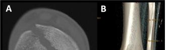


The authors attest that their institution does not require Institutional Review Board approval for publication of this case report. Patient consent has been obtained and filed for the publication of this case report.
Address for Correspondence: Victoria Mae Koniuk, BS, OMS IV, Edward Via College of Osteopathic Medicine – Carolinas, Department of Emergency Medicine, 350 Howard St, Spartanburg, SC 29303. Email: vkoniuk@carolinas.vcom.edu
Conflicts of Interest: By the CPC-EM article submission agreement, all authors are required to disclose all affiliations, funding sources and financial or management relationships that could be perceived as potential sources of bias. The authors disclosed none.
Copyright: © 2025 Koniuk et al. This is an open access article distributed in accordance with the terms of the Creative Commons Attribution (CC BY 4.0) License. See: http://creativecommons.org/ licenses/by/4.0/
1. Saad BN, Yingling JM, Liporace FA, et al. Pilon fractures: challenges and solutions. Orthop Res Rev. 2019;11:149-57.
2. Prabhu S, Neelakrishnan R, Barathiselvan V, et al. Prospective and retrospective study of managing tibial pilon fractures using various modalities of treatment. Int J Orthop Sci. 2020;6(4):554-61.
3. Bastias C and Lagos L. New Principles in Pilon Fracture Management: Revisiting Rüedi and Allgöwer Concepts. Foot Ankle Clin. 2020;25(4):505–21.
4. Jacob N, Amin A, Giotakis N, et al. Management of high-energy tibial pilon fractures. Strategies Trauma Limb Reconstr 2015;10(3):137-47.
5. Mair O, Pflüger P, Hoffeld K, et al. Management of pilon fractures— current concepts. Front Surg. 2021;8:764232.
José Guilherme Assis, MD
Joana Rua, MD
Section Editor: Joel Moll,
MD
Submission history: Submitted June 22, 2024; Revision received September 11, 2024; Accepted September 24, 2024
Electronically published January 1, 2025
Full text available through open access at http://escholarship.org/uc/uciem_cpcem DOI: 10.5811/cpcem.24869
Case Presentation: A 78-year-old male was admitted to the hospital due to acute-on-chronic liver failure with spontaneous bacterial peritonitis. About six liters of a yellow, turbid fluid were collected via indwelling urinary catheter (UC) overnight. He subsequently developed neurological and cardiac dysfunctions. Imaging confirmed bladder perforation and intraperitoneal placement of the UC, establishing the diagnosis of paracentesis-induced circulatory dysfunction due to unintended ascitic fluid drainage. He was stabilized with albumin replacement. The UC was removed, and the bladder injury resolved spontaneously.
Discussion: This case depicts a rare complication of urinary catheterization, which underscores the need for careful monitoring and prompt intervention to effectively manage unexpected catheterrelated issues. [Clin Pract Cases Emerg Med. 2025;19(1):105-106 ]
Keywords: urinary bladder diseases; paracentesis; shock.
CASE PRESENTATION
A 78-year-old male with alcoholic cirrhosis and chronic renal disease with a longstanding indwelling urinary catheter (UC), previously placed because of obstructive uropathy, was admitted to the hospital due to acute-on-chronic liver failure associated with spontaneous bacterial peritonitis. On the fourth day of hospitalization approximately six liters of a yellow, turbid fluid, consistent with infected ascitic fluid, were unexpectedly drained via the UC through the night. This incident resulted in the patient developing somnolence and mild abdominal discomfort. A physical examination revealed pallor, hypotension (blood pressure of 53/29 millimeters of mercury), and sinus tachycardia (heart rate of 105 beats per minute), suggesting significant hemodynamic instability. Besides neurological and cardiac dysfunctions, acute kidney failure was also documented. The imaging studies, including abdominal ultrasonography and computed tomography, revealed extravasation of contrast material injected through the UC into the peritoneal cavity, indicating bladder perforation and confirming the intraperitoneal placement of the catheter (Images 1 and 2).


Urinary catheterization is an essential procedure for the practicing healthcare professional.1 The most common complications include bacterial infection (occurring at a frequency of 1 per 100 to 1 per 1000 catheter days in patients with long-term UC), mechanical trauma (bladder perforation, urethral damage, and urinary leakage, typically affecting onethird of chronic patients) and, rarely, catheter toxicity.2,3 We report a unique case of circulatory dysfunction precipitated by inadvertent, large-volume drainage of ascitic fluid through a bladder perforation caused by a UC. To our knowledge, no prior cases of this specific complication have been described in the literature. Similar reports involve bladder rupture leading to urinary ascites, although these events are still infrequent.4 This unusual complication underscores the importance of careful monitoring, prompt diagnosis, and appropriate therapeutic interventions to manage catheterrelated issues,5 especially in patients with complex medical histories. In this case, early intervention and appropriate management led to a positive outcome even in the face of severe complications.
1. Shackley DC, Whytock C, Parry G, et al. Variation in the prevalence of urinary catheters: a profile of National Health Service patients in England. BMJ Open. 2017;7(6):e013842.
2. Igawa Y, Wyndaele JJ, Nishizawa O. Catheterization: possible complications and their prevention and treatment. Int J Urol. 2008;15(6):481-5.
CPC-EM Capsule
What do we already know about this clinical entity?
Urinary catheters can cause complications such as infection, mechanical trauma (including bladder perforation), and catheter toxicity.
What is the major impact of the image(s)?
The images confirm bladder perforation and fluid extravasation through the urinary catheter, which is critical for diagnosing the cause of the unexpected ascitic drainage.
How might this improve emergency medicine practice?
Early recognition of catheter-related complications, timely use of imaging for diagnosis, and prompt treatment are essential to prevent severe outcomes.
The authors attest that their institution requires neither Institutional Review Board approval, nor patient consent for publication of this case report. Documentation on file.
Address for Correspondence : José Guilherme Gonçalves Pereira de Assis Cardoso, MD, Department of Medicine, Centro Hospitalar de Trás-os-Montes e Alto Douro, Avenida da Noruega, Lordelo, 5000-508, Vila Real, Portugal. Email: jgcardoso@chtmad.min-saude.pt.
Conflicts of Interest: By the CPC-EM article submission agreement, all authors are required to disclose all affiliations, funding sources and financial or management relationships that could be perceived as potential sources of bias. The authors disclosed none.
Copyright: © 2025 Assis et al. This is an open access article distributed in accordance with the terms of the Creative Commons Attribution (CC BY 4.0) License. See: http://creativecommons.org/ licenses/by/4.0/
3. Ortega R, Ng L, Sekhar P, et al. Videos in clinical medicine. Female urethral catheterization. N Engl J Med. 2008;358(14):e15.
4. Ridinger HA, Kavitt RT, Green JK. Urinary ascites and renal failure from unrecognized bladder rupture. Am J Med. 2012;125(9):e1-2.
5. Dellimore KH, Helyer AR, Franklin SE. A scoping review of important urinary catheter-induced complications. J Mater Sci Mater Med. 2013;24(8):1825-35.
Leyan Shalabi, DO
Wesley Eilbert, MD
Section Editor: Anna McFarlin, MD
University of Illinois at Chicago, College of Medicine, Department of Emergency Medicine, Chicago, Illinois
Submission history: Submitted March 4, 2024; Revision received September 11, 2024; Accepted September 7, 2024
Electronically published January 12, 2025
Full text available through open access at http://escholarship.org/uc/uciem_cpcem
DOI: 10.5811/cpcem.20735
Case presentation: A 21-year-old woman with a history of eczema presented to the emergency department with a painful rash over the previous three days spreading from her left axilla to her left arm, left chest and left abdominal wall. The rash consisted of clusters of small, erythematous vesicles on hyperpigmented patches of skin. The patient was treated empirically with intravenous acyclovir for eczema herpeticum with improvement. Polymerase chain reaction testing of the fluid obtained from the rash vesicles later confirmed the presence of herpes simplex virus-1.
Discussion: Eczema herpeticum is a cutaneous superinfection with herpes simplex virus on preexisting sites of eczema. Left untreated, it can have a mortality rate over 50%. Early identification and treatment of this high morbidity condition with antiviral agents is key to improving outcome. [Clin Pract Cases Emerg Med. 2025;19(1):107-108.]
Keywords: eczema herpeticum; eczema; atopic dermatitis; herpes simplex virus.
A 21-year-old woman with a history of eczema presented to the emergency department complaining of a painful rash present for three days. Over the previous week, she had been experiencing worsening eczema symptoms for which she had been applying two corticosteroid creams. The painful rash consisted of grouped clusters of small erythematous vesicles overlying hyperpigmented patches. The rash had started in her left axilla and spread to her left arm, chest, and abdomen (Images 1, 2 and 3). With the rash, she reported associated chills and intermittent emesis.


Due to the extensive skin involvement and associated systemic symptoms, the patient was treated empirically with intravenous acyclovir and admitted to the hospital. Fluid collected from the vesicles of the rash was positive for herpes simplex virus (HSV) -1 deoxyribonucleic acid (DNA) on polymerase chain reaction (PCR) testing. She had significant improvement of the rash and was discharged on hospital day three to complete a 10-day course of valacyclovir.
Atopic dermatitis, or eczema, is the most common inflammatory skin disease, affecting up to 18% of children and 7% of adults.1 Eczema herpeticum (EH) is a cutaneous superinfection with HSV, most commonly HSV-1, on preexisting sites of eczema.2,3 Up to 3% of patients with eczema will experience an episode of EH, and at least 20% of all patients with EH will have a history of recurrent herpes infections.1,4 An initially local disease, EH may progress to a potentially lifethreatening systemic infection.3
Eczema herpeticum typically presents as a sudden eruption of monomorphic, dome-shaped, grouped, 2-3 millimeter vesicles on an erythematous base, superimposed on areas of pre-existing sites of eczema, most commonly the face, neck and upper chest.5 The rash is pruritic and painful and may spread to involve areas of normal skin.5 The rash is often accompanied by systemic symptoms such as fever, malaise, headache, and lymphadenopathy.4 The vesicles rupture and form crusts over underlying erosions after one to two weeks.3
The diagnosis of EH is made on clinical grounds and confirmed by the detection of HSV DNA in vesicle fluid by PCR.5 The sensitivity of PCR testing is between 80-100%.3 If PCR is not available, direct fluorescent antibody testing, a Tzank smear, or viral cultures may be used.5
The mortality rate of EH in the era before antiviral therapy was frequently over 50%.6 Given its potential high morbidity, treatment of EH should begin when it is suspected, without waiting for confirmatory tests.1,3 Acyclovir is the antiviral agent of choice for the treatment of EH.3,4 Mild cases can be treated with oral acyclovir on an outpatient basis.5 Patients with signs of systemic illness, extensive skin involvement, and those less than one year of age should be hospitalized and treated with IV acyclovir.1,5 Up to 30% of patients hospitalized for EH will have bacterial superinfection with Staphylococcus aureus, and some authors recommend empiric treatment with antistaphylococcal antibiotics for cases with extensive skin involvement.7,8
1. Wang V, Boguniewicz J, Boguniewicz M, et al. The infectious complications of atopic dermatitis. Ann Allergy Asthma Immunol. 2021;126(1):3-12.
2. Micali G and Lacarrubba F. Eczema herpeticum. N Engl J Med. 2017;377(7):e9.
3. Damour A, Garcia M, Seneschal J, et al. Eczema herpeticum:
CPC-EM Capsule
What do we already know about this clinical entity?
Eczema herpeticum is a cutaneous superinfection with herpes simplex virus. Left untreated, eczema herpeticum can have a mortality rate over 50%.
What is the major impact of the image(s)?
The images show the characteristic rash of eczema herpeticum: grouped clusters of small erythematous vesicles overlying hyperpigmented patches of skin.
How might this improve emergency medicine practice?
Rapid identification and treatment is necessary to prevent associated mortality.
The authors attest that their institution requires neither Institutional Review Board approval, nor patient consent for publication of this case report. Documentation on file.
Address for Correspondence: Wesley Eilbert, MD, University of Illinois at Chicago, College of Medicine, Department of Emergency Medicine, Room 469 CME, 1819 W Polk St. Chicago, IL 60612. Email: weilbert@uic.edu.
Conflicts of Interest: By the CPC-EM article submission agreement, all authors are required to disclose all affiliations, funding sources and financial or management relationships that could be perceived as potential sources of bias. The authors disclosed none.
Copyright: © 2025 Shalabi et al. This is an open access article distributed in accordance with the terms of the Creative Commons Attribution (CC BY 4.0) License. See: http://creativecommons.org/ licenses/by/4.0/
clinical and pathophysiological aspects. Clin Rev Allergy Imunol. 2020;59(1):1-18.
4. Traidl S, Roesner L, Zeitvogel J, et al. Eczema herpeticum in atopic dermatitis. Allergy. 2021;76(10):3017-27.
5. Xiao A and Tsuchiya A. Eczema herpeticum. In: StatPearls [Internet]. Treasure Island, Florida: StatPearls Publishing; 2024. Available at: http:// www.ncbi.nlm.nih.gov/books/NBK560781/. Accessed March 26, 2024.
6. Wheeler CE Jr and Abele DC. Eczema herpeticum, primary and recurrent. Arch Dermatol. 1966;93(2):162-73.
7. Aronson PL, Yan AC, Mittal MK, et al. Delayed acyclovir and outcomes of children hospitalized with eczema herpeticum. Pediatrics. 2011;128(6):1161-7.
8. Almoalem M, AlAlharith I, Alomer H, et al. Extensive eczema herpeticum in a previously healthy child. Int J Emerg Med. 2022;15(1):21.
Bruce M. Lo, MD, MBA*
Megyn K. Christensen, DO†
Katherine E. Schaffer, MD†
Theodore J. Tzavaras, MD*
Section Editor: Shadi Lahham, MD
* † Sentara Norfolk General Hospital/Eastern Virginia Medical School at Old Dominion University, Norfolk, Virginia
Eastern Virginia Medical School at Old Dominion University, Norfolk, Virginia
Submission history: Submitted September 5, 2024; Revision received October 11, 2024; Accepted October 18, 2024
Electronically published: December 17, 2024
Full text available through open access at http://escholarship.org/uc/uciem_cpcem DOI: 10.5811/cpcem.35284
Case Presentation: A 33-year-old female with a history of antiphospholipid syndrome presented with exertional chest pain and ST-elevation in aVR with diffuse ST-segment depression. An emergent catheterization was performed, which showed an isolated 99% stenosis in the left main coronary artery. The remaining coronary arteries were without any stenosis. Successful stent placement was performed, and the patient was discharged without complications.
Discussion: Previous guidelines have suggested that ST-segment elevation with diffuse STsegment depression should be treated as a ST-elevation myocardial infarction equivalent involving either the left-main or proximal left anterior descending coronary artery. However, recent data suggests that most of these cases may not involve that region. Regardless, this electrocardiogram finding should still be a concern for acute coronary syndrome, with the need for urgent catheterization. [Clin Pract Cases Emerg Med. 2025;19(1):109-110 ]
Keywords: acute coronary syndrome; aVR; ST-segment elevation.
A 33-year-old female with a history of antiphospholipid syndrome (APS), presented with exertional chest pain and shortness of breath. An electrocardiogram (ECG) was performed (Image 1), which showed ST-elevation in aVR with


diffuse ST-depression. The patient was taken emergently for a heart catheterization, which showed a 99% stenosis at the ostial left main artery (Image 2). The remaining coronary arteries showed no disease. The patient had a drug-eluting stent placed with improvement to 0% stenosis, and she was discharged home several days later without event.
ST-segment elevation in aVR with diffuse ST-segment depression has been described to indicate left main or proximal left anterior descending coronary artery stenosis, with previous guidelines suggesting to treat as a ST-elevation myocardial infarction equivalent.1 However, recent data suggests that only 10% will have a culprit lesion with these ECG findings, but approximately 60% will have severe coronary artery disease.2 Although the ECG pattern may not always correspond with a culprit lesion, it should raise concerns for significant coronary artery disease, and an urgent cardiology consultation is needed. History such as APS, which is a multisystem autoimmune disease associated with coronary artery disease, should also increase suspicion for acute myocardial infarction especially in those who are less than 45 years of age.3
Address for Correspondence: Bruce M. Lo, MD, MBA, Sentara Norfolk General Hospital/Eastern Virginia Medical School at Old Dominion University, 600 Gresham Drive, Norfolk, VA 23507. Email: Brucelo1@yahoo.com.
Conflicts of Interest: By the CPC-EM article submission agreement, all authors are required to disclose all affiliations, funding sources and financial or management relationships that could be perceived as potential sources of bias. The authors disclosed none.
Copyright: © 2025 Lo et al. This is an open access article distributed in accordance with the terms of the Creative Commons Attribution (CC BY 4.0) License. See: http://creativecommons.org/ licenses/by/4.0/
The authors attest that their institution requires neither Institutional Review Board approval, nor patient consent for publication of this case report. Documentation on file.
CPC-EM Capsule
What do we already know about this clinical entity?
Evidence is mixed on whether ST-segment elevation in aVR with diffuse ST-segment depression represents a ST-elevation myocardial infarction equivalent.
What is the major impact of the image(s)?
These findings can represent significant coronary artery disease, with this case showing an isolated 99% stenosis in the left main coronary artery.
How might this improve emergency medicine practice?
These electrocardiogram findings should raise concerns for acute coronary syndrome and urgent consultation with cardiology to improve outcomes.
1. Wagner GS, Macfarlane P, Wellens H, et al. AHA/ACCF/HRS recommendations for the standardization and interpretation of the electrocardiogram: part VI: acute ischemia/infarction: a scientific statement from the American Heart Association Electrocardiography and Arrhythmias Committee, Council on Clinical Cardiology; the American College of Cardiology Foundation; and the Heart Rhythm Society. Endorsed by the International Society for Computerized Electrocardiology. J Am Coll Cardiol. 2009;53(11):1003-11.
2. Harhash AA, Huang JJ, Reddy S, et al. aVR ST segment elevation: acute STEMI or Not? Incidence of an acute coronary occlusion. Am J Med. 2019;132(5):622-30.
3. Sood A, Singh A, Gadkari C. Myocardial infarction in young individuals: a review article. Cureus. 2023;15(4):e37102.
Richard White, MD*†
Kaitlyn Mander‡
Christian A. Koziatek, MD*†
Sanjay Mohan, MD§
Section Editor: Jacqueline Le, MD
NYU Grossman School of Medicine, Ronald O. Perelman Department of Emergency Medicine, New York, New York
Bellevue Hospital Center, Department of Emergency Medicine, New York, New York
Geisinger Community Medical Center, Emergency Services PC, Scranton, Pennsylvania
New York University Grossman Long Island School of Medicine, Department of Emergency Medicine, Mineola, New York
Submission history: Submitted July 1, 2024; Revision received October 22, 2024; Accepted November 3, 2024
Electronically published December 17, 2024
Full text available through open access at http://escholarship.org/uc/uciem_cpcem DOI: 10.5811/cpcem.25018
Case Presentation: We describe a case of persistent odynophagia due to a retained foreign body 27 days after emergent intubation.
Discussion: Dentures constitute a potential esophageal foreign body and warrant special consideration during airway management. Odynophagia, dysphagia, and changes in phonation should prompt consideration of retained esophageal foreign bodies, especially in the post-intubation setting. [Clin Pract Cases Emerg Med. 2025;19(1):111-113 ]
Keywords: intubation; foreign body; dentures; odynophagia
A 41-year-old female, with no past medical history, presented to the emergency department (ED) with a chief complaint of odynophagia. The patient initially presented to the ED 29 days prior for back pain after a motor vehicle accident and was found to have an unstable L1 burst fracture. She underwent an uncomplicated L1 surgical fixation after routine endotracheal intubation on the day of presentation and was extubated without any untoward effects. Postoperatively, on hospital day two, computed tomography (CT) of the chest revealed bilateral pulmonary emboli. No foreign bodies were noted in the esophagus or trachea. Later the same day, the patient suffered a cardiac arrest and was emergently intubated. Post-intubation single-view portable chest radiographs did not reveal any obvious foreign bodies in the thoracic cavity (Image 1).
She was eventually weaned off mechanical ventilation on hospital day six with a normal mental status and reports of minor odynophagia immediately post-extubation. She was ultimately discharged to a rehabilitation facility on hospital day 23, on a regular diet, without any subsequent imaging performed.
She returned to the ED six days later with progressively worsening odynophagia since discharge. She noted discomfort with swallowing liquids and solids but denied emesis, changes in phonation, or difficulty breathing. Vital signs were unremarkable. Physical examination revealed an ageappropriate female in no apparent distress and otherwise

tolerating her secretions. Her uvula was midline, and an oropharyngeal examination revealed no evidence of infection or trauma. Her neck was supple, and there was no pain upon palpation of the neck. No upper airway or abnormal breath sounds were noted.
Two-view cervical roentgenograms ultimately revealed a foreign body in the hypopharynx (Image 2). Otolaryngology was consulted. Using flexible nasopharyngoscopy, they visualized a dental plate over the larynx at the level of the cervical esophagus. The patient was emergently taken to the operating room to

views of a foreign body (white arrows) in the hypopharynx.
remove the foreign body and was cleared for discharge to a rehabilitation facility three days later without any changes in phonation, difficulty breathing or swallowing, and she was able to tolerate food and liquids by mouth.
DISCUSSION
After animal bones, dentures are the second most commonly ingested foreign body and account for approximately 4-18% of esophageal foreign bodies.1 Dental prosthetic dislodgement, in general, displays high rates of endoscopic or surgical intervention due to its tendency to become impacted or lead to perforation.2 It is worth noting that dentures, whether total or partial, may or may not be visualized with standard roentgenograms as some are radiolucent 3
Although emergent intubation was suspected to be the sentinel event leading to the patient’s presentation, other etiologies including chest compressions during cardiopulmonary resuscitation leading to dental plate dislodgement, extubation, or unintentional swallowing of the foreign body at any point during the prolonged hospitalization were considered. Dislodgement and retention of dental devices in the context of airway management has been reported previously, but it is worth noting again as a visual reminder of this avoidable and potentially catastrophic mishap during intubation, as well as the duration of time this patient went without identifying the retained foreign body.3-5
Clinicians should be attuned to the risks of dental hardware that may be dislodged unintentionally, particularly in emergent situations such as rapid sequence intubation or cardiac arrest. It is critical to maintain a high index of suspicion for foreign body ingestion in specific patient populations, such as those with psychiatric disorders or cognitive delay, as these groups carry higher rates for such events.5 Furthermore, advanced imaging (such as computed tomography) should be considered to definitively evaluate for the presence of dental hardware, as roentgenograms may miss radiolucent foreign bodies.
CPC-EM Capsule
What do we already know about this clinical entity?
Rapid sequence intubation poses the risk of foreign body aspiration.
What is the major impact of the image(s)?
Dislodged dental foreign bodies should be in the differential for patients presenting with odynophagia after recent airway management.
How might this improve emergency medicine practice?
Prior to intubation, assessing for and documenting the presence of dental prosthetics is essential to minimize the risk of foreign body aspiration.
This case is particularly relevant for emergency, critical care, and anesthesia clinicians whose scope of practice includes airway management, especially in emergent scenarios. Specifically, it highlights the importance of assessing for and documenting the presence of dental prosthetics before and after any procedure that risks dislodgement. Dislodged dental foreign bodies should be in the differential for patients presenting with odynophagia after recent airway management.
The authors attest that their institution requires neither Institutional Review Board approval, nor patient consent for publication of this case report. Documentation on file.
Address for Correspondence: Sanjay Mohan, MD, Department of Emergency Medicine, NYU Grossman Long Island School of Medicine, 259 First Street, Mineola, NY 11501. Email: Sanjay.mohan@nyulangone.org.
Conflicts of Interest: By the CPC-EM article submission agreement, all authors are required to disclose all affiliations, funding sources and financial or management relationships that could be perceived as potential sources of bias. The authors disclosed none.
Copyright: © 2025 White et al. This is an open access article distributed in accordance with the terms of the Creative Commons Attribution (CC BY 4.0) License. See: http://creativecommons.org/ licenses/by/4.0/
White et al. Persistent Odynophagia report and literature review. Dysphagia. 2009;24(1):105-8.
1. Mathew RP, Sarasamma S, Jose M, et al. Clinical presentation, diagnosis and management of aerodigestive tract foreign bodies in the adult population: Part 1. SA J Radiol. 2021;25(1):2022.
2. Bandyopadhyay SN, Das S, Das SK, et al. Impacted dentures in the oesophagus. J Laryngol Otol. 2014;128(5):468-74.
3. Chin RY and Ellul S. Dysphagia after emergency intubation: case
4. Adbullah BJ, Teanh LK, Mahadevan J. Dental prosthesis ingested and impacted in the oesophagus and orolaryngopharynx. J Otolaryngol. 1998;27:190–4
5. Akelma H, Salik F, Başol Ö, et al. Ingestion of partial denture after general anesthesia induction and ventilation: a rare case report. Case Rep Surg. 2019;2019:8726508.
Emily Prentice, BA*
Emily Bartlett, MD, MS†
Jonathan S. Ilgen, MD, PhD‡
Section Editor: Christopher Sampson,
University of Washington School of Medicine, Seattle, Washington
University of New Mexico, Albuquerque, Department of Emergency Medicine, New Mexico
University of Washington, Department of Emergency Medicine, Seattle, Washington
MD
Submission history: Submitted June 13, 2024; Revision received October 2, 2024; Accepted November 3, 2024
Electronically published January 1, 2025
Full text available through open access at http://escholarship.org/uc/uciem_cpcem DOI: 10.5811/cpcem.24845
Case Presentation: A male patient in his thirties with a history of polysubstance use presented to the emergency department (ED) due to an abrasion on his left forehead caused by banging his head against a wall in self-injurious behavior. A non-contrast computed tomography of the head obtained to rule out intracranial injury incidentally demonstrated a radiodense foreign body in the left external ear canal. A round metallic foreign body was subsequently visualized on otoscopic examination. The aural foreign body (AFB) was identified as a metallic bead that the patient had placed into his own ear; however, he reported no associated discomfort, hearing changes, or discharge. Traditional approaches for removing AFBs were considered; however, due to the position and smooth surface of the bead, there was concern they would be unsuccessful. Recognizing the metallic nature of the AFB, the clinician held a ceramic donut magnet adjacent to the patient’s ear and subsequently extracted the AFB without complication or patient discomfort.
Discussion: Aural foreign bodies account for a significant number of visits to EDs annually. Removal of AFBs can be challenging, often requiring specialized equipment or specialty referral for management. Using magnetism over short distances for the purpose of extracting metallic AFBs presents a low-cost, low-risk intervention. When used in applicable scenarios, this technique can decrease the need for specialty referral and can especially benefit patients seeking care in lessresourced settings. [Clin Pract Cases Emerg Med. 2025;19(1):114-116 ]
Keywords: aural foreign body; magnetic bead.
A male patient in his thirties with a history of polysubstance use was evaluated in the emergency department after hitting his head against a wall in self-injurious behavior. He had an abrasion to his left forehead and an otherwise unremarkable physical examination. A non-contrast computed tomography of the head was obtained, which ruled out intracranial injury. This imaging incidentally demonstrated a radiodense foreign body in the left external auditory canal, and a round metallic foreign body was subsequently visualized on otoscopic examination (Image 1).
When questioned, the patient reported placing a string of magnetic beads in his left ear, previously using the magnetic forces of other beads to remove these foreign bodies on his
own. He was aware that a metallic foreign object remained in his external auditory canal but had been unsuccessful in attempts to remove it. He experienced no discomfort or other auditory complaints in the context of this foreign body.
We considered several approaches to remove the aural foreign body (AFB).1 Due to the positioning of the round sphere in the ear canal, a speculum or jet of fluid would not have been able to reach around it and would likely have pushed the AFB further into the canal. Use of cyanoacrylate glue on the tip of a swab was also considered; however, there was concern it would not adhere well to the smooth metal surface. Specialized suction catheters were not readily available in our ED. A ceramic donut magnet from the code cart—typically used to temporarily induce pacemakers into
asynchronous mode—was obtained. When the donut magnet was held adjacent to the patient’s left ear, the magnetic sphere emerged from the ear canal and adhered to the magnet (Image 2). The procedure was well tolerated by the patient with no reported discomfort or complications.

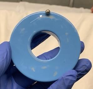
DISCUSSION
Spherical, non-graspable foreign bodies often require specialist referral and potential removal under anesthesia.1 In this case, we quickly and easily removed a magnetic spherical foreign object using a ceramic donut magnet, obviating the need for specialty consultation. Although the use of magnetized instruments for this purpose has been described anecdotally,2
CPC-EM Capsule
What do we already know about this clinical entity?
Removal of aural foreign bodies (AFB) can be challenging, often requiring specialized equipment or specialty referral for management.
What is the major impact of the image(s)?
Using a ceramic donut magnet, a device commonly available in emergency departments, to remove a metallic AFB is a low-cost and easily accessible intervention.
How might this improve emergency medicine practice?
This low-cost, low-risk intervention would be especially useful in settings with less access to specialty care.
to our knowledge this is the first published report of removal of an AFB by magnetic force acting over a distance. Magnets are commonly found in EDs, and this technique could be readily adopted without the need to purchase specialized equipment. Additionally, the use of magnets to remove AFBs may be applicable to objects that are permanent magnets as well as metallic foreign bodies susceptible to induced magnetism.3
A study using the National Electronic Injury Surveillance System reported that over a five-year period 250,000 ED visits were due to AFBs.4 However, access to otolaryngology for specialized management varies widely depending on location, and 65.7% of counties in the United States do not have a practicing otolaryngologist.5 This case describes the use of a low-cost, low-risk intervention for removal of magnetic foreign bodies, which can aid in patient care especially in lower resource settings with less access to specialty care.
Address for Correspondence: Jonathan Ilgen, MD, PhD, Department of Emergency Medicine, University of Washington, Harborview Medical Center, 325 9th Avenue, Box 359702, Seattle, WA, 98104. Email: ilgen@uw.edu.
Conflicts of Interest: By the CPC-EM article submission agreement, all authors are required to disclose all affiliations, funding sources and financial or management relationships that could be perceived as potential sources of bias. The authors disclosed none.
Copyright: © 2025 Prentice et al. This is an open access article distributed in accordance with the terms of the Creative Commons Attribution (CC BY 4.0) License. See: http://creativecommons.org/ licenses/by/4.0/
Documented patient informed consent has been obtained and filed for publication of this case report. The authors attest that their institution does not require Institutional Review Board approval for publication of this case report. Documentation on file.
1. Friedman EM. Removal of Foreign Bodies from the Ear and Nose. N Engl J Med. 2016;374(7):E7-U6.
2. Mellick LB. Using a magnetic pick-up tool for ear foreign body
removal. Available at: https://www.youtube.com/watch?v=HiVtldiwIS8. Accessed October 31, 2024.
3. Brod RC and Schlang HA. Removal of metallic foreign body by magnetic force. JAMA. 1962;179:164-5.
4. Svider PF, Vong A, Sheyn A, et al. What are we putting in our ears? A consumer product analysis of aural foreign bodies. Laryngoscope. 2015;125(3):709-14
5. Vickery TW, Weterings R, Cabrera-Muffly C. Geographic distribution of otolaryngologists in the United States. Ear, Nose, Throat J. 2016;95(6):218-23.
Matthias Barden, MD*†‡
Dustin Marinelli, DO
Kirsten Cable, DO
*§
*§
Section Editor: Austin Smith, MD
* † ‡
Eisenhower Health Emergency Medicine Residency Program, Rancho Mirage, California
§ Hi-Desert Medical Center, Department of Emergency Medicine, Joshua Tree, California
University of California Riverside School of Medicine, Riverside, California Desert Regional Medical Center Emergency Medicine Residency, Palm Springs, California
Submission history: Submitted July 20, 2024; Revision received November 20, 2024; Accepted November 13, 2024
Electronically published January 12, 2025
Full text available through open access at http://escholarship.org/uc/uciem_cpcem
DOI: 10.5811/cpcem.31062
Case Presentation: We present an image and discussion of spontaneous evisceration, or “burst abdomen,” from an anterior abdominal wall hernia. A 61-year-old female with prior history of alcoholic cirrhosis and ascites presented to our emergency department with frank evisceration of multiple loops of small bowel from an open anterior abdominal wall dehiscence. Approximately one year prior to this visit she had also been seen in our department for spontaneous rupture of the skin overlying an umbilical hernia and large-volume external leakage of ascites (Flood syndrome1). She required surgery to repair the abdominal wall at that time but had subsequently developed a new ventral hernia extending from the umbilicus across a large portion of her left lower abdomen as well as several other postoperative complications. On the day of presentation, she suffered dehiscence of that one-year-old surgical site resulting in spontaneous evisceration of her small bowel. She was transferred to a facility with acute care surgical capabilities where she remained in critical condition.
Discussion: Spontaneous evisceration from abdominal wall dehiscence is a devastating surgical complication. It tends to occur in the immediate postoperative period but has been reported to occur years later. This patient likely suffered from delayed burst abdomen due to multiple comorbidities and postoperative complications. [Clin Pract Cases Emerg Med. 2025;19(1):117-119.]
Keywords: case report; images in emergency medicine; burst abdomen; spontaneous evisceration; flood syndrome.
A 61-year-old female presented to our emergency department (ED) with frank evisceration of multiple loops of small bowel from an open anterior abdominal wall dehiscence of a ventral hernia. About one year prior, she had presented with spontaneous rupture of the peritoneum and skin overlying an umbilical hernia and large-volume external leakage of ascites (spontaneous paracentesis, also known as Flood syndrome).1 This is a rare complication of ascites and carries a high morbidity. The mechanism of spontaneous skin rupture is thought to be related to gradual thinning and weakening of the
skin overlying umbilical hernias in patients with longstanding tense ascites.2 She was managed with primary surgical closure and placement of an indwelling peritoneal drain to avoid reaccumulating of ascitic fluid as the surgical site was healing.3 However, her postoperative course after the Flood syndrome repair was complicated. She developed bacterial peritonitis prompting early removal of her indwelling ascites drain. She also developed a larger area of anterior abdominal wall herniation at the site of the prior umbilical hernia that had ruptured. One week prior to presentation she had been admitted at a different facility for incarceration and obstruction of the ventral hernia.
The night prior to presenting to our ED, she had noticed a small, shallow open wound in the surgical scar overlying her hernia. When she awoke on the morning of presentation, she had found that the site was widely open with extruding bowels as pictured in the Image.

Her husband drove her to the ED and helped her inside via our ambulatory entrance. Upon arrival to triage the exposed bowels were covered with sterile, saline-soaked gauze, and she was started on intravenous (IV) fluids and broad-spectrum antibiotics. The general surgeon recommended transfer to another facility for higher level of care. At the receiving facility she was taken emergently to the operating room for exploratory laparotomy and umbilical hernia repair. During the procedure the bowel was reduced, and an absorbable mesh overlay was used to close the fascia. She was managed in the intensive care unit for multiorgan system failure requiring continuous renal replacement therapy and vasopressor support before she unfortunately died one month later.
While intentionally leaving the abdomen open for postoperative management is an increasingly frequent surgical strategy, the unintentional acute postoperative open abdomen is a rare and serious surgical complication.4 Spontaneous evisceration from abdominal wall dehiscence (or burst abdomen) tends to occur in the acute postoperative period but has also been reported to occur years later in the setting of large chronic incisional hernias.5,6
Our patient was about one year out from surgical repair of a prior spontaneous rupture of an umbilical hernia in the setting of cirrhotic ascites, also known as Flood syndrome. Flood syndrome carries a high rate of mortality, and strong evidence is lacking regarding the best technique for its surgical management.7 While this patient survived the initial recovery from Flood syndrome, her postoperative course was quite complicated. These complications, as well as her overall general poor health, likely contributed to weakening and eventual
CPC-EM Capsule
What do we already know about this clinical entity?
Flood syndrome (spontaneous paracentesis) and burst abdomen (spontaneous evisceration) are rare entities with high morbidity and mortality.
What is the major impact of the image(s)?
This image depicts the severity of a presentation of burst abdomen that occurred one year after surgical repair of a case of flood syndrome.
How might this improve emergency medicine practice?
Awareness of flood syndrome and burst abdomen may help clinicians respond appropriately to these dramatic presentations.
rupture of the soft tissue overlying the ventral hernia site. Emergency department management was limited to covering the exposed bowel in sterile, saline-soaked gauze, initiating broad-spectrum IV antibiotics, and transferring her for higher level of care to a facility with acute care surgical capabilities where she was managed surgically and with critical care life support; however, she ultimately died from this event.
The authors attest that their institution requires neither Institutional Review Board approval, nor patient consent forpublication of this case report. Documentation on file.
Address for Correspondence: Matthias Barden, MD, Eisenhower Health, Department of Emergency Medicine, Rancho Mirage, CA. Email: mbarden@eisenhowerhealth.org.
Conflicts of Interest: By the CPC-EM article submission agreement, all authors are required to disclose all affiliations, funding sources and financial or management relationships that could be perceived as potential sources of bias. The authors disclosed none.
Copyright: © 2025 Barden et al. This is an open access article distributed in accordance with the terms of the Creative Commons Attribution (CC BY 4.0) License. See: httpÍ://creativecommons. org/licenses/by/4.0/
Barden et al.
“Burst Abdomen” in Patient with Prior Flood Syndrome Surgical Repair
1. Flood FB. Spontaneous perforation of the umbilicus in Laennec’s cirrhosis with massive ascites. N Engl J Med . 1961;264:72-4
2. Podymow T, Sabbagh C, Turnbull J. Spontaneous paracentesis through an umbilical hernia. CMAJ . 2003;168(6):741
3. Strainiene S, Peciulyte M, Strainys T, et al. Management of Flood syndrome: What can we do better ? World J Gastroenterol 2021;27(32):5297-305.
4. López-Cano M, García-Alamino JM, Antoniou SA, et al. EHS
clinical guidelines on the management of the abdominal wall in the context of the open or burst abdomen. Hernia. 2018;22:921–39.
5. Osei-Tutu E and Osonuga A. Spontaneous evisceration through an incisional hernia: a rare complication in surgical practice. J Acute Disease . 2016;5(4):348-9.
6. Sailaja S, Umadevi K, Rathod PS, et al. Spontaneous evisceration of an incisional hernia presenting one year after primary surgery. Online J Health Allied Scs. 2013;12(4):15.
Alexander W. Lipinski, BSN*
Jordan R. Pollock, MD, MBA†
Nelly Tan, MD†
Douglas Rappaport MD‡
* † ‡
Mayo Clinic Alix School of Medicine, Mayo Clinic, Scottsdale, Arizona
Mayo Clinic, Department of Radiology, Phoenix, Arizona
Mayo Clinic, Department of Emergency Medicine, Phoenix, Arizona
Section Editor: Christopher Sampson, MD
Submission history: Submitted October 2, 2024; Revision received December 11, 2024; Accepted November 25, 2024
Electronically published January 19, 2025
Full text available through open access at http://escholarship.org/uc/uciem_cpcem DOI: 10.5811/cpcem.35602
Case Presentation: An 83-year-old male with a history of prostate cancer and prior prostatectomy presented with lower abdominal pain, urethral leakage, and hematuria after a routine suprapubic catheter exchange, which was found to be incorrectly positioned in the bulbar urethra, leading to obstructive nephropathy with mild hydronephrosis.
Discussion: This case highlights the increased risk of suprapubic catheter misplacement and complications in elderly patients with neurogenic bladder and altered urinary anatomy, particularly after prostatectomy and artificial urethral sphincter placement. It emphasizes the importance of careful management during catheter exchanges in such patients to prevent complications of misplacement. [Clin Pract Cases Emerg Med. 2025;19(1):120-122.]
Keywords: suprapubic catheter; urethral malposition; neurogenic bladder; emergency medicine; obstructive nephropathy.
An 83-year-old male with a history of prostate cancer, treated with prostatectomy and artificial urethral sphincter placement, presented with severe lower abdominal pain, urethral leakage, and hematuria following a routine suprapubic catheter exchange. Pelvic computed tomography revealed that the suprapubic catheter had migrated into the bulbar urethra, resulting in malfunctioning catheter and resultant obstructive physiology with bladder distension and mild hydronephrosis (Image 1A, 1B, and 1C).
This patient’s history presented several risk factors for complications following a routine suprapubic catheter exchange. Notably, his history of prostatectomy and artificial urethral sphincter placement suggests surgical alterations that may have caused anatomical changes, increasing the likelihood of catheter misplacement.1,2 Long-term
catheterization further heightens the risk of complications such as bladder calculi, recurrent infections, granulation tissue formation, and structural changes, which can complicate future catheter exchanges.1,3 Additionally, neurogenic bladder often results in altered bladder dynamics and diminished sensation, masking symptoms of catheter misplacement and predisposing patients to bladder distension and hydronephrosis.2,4 The risk is compounded in older patients, who generally face decreased tissue elasticity, multiple comorbidities, and anatomical changes due to previous surgeries or chronic conditions.1,5
This case underscores the potential for suprapubic catheter malposition and subsequent complications in elderly patients with neurogenic bladder and altered urinary anatomy. The malpositioned catheter placement within the bulbar urethra highlights the need for heightened vigilance and specialized techniques when managing catheter exchanges in patients with altered urinary anatomy,

Image 1A. A coronal computed tomography abdomen and pelvis with intravenous contrast demonstrates a suprapubic catheter with inflated Foley balloon at the bulbar urethra caudal to the urethral anastomosis. There is also bladder distension and mild hydronephrosis from functional urinary obstruction due to malpositioned Foley balloon.

Image 1B. A sagittal computed tomography abdomen pelvis with intravenous contrast demonstrates a suprapubic catheter with inflated Foley balloon at the level of the bulbar urethra, just proximal to the artificial urinary sphincter. The bladder is also distended due to bladder outlet obstruction due to malpositioned catheter.
CPC-EM Capsule
What do we already know about this clinical entity?
Suprapubic catheter misplacement can lead to urinary obstruction and nephropathy, especially in patients with altered urinary anatomy and neurogenic bladder.
What is the major impact of the image(s)?
The images clearly show a suprapubic catheter malpositioned into the bulbar urethra, causing bladder distension and mild hydronephrosis.
How might this improve emergency medicine practice?
Careful catheter management and vigilance for misplacement is vital, improving outcomes in patients with complex urinary anatomy.
specifically in retracting the catheter after balloon is inflated to ensure position in the bladder lumen. This report serves as a valuable reference for clinicians encountering similar cases and encourages further investigation into optimized catheterization strategies for patients with complex urological histories.

Obstructive Nephropathy from Misplaced Suprapubic Catheter Lipinski et al.
The authors attest that their institution requires neither Institutional Review Board approval, nor patient consent for publication of this case report. Documentation on file.
Address for Correspondence: Alexander W. Lipinski, Mayo Clinic Alix School of Medicine, 13400 E Shea Blvd., Phoenix, AZ 85259, Email: Lipinski.Alexander@mayo.edu
Conflicts of Interest: By the CPC-EM article submission agreement, all authors are required to disclose all affiliations, funding sources and financial or management relationships that could be perceived as potential sources of bias. The authors disclosed none.
Copyright: © 2025 Lipinski et al. This is an open access article distributed in accordance with the terms of the Creative Commons Attribution (CC BY 4.0) License. See: http://creativecommons.org/ licenses/by/4.0/
1. Hobbs C, Howles S, Derry F, et al. Suprapubic catheterisation: a study of 1000 elective procedures. BJU Int. 2022;129(6): 760-7.
2. Li JJ and Au CF. Inappropriate placement of urinary catheters into the ureter: a case report and literature review. Medicine (Baltimore) 2024;103(15):e37623.
3. Sullivan N, AlRemeithi R, Pourmand A. Suprapubic catheter encasement by bladder stone. Am J Emerg Med. 2022;56: 395e5-7.
4. Adeyemo B, Makovitch S, Foo D. A peculiar complication of suprapubic catheterization: recurrent ureteral obstruction and hydronephrosis. J Spinal Cord Med. 2013;36(2):166-9.
5. Bos BJ, van Merode NAM, Steffens MG, et al. The patient pathway for men with chronic urinary retention: treatments, complications, and consequences. Urology. 2022;167:185-90.
Luke Wohlford, MD
Richard Bounds, MD
Section Editor: Steven Walsh, MD
The University of Vermont Medical Center, Department of Emergency Medicine, Burlington, Vermont
Submission history: Submitted October 15, 2024; Revision received December 14, 2024; Accepted December 16, 2024
Electronically published January 19, 2025
Full text available through open access at http://escholarship.org/uc/uciem_cpcem DOI: 10.5811/cpcem.35395
Case Presentation: We present a case of a 79-year-old male with gastric outlet obstruction resulting from a stomach herniation through a large left inguinal hernia.
Discussion: Stomach-containing inguinal hernias are a rare cause of gastric outlet obstruction. Treatment options range from conservative to surgical management. Once identified with imaging, prompt treatment should be initiated to prevent incarceration, strangulation, and gastric necrosis.
[Clin Pract Cases Emerg Med. 2025;19(1):123–124.]
Keywords: gastric outlet obstruction; inguinal hernia.
CASE PRESENTATION
A 79-year-old male with a left inguinoscrotal hernia and aortic stenosis presented to the emergency department with an acute syncopal episode. The patient attributed his own weakness and passing out to poor oral intake and abdominal pain for three days. He noted a left inguinal bulge that had never bothered him previously. The gastrointestinal symptoms had improved the day prior to the patient’s syncopal episode, but he started to feel palpitations and generally weaker on the day of presentation. He had no prior surgeries for the inguinal hernia, which had no previous complications.
On examination, vital signs included blood pressure of 123/82 millimeters of mercury, pulse rate 141 and irregularly irregular, respiratory rate 16 breaths per minute, and temperature 36.8° Celsius. The abdominal exam demonstrated a distended but non-tender abdomen, with a palpable left inguinal mass (Image 1). Further workup with computed tomography demonstrated a large and distended stomach with extension of the distal portion including the pylorus into the inguinal hernia (Images 2 and 3).
Incarceration of a portion of the stomach is a rare cause of gastric outlet obstruction, with fewer than 20 cases documented in the literature.1 The rarity of stomach-containing groin hernias is remarkable, considering that the lifetime incidence of groin hernias is estimated to be between 27-43% for men and 3-6% for women.2 The inferior portion of the stomach attaches to

the omentum by the gastrocolic ligament, making it vulnerable to herniation through particularly large and chronic inguinal hernias.3 Acquired inguinal hernias are typically direct, resulting from the chronic pressure of a hernia sac just medial to the inferior epigastric vessels.4 Less common than direct hernias, but seen in our patient, is the acquired, indirect hernia protruding lateral to the inferior epigastric vessels, which travels through the inguinal canal and often extends into the scrotum. Nasogastric

the pylorus.

3. Computed tomography with sagittal images demonstrating a large and distended stomach partially in an inguinal hernia.
tube placement may adequately relieve the most severe symptoms, which can be followed by operative hernia repair, or even percutaneous endoscopic gastronomy tube placement.5 Physicians should be aware of the ability of the stomach to herniate into large inguinal hernia defects and act promptly to avoid incarceration, strangulation, and stomach necrosis.
Patient consent has been obtained and filed for the publication of this case report.
CPC-EM Capsule
What do we already know about this clinical entity?
Stomach-containing inguinal hernias are rare and can potentially lead to gastric outlet obstruction if left untreated.
What is the major impact of the image(s)?
The computed tomography images depict herniation of the stomach through the inguinal canal, including the gastric antrum, demonstrating a unique cause of gastric outlet obstruction.
How might this improve emergency medicine practice?
This case highlights the importance of thorough physical examination and consideration of imaging to prevent serious, hernia-related complications.
Address for Correspondence: Luke Wohlford, MD, The University of Vermont Medical Center, Department of Emergency Medicine, 111 Colchester Ave, Burlington, VT, 05401. Email: luke.wohlford@ uvmhealth.org.
Conflicts of Interest: By the CPC-EM article submission agreement, all authors are required to disclose all affiliations, funding sources and financial or management relationships that could be perceived as potential sources of bias. The authors disclosed none.
Copyright: © 2025 Wohlford et al. This is an open access article distributed in accordance with the terms of the Creative Commons Attribution (CC BY 4.0) License. See: http://creativecommons.org/ licenses/by/4.0/
1. Favela JG, Argo MB, McAllister J, et al. Gastric outlet obstruction from stomach-containing groin hernias: case report and a systematic review. J Clin Med. 2023;13(1):155.
2. HerniaSurge Group. International guidelines for groin hernia management. Hernia. 2018;22(1):1-165.
3. Grantham TA, Ramachandran R, Parvataneni S, et al. Stomach within a large inguinal hernia. Cureus. 2022;14(5):e24783.
4. Morrison Z, Kashyap S, Nirujogi VL. Adult inguinal hernia. In: StatPearls. Treasure Island, FL: StatPearls Publishing. 2023. Available at: https:// www.ncbi.nlm.nih.gov/books/NBK537241/. Accessed October 15, 2024.
5. Blair SM, Cournoyer RM, Newcomb MR, et al. Novel treatment of gastric outlet obstruction secondary to incarcerated inguinal hernia: percutaneous endoscopic gastrostomy tube. J Surg Case Rep. 2023;2023(6):rjad294.

- Western Journal of Emergency Medicine Subscription -CAL/AAEM News Service email updates
-Discounted AAEM pre-conference fees -And more!
CAL/AAEM NEWS SERVICE
-Healthcare industry news
-Public policy
-Government issues
-Legal cases and court decisions
