


8, Number 4, November


8, Number 4, November

Case Reports
318 Achenbach Syndrome: A Case Report
Chen M, Yanes D, Manvar-Singh P, Iamonaco C, Jerome CJ, Supariwala A
322 Loculated Fluid Visualized in Hepatorenal Space with Point-of-care Ultrasound in Patient with Pelvic Inflammatory Disease Caused by Group A Streptococcus: Case Report Makhhijani N, Sondheim S, Turandot Saul T, Yetter E
326 Burst that Bubble. Gastric Perforation from an Ingested Intragastric Balloon: A Case Report
Martinez M, Faiss K, Sehgal N
329 Exploring the Palmar Surface: A Critical Case Report for Emergency Physicians
Van Ligten M, Rappaport D, Martini W
332 Metal Pneumonitis from “Non-toxic” Decorative Cake Dust Aspiration: A Case Report Sanders T, Hymowitz M, Murphy C
336 Autophagia in a Patient with Dementia and Hemineglect: A Case Report
Bragg B, Bragg K
339 Immune Checkpoint Inhibitor - Induced Primary Adrenal Insufficiency: A Case Report
Gupta R, Lubkin C
343 Esophageal Obstruction from Food Bolus Impaction Successfully Managed with the “Upright Posture, Chin Tuck, Double Swallow” Maneuver: A Case Report Barden M, Schwartz ME
346 Traumatic Atrial Septal Defect with Tricuspid Regurgitation Following Blunt Chest Trauma Presenting as Hypoxemia: A Case Report
Rashid A, Ganie FA, Rather H, Ajaz S, Shounthoo RS



Contents continued on page iii


Penn State Health is a multi-hospital health system serving patients and communities across central Pennsylvania. We are the only medical facility in Pennsylvania to be accredited as a Level I pediatric trauma center and Level I adult trauma center. The system includes Penn State Health Milton S. Hershey Medical Center, Penn State Health Children’s Hospital, and Penn State Cancer Institute based in Hershey, Pa.; Penn State Health Hampden Medical Center in Enola, Pa.; Penn State Health Holy Spirit Medical Center in Camp Hill, Pa.; Penn State Health St. Joseph Medical Center in Reading, Pa.; Penn State Health Lancaster Pediatric Center in Lancaster, Pa.; Penn State Health Lancaster Medical Center (opening fall 2022); and more than 3,000 physicians and direct care providers at more than 126 outpatient practices in 94 locations. Additionally, the system jointly operates various health care providers, including Penn State Health Rehabilitation Hospital, Hershey Outpatient Surgery Center, Hershey Endoscopy Center, Horizon Home Healthcare and the Pennsylvania Psychiatric Institute.
We foster a collaborative environment rich with diversity, share a passion for patient care, and have a space for those who share our spark of innovative research interests. Our health system is expanding and we have opportunities in both academic hospital as well community hospital settings.

Benefit highlights include:
• Competitive salary with sign-on bonus
• Comprehensive benefits and retirement package
• Relocation assistance & CME allowance
• Attractive neighborhoods in scenic central Pa.

Indexed in PubMed and full text in PubMed Central
Rick A. McPheeters, DO, Editor-in-Chief Kern Medical/UCLA- Bakersfield, California
R. Gentry Wilkerson, MD, Deputy Editor University of Maryland School of Medicine
Mark I. Langdorf, MD, MHPE, Senior Associate Editor University of California, Irvine School of Medicine- Irvine, California
Shahram Lotfipour, MD, MPH, Senior Associate Editor University of California, Irvine School of Medicine- Irvine, California
Shadi Lahham, MD, MS, Associate Editor Kaiser Permanente- Orange County, California
John Ashurst, DO, Decision Editor/ ACOEP Guest Editor Kingman Regional Health Network, Arizona
Anna McFarlin, MD, Decision Editor Louisiana State University Health Science Center- New Orleans, Louisiana
Lev Libet, MD, Decision Editor Kern Medical/UCLA- Bakersfield, California
Amin A. Kazzi, MD, MAAEM
The American University of Beirut, Beirut, Lebanon
Anwar Al-Awadhi, MD
Mubarak Al-Kabeer Hospital, Jabriya, Kuwait
Arif A. Cevik, MD United Arab Emirates University College of Medicine and Health Sciences, Al Ain, United Arab Emirates
Abhinandan A.Desai, MD University of Bombay Grant Medical College, Bombay, India
Bandr Mzahim, MD
King Fahad Medical City, Riyadh, Saudi Arabia
Barry E. Brenner, MD, MPH Case Western Reserve University
Brent King, MD, MMM University of Texas, Houston
Daniel J. Dire, MD University of Texas Health Sciences Center San Antonio
David F.M. Brown, MD Massachusetts General Hospital/Harvard Medical School
Edward Michelson, MD Texas Tech University
Edward Panacek, MD, MPH University of South Alabama
Erik D. Barton, MD, MBA Icahn School of Medicine, Mount Sinai, New York
Francesco Dellacorte, MD
Azienda Ospedaliera Universitaria “Maggiore della Carità,” Novara, Italy
Francis Counselman, MD Eastern Virginia Medical School
Gayle Galleta, MD
Sørlandet Sykehus HF, Akershus Universitetssykehus, Lorenskog, Norway
Hjalti Björnsson, MD Icelandic Society of Emergency Medicine
Jacob (Kobi) Peleg, PhD, MPH Tel-Aviv University, Tel-Aviv, Israel
Jonathan Olshaker, MD Boston University
Katsuhiro Kanemaru, MD University of Miyazaki Hospital, Miyazaki, Japan
Amal Khalil, MBA UC Irvine Health School of Medicine
Elena Lopez-Gusman, JD
California ACEP
American College of Emergency Physicians
DeAnna McNett, CAE
American College of Osteopathic Emergency Physicians
John B. Christensen, MD California Chapter Division of AAEM
Randy Young, MD
California ACEP
American College of Emergency Physicians
Mark I. Langdorf, MD, MHPE UC Irvine Health School of Medicine
Jorge Fernandez, MD
California ACEP
American College of Emergency Physicians University of California, San Diego
Peter A. Bell, DO, MBA
American College of Osteopathic Emergency Physicians
Baptist Health Science University
Robert Suter, DO, MHA
American College of Osteopathic Emergency Physicians UT Southwestern Medical Center
Shahram Lotfipour, MD, MPH UC Irvine Health School of Medicine
Brian Potts, MD, MBA
California Chapter Division of AAEM Alta Bates Summit-Berkeley Campus
Christopher Sampson, MD, Decision Editor University of Missouri- Columbia, Missouri
Joel Moll, MD, Decision Editor
Virginia Commonwealth University School of Medicine- Richmond, Virginia
Steven Walsh, MD, Decision Editor Einstein Medical Center Philadelphia-Philadelphia, Pennsylvania
Melanie Heniff, MD, JD, Decision Editor University of Indiana School of Medicine- Indianapolis, Indiana
Austin Smith, MD, Decision Editor Vanderbilt University Medical Center-Nashville, Tennessee
Rachel A. Lindor, MD, JD, Decision Editor Mayo Clinic College of Medicine and Science
Jacqueline K. Le, MD, Decision Editor Desert Regional Medical Center
Christopher San Miguel, MD, Decision Editor Ohio State Univesity Wexner Medical Center
Khrongwong Musikatavorn, MD King Chulalongkorn Memorial Hospital, Chulalongkorn University, Bangkok, Thailand
Leslie Zun, MD, MBA Chicago Medical School
Linda S. Murphy, MLIS University of California, Irvine School of Medicine Librarian
Nadeem Qureshi, MD
St. Louis University, USA Emirates Society of Emergency Medicine, United Arab Emirates
Niels K. Rathlev, MD Tufts University School of Medicine
Pablo Aguilera Fuenzalida, MD Pontificia Universidad Catolica de Chile, Región Metropolitana, Chile
Peter A. Bell, DO, MBA Baptist Health Science University
Peter Sokolove, MD University of California, San Francisco
Robert M. Rodriguez, MD University of California, San Francisco
Robert Suter, DO, MHA UT Southwestern Medical Center
Robert W. Derlet, MD University of California, Davis
Rosidah Ibrahim, MD
Hospital Serdang, Selangor, Malaysia
Samuel J. Stratton, MD, MPH Orange County, CA, EMS Agency
Scott Rudkin, MD, MBA University of California, Irvine
Scott Zeller, MD University of California, Riverside
Steven Gabaeff, MD Clinical Forensic Medicine
Steven H. Lim, MD Changi General Hospital, Simei, Singapore
Terry Mulligan, DO, MPH, FIFEM ACEP Ambassador to the Netherlands Society of Emergency Physicians
Vijay Gautam, MBBS University of London, London, England
Wirachin Hoonpongsimanont, MD, MSBATS Siriraj Hospital, Mahidol University, Bangkok, Thailand
Isabelle Nepomuceno, BS Executive Editorial Director
Ian Oliffe, BS WestJEM Editorial Director
Emily Kane, MA WestJEM Editorial Director
Tran Nguyen, BS CPC-EM Editorial Director
Stephanie Burmeister, MLIS WestJEM Staff Liaison
Cassandra Saucedo, MS Executive Publishing Director
Nicole Valenzi, BA WestJEM Publishing Director
Alyson Tsai CPC-EM Publishing Director
June Casey, BA Copy Editor
Official Journal of the California Chapter of the American College of Emergency Physicians, the America College of Osteopathic Emergency Physicians, and the California Chapter of the American Academy of Emergency Medicine


Available in MEDLINE, PubMed, PubMed Central, Google Scholar, eScholarship, DOAJ, and OASPA.

Editorial and Publishing Office: WestJEM/Depatment of Emergency Medicine, UC Irvine Health, 3800 W. Chapman Ave. Suite 3200, Orange, CA 92868, USA Office: 1-714-456-6389; Email: Editor@westjem.org
Volume 8, no. 4: November 2024 i Clinical Practice and Cases in Emergency
Indexed in PubMed and full text in PubMed Central
This open access publication would not be possible without the generous and continual financial support of our society sponsors, department and chapter subscribers.
Professional Society Sponsors
American College of Osteopathic Emergency Physicians
California ACEP
Academic Department of Emergency Medicine Subscribers
Albany Medical College Albany, NY
American University of Beirut Beirut, Lebanon
Arrowhead Regional Medical Center Colton, CA
Augusta University Augusta GA
Baystate Medical Center Springfield, MA
Beaumont Hospital Royal Oak, MI
Beth Israel Deaconess Medical Center Boston, MA
Boston Medical Center Boston, MA
Brigham and Women’s Hospital Boston, MA
Brown University Providence, RI
Carl R. Darnall Army Medical Center Fort Hood, TX
Conemaugh Memorial Medical Center Johnstown, PA
Desert Regional Medical Center Palm Springs, CA
Doctors Hospital/Ohio Health Columbus, OH
Eastern Virginia Medical School Norfolk, VA
Einstein Healthcare Network Philadelphia, PA
Emory University Atlanta, GA
Genesys Regional Medical Center Grand Blanc, Michigan
Hartford Hospital Hartford, CT
Hennepin County Medical Center Minneapolis, MN
Henry Ford Hospital Detroit, MI
State Chapter Subscribers
Arizona Chapter Division of the American Academy of Emergency Medicine
California Chapter Division of the American Academy of Emergency Medicine
Florida Chapter Division of the American Academy of Emergency Medicine
International Society Partners
INTEGRIS Health
Oklahoma City, OK
Kaweah Delta Health Care District Visalia, CA
Kennedy University Hospitals Turnersville, NJ
Kern Medical Bakersfield, CA
Lakeland HealthCare
St. Joseph, MI
Lehigh Valley Hospital and Health Network Allentown, PA
Loma Linda University Medical Center Loma Linda, CA
Louisiana State University Health Sciences Center New Orleans, LA
Madigan Army Medical Center Tacoma, WA
Maimonides Medical Center Brooklyn, NY
Maricopa Medical Center Phoenix, AZ
Massachusetts General Hospital Boston, MA
Mayo Clinic College of Medicine Rochester, MN
Mt. Sinai Medical Center Miami Beach, FL
North Shore University Hospital Manhasset, NY
Northwestern Medical Group Chicago, IL
Ohio State University Medical Center Columbus, OH
Ohio Valley Medical Center Wheeling, WV
Oregon Health and Science University Portland, OR
Penn State Milton S. Hershey Medical Center Hershey, PA
Presence Resurrection Medical Center Chicago, IL
California Chapter Division of AmericanAcademy of Emergency Medicine
Robert Wood Johnson University Hospital New Brunswick, NJ
Rush University Medical Center Chicago, IL
Southern Illinois University Carbondale, IL
St. Luke’s University Health Network Bethlehem, PA
Stanford/Kaiser Emergency Medicine Residency Program Stanford, CA
Staten Island University Hospital Staten Island, NY
SUNY Upstate Medical University Syracuse, NY
Temple University Philadelphia, PA
Texas Tech University Health Sciences Center El Paso, TX
University of Alabama, Birmingham Birmingham, AL
University of Arkansas for Medical Sciences Little Rock, AR
University of California, Davis Medical Center Sacramento, CA
University of California Irvine Orange, CA
University of California, Los Angeles Los Angeles, CA
University of California, San Diego La Jolla, CA
University of California, San Francisco San Francisco, CA
UCSF Fresno Center Fresno, CA
University of Chicago, Chicago, IL
University of Colorado, Denver Denver, CO
University of Florida Gainesville, FL
University of Florida, Jacksonville Jacksonville, FL
University of Illinois at Chicago Chicago, IL
University of Illinois College of Medicine Peoria, IL
University of Iowa Iowa City, IA
University of Louisville Louisville, KY
University of Maryland Baltimore, MD
University of Michigan Ann Arbor, MI
University of Missouri, Columbia Columbia, MO
University of Nebraska Medical Center Omaha, NE
University of South Alabama Mobile, AL
University of Southern California/Keck School of Medicine Los Angeles, CA
University of Tennessee, Memphis Memphis, TN
University of Texas, Houston Houston, TX
University of Texas Health San Antonio, TX
University of Warwick Library Coventry, United Kingdom
University of Washington Seattle, WA
University of Wisconsin Hospitals and Clinics Madison, WI
Wake Forest University Winston-Salem, NC
Wright State University Dayton, OH
Uniformed Services Chapter Division of the American Academy of Emergency Medicine
Virginia Chapter Division of the American Academy of Emergency Medicine
To become a WestJEM departmental sponsor, waive article processing fee, receive print and copies for all faculty and electronic for faculty/residents, and free CME and faculty/fellow position advertisement space, please go to http://westjem.com/subscribe or contact: Emergency Medicine Association of Turkey Lebanese Academy of Emergency Medicine MediterraneanAcademyofEmergencyMedicine
Stephanie Burmeister
WestJEM Staff Liaison
Phone: 1-800-884-2236
Email: sales@westjem.org
Sociedad Chileno Medicina Urgencia ThaiAssociationforEmergencyMedicine
Indexed in PubMed and full text in PubMed Central
Clinical Practice and Cases in Emergency Medicine (CPC-EM) is a MEDLINE-indexed internationally recognized journal affiliated with the Western Journal of Emergency Medicine (WestJEM). It offers the latest in patient care case reports, images in the field of emergency medicine and state of the art clinicopathological and medicolegal cases. CPC-EM is fully open-access, peer reviewed, well indexed and available anywhere with an internet connection. CPC-EM encourages submissions from junior authors, established faculty, and residents of established and developing emergency medicine programs throughout the world.
349 Acute Cerebellar Infarct in A Patient with Undiagnosed Fahr’s Syndrome: A Case Report
RW Slaven, M Huecker, D Kersting
353 Feculent Drainage from Percutaneous Endoscopic Gastrostomy Tube due to Gastrocolocutaneous Fistula Found in Emergency Department: A Case Report
I Muchiutti, E Samones, T Phan, E Barrett
357 Wernicke Encephalopathy Associated with Hyperemesis Gravidarum: A Case Report
B Kreutzer, B Buehrer, P Rohde, A Pelikan
361 Painless Aortic Syndrome in a Patient with Syncope and Globus Sensation: A Case Report
G Rosenfeld-Barnhard, JR Jackson, KA Mendez, KE Schreyer
365 Left, Then Right Internal Carotid Artery Dissection: A Case Report
JM Kalczynski, J Douds, ME Silverman
369
Paradoxical Agitation and Masseter Spasm During Propofol Procedural Sedation: A Case Report
AS Padaki, P Uppal, M Perza
Images in Emergency Medicine
372 Contrast Agent Pooling in the Descending Aorta Due to Severe Heart Failure
Y Maezawa, K Nagasaki, H Kobayashi, S Sakai, T Irie
375 A Man with Sudden Onset Leg Pain and Weakness
J DeChiara, L Skinner
377 Undiagnosed Schizencephaly Presenting as Breakthrough Seizures
J Coacci, P Viccellio
379 RUSH to the Diagnosis: Identifying Occult Pathology in Hypotensive Patients
M Berger, J Hussain, M Anshien
381 A Diagnosis Fit for a Queen: Crowned Dens Syndrome
EN Dankert Eggum, SA Schroeder Hevesi, B Sandefur
384 Point-of-care Ultrasound Used in the Diagnosis of Reverse Takotsubo Cardiomyopathy P Dave, A Daecher, C Abramoff
Policies for peer review, author instructions, conflicts of interest and human and animal subjects protections can be found online at www.cpcem.org.
Indexed in PubMed and full text in PubMed Central
Clinical Practice and Cases in Emergency Medicine (CPC-EM) is a MEDLINE-indexed internationally recognized journal affiliated with the Western Journal of Emergency Medicine (WestJEM). It offers the latest in patient care case reports, images in the field of emergency medicine and state of the art clinicopathological and medicolegal cases. CPC-EM is fully open-access, peer reviewed, well indexed and available anywhere with an internet connection. CPC-EM encourages submissions from junior authors, established faculty, and residents of established and developing emergency medicine programs throughout the world.
of Contents continued
386 A 32 Year-Old Male with Corneal Hydrops
JE Anderson, R Grinnell, K Domanski, J Baydoun
388 A Case of Severe Erythroderma in a Patient with Pustular Psoriasis
J Berko, C Raps, Q Cacic, R Stephen, M Fix, A Beaulieu
391 Severe Hyperkalemia in a Child with Vomiting and Diarrhea
A Khan
Policies for peer review, author instructions, conflicts of interest and human and animal subjects protections can be found online at www.cpcem.org.

MichelleChen,DO*
DebbyYanes,MD*
PallaviManvar-Singh,MD†
ChristopherIamonaco,DO‡
ChristopherJohnJerome,PA-C,MBA,MS§ AzharSupariwala,MD∥
*NorthwellHealth,SouthshoreUniversityHospital, DepartmentofEmergencyMedicine,BayShore,NewYork † NorthwellHealth,SouthshoreUniversityHospital, DepartmentofVascularSurgery,BayShore,NewYork ‡ NorthwellHealth,SyossetHospital,DepartmentofEmergency Medicine,Syosset,NewYork
§ NorthwellHealth,SouthshoreUniversityHospital, DepartmentofOrthopedics,BayShore,NewYork
∥ NorthwellHealth,SouthshoreUniversityHospital, DepartmentofCardiology,BayShore,NewYork
SectionEditor:RachelLindor,MD,JD
Submissionhistory:SubmittedOctober2,2023;RevisionreceivedJanuary17,2024;AcceptedJanuary29,2024
ElectronicallypublishedNovember9,2024
Fulltextavailablethroughopenaccessat http://escholarship.org/uc/uciem_cpcem DOI: 10.5811/cpcem.1917
Introduction: Achenbach syndrome is a rare, benign condition characterized by painful discoloration of a finger. Recognition of this syndrome prevents unnecessary costly workup and risky interventions.
Case Report: A healthy, 54-year-old female was transferred to our emergency department (ED) from a community ED for vascular evaluation of discoloration and numbness to her finger. After extensive workup, medical intervention, and consultation with multiple specialists, she was diagnosed with Achenbach syndrome.
Conclusion: Emergency physicians may practice good healthcare stewardship and limit invasive, potentially harmful, and expensive workup by reassuring patients of the benign nature of this condition. [Clin Pract Cases Emerg Med. 2024;8(4)318–321.]
Keywords: Achenbach syndrome; finger discoloration; case report.
Achenbachsyndromeisarelativelyrare,likelyunderreported,benign,painfuldiscolorationofa fingerthatisselfresolving.Despitebeing firstreportedin1958byDr.Walter Achenbach,thisconditionhasprimarilybeendocumentedin casereportsandsmallcaseseries.Mostcommonly,patients aremiddle-agedfemalespresentingwithatraumatic ecchymosisofthevolaraspectofasingledigitwithoutany identifiableriskfactorssuchassmoking,coagulopathies, trauma,druguse,andrheumatologicdisorders.1–3 The etiologyisunknownanddoesnotappeartohavepermanent sequelae.1 Despitethebenigncourseofthiscondition,the presentationofAchenbachsyndromeisoftendistressingto patientsandcliniciansalike.Asthisisalittle-known syndrome,patientsareattimessubjecttounnecessary workupleadingtoincreasedanxiety,cost,andrisksof
interventions.Herewepresentacaseofanotherwisehealthy femalewhowastransferredtoourtertiarycarecenter forhigherlevelofcaretoevaluateanatraumatic finger withdiscolorationandnumbness,concerningfor ischemicetiology.
A54-year-oldWhitefemalewithnopastmedicalhistorywas transferredtoouremergencydepartment(ED)froma communityEDinthesummerseasonforfurthervascular evaluationofthediscolorationandnumbnesstoherleftfourth digit.Thepatientreportedfeelingasuddensharppinchinthe middlephalanxtheprioreveningandthennotingthe developmentofabluecircle(Image1).Sheawokethenext morningto findthatherentiremiddlephalanxappearedbruised withsometendernesstopalpationandnumbness(Image2).
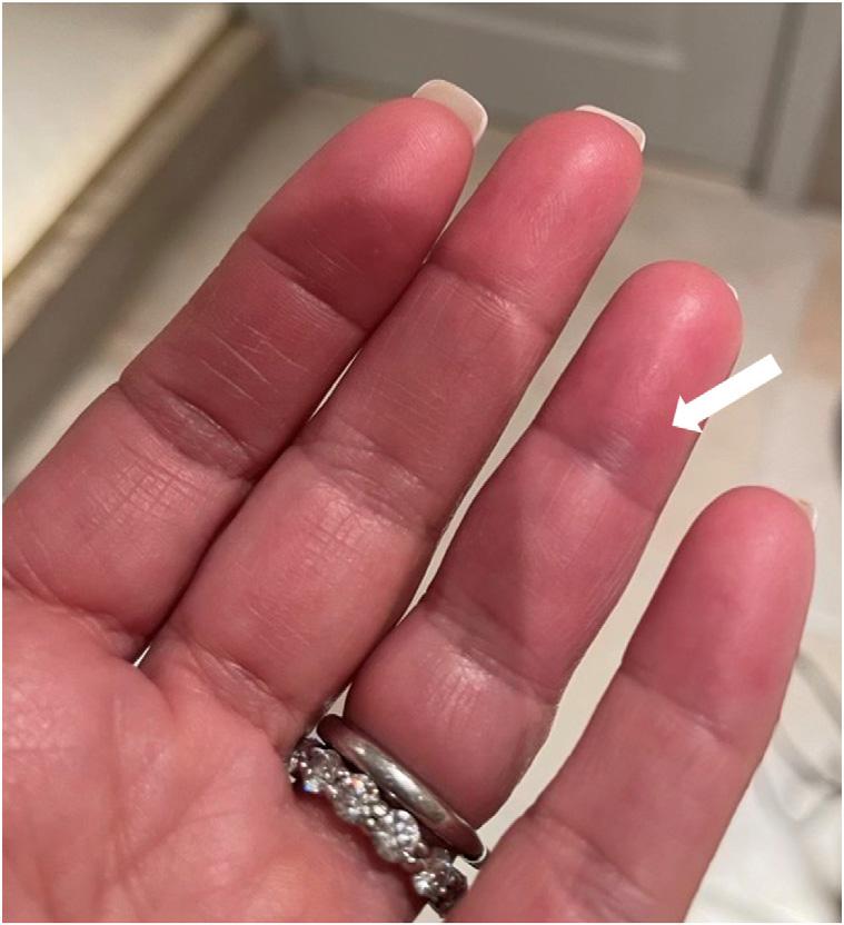
Image1. Abluishdiscolorationresemblingacirclenotedtothe volaraspectoftheleftfourthdigitjustdistaltothecreaseofthe distalphalanx.Ofnote,thedarkdiscolorationseeninthesecond, third,andproximalaspectofthefourththdigitaretheresultofa shadowfromthepatient’sphone.
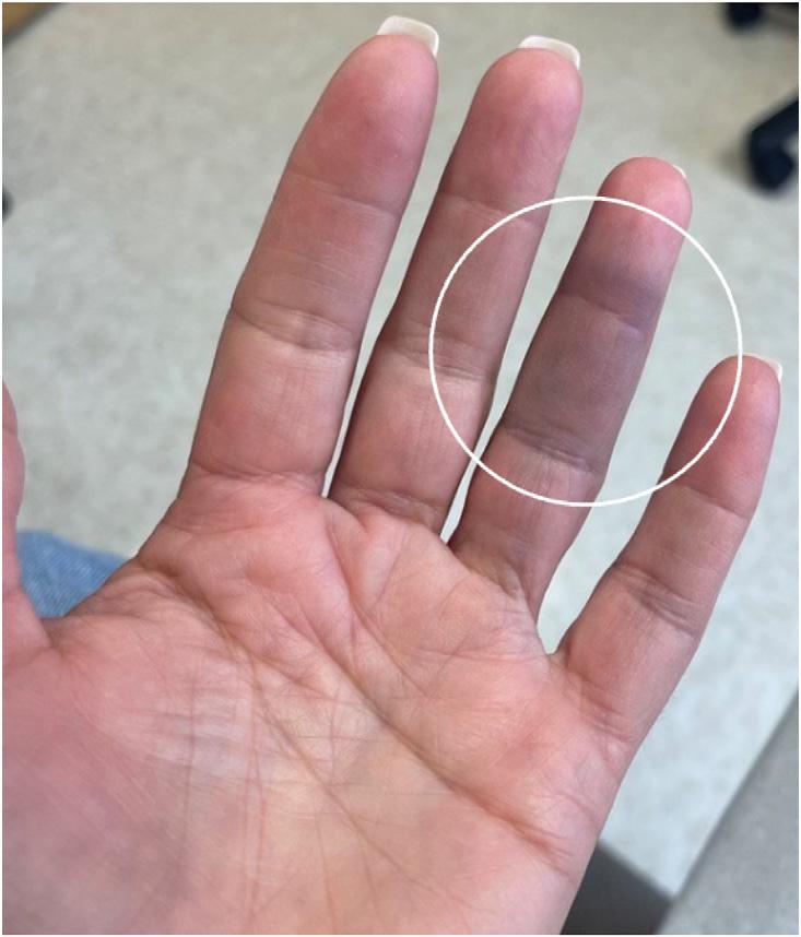
Image2. Awell-demarcatedareaofecchymosistothevolar aspectofthefourthdigitontheleftencompassingtheentire middlephalanxwithsomeextensionintothedistalphalanx.
Thepatientwasaformersmokerofnineyearsandhadquit 30yearsearlier.Shedeniedpriorhistoryofsimilarevents.
Onphysicalexam,herleftfourthdigitdemonstrateda bluishdiscolorationlimitedtothevolaraspectofhermiddle phalanx;itwaswarmtotouch,withdecreasedsensationand slighttendernesstotheaffectedarea.Thedistalphalanxhad normalcolorwithgoodcapillaryrefill.Thepatienthadfull rangeofmotionofalldigits.Theremainderofherlefthand
PopulationHealthResearchCapsule
Whatdowealreadyknowaboutthis clinicalentity?
Achenbachsyndrome,characterizedby painfuldiscolorationofa fi nger,isarare disorderthatiscompletelybenign andself-resolving.
Whatmakesthispresentationof diseasereportable?
Achenbachsyndromeoftengoes unrecognized,leadingtocostlyand potentiallyharmfulworkups.
Whatisthemajorlearningpoint?
Physiciansshouldfeelcomfortable diagnosingAchenbachsyndromeand reassuringpatientsofitsbenignnature.
Howmightthisimproveemergency -medicinepractice?
Awarenessofthisdisorderallowsemergency physicianstoconsideritinadifferential, diagnoseasappropriate,andsavethepatient time,cost,andanxiety.
appearedwithinnormallimits.Herradialpulseswereequal andstrongbilaterally.
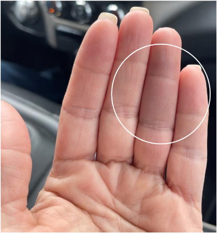
ThepatientwasinitiallyevaluatedatacommunityED wheretherewasaconcernforvascularinjury.Priorto transfertoobtainspecialtyconsultation,sheunderwentan ultrasoundarterialduplexoftheleftupperextremity,which wasfoundtobenormal,andshewasstartedonfull anticoagulationviaheparindrip.Thepatientwasadmitted intoourhospitalandwascontinuedontheheparindrip.She receivedatransthoracicechowithbubblestudy,which demonstratedanintactinteratrialseptum,andshewas evaluatedbymultiplespecialistsincludingvascularsurgery andcardiologytoassessforacardioembolicsourceof ischemiaandlateranorthopedic-handspecialistforother possiblediagnoses.Alllabworkincludingerythrocyte sedimentationrate(ESR)andC-reactiveprotein(CRP)were normal.Ultimately,thepatientwasdiagnosedwith Achenbachsyndromeanddischargedhome.Hersymptoms self-resolvedafterfourdays(Image3).
Achenbachsyndromeisabenignandself-resolving conditionoftenpresentingasacutediscoloration,pain,and
numbnessofoneormoredigits.Despite firstbeingreported in1958,thisremainsalittle-knowndiseasewhich,onreview ofcurrentavailablecasereports,oftenresultsinunnecessary testing,consultations,andanxietyforpatients.Theemergent concernintheinitialpresentationofAchenbachsyndromeis acutelimbischemiawherethrombosis,embolism,or dissectionmayleadtosuddenarterialocclusion.Ifthiswere thecase,patientswouldcommonlyexperiencepain,cold skin,pallor,orpulselessness.4 However,inAchenbach syndrome,pulsesandtemperatureareintact,andpatients tendtopresentwithecchymosisofjustthevolaraspectofa digit.Reportsindicateapropensityforthissyndrometo occuronthevolaraspectofthethirdorfourthdigitsin womenbuthavealsobeenreportedtooccurinmenandon nearlyallotherdigits,thedorsalaspectofdigits,thepalm, andeventoes.1 Inourpatient,sparingofthedistalphalanx wasveryprominent;this findingmaynotbethecaseinall patientswiththissyndrome.
PatientsoftenundergovascularstudiessuchasDoppler ultrasonography,echocardiograms,andcomputed tomographyangiography,whicharenormal.1 Areportofa patientwithAchenbachsyndromewhounderwentpunch biopsyrevealedmultipleectaticcapillariesinthedermal layer,inadditiontosomeextravasationofredbloodcellsout ofthesevesselsintothedermis,whichwouldsuggestsome degreeofvesselfragility.5 Aseparatereportofapatientwho underwentcapillaroscopydemonstratedhemorrhages withoutchangeincapillarymorphologyorblood flow, indicatingthatcapillarymicrohemorrhagescanbeobserved inAchenbachsyndrome.6
Vasculitisisanotherdifferentialtoconsiderwiththis presentation.Invasculitis,symptomresolutiongenerally takesweekstomonths,ratherthandaysasinAchenbach syndrome.1 Thereareoftennotablelaboratoryfeaturesin
vasculitissuchaselevatedESR,CRP,andantinuclear antibodylevels.5 Inaddition,therearefrequentlyother clinicalcluestosuggestthesediagnoses.Astronghistoryof tobaccousemaysuggestthromboangiitisobliterans,which mostcommonlymanifestsassignsandsymptomsoflimb ischemiainthemostdistalportionsofanextremityand progressesproximallyinthesettingofpersistenttobaccouse, ofteninvolvingmultipledigitsandextremities.7
ExposuretocoldtemperaturescouldsuggestRaynaud phenomenonorpernio.Classically,patientsexperiencing Raynaudphenomenoncomplainofsymmetriccolor changesinmultipledigits,generallybeginningaswhitedue totissueischemiaresultingfromvasospasmofthearteries, thenblueorpurple,and finallyredasthetissuesreperfuse oncerewarmed.Perniooccursafterprolongedexposureto temperaturesabovefreezing.Patientsdeveloppainful, violaceouspapulesusuallyonthedorsalaspectofmultiple fingersortoes,withoutthecolorchangeasnotedin Raynaud.8 Recentstressoranxietycouldsuggest psychogenicpurpura.9 Inpsychogenicpurpura,patients developecchymosesinvariouslocations,generallyonthe extremities,thatareassociatedwithdifferentprodromes rangingfromitchinesstopainorwarmth.10
Ingeneral,Achenbachsyndromeinvolvesecchymosis ofasingledigitorpartofasingledigit,whichdetractslargely fromtheaforementionedconditionsthatofteninvolve multipledigitsordifferentbodilylocations.Thisisbyno meansanexhaustivelistofdifferentials,butjustafewto illustratetheimportanceofathoroughhistoryandphysical examtohelpdifferentiatepotentialcausesofthis presentation.Additionally,arecentreportrevealeda possiblegeneticcomponenttoAchenbachsyndrome, detailingthreecaseswithintwosuccessivegenerationsof middle-toolder-agedwomen.11 Obtainingacarefulfamily historymayaidcliniciansinmakingthisdiagnosis.Further evaluationwouldbeusefulinelucidatingwhetherthese geneticfactorsmaybeassociatedwithotherpathologies ofconcern.
Inanearlysystematicreviewofthemedicalliteratureon Achenbachsyndrome,Kordzadehformulatedan algorithmicapproachfordiagnosisandmanagement basedupon12casereports.Kordzadehsuggestsinitially assessingpulsesinanypatientpresentingwithbluish discoloration,pain,edema,andparesthesiaofadigit.If pulsesarenotpalpable,theclinicianshouldworkupthe patientforacutelimbischemia.Ifpulsesarepalpable,then theclinicianshouldassessfordifferentiatingfactorswhen consideringlimbischemia,vascularpathologies,and Achenbachsyndrome.
Kordzadeh’sreviewfoundthatthemostcommonpatient demographicsinAchenbachsyndromewerefemalepatients undertheageof60.Usingtheclinicalpicture,patient demographics,physicalexam,andlackofsignificantrisk factorsforothervasculitisdiagnoses,cliniciansmayreliably
diagnoseAchenbachsyndromewithoutfurther investigations.Iftherearedoubtfulcircumstances,the patientmayundergoDopplerultrasonography.12 This algorithmicapproachoffersastartingpointfromwhicha clinicianmaybasetheirdecisions.However,itisimportant tonotethatthisalgorithmhasnotbeenvalidatedandisa theoreticaldecisiontoolbasedupon12casereports.
Insummary,Achenbachsyndromeisabenignandselfresolvingdisorderthatusuallypresentsasanatraumatic ecchymosisofa finger.Thetreatmentisreassurance.Our patientwasanotherwisehealthy54-year-oldWhitefemale witharemotehistoryoftobaccouseandanormalultrasound arterialduplexwhounderwentahospital-to-hospitaltransfer byambulance,wastreatedwithacontinuousinfusionof medicationandevaluatedbymultiplespecialists,and underwentadvancedimagingwiththeresultantdiagnosisof Achenbachsyndrome.Eachinterventionposedan additionalriskandcostforthepatient.Ourhopeisthatby increasingawarenessofthisdiagnosis,wemaylimitfuture potentiallyharmfulinterventions.
Itisessentialforclinicianstobeawareofthedifferential diagnosisofAchenbachsyndrome.Onceacutelimb ischemiaisruledout,andtheclinicalpictureisconsistent, cliniciansmaypracticegoodhealthcarestewardship andlimitinvasive,potentiallyharmful,andexpensive workupbyreassuringpatientsofthebenignnatureof thiscondition.
TheauthorsattestthattheirinstitutionrequiresneitherInstitutional ReviewBoardapproval,norpatientconsentforpublicationofthis casereport.Documentationon file.
AddressforCorrespondence:AzharSupariwala,MD,SouthShore UniversityHospital,DepartmentofCardiology,301EMainSt.,Bay Shore,NY11706.Email: Asupariwala@northwell.edu
ConflictsofInterest:Bythe CPC-EM articlesubmissionagreement, allauthorsarerequiredtodiscloseallaffiliations,fundingsources
and financialormanagementrelationshipsthatcouldbeperceived aspotentialsourcesofbias.Theauthorsdisclosednone.
Copyright:©2024Chenetal.Thisisanopenaccessarticle distributedinaccordancewiththetermsoftheCreativeCommons Attribution(CCBY4.0)License.See: http://creativecommons.org/ licenses/by/4.0/
1.GodoyAandTabaresAH.Achenbachsyndrome(paroxysmal finger hematoma). VascMed. 2019;24(4):361–6.
2.AzarfarAandBegS.Achenbachsyndrome:acaseseries. Cureus. 2022;14(3):e22824.
3.ChiriacA,WollinaU,MiulescuR,etal.Achenbachsyndrome-case reportanddiscussionontheinterdisciplinaryapproachofapatient. Maedica(Bucur). 2022;17(3):740–2.
4.TurnerEJH,LohA,HowardA.Systematicreviewoftheoperativeand non-operativemanagementofacuteupperlimbischemia. JVascNurs. 2012;30(3):71–6.
5.HarnarayanP,RamdassMJ,IslamS,etal.Achenbach’ssyndrome revisited:theparoxysmal fingerhematomamayhaveageneticlink. VascHealthandRiskManag. 2021;17:809–16.
6.FrerixM,RichterK,Müller-LadnerU,etal.Achenbach’ssyndrome (paroxysmal fingerhematoma)withcapillaroscopicevidenceof microhemorrhages. ArthritisRheumatol. 2015;67(4):1073.
7.PiazzaGandCreareMA.Thomboangiitisobliterates. Circulation. 2010;121(16):1858–61.
8.BrownPJ,ZirwasMJ,EnglishJC3rd.Thepurpledigit:analgorithmic approachtodiagnosis. AmJClinDermatol. 2010;11(2):103–16.
9.ToddNL,BowlingS,JengoM,etal.Achenbachsyndrome:casereport anddiscussion. Cureus. 2022;14(3):e23448.
10.SridharanM,AliU,HookCC,etal.TheMayoClinicexperiencewith psychogenicpurpura(Gardner-Diamondsyndrome). AmJMedSci. 2019;357(5):411–20.
11.HelmRH.Achenbachsyndrome:areportofthreefamilialcases. JHandSurgEurVol. 2022;47(2):214–5.
12.KordzadehA,CainePL,JonasA,etal.IsAchenbach’ssyndromea surgicalemergency?Asystematicreview. EurJTraumaEmergSurg. 2016;42(4):439–43.
NeilMakhijani,MD
SamuelE.Sondheim,MD,MBA
TurandotSaul,MD,MBA
ElizabethYetter,MD,MHPE
Section Editor: R. Gentry Wilkerson, MD
MountSinaiMorningside-MountSinaiWest,IcahnSchoolofMedicineatMount Sinai,DepartmentofEmergencyMedicine,NewYork,NewYork
Submission history: Submitted January 6, 2024; Revision received February 21, 2024; Accepted March 7, 2024
Electronically published August 9, 2024
Full text available through open access at http://escholarship.org/uc/uciem_cpcem DOI: 10.5811/cpcem.6663
Introduction: Point-of-care ultrasound (POCUS) is a screening and diagnostic modality frequently used in the emergency department to assess patients with abdominal pain.
Case Report: We present a case describing the unusual finding of intraperitoneal fluid with loculations visualized in the right upper quadrant of the abdomen in a patient ultimately diagnosed with pelvic inflammatory disease (PID) with ruptured tubo-ovarian abscess caused by group A streptococcus (GAS), a pathogen rarely implicated in the disease.
Conclusion: Uncommon findings on abdominal POCUS should trigger further investigation. In a patient not responding to antibiotics administered for typical PID coverage, GAS should be considered as a possible etiology and a penicillin-based antibiotic administered to prevent progression to tubo-ovarianabscess formation, peritonitis, and sepsis. [Clin Pract Cases Emerg Med. 2024;8(4)322–325.]
Keywords: point-of-care ultrasound; pelvic inflammatory disease; peritonitis; case report.
Pelvicinflammatorydisease(PID)isaninfectionofthe uppergenitourinarytracttypicallyseeninsexuallyactive women.Whilesomepatientsareasymptomatic,signsand symptomsmayincludeabdominalpain,dyspareunia, cervicalmotiontenderness,andvaginaldischarge.Pelvic inflammatorydiseaseismostoftencausedby Neisseria gonorrhoeae,Chlamydiatrachomatis ,orothercolonizing floraofthegenitourinarytract.1 NationalHealthand NutritionExaminationSurveydatafrom2013–2014 revealedthat4.4%ofwomenbetweentheagesof18–44years whoweresexuallyactivereportedapastmedicalhistoryofa PIDdiagnosis.2 Infertilityandtubo-ovarianabscesses (TOA)areknowncomplicationsofPID. Point-of-careultrasound(POCUS)isascreeningand diagnosticmodalityfrequentlyusedintheemergency department(ED)toassesspatientswithabdominalpain.
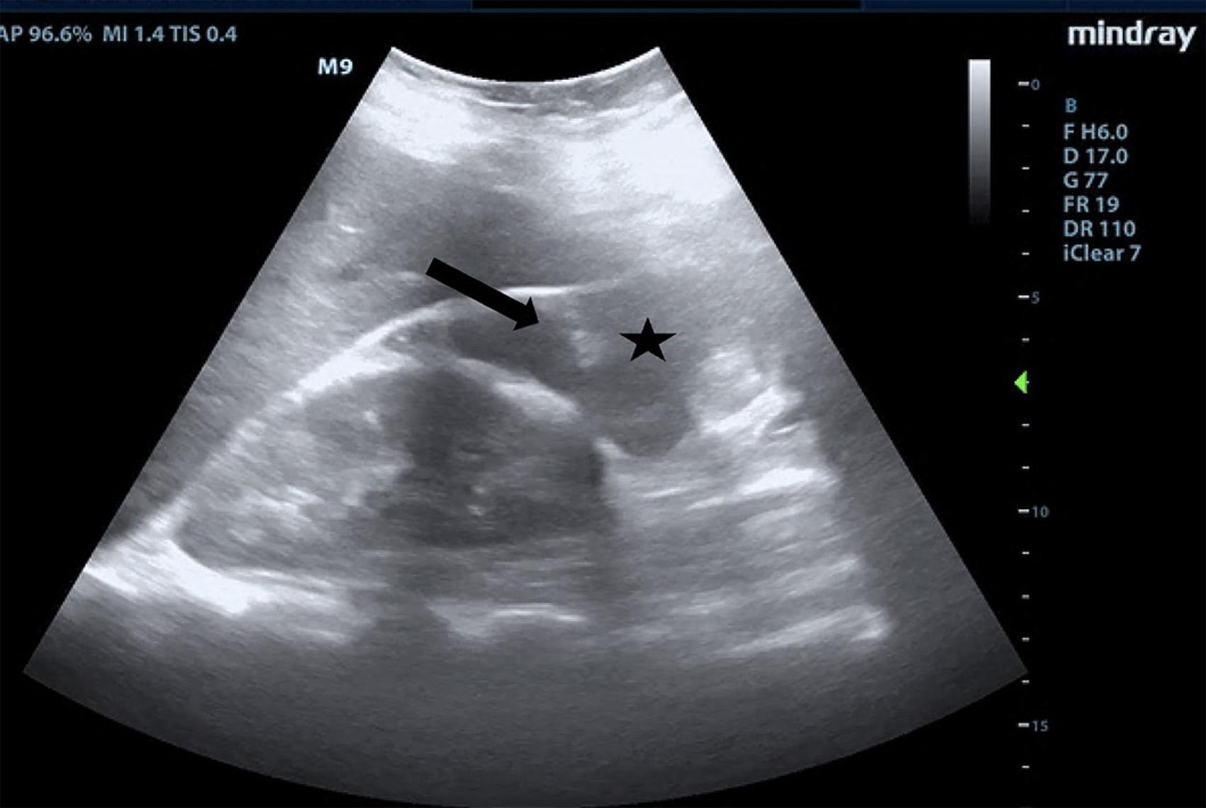
Inthiscasereportwedescribetheunusual findingof intraperitoneal fluidwithloculationsvisualizedintheright upperquadrantoftheabdomen.This finding,whencoupled withtheclinicalhistoryandphysicalexamination,ledto additionaldiagnosticimagingandultimatelyadiagnosisofa rupturedTOA.Grosslypurulent fluidwasdiscoveredduring peritonealwashoutintheoperatingroom.Cultureofthe peritoneal fluidgrewgroupAstreptococcus(GAS),a pathogenrarelyimplicatedinPID.Wefoundnoother reportedcasesofPIDcausedbyGASwithassociated POCUS findingsofseptatedorloculatedintraperitoneal fluidinthehepatorenalspaceinareviewofthe medicalliterature.
A24-year-oldfemaleG1P0010withapastmedicalhistory ofarightovariancystandamedicalterminationof
pregnancythreemonthspriorpresentedtotheEDwith suddenonsetofabdominalpainthatwokeherfromsleep. Shereportedthepainassevereanddiffuse,notablyworsein theupperabdomen,withassociatednauseaandlowerback pain.Additionally,thepatientreportedwhitevaginal discharge.Shedeniedanyfever,chills,vomiting,chestpain, shortnessofbreath,diarrhea,orconstipation.
Onphysicalexamination,thepatienthadanoral temperatureof99° Fahrenheit(37.2° Celsius),tachycardiaof 108beatsperminute,bloodpressureof106/71millimetersof mercury,97%oxygensaturationonroomair,andadiffusely tenderabdomenwithvoluntaryguardingandrebound tendernesssuggestiveofperitonitis.APOCUSwas performedtoassessforintraperitonealfree fluid.Itrevealeda complexcollectionof fluidwithinternalechoesinthe hepatorenalspaceraisingconcernforaloculatedinfection (See Image and Video).
Bowelgasobscuredthetransabdominalsuprapubic imagesofthepelvicorgans.Intravenous(IV)morphinewas administeredforpaincontrol.Pelvicexaminationrevealed cervicalmotiontendernessandwhitedischargecomingfrom theexternalcervicalos.Laboratoryanalysiswasremarkable forawhitebloodcellcountof18.9 × 103 permicroliter (K/μL)(referencerange4.5–11.0K/μL)withaneutrophil predominanceof93%.Electrolytes,lipase,hepaticfunction panel,andlactatewerewithinnormallimits.Serum β-human chorionicgonadotropinwasnegative,andtherewas anunremarkableurinalysis.Broadspectrumantibiotics wereinitiated.
Transvaginalultrasoundperformedbyradiologyreported asymmetricenlargementoftherightovarywithacomplex,
PopulationHealthResearchCapsule
Whatdowealreadyknowaboutthisissue? Point-of-careultrasound(POCUS)isa readilyavailabletooltoassistin differentiatingabdominalpain.
Whatmakesthispresentationof diseasereportable?
UnusualPOCUS fi ndingsledtofurther workuprevealingarupturedtubo-ovarian abscess(TOA)thatultimatelygrewgroupA streptococcus(GAS)despitenoexpected riskfactors.
Whatwasthemajorlearningpoint?
UnusualPOCUS fi ndingsshouldtrigger furtherinvestigation;considerGASasan etiologyinpatientswhodonotrespond totypicalpelvicin fl ammatorydisease (PID)coverage.
Howdoesthisimproveemergency medicinepractice?
InPID/TOApatientswhohavebeen appropriatelytreatedbutcontinuetohave infectioussymptomsandunusualPOCUS fi ndings,considerGAS.
4.2-centimeter(cm)cyst.Therewasasymmetricincreased flowtotherightovaryonDopplerevaluation.Gynecologic consultationwasobtained,andduetoconcernforright ovariantorsion/detorsionthepatientwastakenfor diagnosticlaparoscopy.Thepatientwasfoundtohavea rupturedTOAwith200milliliters(mL)ofpurulent fluidin theperitonealspace.Aperitonealwashoutandcystectomy wereperformed,andthepatientwasadmittedforIV ceftriaxone,doxycycline,andmetronidazole.Shewas dischargedonpostoperativedaythreewitha14-day courseoforaldoxycyclineandmetronidazole.The intraperitoneal fluidsamplecollectedduringlaparoscopy grewGAS.
Thepatientreturnedonemonthlaterwithfever,chills, yellowvaginaldischarge,andlowerabdominalpain. Contrast-enhancedcomputedtomographyconfirmeda recurrentTOA,whichwastreatedwithIVpiperacillintazobactamandclindamycinperrecommendationbythe infectiousdiseaseconsultant.Theinfectionrespondedwellto antibiotics,andthepatientwasdischargedwithoutfurther recurrencetodate.
Abdomino-pelvicPOCUSmayguidetriage,diagnosis, andmanagement,assistingtheclinicianininvestigatinga rangeofdiseaseentitiesincludingbiliarypathology, abdominalaorticaneurysm,orTOA.3–5 Bedsideultrasound isgenerallyreadilyavailableintheEDsettingandmayserve asanadditionalmodalitytoidentifyunusual findingsearlyin thepatient’sclinicalcourse.Itdoesnotrequireionizing radiationandisnottimeintensivetoperform.Cliniciansmay considerearlyuseofPOCUSforperitoneal findings,as evidencedinthiscasepresentation.Inconjunctionwith informationobtainedonhistoryandphysicalexamination, theunusual,rightupperquadrantPOCUS findingsof intraperitoneal fluidwithloculationscausedconcernfora disseminatedpelvicinfection.Thiswasconfirmedasa rupturedTOAwithamoderateamountofpurulent, intraperitonealfree fluidontheoperativereport.Fitz-HughCurtissyndromewasaconcerngiventhecomplicated fluidvisualizedwithPOCUS,buttherewasnocommentof violin-stringadhesions,adhesions,or fibrousadhesions betweentheanteriorhepaticcapsuleandparietal peritoneumnotedonthelaparoscopicoperativereport, althoughitisunknownwhetherthisanatomicarea wasevaluated.
Intraperitoneal fluidiseasilyvisualizedonultrasound, andwhencomplex fluidwithloculationsisencountered,the differentialdiagnosisincludesmalignancy,inflammation, andinfectiousprocesses.6 Similar findingsofseptated intraperitoneal fluidhavebeendocumentedincases of Ctrachomatis inPID,7 cholecystitis,8 and tubercularperitonitis.9
InadditiontotheuncommonPOCUSexamination findings, fluidculturedfromtheabdominalcavitygrew GAS.Outsideofperipartuminfections,thoseassociatedwith anintrauterinedevice(IUD)andotherinvasivegynecologic procedures,GASinfectionsarerare.Thispatientreportedno recentgynecologicinstrumentation.SnyderandSchmalzle describedacaseofapatientinsepticshockfromPID,where GASwasthecausativeorganism.10 Theythenconducteda literaturereviewof13othercasesofPIDduetoGAS infection.Allbutoneofthecasesreportedabdominalpain, and8/13reportedgenitourinarysymptomssuchasvaginal dischargeorgastrointestinalsymptomssuchasnausea, vomiting,and/ordiarrhea.
Thispatient’sclinicalcoursewascomplicatedbya recurrentTOAandsepsisonareturnEDvisitafterinitially respondingtostandardPIDantibioticcoverage.She ultimatelyrequiredpenicillintotreatGAS.WhilebothPID andGASareeasilytreatedbyantibiotics,ifleftuntreated theymayprogresstosepticshockanddeath.Inpatients diagnosedwithPIDbutnotimprovingonstandard antibioticregimens,cliniciansshouldconsider othercausessuchasGASandstreptococcaltoxic shocksyndrome.
GroupAstreptococcusisararecauseofpelvic in fl ammatorydisease,usuallyseenintheperipartum period,inpatientswithanIUDorwhohavehadother recent,invasivegynecologicprocedures.Inapatientnot respondingtoantibioticsadministeredfortypicalPID coverage,GASshouldbeconsideredasapossibleetiology andapenicillin-basedantibioticadministeredtoprevent progressiontotubo-ovarianabscessformation,peritonitis, andsepsis,particularlyincasesofreturningpatients previouslytreatedwithantibiotics.Bedsideultrasound shouldbeperformedinpatientspresentingwithabdominal pain,anduncommon fi ndingsshouldtriggerfurther investigation.Toourknowledge,noothercasesofloculated fl uidinthehepatorenalspaceassociatedwithPIDhave beenreported.
Video. Rightupperquadrantultrasonographyusingcurvilinear probefanningthroughthecoronalplanedemonstratingascitesand septationsinapatientultimatelydiagnosedwithpelvic inflammatorydisease.
Patientconsenthasbeenobtainedand filedforthepublicationofthis casereport.
AddressforCorrespondence:NeilMakhijani,MD,MountSinai Morningside-MountSinaiWest,IcahnSchoolofMedicineatMount Sinai,DepartmentofEmergencyMedicine,515W59thSt.,Apt20M, NewYork,NY10019.Email: Neil.makhijani@gmail.com
ConflictsofInterest:Bythe CPC-EM articlesubmissionagreement, allauthorsarerequiredtodiscloseallaffiliations,fundingsources and financialormanagementrelationshipsthatcouldbeperceived aspotentialsourcesofbias.Theauthorsdisclosednone.
Copyright:©2024Makhijanietal.Thisisanopenaccessarticle distributedinaccordancewiththetermsoftheCreativeCommons Attribution(CCBY4.0)License.See: http://creativecommons.org/ licenses/by/4.0/
1.CurryA,WilliamsT,PennyML.Pelvicinflammatorydisease: diagnosis,management,andprevention. AmFamPhysician. 2019;100(6):357–64.
2.KreiselK,TorroneE,BernsteinK,etal.Prevalenceofpelvic inflammatorydiseaseinsexuallyexperiencedwomenofreproductive age – UnitedStates,2013–2014. MMWRMorbMortalWklyRep. 2017;66(3):80–3.
3.AdhikariS,BlaivasM,LyonM.Roleofbedsidetransvaginal ultrasonographyinthediagnosisoftubo-ovarianabscess intheemergencydepartment. JEmergMed. 2008;34(4):429–33.
4.GragliaS,ShokoohiH,LoescheMA,etal.Prospectivevalidationofthe bedsidesonographicacutecholecystitisscoreinemergency departmentpatients. AmJEmergMed. 2021;42:15–9.
5.FernandoSM,TranA,ChengW,etal.Accuracyofpresenting symptoms,physicalexamination,andimagingfordiagnosisofruptured abdominalaorticaneurysm:systematicreviewandmeta-analysis. Acad EmergMed. 2022;29(4):486–96.
6.RudralingamV,FootittC,LaytonB.Ascitesmatters. Ultrasound. 2017;25(2):69–79.
7.BarrosLL,daSilvaJC,DantasACB,etal.Peritoneal Chlamydia trachomatis infectionasacauseofascites:adiagnosisnottobemissed. CaseRepGastroenterol. 2021;15(3):898–903.
8.BouffardJ,LaLoucheK,GierengerS,etal.Septatedascitesand cholelithiasis:anunusualclinicalpresentationforgallbladderrupture. JDiagnMedSonogr. 2002;18(2):84–6.
9.JainS,PhatakS,DeshpandeS,etal.Septatedascites:animportant signoftubercularperitonitis. JDattaMegheInstituteofMedical SciencesUniversity. 2021;16(3):596.
10.SnyderAandSchmalzleSA.Spontaneous Streptococcuspyogenes pelvicinflammatorydisease;casereportandreviewoftheliterature. IDCases. 2020;20:e00785.
MiriamMartinez,MD,DrPH,MPH KhristopherFaiss,DO NehaSehgal,DO
Section Editor: Lev Libet, MD
TexasTechUniversityHealthSciencesCenteratElPaso, DepartmentofEmergencyMedicine,ElPaso,Texas
Submission history: Submitted September 13, 2023; Revision received March 21, 2024; Accepted April 26, 2024
Electronically published September 29, 2024
Full text available through open access at http://escholarship.org/uc/uciem_cpcem DOI: 10.5811/cpcem.1668
Introduction: More than 40% of Americans are considered obese, resulting in annual healthcare costs estimated at $173 billion.1,2 Various interventions exist to address obesity including lifestyle modification, medications, and several surgical options. A novel ingestible intragastric balloon that self-deflates and is excreted approximately four months post-ingestion is being used in other countries such as Australia, Mexico, and several European countries. Currently, however, there are no US Food and Drug Administration-approved, commercially available options like this in the United States.
Case Report: We present a case of a 31-year-old, obese male who presented to the emergency department for abdominal pain approximately 10 weeks after the ingestion of an inflatable balloon for weight loss treatment in Mexico. He was found to have a gastric perforation and required an emergent exploratory laparotomy.
Conclusion: While ingestible, weight-loss balloons are not yet commercially available in the United States, emergency physicians may still encounter complications of such devices. [Clin Pract Cases Emerg Med. 2024;8(4)326–328.]
Keywords: intragastric balloon; gastric perforation; weight loss; case report.
Obesityisanepidemicthatisatthecoreofseveral commonchronichealthconditionsincludingheartdisease, type2diabetes,andhypertension.In2022,theprevalenceof obesityamongadultsintheUnitedStateswas41.9%.Black adultshadthehighestrateat49.9%,followedbyHispanic adultsat45.6%.1 Nonsurgicalandnon-endoscopic ingestible,intragastricballoons(IGB)arebeingusedinmany countriesincludingMexico,Australia,andacrossEuropeto assistpatientswithweightloss.
TherearecurrentlynoUSFoodandDrug Administration-approvedcommercialoptionsintheUS. TheprimarybenefitsofingestibleIGBsarethattheydonot requireanesthesia,sedation,orendoscopyforplacementand subsequentretrieval.Theballoonisingestedwithacatheter attached;itis filledandthenexcretedinthestool approximatelyfourmonthspost-ingestion.2–4 Forpatients,
thismaybeanattractiveoptionasthedeviceremovesthe risksassociatedwithsedation,anesthesia,andendoscopy.5–8
A31-year-oldobese,Hispanicmalepresentedtothe emergencydepartmentwithprogressivelyworseningleft upperquadrantabdominalpainfor fivedays.Thepainwas describedascramping,intermittent,andexacerbatedby bendingforward.Hedeniedpastmedicalhistory. Severaldayspriortopresentationthepatienthadbeen evaluatedbyhisprimarycarephysician.Hewasdiagnosed withmusculoskeletalpainandprescribedcyclobenzaprine. Sincetheevaluationbytheprimarycarephysician,thepain hadincreasedinfrequencyandintensity.Thepatientdenied anynausea,vomiting,fevers,diarrhea,orhematochezia.He did,however,reportintentionalweightlossof15kilograms. Tenweeksprior,thepatienthadingestedanIGB(Allurion,
formerlyknownasElipse),forweight-losspurposesunder thecareofaphysicianinJuárez,Mexico.
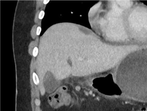
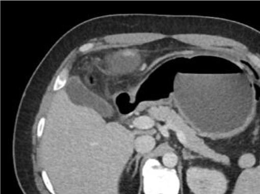
Oninitialevaluation,vitalsignswerenormalexceptfor sinustachycardiaat105beatsperminute.Hewasinmild distresswithmoderatetendernesstopalpationintheleftupper quadrant.Therewasnoguarding,rebound,orrigidity. Laboratoryevaluationshowedawhitebloodcellcountof 21.7 × 10³ cellspercubicmillimeter(mm3)(referencerange 4.5 11 × 103 cells/mm3)withaneutrophilpredominanceof 18.9 × 10³ cells/mm3 (2 7.8 × 10³ cells/mm3)andanelevated bloodureanitrogenat25milligrams(mg)/deciliter(dL) (9–20mg/dL).Thepatient’selectrolytes,liverfunction tests,andserumcreatininewerewithinnormallimits. Acomputedtomography(CT)oftheabdomenandpelviswith intravenouscontrastwasorderedtoassessforbowel obstruction,perforation,andballoonintegrity.TheCT indicatedintraluminalgastricballoonwithanteriorgastricwall perforationwithoutevidenceofintestinalpathology(Image).
GeneralsurgerywasconsultedandobtainedaCT abdomenandpelviswithoralcontrast,whichdirectly showedextravasationintotheperitonealcavity.Thepatient wasemergentlytakentotheoperatingroomforan exploratorylaparotomy,abdominalwashout,removalofthe IGB,andgastricperforationrepair.Hewasstartedon piperacillin/tazobactamand fluconazoleforpurulent peritonitis.Afteranovernightstayinthesurgicalintensive careunit,hewastransferredtothesurgical floorwherehe remainedforthefollowing10days.Onday5,afollow-up uppergastrointestinalserieswithgastrografinwasobtained, andnocontrastextravasationwasnoted.Thepatient’sdiet wasadvanced,andhewasdischargedonpostoperativeday 10withamoxicillin-clavulanatefor10days.Forty-twodays afterdischargethepatientwasnotedtoberecoveringwellon afollowupvisit.
Ourcasedemonstratesapotentiallylethalcomplicationof thisnovelingestibleIGB.Thedeviceisdifferentfromother toolsforweightlossinthatitdoesnotrequiresurgery, endoscopy,oranesthesiaforplacementandremoval.Italso

Whatdowealreadyknowaboutthis clinicalentity?
Variousinterventionsexisttoaddressobesity. Aningestible,intragastricballoonthat self-de fl atesandisexcretedfourmonths post-ingestionisnotyetavailableinthe UnitedStates(US).
Whatmakesthispresentationof diseasereportable?
Anobesemalepresentedtoouremergency departmentwithagastricperforation 10weeksafteringestionofaweight-loss ballooninMexico.
Whatisthemajorlearningpoint?
Complicationsfromingestible,intragastric balloonsincludingperforationsaremore likelytooccurontheUS-Mexicoborder orinplaceswithalargemedical tourismpopulation.
Howmightthisimproveemergency medicinepractice?
Emergencyphysiciansshouldconsidergastric perforationinpatientswithintragastric balloons.Earlyspecialistinvolvementis keytomanagingsuchpatients.
ismoreconvenientinthatplacementcanbedoneina 20-minutevisit.5 Theballoontakesupvolumewithinthe stomach,promotingearlysatietyandweightloss.3–8
TheIGBballoonismadefrombiodegradablematerials andfoldedintoavegancapsule.Whilestillconnectedtoa thin fillingcatheterthecapsulewiththeIGBinsideis swallowed.Positioninginthestomachisconfirmedwitha



radiograph,andtheballoonis filledwith550millilitersof distilledwater,potassiumsorbate,andcitricacidas preservativeswithareconfirmationofpositioningfollowing the fillingoftheballoon.Oncetheballoonis filled,the catheterispulledoutandthe fillvalvemadefromathin film issealedshut.TheIGBhasaself-releasevalvethatisclosed bya filamentthatisonlyincontactwiththeinsideofthe balloon.This filamentweakensgraduallyandbreaksat approximatelyfourmonths.Breakingofthe filamentcauses thecontentsoftheballoontoemptyintothestomach.Once theballoonisemptieditisexcretedinabowelmovement.4–8
Literatureonthedeviceislimitedasthedeviceismostly beingusedintheMiddleEastandEurope.Sixuniquestudies comprisedof2,013patientswereevaluatedinasystematic reviewandmeta-analysisontheefficacyandsafetyofthe device.Thisstudyfoundthatalltheballoonshadsuccessful placementexceptforonethatwasretainedinthelowerportion oftheesophagus.Whiletherewasasmallnumberofserious complicationsnomortalitieswerereported.8 Amongthe2,013 casestherewasonlyonewithagastricperforationthat requiredsurgery.5–8 Othercomplications,whichwerealsorare, includedsmallbowelobstructionsinthreepatients, spontaneousballoonhyperinflationinfourpatients,andgastric outletobstruction,pancreatitis,andesophagitis,eachinone patient.4,8 Whilethisballoonhasanoverallfavorablesafety profileitisimportanttonotethatthereisstillthepossibilityof seriouscomplications.Toourknowledgethiscasepresentsonly thesecondinstanceofagastricperforationfromanIGB.
Gastricperforationisararebutseriouscomplication associatedwithahighmortalityrateinpatientswith intragastricballoonplacement.Complicationsfromsuch devicesaremorelikelytooccurontheUS-Mexicoborderor incommunitieswithalargemedicaltourismpopulation,as theprocedureisnotavailableintheUnitedStates.Itis prudentforemergencyphysicianstoconsidergastric perforationinpatientswithintragastricballoons.Early specialistinvolvementandpromptresuscitationarekeyto successfullymanagingsuchpatients.
TheauthorsattestthattheirinstitutionrequiresneitherInstitutional ReviewBoardapproval,norpatientconsentforpublicationofthis casereport.Documentationon file.
AddressforCorrespondence:NehaSehgal,DO,TexasTech UniversityHealthSciencesCenteratElPaso,Departmentof EmergencyMedicine,4800AlbertaAve.,SuiteB3200,ElPasoTX, 79905.Email: Neha.Sehgal@ttuhsc.edu
ConflictsofInterest:Bythe CPC-EM articlesubmissionagreement, allauthorsarerequiredtodiscloseallaffiliations,fundingsources and financialormanagementrelationshipsthatcouldbeperceived aspotentialsourcesofbias.Theauthorsdisclosednone.
Copyright:©2024Martinezetal.Thisisanopenaccessarticle distributedinaccordancewiththetermsoftheCreativeCommons Attribution(CCBY4.0)License.See: http://creativecommons.org/ licenses/by/4.0/
1.CentersforDiseaseControlandPrevention.Adultobesityfacts.2022. Availableat: https://www.cdc.gov/obesity/data/adult.html AccessedMarch11,2023.
2.HalesCM,CarrollMD,FryarCD,etal.Prevalenceofobesityandsevere obesityamongadults:UnitedStates,2017–2018. NCHSDataBrief. 2020;(360):1–8.
3.Al-SubaieS,Al-BarjasH,Al-SabahS,etal.Laparoscopic managementofasmallbowelobstructionsecondarytoElipse intragastricballoonmigration:acasereport. IntJSurgCaseRep. 2017;41:287–91.
4.MachytkaE,ChuttaniR,BojkovaM,etal.Elipse™,aprocedureless gastricballoonforweightloss:aproof-of-conceptpilotstudy. ObesSurg. 2016;26(3):512–6.
5.IencaR,AlJarallahM,CaballeroA,etal.TheprocedurelessElipse gastricballoonprogram:multicenterexperiencein1770consecutive patients[publishedcorrectionappearsin ObesSurg 2020;30(11):4691–2]. ObesSurg. 2020;30(9):3354–62.
6.AngrisaniL,SantonicolaA,VitielloA,etal.Elipseballoon:thepitfallsof excessivesimplicity. ObesSurg. 2018;28(5):1419–21.
7.RaftopoulosIandGiannakouA.TheElipseballoon,aswallowable gastricballoonforweightlossnotrequiringsedation,anesthesiaor endoscopy:apilotstudywith12-monthoutcomes. SurgObesRelatDis. 2017;13(7):1174–82.
8.VantanasiriK,MatarR,BeranA,etal.Theefficacyandsafetyofa procedurelessgastricballoonforweightloss:asystematicreviewand meta-analysis. ObesSurg. 2020;30(9):3341–6.
Matthew Van Ligten, MD*
Douglas E. Rappaport, MD†
Wayne A. Martini, MD†
Mayo Clinic Alix School of Medicine, Phoenix, Arizona
Mayo Clinic Arizona, Department of Emergency Medicine, Phoenix, Arizona
Section Editor: Christopher Sampson, MD
Submission history: Submitted January 4, 2024; Revision received May 14, 2024; Accepted May 21, 2024
Electronically published September 6, 2024
Full text available through open access at http://escholarship.org/uc/uciem_cpcem DOI: 10.5811/cpcem.6657
Introduction: Tendon injuries of the hand present a diverse spectrum of challenges in emergency medicine, ranging from minor strains to catastrophic ruptures. The superficial anatomy of hand tendons predisposes them to various mechanisms of injury, leading to complex medical scenarios. Here, we present a unique case of flexor tendon exposure secondary to abscess formation and spontaneous rupture, emphasizing the importance of prompt recognition and management of such injuries in the emergency department.
Case Report: A 69-year-old male with multiple comorbidities presented with diffuse pain and a pale, pulseless right lower extremity, alongside a left hand exhibiting exposed flexor tendons due to recent abscess drainage. Despite broad-spectrum antibiotics and pain management, the patient underwent above-knee amputation due to vascular compromise. Evaluation revealed a complete flexor tendon rupture likely attributable to infection, necessitating emergent hand surgery at the bedside.
Conclusion: Understanding the nuances of tendon injuries is paramount for emergency physicians, given their potential for lifelong disability if inadequately addressed. Awareness of risk factors and appropriate management strategies, including early surgical intervention when indicated, is essential in optimizing patient outcomes. This case serves as a reminder of the complexities involved in hand injuries and underscores the need for vigilance and tailored care in the emergency setting. [Clin Pract Cases Emerg Med. 2024;8(4):329–331.]
Keywords: tendon injuries; flexor tendon rupture; emergency department; hand surgery; comorbidities; case report.
Tendon injuries are commonly seen in the emergency department (ED) with causes ranging from minor falls and lacerations to major trauma. The substantial number of mechanisms of injury to the hand coupled with the superficial anatomy of the tendons that allow movement leads to a broad range of medical complexities. Closed injuries such as the commonly seen “mallet finger” can be splinted to allow for healing, while complete tearing and/or exposure of the tendon may require surgical intervention to prevent lifelong disability.1 Moreover, while incomplete ruptures may allow the patient to remain functional, these injuries can evolve
over time leading to a complete rupture later. Diagnoses of these injuries require a thorough physical exam to test for neurological function, imaging with radiographs to rule out bone abnormalities, and ultrasound and/or magnetic resonance imaging for definitive diagnosis of a soft tissue injury. Here, we present a unique presentation of a patient with flexor tendon exposure in the setting of recent abscess formation with spontaneous rupture.
The patient was a 69-year-old, Russian-speaking male with a significant medical history including type 2 diabetes,
peripheral arterial disease (PAD) status post multiple bilateral lower extremity revascularization procedures, coronary artery disease (CAD), hypertension, rheumatoid arthritis (RA), and recently diagnosed gastric adenocarcinoma. He presented to our ED with diffuse pain in his body, but primarily in his right leg and left hand.
He had been seen two days earlier at an outside ED where he had an abscess on the left hand that had spontaneously ruptured. At that time, he was treated with metronidazole, doxycycline, and trimethoprim-sulfamethoxazole.
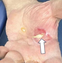
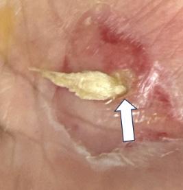
On his evaluation in our ED, the patient was in distress secondary to his pain. The right lower extremity was identified as pale, cold, and pulseless. His left hand showed exposed shiny, ribbon-like substance protruding from the palmar aspect with serosanguinous fluid draining (Images 1 and 2). We attempted to maneuver the substance with 4x4 cotton, which revealed it to be anchored beneath the surface. Flexion of fingers and palm caused the substance to withdraw nearly entirely beneath the surface.
He was diagnosed with acute limb ischemia suspected to be secondary to an acute arterial occlusion, as well as spontaneous flexor tendon rupture secondary to an abscess within the
CPC-EM Capsule
What do we already know about this clinical entity?
Tendon injuries in the hand vary from minor strains to catastrophic ruptures, necessitating prompt recognition and management due to potential lifelong disability.
What makes this presentation of disease reportable?
This rare case of flexor tendon exposure due to abscess and spontaneous rupture, highlights complexities in diagnosis and management.
What is the major learning point?
It is vital to recognize risk factors such as comorbidities (eg, rheumatoid arthritis, peripheral arterial disease), and the need for cautious intervention in hand pathologies, and timely surgical management.
How might this improve emergency medicine practice?
Emergency physicians should be aware of the nuanced presentations of hand injuries, facilitating tailored and timely interventions to optimize patient outcomes.
left hand. He was started on broad spectrum antibiotics (as treatment for tendon infection and prophylaxis for the operating room), pain medication, and high-dose heparin drip. Emergent evaluation by vascular surgery and hand surgery teams were called to the ED for evaluation. Due to the vascular emergency of his right lower extremity, the patient was sent to vascular surgery for attempted salvation; however, he was deemed inoperable, which resulted in an above the knee amputation. Evaluation of his left hand showed a full tendon rupture, thought to be due to infection and spontaneous abscess drainage before arriving in the ED two days prior when the abscess was still in place. Due to his medical complications, the patient’s hand surgery was performed at bedside with irrigation and washout with consideration of further intervention in the future.
Emergency physicians need to promptly recognize and manage tendinous injuries due to their diverse presentation, severity, and treatment requirements. Initial treatment requires a thorough physical exam, focusing on neurological status and motor function. The treatment depends on the severity with most partial ruptures being treated conservatively. Some complete extensor tendon partial tears or ruptures, such
as mallet and Jersey finger, can be treated in the ED with splinting and outpatient follow-up.2 Other clear indications for surgical management would be complete rupture of more proximal tendons and consideration of the patient’s recovery and ultimate outcomes after hand surgery consultation.2,3
Considering pre-existing conditions is crucial in risk management assessments for hand interventions. Using our case as an example, the patient had RA. Rheumatoid arthritis increases the likelihood of both flexor and extensor tendon rupture due to recurrent episodes of tendon inflammation (tenosynovitis), tendon erosion, and malformed bone.5 The previous spontaneous abscess drainage or multiple antibiotics may explain the atypical presentation of the tendon itself, with it protruding from the abscess area likely due to the infection weakening the tendon.3,4 Additionally, looking at the patient’s leg, we saw that he suffered from severe PAD with the development acute right lower leg ischemia as well as left lower leg developing a stage 4 ulcer involving tendon and bone. This PAD in the lower extremities was also contributory to the upper extremity tendon rupture.
This case demonstrates that emergency physicians should take care when treating pathologies of the hand in patients with comorbidities such as RA, PAD, and CAD. Closed injuries such as sprains and extensor avulsions such as mallet finger can be splinted, but leaving exposed skin on a patient with known PAD and healing issues may lead to increased risk of rupture and subsequent complications. We suspect that if the outside hospital physician who saw the patient had performed incision and drainage of the hand without guidance from hand surgery, tendon injury may have inadvertently occurred. The outside hospital physician attempted to hospitalize the patient for antibiotics and further management; however, the patient refused admission and was discharged home.
A lesson to be taken from this case is to proceed with caution when intervening in the hand in the ED. Instrumentation for abscess drainage without proper imaging, consultation, and follow-up can lead to rupture and further complications. We recommend consideration of broad spectrum empiric antibiotic coverage for Staphylococcus aureus and Streptococcus species coverage6 (the most common causes of tenosynovitis, septic arthritis, and osteomyelitis) such as cefazolin or clindamycin with vancomycin added. For patients who will ultimately be discharged we recommend cephalexin or clindamycin. Lastly, one should always be cognizant of any comorbidities that a patient may have that can affect their ability to heal and or increase their risk for certain complications.
In 2016 there were 42.3 million injury-related ED encounters nationwide, according to the National Center for Health Statistics.3 Of these, hand and upper extremity injuries accounted for 26%. While tendon injuries are not
the most common, they can lead to lifelong disability if missed. This case illustrates the importance for emergency physicians to be aware of risk factors for tendon rupture, such as rheumatoid arthritis, and to tailor treatment accordingly. Tendon ruptures, partial or complete, that are open require emergent surgical intervention with wash out.7 This case highlights that one should use caution, with patients who do not have spontaneous rupture when intervening on the hand, due to the anatomy of tendons, muscles, and nerves that run superficially. Hand surgery is recommended in these cases due to complexity and high risk for poor outcomes.
Documented patient informed consent has been obtained and filed for publication of this case report.
Address for Correspondence: Wayne Martini, MD, Mayo Clinic Arizona, Department of Emergency Medicine, Attn: Wayne Martini, MD & Lori Robinson, 5777 E Mayo Blvd, Phoenix, AZ 85054. Email: Martini.Wayne@Mayo.edu
Conflicts of Interest: By the CPC-EM article submission agreement, all authors are required to disclose all affiliations, funding sources and financial or management relationships that could be perceived as potential sources of bias. The authors disclosed none.
Copyright: © 2024 Ligten et al. This is an open access article distributed in accordance with the terms of the Creative Commons Attribution (CC BY 4.0) License. See: http://creativecommons.org/ licenses/by/4.0/
1. Schöffl V, Heid A, Küpper T. Tendon injuries of the hand. World J Orthop. 2012;3(6):62–9.
2. Park CW, Juliano ML, Woodhall D. Tendon lacerations. In: Knoop KJ, Stack LB, Storrow AB, Thurman R (Eds.). The Atlas of Emergency Medicine, 5th ed. New York: McGraw Hill (2021)
3. Rui P, Kang K, Ashman JJ. National Hospital Ambulatory Medical Care Survey: 2016 Emergency Department Summary Tables. Available at: https://www.cdc.gov/nchs/data/nhamcs/web_ tables/2016_ed_web_tables.pdf. Accessed December 22, 2023.
4. Ertel AN. Flexor tendon ruptures in rheumatoid arthritis. Hand Clin. 1989;5(2):177–90.
5. Biehl C, Rupp M, Kern S, et al. Extensor tendon ruptures in rheumatoid wrists. Eur J Orthop Surg Traumatol. 2020;30(8):1499–1504.
6. Gelfand JM, Patel DN. Bacterial tenosynovitis. In: StatPearls [Internet]. Treasure Island, FL: StatPearls Publishing, 2022. Available at: https://www.ncbi.nlm.nih.gov/books/NBK544324/. Accessed September 14th, 2023.
7. Ishak A, Rajangam A, Khajuria A. The evidence-base for the management of flexor tendon injuries of the hand: review. Ann Med Surg. 2019;48:1–6.
Taylor Sanders, MD*†
Mitchell Hymowitz, MD†
Christine Murphy, MD*
Section Editor: Anna McFarlin, MD
Atrium Health’s Carolinas Medical Center, Department of Emergency Medicine, Division of Medical Toxicology, Charlotte, North Carolina Louisiana State University Health Sciences Center, School of Medicine, Emergency Medicine Residency Program, Baton Rouge Campus, Baton Rouge, Louisiana
Submission history: Submitted January 20, 2024; Revision received May 17, 2024; Accepted May 22, 2024
Electronically published: July 28, 2024
Full text available through open access at http://escholarship.org/uc/uciem_cpcem
DOI: 10.5811/cpcem.7220
Introduction: Metallic luster dusts are decorative agents for cakes and other confections. While some powders are labeled “non-edible,” they are also marketed as “non-toxic.” We present a case of a child who developed acute metal pneumonitis after accidental aspiration of metallic luster dust.
Case Report: A four-year-old presented to the emergency department (ED) in respiratory distress after attempting to ingest gold decorative metallic luster dust. In the ED she was placed on supplemental oxygen. Her initial chest radiograph (CXR) was unremarkable. Her condition worsened despite high-flow nasal cannula oxygen, and she was intubated. A repeat CXR revealed patchy perihilar and peribronchial opacities. While receiving aggressive ventilatory support, her CXR worsened over the next 48 hours as bilateral interstitial and alveolar opacities progressed, likely representing acute metal pneumonitis with acute respiratory distress syndrome (ARDS). She remained intubated until hospital day (HD) 5, requiring supplemental oxygen until HD 9. She was discharged home on HD 10. A CXR obtained four months later demonstrated increased interstitial markings throughout both lungs with overinflation and subsegmental atelectasis. The patient had persistent dyspnea upon exertion, with pulmonology documenting that her symptoms were likely sequelae from inhalation of the cake luster dust.
Conclusion: Non-edible metallic cake dusts are toxic. “Non-edible” labeling does not convey the health risks associated with handling by children, as evidenced by this case of metal pneumonitis with associated ARDS and chronic pulmonary disease. Accordingly, this descriptor should be abandoned for these products, and physicians should be aware of this potential complication. [Clin Pract Cases Emerg Med. 2024;8(4):332–335.]
Keywords: case report; cake dust; bronze; metal pneumonitis; pediatric.
INTRODUCTION
Luster dusts are increasingly popular decorative agents for cakes and other confections. Like decorative glitters, some luster dusts are safe for use on food and contain ingredients such as sugar, cornstarch, maltodextrin, and color additives specifically approved for use as food by the United States Food and Drug Administration (FDA).1 These products will typically display the descriptor “edible” on package labeling
and must include a list of ingredients per the Federal Food, Drug, and Cosmetic Act and the Fair Packaging and Labeling Act.2 Other luster dusts are simply shavings of one or more metals, including aluminum, barium, chromium, copper, iron, lead, manganese, nickel, and zinc.3 These metallic luster dusts are not intended for human consumption but are sold online, in grocery stores, and in bakeries, often adjacent to the edible varieties. Due to their intended use as decorations only,
these metallic cake dusts do not meet the FDA definition for food additives and are not subject to its regulations.4 While most manufacturers will include “non-edible” labeling, they are also marketed as “non-toxic” and can be quite similar in appearance to the edible decorative agents. We present a case of a child who developed acute metal pneumonitis after accidental aspiration of metallic luster dust.
A four-year-old female with no past medical history presented to the emergency department (ED) shortly after ingesting gold decorative metallic luster dust (Image 1). The patient’s mother reported she was in a nearby room when she heard the patient suddenly begin coughing and choking. When she came to investigate, the patient was holding the bottle with evidence of the cake dust both surrounding and within her nose and mouth, prompting the mother to immediately bring her to the ED.

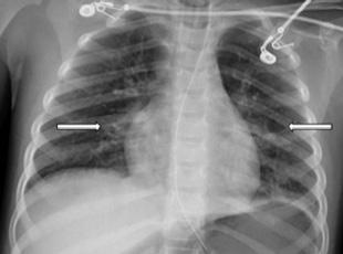
On arrival, the patient was tachypneic (respiratory rate of 27 breaths per minute) and coughing, and appeared to be in distress. She had coarse breath sounds and a room air oxygen saturation of 88% (reference range 95-100%). She was placed on heated high-flow nasal cannula delivering 40% oxygen at 16 liters per minute (L/min), which initially increased her oxygen saturation to 99%. A chest radiograph (CXR) was obtained and interpreted as unremarkable. An initial venous blood gas (VBG) revealed a partial pressure of carbon dioxide (pCO2) of 29 millimeters of mercury (mm Hg) (35-45 mm Hg) with pH 7.39 (7.35-7.45). While in the ED, her respiratory rate increased to 36 breaths per minute, her mental status declined, and she developed diffuse crackles on chest auscultation. A repeat VBG 65 minutes after arrival (45 minutes after initial VBG) revealed pCO2 of 58 mm Hg with pH of 7.22.
With obtundation and a worsening respiratory acidosis, she was intubated and placed on synchronized intermittent mandatory ventilation with a fraction of inspired oxygen (FiO2) of 75% and positive end-expiratory pressure of 7 mm Hg. A post-intubation CXR, performed approximately
CPC-EM Capsule
What do we already know about this clinical entity? Metal pneumonitis is the most severe acute complication of metal inhalation, and its occurrence is not confined to the workplace or one specific metal.
What makes this presentation of disease reportable? The case highlights the risk of severe lung injury from inhalation of a household product, for which no warning to consumers is disclosed by the manufacturer or the US Food and Drug Administration (FDA).
What is the major learning point?
Not all food items are regulated by the FDA or safe for handling by children. Severe inhalational injury can occur from metallic cake-dust products.
How might this improve emergency medicine practice?
Knowledge of lesser-known household agents and their potential to produce significant injury is important for emergency physicians.
148 minutes after arrival (98 minutes after the initial CXR), revealed bilateral patchy perihilar and peribronchial opacities (Image 2). There were no acute complications with the intubation, and the patient remained hemodynamically stable while being transferred to the intensive care unit (ICU).
In the ICU she received continued mechanical respiratory support and intermittent diuresis with furosemide to maintain neutral fluid balance. The infiltrates seen on CXR worsened over the next three days with progression of mixed interstitial
and alveolar opacities (Image 3). Despite a worsening CXR appearance, the patient maintained adequate oxygenation with ventilator settings of FiO2 of 30% and positive endexpiratory pressure of 5 mm Hg. She had intermittent fevers and leukocytosis, with a maximum white blood cell count of 26 x 103 cells per microliter (μL) (reference range 4.5 x 10³ - 11 x 103 cells/μL); however, antibiotics were deferred. There was no evidence of extrapulmonary organ damage on clinical exam or laboratory testing throughout the duration of her hospitalization. Two respiratory pathogen panels testing for 22 pathogens obtained on hospital days (HD) 2 and 4 were negative, as were blood, urine, and sputum cultures. She was extubated on HD 5. Post-extubation, she required 35-40% FiO2 via heated high-flow nasal cannula at 8-10 L/min for an additional two days as her CXR improved. She required supplemental oxygen until HD 9 and was discharged home the following day.
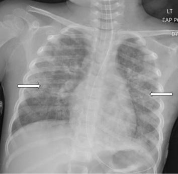
Chest radiograph on hospital day three showing worsening bilateral infiltrates and progression of mixed interstitial and alveolar opacities
The patient was referred to both cardiology and pulmonology for outpatient evaluation by her primary care physician four months after her hospitalization. Her chief complaints at these follow-up visits included decreased oxygen saturation readings at home, dyspnea on exertion, decreased appetite, and fatigue. Cardiology performed a cardiac ultrasound, which was normal, and did not believe her symptoms were cardiac in nature. A CXR obtained five months later demonstrated increased perihilar and peribronchial markings bilaterally and mild reticular nodular opacity involving both lungs (Image 4). She was diagnosed with moderate persistent reactive airway disease which, per her treating pulmonologist, was likely a sequela of her acute lung injury from inhalation of the cake luster dust. At greater than one year since hospitalization, she continued to be prescribed budesonide/ formoterol twice daily and rescue albuterol as needed.
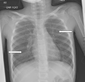
4. Chest radiograph at five months after discharge demonstrating increased perihilar and peribronchial markings bilaterally and mild reticular nodular opacity involving both lungs (arrows).
The cake luster dust in this case consists of a mixture of gold-appearing flakes and dust with a fine texture (Image 1). Online vendors note this product is “non-toxic”; “bronze based powde rs” are listed under ingredients. Bronze alloys, as a component of the cake dust, commonly consist of roughly 88% copper, 12% tin, and a trace amount of other metallic components. While this patient did not develop signs of heavy metal toxicity, there are at least seven reported cases of acute metal toxicity following ingestion of decorative cake dust.3 As in this case, there have been other published reports of inhalational exposures to metallic cake dusts. A recent case series describes three inhalational exposures leading to metal fume fever-like syndromes. 5 In all three cases, symptoms improved with bronchodilators and resolved 48 hours after exposure. A 2022 case report of metal fume fever from cake dust described a preschool-aged male with acute inhalational lung injury who required high-flow nasal cannula (18 L/ min, 43% oxygen) for 24 hours. He also had a persistent oxygen requirement for nine days after exposure.6
Metal fume fever is typically associated with inhalation of metal oxides like zinc and aluminum oxides that are formed when metals are heated through processes such as welding. Metal fume fever is one of several terms describing what is classically a “subclinical alveolitis not apparent on chest radiograph or pulmonary function tests except in unusually severe cases.”7 It is “associated with a neutrophilic predominance on bronchoalveolar lavage and a systemic leukocytosis” with short-lived clinical effects, similar to a flulike illness, and is unlikely to lead to sequelae.7 Alternatively, exposures resulting in a prolonged course (greater than 72 hours) with pulmonary infiltrates on CXR and respiratory failure more likely represent acute metal pneumonitis with or
Sanders et al Metal Pneumonitis from Cake Dust Aspiration
without acute respiratory distress syndrome (ARDS). High concentrations of several inhaled metallic dusts containing copper and mercury have been shown to cause acute pulmonary damage, resulting clinically in chemical pneumonitis and, in some cases, ARDS.8-9 Case reports have also described chronic pulmonary manifestations including pulmonary fibrosis and hyperreactive airway disease.10 Our case involves a notably severe clinical course with significant radiologic evidence of acute lung inflammation and both clinical and radiographic evidence of persistent pulmonary abnormalities, which we believe is consistent with metal pneumonitis.
This case highlights that non-edible metallic cake dusts are easily perceived as lacking toxicity. These products are sometimes difficult to distinguish from edible cake dusts made from sugar and are often sold alongside them in retail settings. While “non-edible” labeling is frequently used, these same containers also display the words “non-toxic.” This does not adequately convey the health risks associated with improper handling or provide purchasers with a warning regarding which portions of a confection may be dangerous to eat. The inadequacy of this labeling is evidenced by this case of metal pneumonitis with associated ARDS and chronic pulmonary disease. Accordingly, “non-toxic” should be abandoned as a descriptor of these products, and both consumers and treating physicians made aware of potential complications from inadvertent exposures.
1. U.S. Food and Drug Administration. 2018. FDA advises home and commercial bakers to avoid use of non-edible food decorative products. Food and Drug Administration. Available at: https:// www.fda.gov/food/food-additives-petitions/fda-advises-home-andcommercial-bakers-avoid-use-non-edible-food-decorative-products. Accessed July 22, 2022.
2. U.S. Food and Drug Administration. 2018. Compliance policy guide (CPG) Sec. 545.200. confectionary decorations (nutritive and nonnutritive). Food and Drug Administration. Available at: https://www. fda.gov/regulatory-information/search-fda-guidance-documents/cpgsec-545200-confectionery-decorations-nutritive-and-non-nutritive. Accessed July 22, 2022.
Documented patient informed consent and/or Institutional Review Board approval has been obtained and filed for publication of this case report.
Address for Correspondence: Christine Murphy, MD, Atrium Health’s Carolinas Medical Center, Department of Emergency Medicine, Division of Medical Toxicology, PO Box 32861, Charlotte, NC 28232-2861. Email: christine.murphy@atriumhealth.org
Conflicts of Interest: By the CPC-EM article submission agreement, all authors are required to disclose all affiliations, funding sources and financial or management relationships that could be perceived as potential sources of bias. The authors disclosed none.
Copyright: © 2024 Sanders et al. This is an open access article distributed in accordance with the terms of the Creative Commons Attribution (CC BY 4.0) License. See: http://creativecommons.org/ licenses/by/4.0/
3. Viveiros B, Caron G, Barkley J, et al. Cake decorating luster dust associated with toxic metal poisonings - Rhode Island and Missouri, 2018–2019. MMWR Morb Mortal Wkly Rep. 2021;70(43):1501–4.
4. U.S. Food and Drug Administration. 2016. Code of Federal Regulations, Title 21, Food and Drugs, Chapter 1. Electronic Code of Federal Regulations. Available at: https://www.ecfr.gov/current/ title-21/chapter-I/subchapter-B/part-170/subpart-A/section-170.3. Accessed July 22, 2022.
5. Pouget A, Evrard M, Le Visage L, et al. Metal fume fever-like syndrome after inhalation of cake decorating luster dust: Beware, dangerous frosting! Clin Toxicol. 2022;60(11):11:1290–1.
6. Moss J, Sinha A, Messahel S, et al. Acute inhalation lung injury secondary to zinc and copper aspiration from food contact dust. BMJ Case Rep. 2022;15(6):e250152.
7. Maddry JK. Irritant and toxic pulmonary injuries. In: Brent J, et al. (Eds.), Critical Care Toxicology, 2nd Ed (1973–2002). Cham, Switzerland: Springer, 2017.
8. Donoso A, Cruces P, Camacho J, et al. Acute respiratory distress syndrome resulting from inhalation of powdered copper. Clin Toxicol. 2007;45(6):714–6.
9. Moromisato DY, Anas NG, Goodman G. Mercury inhalation poisoning and acute lung injury in a child: use of high-frequency oscillatory ventilation. Chest. 1994;105(2):613–5.
10. Nemery B. Metal toxicity and the respiratory tract. Eur Respir J. 1990;3(2):202–19.
Bradley N. Bragg, MD
Kara J. Bragg, DNP, APRN
Mayo Clinic, Department of Emergency Medicine, Jacksonville, Florida
Section Editor: Christopher Sampson, MD
Submission history: Submitted January 24, 2024; Revision received April 14, 2024; Accepted May 24, 2024
Electronically published August 16, 2024
Full text available through open access at http://escholarship.org/uc/uciem_cpcem DOI: 10.5811/cpcem.7228
Introduction: Patients living with dementia as well as patients with neurological deficits are at significant risk for injury from multiple sources. Injuries may include falls, neglect, and, in some cases, self-injury. These patients require significant observation and closely monitored care.
Case Report: A 90-year-old man presented to a suburban emergency department (ED) by his family, who cared for him at home. The following case report describes a patient with dementia, hemineglect, and bruxism from a previous stroke who suffered a self-induced, partial amputation of his own thumb on the neglected side of his body.
Conclusion: Patients with dementia and neurologic deficits present frequently in the ED. These patients are at considerable risk of self-injury. The emergency physician should maintain vigilance in both screening for injuries and being aware of these risks when planning living arrangements after disposition from the ED. [Clin Pract Cases Emerg Med. 2024;8(4):336–338.]
Keywords: case report; patients living with dementia; self-injury; autophagia; hemineglect.
Emergency departments (ED) frequently care for patients living with dementia (PLWD), and there is a significant need to develop care plans and research protocols for better management of this patient population.2 Patients living with dementia have additional risks for comorbidities associated with functional dependence, behavioral challenges, and psychological manifestations of this disease.2 Patients with brain lesions or strokes can suffer from hemineglect, which shows a general lack of awareness of response to stimuli on the contralateral side from the lesion.3 This case illustrates an example of personal neglect due to a form of hemiinattention with a deficit of tactile sensation and pain, as well as proprioceptive awareness on the affected side of the body.3 There is little research on PLWD with hemineglect and additional risk factors for comorbidities. Treatment requires a comprehensive approach to evaluation and discharge planning.
A 90-year-old man presented to a suburban ED by his
family, who cared for him at home. He had a history of dementia and right-sided neglect from a previous stroke four years prior. As a result of the previous stroke, he also had an oral fixation and a predisposition to grinding his teeth (bruxism). According to the history provided by family, the patient would intermittently grind his teeth, profoundly at times, but it appeared to worsen when he was sleeping. The patient reportedly fell asleep at 6:30 pm in the evening. His family checked on him at approximately 7:30 pm and found a large volume of blood on his bed with a partial amputation of his right thumb. Given the patient’s history of bruxism and dementia, the physician concluded that he inadvertently chewed through his own thumb.
Physical examination revealed multiple jagged lacerations surrounding the distal half of the right thumb, beginning at the proximal phalanx. The nail plate was disrupted with these lacerations. The proximal portion of the nail plate was removed from the nail fold. The most proximal portion of the proximal phalanx both palmar and dorsally had jagged lacerations such that the only thing attaching the distal thumb
Bragg et al Autophagia with Dementia and Hemineglect
to the hand was a 1centimeter skin bridge on either side of the medial and lateral aspects of the thumb (Image).
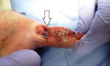
The distal thumb appeared dusky in color and was cool to the touch. A pulse oximeter probe placed on the thumb did not register any oxygen saturation. The capillary refill in the thumb was sluggish, measuring over two seconds in duration. Due to the patient’s impaired mentation and hemineglect, sensation of the thumb was unable to be assessed.
A radiograph of the right thumb revealed a comminuted fracture through the base of the first proximal phalanx. There was mild radial subluxation with minimal displacement of the distal fracture fragment. Gas in the soft tissues surrounding the proximal phalangeal fracture was also noted adjacent to the laceration. No foreign bodies were visualized.
The physician consulted orthopedic surgery. The surgeon discussed options with the family with consideration to the cleanliness of the laceration, bony exposure, and poor viability of the distal digit, the orthopedic surgeon performed a complete amputation. The patient was given ampicillinsulbactam intravenously in the ED for prophylaxis, and the procedure was performed by the orthopedic surgery resident at the bedside. The distal thumb was amputated followed by copious irrigation and closure of skin over the residual thumb. The patient was discharged from the ED with amoxicillinclavulanate with follow-up in the orthopedic hand surgery clinic. The wound healed well without complication.
Self-injurious behavior has been reported in the elderly on multiple occasions, making this patient population at particularly high risk, particularly when cared for at home by family members.4,5 Self-amputation of the hand and selfcannibalism have been reported in the past, customarily in the setting of psychiatric illness and not as the result of dementia.6,7 Schizophrenia and associated depersonalization or dissociation can account for this behavior.8 Self-mutilation
CPC-EM Capsule
What do we already know about this clinical entity?
Little has been reported about patients living with dementia with hemineglect and risk factors for self-injury. While patients with dementia are recognized to be at increased risk for injury from trauma such as falls, cases of unintentional self-harm are rarely reported.
What makes this presentation of disease reportable?
This case demonstrates a rarely-reported incident of self-harm in a patient with dementia and hemineglect.
What is the major learning point?
Patients with dementia can present significant risk of self injury. Emergency physicians should keep these potential risks in mind when managing patients with dementia and hemineglect.
How might this improve emergency medicine practice?
This case provides an example of an injury that can result from patients with dementia where adequate precautions were not taken to prevent self-harm. Having insight about this risk may help guide decisions about patient care.
is commonly seen in clinical practice, although studies are limited and often associated with abuse or opioids.8 In PLWD, the specific prevalence of self-induced injuries is not well documented, and most involve excoriations in the skin secondary to formication.9 This case would indicate that the hemineglect from the stroke combined with dementia-induced behaviors put this patient at significant risk for autophagia. Injury precautions for patients at risk of self-injury should be strongly adhered to. Close observation and frequent physical assessments are indicated by caregivers. In Lesch-Nyhan patients with compulsive self-injury behavior, often related to biting, preventive mouthguards have been proposed as an option to prevent self-mutilation.10 In this situation, restraint mitts may aid in prevention of self-mutilation of hands.11
Recent trends in end-of-life care have resulted in the United States using more supportive care and has found that patients aged 65 years and older are more likely to
Autophagia with Dementia and Hemineglect Bragg et al
die at home.1 With an increasing number of patients with dementia being cared for at home, this places an increased responsibility on the family and more support with caregiver support and palliative home-based care.1 The emergency clinician should be aware of these issues and keep them in mind when arranging for home care of patients with dementia. The physician should maintain a high degree of vigilance to screen and prevent self-mutilation injuries in the ED given the substantial risk in this patient population. Additionally, noted risk of self-mutilation in a population with combined stroke deficits and dementia must be considered in discharge planning from the ED or hospital.
1. Cross SH, Kaufman BG, Taylor Jr. DH, et al. Trends and factors associated with place of death for individuals with dementia in the United States. J Am Geriatr Soc. 2020;68(2):250–5.
2. Dresden SM, Taylor Z, Serina P, et al. Optimal emergency department care practices for persons living with dementia: a scoping review. J Am Med Dir Assoc. 2022;23(8):1314.e1–e29.
3. Caggiano P and Jehkonen M. The ‘neglected’ personal neglect. Neuropsychol Rev. 2018;28(4):417–35.
4. Parks SM and Feldman S. Self-injurious behavior in the elderly. Consultant Pharm. 2006;21(11):905–10.
5. Warnock JK, Burke WJ, Huerter C. Self-injurious behavior in elderly patients with dementia: four case reports. Am J Geriatr Psychiatry. 1999;7(2):166–70.
6. Sorene ED, Heras-Palou C, Burke FD. Self-amputation of a healthy hand: a case of body integrity identity disorder. J Hand Surg Am. 2006;31(6):593–5.
7. Yilmaz A, Uyanik E, Balci Şengül MC, et al. Self-cannibalism: the
Documented patient informed consent has been obtained and filed for publication of this case report.
Address for Correspondence: Bradley N. Bragg, MD, Mayo Clinic, Department of Emergency Medicine, 4500 San Pablo Rd., Jacksonville, FL. Email: bragg.bradley@mayo.edu
Conflicts of Interest: By the CPC-EM article submission agreement, all authors are required to disclose all affiliations, funding sources and financial or management relationships that could be perceived as potential sources of bias. The authors disclosed none.
Copyright: © 2024 Bragg et al. This is an open access article distributed in accordance with the terms of the Creative Commons Attribution (CC BY 4.0) License. See: http://creativecommons.org/ licenses/by/4.0/
man who eats himself. West J Emerg Med. 2014;15(6):701–2.
8. Aras N. A case of self-cannibalism: an extremely rare typeof self-mutilation with multiple risk factors. J Nerv Ment Dis. 2021;209(4):237–9.
9. Desai AK and Grossberg GT. Recognition and management of behavioral disturbances in dementia. Prim Care Companion J Clin Psychiatry. 2001;3(3):93–109.
10. Ragazzini G, Delucchi A, Calcagno E, et al. A modified intraoral resin mouthguard to prevent self-mutilations in Lesch-Nyhan patients. Int J Dent. 2014;2014:396830.
11. Lane E, Kuzow H, Manguera A, et al. 1142: Mitts before wrists: Safety mitts reduce restraint usage and can decrease unplanned self-extubations. Crit Care Med. 2016;44(12):361.
Rahul Gupta, MD
Cary Lubkin, MD
Section Editor: R. Gentry Wilkerson, MD
Cooper University Hospital, Department of Emergency Medicine, Camden, New Jersey
Submission history: Submitted March 24, 2024; Revision received July 3, 2024; Accepted June 29, 2024
Electronically published August 26, 2024
Full text available through open access at http://escholarship.org/uc/uciem_cpcem DOI: 10.5811/cpcem.20448
Introduction: One of the less common and more life-threatening etiologies of adrenal insufficiency is immune checkpoint inhibitor (ICI)-induced primary adrenal insufficiency (PAI). Patients typically present with fatigue, malaise, and nausea and are treated empirically with hydrocortisone.
Case Report: We present the case of a 59-year-old female who presented with hypotension, which initially was thought to be due to hypovolemia or medication-related, but was ultimately found to have PAI.
Conclusion: This case highlights the importance of early detection of ICI-induced primary adrenal insufficiency, given its associated morbidity and mortality and its incidence in patients with a history of immunotherapy. [Clin Pract Cases Emerg Med. 2024;8(4):339–342.]
Keywords: immunotherapy; shock; adrenal insufficiency; immune checkpoint inhibitor; case report.
Adrenal insufficiency is a well-known cause of shock in patients, with various sub-etiologies. One of the less common etiologies includes immune checkpoint inhibitor (ICI)-induced primary adrenal insufficiency (PAI).1-4 Patients who are on ICIs are susceptible to many endocrine adverse events, many of which are thought to be secondary to autoimmune effects on various organs.3 Based on current literature, one of the rarer ICIinduced endocrinopathies is thought to be PAI.2 It is important to recognize and treat ICI-induced PAI early given the associated morbidity and mortality.2-3,5 Patients with ICI-induced PAI typically present with malaise, fatigue, and nausea.1-2,6
Evaluation of adrenocorticotropic hormone (ACTH) and cortisol levels is important when PAI is clinically suspected, although this should not delay empiric treatment with hydrocortisone in acutely ill patients.1,5-6 All ICI therapies have had some association with PAI. Immune checkpoint inhibitors are a relatively new form of immunotherapy and include the following: cytotoxic T-lymphocyte-associated protein inhibitor (eg, ipilimumab), programmed cell death protein 1 inhibitors (eg, nivolumab and pembrolizumab), and
programmed cell death ligand 1 inhibitors (eg, atezolizumab, avelumab, and durvalumab).3 Given the rising use of ICI therapies, it is important that emergency physicians be aware of the incidence of ICI-induced PAI when evaluating a patient with undifferentiated shock with a history of treatment with these medications.
We describe a case spanning two emergency department (ED) presentations, coincidentally seen by the same resident and attending physician each time, and ultimately deemed to be ICI-induced PAI.
A 59-year-old female presented to a tertiary ED with lightheadedness for one day. Per emergency medical services, the patient was found to be hypotensive by the home health aide, with a blood pressure of 60s/30s millimeters of mercury (mm Hg), but otherwise no mental status changes or other acute complaints. Of note, the patient had been discharged two weeks prior from an outside hospital after a two-week stay requiring ventilator support due to concern for a sinceresolved cardiogenic shock state (ejection fraction 15-20%),
thought to be secondary to stress-induced cardiomyopathy or secondary to use of ICIs, ipilimumab and nivolumab, which she had last received approximately two months before. She had a past medical history of hypertension, atrial fibrillation, hyperlipidemia, and mesothelioma for which she had recently been on ICIs, as discussed above. The patient reported no significant family or social history. Additionally, she was prescribed apixaban due to her atrial fibrillation. The patient was also on metoprolol succinate XL 50 milligrams (mg) daily and atorvastatin 40 mg daily.
On arrival, her blood pressure was 89/54 mm Hg, with a repeat blood pressure of 63/38 mm Hg. Other initial vitals were as follows: heart rate 55 beats per minute; temperature 36.6°Celsius (C); respiratory rate 18 breaths per minute; and oxygen saturation 96% on room air. The patient’s physical examination was notable for a chest wall port with no evidence of infection, but otherwise was benign. Electrocardiogram demonstrated sinus bradycardia to 55 with no changes from prior baseline. Point-of-care cardiac ultrasound demonstrated a mildly depressed ejection fraction, normal right ventricle/ left ventricle ratio, and no pericardial effusion. No B-lines were visualized on point-of-care lung ultrasound.
Laoratory results were notable for lactic acid of 1.1 millimoles per liter (mmol/L) (reference range 0.5-2.2 mmol/L), hemoglobin of 11.2 grams per deciliter (g/ dL) (14-18 g/dL), white blood cell count of 7.87x10³ per microliter (μL) (5.50-11x103/μL), and 0- and 1-hour high sensitivity troponin trend was 59 to 46 nanograms (ng)/L (0-19 ng/L). Given the patient’s history of steroid use in the past (although it was unclear when it had last been taken) a random cortisol level was drawn and found to be 0.3 micrograms (μg)/dL (5.0-23.0 μg/dL). Basic metabolic panel had no electrolyte derangements. Her chest radiograph was unremarkable. The patient was started on broad spectrum antibiotics empirically given her hypotension with a potentially immunocompromised state.
The patient was given 500 milliliters of intravenous (IV) lactated Ringer’s with subsequent improvement in blood pressure to 119/58 mm Hg. She was subsequently admitted and was evaluated by the hospitalist and cardiology services, who believed hypotension was secondary to medications or hypovolemia. The patient was discharged the following day. Transthoracic echocardiogram (TTE) demonstrated an ejection fraction of 45-49% with mildly dilated left ventricle and left atrium. No changes were made to the patient’s medications.
The patient re-presented to the same ED seven days after her first presentation with hypotension and lightheadedness. On this visit, the patient reported chills and diarrhea but no other associated symptoms. Emergency medical services reported a blood pressure of 50/20 mm Hg en route. On arrival, she had a blood pressure 60/45 mm Hg, heart rate 87 beats per minute, temperature 36.7°C, respiratory rate 23 breaths per minute, and oxygen saturation 91% on room air. Point-of-care ultrasound showed no change from prior TTE. Computed tomography
CPC-EM Capsule
What do we already know about this clinical entity?
Immune checkpoint inhibitors (ICI) can cause primary adrenal insufficiency, a rare but lifethreatening condition typically presenting with fatigue, malaise, and nausea.
What makes this presentation of disease reportable?
This presentation of primary adrenal insufficiency is due to ICIs, a treatment that is becoming more prevalent.
What is the major learning point?
It is vital to consider primary adrenal insufficiency as a cause of shock in patients who are currently being treated with ICIs.
How might this improve emergency medicine practice?
Being aware of this life-threatening condition will allow emergency physicians to recognize and treat the condition earlier in its course.
of the chest and abdomen/pelvis were significant for trace left pleural effusion, small right pleural effusion with baseline pleural thickening, and enlarged subcarinal lymph nodes. After no improvement with two liters of IV lactated Ringer’s, the patient was started on norepinephrine with a mean arterial pressure goal of 65 mm Hg, necessitating an infusion at a rate of 15 μg/kilogram (kg) per minute (min). As at the previous visit, she was empirically started on broad spectrum antibiotics. There were no significant laboratory findings including the basic metabolic panel with no electrolyte derangements. Baseline cortisol drawn at the time of her admission was 1.7 μg/ dL (reference range 5.0-23.0 μg/dL).
After intensive care unit admission, evening cortisol was found to be 0.3 μg/dL. Endocrinology was consulted, and as per their recommendations a cosyntropin stimulation test was performed without an appropriate response: 30min cortisol = 6.4 μg/dL (>18 μg/dL) and 60-min cortisol = 8.6 μg/dL (>18 μg/dL). Adrenocorticotropic hormone (ACTH) noted to be elevated. Based on these results, endocrinology was concerned for PAI due to ICI use and stress dose hydrocortisone 50 mg every eight hours was initiated with improvement in baseline blood pressure and decreasing pressor requirements. This dose was tapered, and the patient was subsequently discharged on
hydrocortisone 15 mg every morning and 5 mg nightly. She did later have a repeat admission for hypotension secondary to running out of hydrocortisone at home, but her blood pressure quickly improved with the administration of hydrocortisone and she was discharged the day after admission with no complications.
This case demonstrates a case of ICI-induced PAI, a rare complication of ICI therapy. The pathophysiology is poorly understood, but it is thought to be secondary to T-cell mediated destruction of the adrenal cortex.1-2 Laboratory testing will typically reveal decreased random or morning cortisol levels, inappropriate cosyntropin stimulation test response, and elevated ACTH levels.1,5-6 If ACTH is noted to be decreased, secondary adrenal insufficiency should be considered, which is postulated to be due to ICI-induced hypophysitis, inflammation of the pituitary gland.6 Electrolyte derangements may also occur if mineralocorticoid-producing cells are also affected, resulting in hyperkalemia and hyponatremia.1,5 Patients typically present with malaise, fatigue, and nausea, with associated hypotension, much like the patient in this case.1-2,6 As these patients are often immunocompromised and present with undifferentiated shock, it is important to thoroughly rule out other causes of shock, including septic shock and hypovolemic shock.
Treatment typically consists of corticosteroid therapy immediately with consideration for mineralocorticoid therapy if there is evidence of hyperkalemia and hyponatremia, which demonstrates involvement of mineralocorticoid-producing cells. Given the life-threatening nature of ICI-induced PAI, if there is a heightened clinical suspicion for this condition, it is vital to treat empirically in the ED with corticosteroids while laboratory tests are pending for confirmation of diagnosis. Unfortunately, though, it is common for the diagnosis to be delayed given the initially insidious nature and non-specific symptomatology.2 Per the current literature, the recommended treatment is hydrocortisone as an empiric treatment in critically ill patients.1-2,6 Typically, an initial dose of 100 mg hydrocortisone IV should be administered followed by 50 mg IV every six hours. If available, an endocrinology consult should be obtained for further guidance.5
Ultimately, ICI-induced PAI is a very rare complication of ICI therapy. In their literature review in 2018, Barroso-Sousa et al reported 43 total cases of any-grade PAI among 5,381 patients given ICI monotherapy, representing an incidence of 0.7%.3 They also found the rate of PAI in all patients treated with combination ICI therapy, as in our patient, to be 4.2%.3 Other studies report 451 instances found in the literature of ICI-induced PAI, but report that only 45 of them were definite, laboratory-confirmed cases while the others were assumed cases.7-8 Interestingly, more than 90% of the cases of ICI-induced adrenal insufficiency are classified as secondary adrenal insufficiency.9
Ultimately, the incidence of ICI-induced PAI is rare but varies widely, depending on the type and dose of ICI. Further complicating the diagnosis is the variability of the timeline that patients may present with ICI-induced PAI, with a median onset of about 10 weeks. There are also reported cases ranging from 2-4 months, and many cases are noted to be found long after the end of treatment, as in this patient.10-13
Emergency physicians often encounter patients in a shock state and are well-versed in differentiation and treatment of the most common causes of shock. Unfortunately, there are rare occasions of refractory shock where other diagnoses and etiologies should be considered. Given the rising use of immune checkpoint inhibitor therapy, ICI-induced primary adrenal insufficiency is a diagnosis that should be considered when there is a high clinical suspicion and treated expeditiously given the life-threatening nature of the disease.
The authors attest that their institution requires neither Institutional Review Board approval, nor patient consent for publication of this case report. Documentation on file.
Address for Correspondence:Rahul Gupta, MD, Cooper University Hospital, Department of Emergency Medicine, Education and Research Building, 401 Haddon Avenue, Camden, NJ 08103. Email: rgrahul1234@gmail.com.
Conflicts of Interest: By the CPC-EM article submission agreement, all authors are required to disclose all affiliations, funding sources and financial or management relationships that could be perceived as potential sources of bias. The authors disclosed none.
Copyright: © 2024 Gupta et al. This is an open access article distributed in accordance with the terms of the Creative Commons Attribution (CC BY 4.0) License. See: http://creativecommons.org/ licenses/by/4.0/
1. Wright JJ, Powers AC, Johnson DB. Endocrine toxicities of immune checkpoint inhibitors. Nat Rev Endocrinol. 2021;17(7):389-99.
2. Cherry G. Immune checkpoint inhibitor-related adrenal insufficiency Semin Oncol Nurs. 2021;37(2):151131.
3. Barroso-Sousa R, Barry WT, Garrido-Castro AC, et al. Incidence of endocrine dysfunction following the use of different immune checkpoint inhibitor regimens. JAMA Oncol. 2018;4(2):173.
4. Shi Y, Shen M, Zheng X, et al. Immune checkpoint inhibitor-induced adrenalitis and primary adrenal insufficiency: systematic review and optimal management. Endocr Pract. 2021;27(2):165-9.
5. Stelmachowska-Banaś M and Czajka-Oraniec I. Management of endocrine immune-related adverse events of immune checkpoint inhibitors: an updated review. Endocr Connect. 2020;9(10):R207-28.
6. Cui K, Wang Z, Zhang Q, et al. Immune checkpoint inhibitors and adrenal insufficiency: a large-sample case series study. Ann Transl Med. 2022;10(5):251.
7. Grouthier V, Lebrun-Vignes B, Moey M, et al. Immune checkpoint inhibitor-associated primary adrenal insufficiency: WHO VigiBase report analysis. Oncologist. 2020;25:696-701.
8. Lu D, Yao J, Yuan G, et al. Immune checkpoint inhibitor-associated new-onset primary adrenal insufficiency: a retrospective analysis using the FAERS. J Endocrinol Invest. 2022;45(11):2131-7.
9. Salinas C, Renner A, Rojas C, et al. Primary adrenal insufficiency during immune checkpoint inhibitor treatment: case reports and review of the literature. Case Rep Oncol. 2020;13(2):621-6.
10. Martella S, Lucas M, Porcu M, et al. Primary adrenal insufficiency induced by immune checkpoint inhibitors: biological, clinical, and radiological aspects. Semin Oncol. 2023;50(6):144-8.
11. Paepegaey A-C, Lheure C, Ratour C, et al. Polyendocrinopathy resulting from pembrolizumab in a patient with a malignant melanoma. J Endocr Soc. 2017;1(6):646-9.
12. Tan MH, Iyengar R, Mizokami-Stout K, et al. Spectrum of immune checkpoint inhibitors-induced endocrinopathies in cancer patients: a scoping review of case reports. Clin Diabetes Endocrinol. 2019;5(1):1.
13. Cukier P, Santini FC, Scaranti M, et al. Endocrine side effects of cancer immunotherapy. Endocr Relat Cancer. 2017;24(12):T331-47.
Matthias Barden, MD*† Michael E. Schwartz, DO‡
Section Editor: Lev Libet, MD
Eisenhower Health, Emergency Medicine Residency Program, Racho Mirage, California
Hi-Desert Medical Center, Department of Emergency Medicine, Joshua Tree, California
Desert Care Network, Section Gastroenterology, Coachella Valley, California
Submission history: Submitted February 5, 2024; Revision received May 17, 2024; Accepted May 23, 2024
Electronically published July 28, 2024
Full text available through open access at http://escholarship.org/uc/uciem_cpcem DOI: 10.5811/cpcem.19397
Introduction: An attempt at medical management is often the initial step in addressing esophageal obstruction from an impacted food bolus. Medical management, however, has limited success and often requires urgent endoscopy. We present a case in which standard medical treatment failed, but a swallowing augmentation maneuver resolved the obstruction.
Case Report: A 67-year-old female presented with esophageal obstruction after eating steak. Transfer to higher level of care for endoscopy was initiated; however, the receiving gastroenterologist suggested an “upright posture, chin tuck, double swallow” maneuver. This immediately resolved the patient’s symptoms, and she was discharged home.
Conclusion: This case suggests a novel, non-endoscopic technique for esophageal obstruction from food bolus impaction. [Clin Pract Cases Emerg Med. 2024;8(4):343–345.]
Keywords: gastrointestinal; esophageal obstruction; meat impaction; food bolus impaction.
Esophageal obstruction from impacted food bolus can cause significant discomfort, prevent oral intake, and be associated with serious complications.1 The failure of medical management may use significant resources, including transfer to an endoscopy-capable institution.2 Thus, many medical treatments have been proposed as ways to avoid the need for urgent endoscopy. In this case, some previously reported swallowing augmentation techniques generally used in dysphagia patients were combined to successfully resolve the obstruction and avoid a transfer for urgent endoscopy.
A 67-year-old female presented to our emergency department with the complaint of esophageal obstruction. She denied any prior episodes but did report a history of known hiatal hernia. She was not taking any medications
routinely and denied any past surgical history. The night prior to her presentation, she had been eating steak when she felt a bolus of meat become lodged in her upper esophagus. She made several attempts at swallowing and felt the bolus move lower in her chest but remain impacted there. Afterward she had odynophagia with any attempt to drink and experienced intermittent episodes of regurgitation. Her examination was benign, with stable vital signs and a soft, non-tender, nondistended abdomen.
Our facility lacks gastroenterology specialists on staff, and on the day of presentation general surgery was not available. We attempted medical management with normal saline 1 liter intravenous (IV), ondansetron 4 milligrams (mg) IV, and glucagon 2 mg IV. Thirty minutes after receiving glucagon, the patient was provided a carbonated beverage to drink. This worsened her discomfort and was followed by immediate regurgitation, but the sensation of obstruction was unchanged.
We contacted our health system’s transfer center and were connected with the gastroenterologist on call at one of our partner hospitals about 60 miles away. He agreed to receive the patient but suggested that prior to transfer a specific series of swallowing augmentation maneuvers be attempted. He suggested that the patient perform an “upright posture, chin tuck, double swallow” with water. The patient was provided a glass of water, instructed to stand in a straight upright position, flex her neck to tuck her chin to her chest, swallow some of the water, and then once it started to go down, initiate a second effort of swallowing.
This technique resulted in immediate and complete resolution of the patient’s symptoms. Afterward, she tolerated intake by mouth without difficulty. She was discharged home with a prescription for 40 mg pantoprazole daily and was given instructions for out-patient gastroenterology followup. Further information about her subsequent course was unavailable.
Many methods have been proposed to avoid resorting to urgent endoscopy with esophageal obstruction from impacted food bolus. There is a low rate of success reported for the commonly attempted options, such as benzodiazepines, proteolytic enzymes, glucagon,3 nitroglycerin,4 and carbonated beverages.5 All these methods rely on the patient ultimately being able to complete the act of swallowing to relieve the obstruction. There are numerous techniques described to augment the process of swallowing for patients with neurologic, anatomic, or post-surgical causes of dysphagia.6 However, similar techniques have not previously been reported for esophageal obstruction from food bolus impactions.
Some of the specific techniques that have been described include upright posture,7 chin tuck position,8 and “double swallowing”.9 The consulting gastroenterologist on this case suggested that all three of those modalities be combined at the same time. While pretreatment with glucagon and providing a carbonated beverage had failed, the addition of instructing the patient on this “upright posture, chin tuck, double swallow” technique succeeded in relieving the obstruction, avoiding the need for transfer for urgent endoscopy.
This case suggests the “upright posture, chin tuck, double swallow” technique may benefit patients with esophageal meat impaction and potentially avoid urgent endoscopy. Some of the usual medical interventions were attempted in this case and were unsuccessful in resolving the obstruction. This is only one case of a successful, augmented swallowing maneuver to relieve an esophageal obstruction, but the technique is easy to perform and, if successful, can avoid the need for urgent endoscopy.
CPC-EM Capsule
What do we already know about this clinical entity?
There is a low rate of success for medical management of esophageal meat impaction. Failed medical treatment requires urgent endoscopy.
What makes this presentation of disease reportable?
A novel application of dysphagia treatment strategies was used successfully for esophageal meat impaction after standard medical management had failed.
What is the major learning point?
Dysphagia patients benefit from instruction on positioning and swallowing techniques. Those same instructions may help patients with esophageal meat impaction.
How might this improve emergency medicine practice?
Instruction on the “upright posture, chin tuck, double swallow” technique is simple and could potentially avoid the need for urgent endoscopy
Documented patient informed consent and/or Institutional Review Board approval has been obtained and filed for publication of this case report.
Address for Correspondence: Matthias Barden, MD, Hi-Desert Medical Center Emergency Department, 6601 White Feather Rd, Joshua Tree CA 92252. Email: mbarden@eisenhowerhealth.org
Conflicts of Interest: By the CPC-EM article submission agreement, all authors are required to disclose all affiliations, funding sources and financial or management relationships that could be perceived as potential sources of bias. The authors disclosed none.
Copyright: © 2024 Barden et al. This is an open access article distributed in accordance with the terms of the Creative Commons Attribution (CC BY 4.0) License. See: http://creativecommons.org/ licenses/by/4.0/
1. Schupack DA, Lenz CJ, Geno DM, et al. The evolution of treatment and complications of esophageal food impaction. United European Gastroenterol J. 2019;7(4):548–56.
2. Crockett SD, Sperry SL, Miller CB, et al. Emergency care of esophageal foreign body impactions: timing, treatment modalities, and resource utilization. Dis Esophagus. 2013;26(2):105–12
3. Hardman J, Sharma N, Smith J, et al. Conservative management of oesophageal soft food bolus impaction. Cochrane Database Syst Rev. 2020;2020(5):CD007352.
4. Willenbring BA, Schnitker CK, Stellpflug SJ. Oral nitroglycerin solution for oesophageal food impaction: a prospective single-arm pilot study. Emerg Med J. 2020;37(7):434–6.
5. Tiebie EG, Baerends EP, Boeije T, et al. Efficacy of cola ingestion for oesophageal food bolus impaction: open label, multicentre, randomised controlled trial. BMJ. 2023;383:e007294.
6. López-Liria R, Parra-Egeda J, Vega-Ramírez FA, et al. Treatment of dysphagia in Parkinson’s disease: a systematic review. Int J Environ Res Public Health. 2020;17(11):4104.
7. Alghadir AH, Zafar H, Al-Eisa ES, et al. Effect of posture on swallowing. Afr Health Sci. 2017;17(1):133–7.
8. Li M, Huang S, Ding Y, et al. The effectiveness of chin-down manoeuvre in patients with dysphagia: a systematic review and meta-analysis. J Oral Rehabil. 2024;51(4):762–74.
9. Postma GN, Burkhead LM, Moretz WH 3rd. Double swallow. Ear Nose Throat J. 2006;85(8):488.
Aamir Rashid, MD, DM*
Farooq Ahmad Ganie, MS, MCH†
Hilal Rather, MD, DM*
Shahood Ajaz Kakroo, MD, DM*
Raja Suhail Shounthoo, MD‡
Section Editor: Austin Smith, MD
Sher-i-Kashmir Institute of Medical Sciences Soura, Department of Cardiology, Srinagar, Jammu and Kashmir, India
Sher-i-Kashmir Institute of Medical Sciences Soura, Department of Cardiovascular and Thoracic Surgery, Srinagar, Jammu and Kashmir, India
Sher-i-Kashmir Institute of Medical Sciences Soura, Department of Anesthesia, Srinagar, Jammu and Kashmir, India
Submission history: Submitted February 23, 2024; Revision received June 20, 2024; Accepted June 20, 2024
Electronically published September 21, 2024
Full text available through open access at http://escholarship.org/uc/uciem_cpcem
DOI: 10.5811/cpcem.20279
Introduction: Although myocardial injury is common after blunt chest trauma, tricuspid valve injury associated with traumatic atrial septal defect resulting in acute hypoxia is an infrequent event. We report an unusual case of blunt chest trauma referred to us for unexplained hypoxemia, emphasizing the unusual nature of injury and the importance of comprehensive cardiac evaluation in such cases.
Case Report: A 35-year-old male presented to the emergency department after falling from a tree from an approximate height of 15 feet. He sustained multiple rib fractures and a left hemopneumothorax. Examination revealed decreased air entry over the left hemithorax and a systolic murmur over the left sternal border. Electrocardiography showed a junctional rhythm, and troponin levels were significantly elevated. Despite tube thoracostomy, the patient remained hypoxemic. Cardiology evaluation revealed a flail tricuspid valve with severe regurgitation and a traumatic atrial septal defect (ASD). Bidirectional shunting across the atrial septal defect was causing hypoxemia. The patient underwent surgical repair of the ASD and tricuspid valve, which resulted in a successful outcome.
Conclusion: Our case highlights the need for comprehensive cardiac evaluation in such patients. In addition to sonography for trauma, point-of-care echocardiographic examination should be a part of the focused assessment. [Clin Pract Cases Emerg Med. 2024;8(4):346–348.]
Keywords: cardiac blunt trauma; traumatic ASD; tricuspid regurgitation; hypoxia; echocardiographic examination; case report.
INTRODUCTION
Traumatic injuries result in close to six million fatalities worldwide annually.1 Thoracic injuries account for the highest morbidity and mortality among trauma patients. Up to 10-25% of all traumatic fatalities involve cardiac or aortic injuries.1 The right ventricle (RV) and right atrium (RA) are more commonly involved than the left ventricle and left atrium, due to their anatomical location and wall thickness. The RV and
RA are positioned anteriorly in the chest, making them more susceptible to direct trauma. Additionally, the walls of the RV and RA are thinner compared to the thicker, more muscular walls of the left ventricle, making them more vulnerable to injury upon impact.1
Valve injury secondary to blunt trauma is uncommon. Ventricular septal defects are extremely rare, and atrial septal defects (ASD) are even less frequent, with only a few case
reports of traumatic ASDs.2 While acute traumatic right-toleft cardiac shunts have been described following penetrating trauma,2 their occurrence following blunt chest trauma is exceedingly rare.3 We describe the case of a young male referred to us for unexplained refractory hypoxemia following blunt chest trauma. Our case highlights a traumatic blunt chest injury resulting in an unusual presentation.
A 35-year-old male presented to our emergency department following a fall from a tree. He had no significant past history. He suffered multiple rib fractures and left hemopneumothorax. The physical examination showed decreased air entry over the left hemithorax and a systolic murmur over the left sternal border. Electrocardiography showed a junctional rhythm. The troponin levels were significantly increased to 56 nanograms per milliliter (ng/ mL) (reference 0-1 ng/mL). The patient underwent a tube thoracostomy; however, he continued to be hypoxemic despite high-flow oxygen and appropriate management of his hemopneumothorax. Cardiology consultation was sought, and echocardiography demonstrated a flail tricuspid valve due to a rupture of the sub-valvular segment (chordal rupture) of the anterior tricuspid leaflet, resulting in severe tricuspid regurgitation (TR) (Image 1).
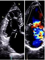
Image 1. Transthoracic echocardiography showing severe tricuspid regurgitation with flail anterior leaflet of tricuspid valve. White arrow showing flail anterior leaflet of tricuspid valve and black arrow showing color Doppler of severe tricuspid regurgitation.
The echocardiography further showed a significant ASD with traumatic flail margins (Image 2) and a TR jet directing flow across the ASD, resulting in bidirectional shunting across the ASD, explaining the hypoxemia.
CPC-EM Capsule
What do we already know about this clinical entity?
Although blunt myocardial injury is common, tricuspid valve injury associated with traumatic atrial septal defect is extremely rare.
What makes this presentation of disease reportable?
This case highlights a rare combination of traumatic tricuspid regurgitation and atrial septal defect leading to unexplained hypoxemia.
What is the major learning point?
Comprehensive cardiac evaluation, including echocardiography, is crucial in patients with blunt chest trauma and unexplained hypoxemia.
How might this improve emergency medicine practice?
Routine use of point-of-care echocardiography in blunt chest trauma can detect rare cardiac injuries, improving diagnosis and outcomes.
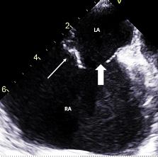
Image 2. Transesophageal view at 140° showing ostium secondum atrial septal defect with flimsy torn atrial septum. White thin arrow showing flimsy torn atrial septum and white thick arrow showing atrial septal defect. LA, left atrium; RA, right atrium.
Considering the flail traumatic margins of the ASD and the non-dilatation of the pulmonary arteries, the ASD was assumed to be traumatic rather than congenital. The patient
Traumatic Atrial Septal Defect with Tricuspid Regurgitation
was urgently referred to surgery and underwent a successful ASD closure with tricuspid valve repair on the second day of hospital admission.
Our case underscores the importance of dedicated cardiac evaluation in patients with blunt chest trauma who present with unexplained hypoxemia. Although blunt myocardial injury is common, tricuspid valve injury associated with traumatic ASD is extremely rare. Valve injury secondary to blunt cardiac trauma is uncommon.4 The aortic valve is the most commonly involved cardiac valve following trauma, followed by the mitral and the tricuspid.5 Traumatic TR is usually well tolerated. However, in our patient, the presence of an associated ASD resulted in a right-to-left shunt causing severe hypoxemia.
Traumatic TR can be caused by compression and decompression of the thorax. The resulting increase in RV pressure, especially if it occurs during the isovolumic contraction phase when all valves are closed, increases the risk of injury to the atrioventricular valves with rupture of the chordae or papillary muscle from the sudden increase in intracardiac pressure.4 The aortic and pulmonary valves are more susceptible to injury in diastole. The mechanisms of valve injury include papillary muscle rupture, chordal tear, leaflet injury, and delayed papillary muscle necrosis secondary to contusion.4 Our patient suffered from a chordal tear of the anterior tricuspid leaflet and papillary muscle rupture. Tricuspid valve rupture with varying degrees of heart block has been described.6 Our patient had a junctional rhythm, which improved spontaneously two weeks after surgical repair.
Although traumatic ventricular septal defect has been reported to have a prevalence of 5% following blunt thorax trauma, the occurrence of traumatic ASD is rare.7 Getz et al8 postulated different mechanisms of injury in cardiac chamber rupture. These include a direct blow to the chest, indirect trauma due to a crush injury of the abdomen with a resultant increase in intracardiac pressure, bidirectional force from compression of the heart between the sternum and vertebral bodies, acceleration and deceleration injury, blast forces, and penetrating trauma. Our patient had a direct blow to the chest as a result of a fall from a height.
The atrial septum is most susceptible to injury in late systole when the atria are full and all valves are closed. In contrast, the ventricular septum is more susceptible to injury in early systole and late diastole.8 Delayed perforation can also occur secondary to inflammatory reaction due to septal contusion.9 The right-to-left shunt due to the severe TR jet across the ASD caused the hypoxemia in our patient. The selective streaming of the severe TR jet across the ASD was thought to be responsible for central cyanosis and has been described previously.2,3 Furthermore, acute TR can increase the RA pressure to more than the left atrium pressure, resulting in a right-to-left shunt across the ASD. The right-to-left shunt has been described in patients with congenital ASD and TR.10
The patient described sustained a traumatic Tricuspid valve injury resulting in severe TR and an ASD with shunting resulting in hypoxemia after a fall from height. This case highlights the need for comprehensive cardiac evaluation in blunt chest trauma. Point-of-care echocardiographic examination should be an essential part of focused assessment along with sonography for cardiac blunt trauma.
Patient consent has been obtained and filed for the publication of this case report. The author attests that their institution requires does not require Institutional Review Board approval. Documentation on file.
Address for Correspondence: Aamir Rashid, MD, DM, SKIMS Soura, Department of Cardiology, Sheri-i-Kashmir, Institute of Medical science, Post bag No: 27, Srinagar, 190 011 Kashmir India, Srinagar, Jammu and Kashmir, India. Email: aamirrashid1111@gmail.com.
Conflicts of Interest: By the CPC-EM article submission agreement, all authors are required to disclose all affiliations, funding sources and financial or management relationships that could be perceived as potential sources of bias. The authors disclosed none.
Copyright: © 2024 Rashid et al. This is an open access article distributed in accordance with the terms of the Creative Commons Attribution (CC BY 4.0) License. See: http://creativecommons.org/ licenses/by/4.0/
1. Huis MA, Craft CA, Hood RE, at al. Blunt cardiac trauma review. Cardiol Clin. 2018;36:183-91.
2. Oliemy A, Mahesh B, Pathi V, et al. Acute traumatic right to left cardiac shunt. Ann Thorac Surg. 2016;102:e303-4.
3. Rao G, Garvey J, Gupta M, et al. Atrial septal defect due to blunt thoracic trauma. J Trauma. 1977;17:405-6.
4. Varahan SL, Farah GM, Caldeira CC, et al. The double jeopardy of blunt chest trauma: a case report and review. Echocardiography. 2006;23:235-9.
5. Liedtke AJ and DeMuth WE Jr. Nonpenetrating cardiac injuries: a collective review. Am Heart J. 1973;86:687-97.
6. Hasdemir H, Arslan Y, Alper A, et al. Severe tricuspid regurgitation and atrioventricular block caused by blunt thoracic trauma in an elderly woman. J Emerg Med. 2012;43:445-7.
7. Menaker J, Tesoriero RB, Hyder M, et al. Traumatic atrial septal defect and papillary muscle rupture requiring mitral valve replacement after blunt injury. J Trauma. 2009;67:1126.
8. Getz BS, Davies E, Steinberg SM, et al. Blunt cardiac trauma resulting in right atrial rupture. JAMA. 1986;255:761-3.
9. Ortiz Y, Waldman AJ, Bott JN, et al. Blunt chest trauma resulting in both atrial and ventricular septal defects. Echocardiography. 2015 Mar;32(3):592-4.
10. Swan HJ, Burchell HB, Wood EH, et al. The presence of venoarterial shunts in patients with interatrial communications. Circulation. 1954;10:705-13.
R. Wesley Slaven, MD* Martin Huecker, MD†
David Kersting, MSN†
Section Editor: Austin Smith, MD
University of Louisville, Department of Emergency Medicine, Louisville, Kentucky University of Louisville Health, Louisville, Kentucky
Submission history: Submitted April 23, 2024; Revision received July 11, 2024; Accepted June 29, 2024
Electronically published October 14, 2024
Full text available through open access at http://escholarship.org/uc/uciem_cpcem DOI: 10.5811/cpcem.20926
Introduction: Fahr disease and Fahr syndrome represent clinical entities that result in diffuse intracranial brain calcification, either by way of genetic mutation in the case of the former or by secondary endocrine dysfunction in the latter.
Case Report: We present a case of a middle-aged male with undiagnosed Fahr syndrome, identified during evaluation for symptoms of an acute posterior circulation cerebrovascular accident.
Conclusion: Fahr syndrome is a clinical constellation of symptoms and radiographic findings often seen in late-stage hypoparathyroidism. The intracranial calcifications associated may be related to an increased risk for intracranial cerebrovascular disorders such as ischemic or hemorrhagic infarct.
[Clin Pract Cases Emerg Med. 2024;8(4):349–352.]
Keywords: Fahr syndrome; case report; posterior circulation stroke; cerebellar infarct.
Primary familial brain calcification, also known as Fahr disease, is a disorder characterized by calcification of the bilateral basal ganglia as well as a gradual-onset movement disorder. It is inherited in an autosomal dominant or sporadic fashion and usually presents in the fourth or fifth decade of life.1 Its manifestations typically involve clumsiness, gait disturbance, and involuntary movements or cramping. However, the constellation of symptoms can be variable in timing, severity, and character.2 Disease prevalence is noted at <1 per 1,000,000.3 Although calcifications primarily involve basal ganglia, thalamus, and subcortical white matter, the presence of symptoms varies greatly between patients.
Differentiating between Fahr disease and Fahr syndrome lies in the ability to identify secondary causes of brain calcification. Most commonly, underlying endocrine disorders, including hypoparathyroidism or hyperparathyroidism, represent the underlying cause of secondary Fahr syndrome.4 FLowenthal and Bruyn highlight that 21.5% of patients with idiopathic hypoparathyroidism develop Fahr syndrome,5 and multiple studies have demonstrated basal ganglia calcification
following thyroidectomy.6,7 Case reports have shown a potential relationship between intracranial calcifications and cerebrovascular accidents, both hemorrhagic and ischemic;8,9 however, to our knowledge, no cases of cerebellar or posterior circulation infarcts have been described. In several reports, these events occur in patients with an otherwise favorable risk profile, including those of young age and without secondary risks of stroke.
A 54-year old male with history of hypertension, diabetes, heart failure, and transient ischemic attack presented to the emergency department (ED) for several days of dizziness with a dull headache of moderate intensity. Symptoms began approximately three days prior to arrival, accompanied by blurry vision. Given his medical history and due to a concern for stroke or other intracranial pathology, a computed tomography (CT) of the head was obtained, which was notable for diffuse intracranial calcifications as well as a hypodensity in the posterior right cerebellar hemisphere (Image 1). Magnetic resonance imaging (MRI) showed an acute to
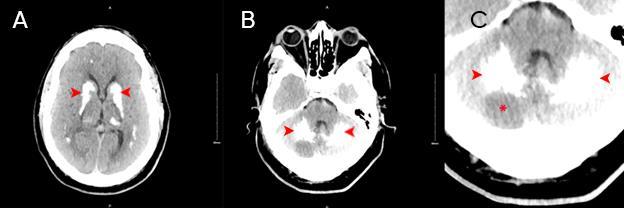

Image 1. A) Non-contrast computed tomography of the head demonstrates diffuse calcification of the bilateral basal ganglia, thalami, and subcortical white matter of cerebral hemispheres (arrowheads). B) Calcification of the cerebellum bilaterally with area of hypodensity (arrowheads). C) Cerebellum with bilateral calcification (arrowheads) with area of hypodensity noted (asterisk).
CPC-EM Capsule
What do we already know about this clinical
Fahr syndrome is a clinical syndrome present in late-stage hypoparathyroidism patients that may increase the risk of associated cerebrovascular
What makes this presentation of disease reportable?
This is the first demonstrated case in the literature of posterior circulation cerebrovascular accident in the Fahr syndrome population.
What is the major learning point?
The identification of radiographic findings suggestive of Fahr syndrome should prompt timely evaluation of secondary causes, namely endocrine disorders.
How might this improve emergency medicine practice?
We highlight a rare sequela of endocrine disorder that warrants careful optimization, often with a multidisciplinary team.
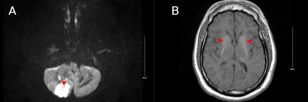

Image 2. A) A magnetic resonance image of the brain shows an area of diffusion restriction in posterior-inferior right cerebellar hemisphere consistent with acute to subacute infarct, with susceptibility blooming (an artifact often seen surrounding prior hemorrhage or calcification, noted with arrowhead) along inferolateral margin consistent with petechial hemorrhagic transformation. B) Bilateral susceptibility blooming (arrowheads) in basal ganglia and cerebral white matter.
subacute right posterior-inferior cerebellar artery stroke with early hemorrhagic transformation (Image 2). He was subsequently admitted to the neurology stroke service for further evaluation and management.
Additional history obtained following admission showed that this patient reported a previous history of thyroid carcinoma with subsequent resection at age six, not disclosed at the time of his initial encounter with ED staff, with resultant post-procedural hypothyroidism and hypoparathyroidism. He also reported multiple bouts with ureterolithiasis bilaterally, and cataracts that were to be intervened upon at a later date. Following admission, angiogram studies revealed right vertebral artery occlusion with reconstitution, as well as severe left vertebral artery stenosis. The patient subsequently underwent vertebral artery stenting and risk factor management including dual antiplatelet therapy and initiation of a high-intensity statin. He was discharged four days following admission to outpatient follow-up.
Following discharge, the patient was seen in neurology clinic where his National Institutes of Health Stroke Scale score was zero. He did report issues with forgetfulness, but otherwise had no residual neurologic deficit. In addition, he has had multiple admissions for treatment of symptomatic nephrolithiasis, requiring multiple ureteral stents and
lithotripsy procedures. To date, he is doing well in regard to his neurologic status and is currently followed on an outpatient basis by neurology, as well as by nephrology, otolaryngology, and cardiology.
Fahr disease is a rare cause of neurologic symptoms of variable severity and quality. Idiopathic calcifications most commonly affect the basal ganglia, thalamus, and cerebral white matter.10 Computed tomography remains a reliable initial test for evaluation, given that these calcium deposits are hyperdense and their locations and abundance often point toward the distinct disease entity of Fahr disease/syndrome. MRI findings can vary depending on the composition of deposits and stage of disease. As a result, MRI may exhibit a near-normal appearance in a patient whose CT imaging is grossly abnormal.11
While Fahr disease represents a distinct, primary pathology leading to the finding of basal ganglia calcification, the disease prevalence is less than one per million. Highlighting this rarity, an evaluation into secondary causes of these diffuse calcifications is warranted, with special attention paid to endocrine disorders as an underlying driving force.
In a 1981 report by Harrington et al, 42 of 7,000 patients were retrospectively identified as having basal ganglia calcifications on CT of the head, an incidence of 0.6%. Of these patients, 26 were available for follow-up, and two of the 26, or 7.7%, were found to have evidence of parathyroid dysfunction.12 Emergency physicians might encounter this disease process over the course of their career. Recognition of this clinical entity is of paramount importance, as these patients warrant prompt evaluation for underlying causes, such as endocrine disorders. While this may not take place in an emergency setting, subspecialist referral is necessary to ensure appropriate long-term symptom management.
In this patient, his initial presentation and workup focused on headache and dizziness. While the spectrum of symptoms in Fahr syndrome is broad, most patients do not experience headaches or dizziness. However, this may be confounded in that patients may experience gait disturbance and Parkinsonian symptomatology,13 which may mimic sensations of dizziness or, alternatively, limit history. Unfortunately, he had a concurrent acute pathology in the form of an acute cerebellar stroke contributing to his symptoms. While previous case reports have described both hemorrhagic and ischemic cerebrovascular accidents in patients found to have radiographic evidence of Fahr syndrome/disease, none have shown presence of a posterior circulation stroke.
Regardless, in this patient it remains a possibility that his underlying endocrine disorder played a role in the development of his cerebrovascular accident and, potentially, his prior transient ischemic attack. Unbeknownst to ED staff at the time, this patient represented a classic array of symptomatic hypoparathyroidism, with ureterolithiasis and resultant kidney disease, coronary artery disease, hypothyroidism, and Fahr syndrome, diagnosed during
his initial visit. How this syndrome translates to clinically significant cerebrovascular conditions is a subject of speculation. It has been shown that calcium deposits into the walls of arterioles, capillaries, and veins.14 Additionally, a postmortem examination of a patient with Fahr disease published in 2004 shows extensive calcifications of small blood vessel walls on the dentate nucleus of the cerebellum. Inflammatory changes and deposition in endothelial and stromal vascular cells were also observed.15 The combination of intravascular calcium deposition and associated inflammation may increase risk of regional ischemia, aneurysm formation, or associated hemorrhagic insult.
We present a patient with a phenotypic presentation of symptomatic, idiopathic hypoparathyroidism with resultant development of diffuse intracranial calcification, known as Fahr syndrome. This constellation was identified during evaluation for a concurrent, acute right posterior inferior cerebellar artery infarct.
The authors attest that their institution requires neither Institutional Review Board approval, nor patient consent for publication of this case report. Documentation on file.
Address for Correspondence: R. Wesley Slaven, MD, University of Louisville, Department of Emergency Medicine, 530 S. Jackson St., C1H17, Louisville, KY 40202. Email: rwesleyslaven@gmail.com.
Conflicts of Interest: By the CPC-EM article submission agreement, all authors are required to disclose all affiliations, funding sources and financial or management relationships that could be perceived as potential sources of bias. The authors disclosed none.
Copyright: © 2024 Slaven et al. This is an open access article distributed in accordance with the terms of the Creative Commons Attribution (CC BY 4.0) License. See: http://creativecommons.org/ licenses/by/4.0/
1. Ramos EM, Oliveira J, Sobrido MJ, et al. (2004). Primary familial brain calcification. [Updated 2017 Aug 24]. In: Adam MP, Mirzaa GM, Pagon RA, et al., editors. GeneReviews [Internet]. Seattle, WA: University of Washington, Seattle.
2. Chiu HF, Lam LC, Shum PP, et al. Idiopathic calcification of the basal ganglia. Postgrad Med J. 1993;69(807):68-70.
3. Ellie E, Julien J, Ferrer X. Familial idiopathic striopallidodentate calcifications. Neurology. 1989;39(3):381–5.
4. Saleem S, Aslam HM, Anwar M, et al. Fahr syndrome: literature review of current evidence. Orphanet J Rare Dis. 2013;8:156.
5. Lowenthal A and Bruyn G. Calcification of the striopallidodentate system. Handb Clin Neurol. 1968;6:703–25.
6. Danowski TS, Lasser EC, Wechsler RL. Calcification of basal ganglia in post-thyroidectomy hypoparathyroidism. Metabolism. 1960;9:1064-5.
7. Smith KD, Geraci A, Luparello FJ. Basal ganglia calcification
Acute Cerebellar Infarct in Patient with Undiagnosed Fahr’s Syndrome Slaven et al. in postoperative hypoparathyroidism. NY State J Med 1973;73(13):1807-9.
8. Yang CS, Lo CP, Wu MC. Ischemic stroke in a young patient with Fahr disease: a case report. BMC Neurol. 2016;16:33.
9. Sgulò FG, di Nuzzo G, de Notaris M, et al. Cerebrovascular disorders and Fahr disease: report of two cases and literature review. J Clin Neurosci. 2018;50:163-4.
10. Lester J, Zúniga C, Díaz S, et al. Diffuse intracranial calcinosis (Fahr disease) Arch Neurol. 2006;63:1806.
11. Scotti G, Sciafla G, Tampieri D, et al. MR imaging in Fahr disease. J Comput Assist Tomogr. 1985;9:790–2
12. Harrington MG, MacPherson P, McIntosh WB, et al. The significance of the incidental finding of basal ganglia calcification on computed tomography. J Neurol Neurosurg Psychiatry. 1981;44:1168–70.
13. U.S. Department of Health and Human Services. (n.d.). Fahr syndrome. National Institute of Neurological Disorders and Stroke. Available at: https://www.ninds.nih.gov/health-information/disorders/fahrs-syndrome Accessed June 22, 2024.
14. Chalkias SM, Magnaldi S, Cova MA, et al. Fahr disease: significance and predictive value of CT and MR findings. Eur Radiol. 1992;2:570–5.
15. Elshimali YI. The value of differential diagnosis of Fahr disease by radiology. J Radiol. 2005 4:1.
Ivan Muchiutti, BS*
Emmelyn J. Samones, BS†
Tammy Phan, BS†
Emily Barrett, MD†
Section Editor: Joel Moll, MD
Loma Linda University Health School of Medicine, Loma Linda, California Loma Linda University Medical Center, Department of Emergency Medicine, Loma Linda, California
Submission history: Submitted May 31, 2024; Revision received June 5, 2024; Accepted July 8, 2024
Electronically published: October 22, 2024
Full text available through open access at http://escholarship.org/uc/uciem_cpcem DOI: 10.5811/cpcem.21286
Introduction: Percutaneous endoscopic gastrostomy (PEG) placement is a common procedure for patients requiring non-oral feeding. One rare complication of PEG placement is the formation of a gastrocolocutaneous fistula that develops when the bowel is caught between the stomach and abdominal wall during placement. This report explores an elderly patient’s gastrocolocutaneous fistula development months post-PEG placement who presented with malodorous leakage from the gastrostomy tube to the emergency department (ED).
Case Report: A 73-year-old male on hospice presented to the ED with malodorous leakage from his PEG tube. He had received the PEG tube four months prior to this presentation and had it replaced once at an outside hospital due to blockages. In the ED, his PEG tube was found to have a deflated balloon stopper. The PEG tube was replaced, but the feculent discharge persisted. Imaging showed the tube’s position in the transverse colon. The patient underwent non-surgical management, with PEG tube removal and nutritional support via nasogastric tube. He was discharged with improvement of PEG site.
Conclusion: Gastrocolocutaneous fistula should be considered in patients experiencing unexpected PEG tube drainage or feeding-related complications such as diarrhea. Careful replacement techniques after dislodgement or blockage are important. Radiologic confirmation should be considered after replacement of tubes with feculent drainage. The rarity of gastrocolocutaneous fistula cases in the literature explains the lack of standardized management approaches. Clinical signs such as feculent leakage through the PEG tube site should prompt recognition and diagnosis by the emergency clinician. [Clin Pract Cases Emerg Med. 2024;8(4):353–346.]
Keywords: Percutaneous endoscopic gastrostomy; gastrocolocutaneous fistula; PEG replacement; case report.
Percutaneous endoscopic gastrostomy (PEG) is one of the most common endoscopic procedures and the gold standard for feeding in patients with viable enteric tracts and difficulty maintaining oral intake.1 Like any procedure, PEG placement comes with complications both during placement
and much later afterward. Formation of a gastrocolocutaneous fistula is a rare complication of PEG placement that occurs in 0.5% of adults and 3.5% of children.2,3 Fistula formation is theorized to occur when an interposed segment of small or large bowel is caught between the stomach and abdominal wall during placement
and is generally asymptomatic until the tube is dislodged in some manner.4,5
Patients with a gastrocolocutaneous fistula can stay asymptomatic for months, but when the PEG tube is dislodged either by malfunction or replacement, patients can present with a variety of symptoms including diarrhea after food administration, weight loss, malnutrition, tube blockage, and fecal leakage.6,7,8 Optimal treatment and management has not been clearly determined in the current literature. We present a case of this rare complication in an elderly patient presenting months after initial placement to the emergency department (ED) with foul-smelling leakage from his PEG tube who was ultimately treated with non-surgical management.
A 73-year-old man presented to the ED with a leaking PEG tube with foul-smelling drainage for three days. The patient had a history of Alzheimer dementia and was on hospice at home. He had received a PEG tube four months prior at an outside hospital due to poor oral intake. One month after initial placement, the PEG tube was replaced due to a blockage. The patient improved after PEG replacement and began tolerating oral intake, only requiring tube feeds at night. Three days prior to his visit to the ED, the patient became more lethargic and less interactive. The patient’s family began at this time to note some brown, feculent-appearing leakage coming out from the PEG tube. On physical exam, the patient’s PEG tube had mild leakage at the site and mild skin irritation of the abdominal wall. The existing PEG tube was found to have a deflated balloon. Initially, a bedside exchange of the patient’s PEG tube was performed in the ED. However, after the exchange, persistent foul-smelling feculent drainage was still noted coming from and around the PEG tube. Computed tomography (CT) of the abdomen revealed that “[the] percutaneous gastrostomy tube appears positioned within the transverse colon” and “no bowel dilatation to suggest obstruction” (Image).
The patient was admitted to surgical floor the following day, but the team along with family input opted for medical management. The PEG tube was removed, and a nasogastric tube was placed for nutritional support until the patient began tolerating oral intake again. The patient was discharged to a skilled nursing facility with his PEG tube site healing well.
Formation of a gastrocolocutaneous fistula is a rare complication of PEG tube placement that is theorized to occur when a decompressed segment of small or large bowel is caught between the stomach and the abdominal wall during tube insertion. The PEG placement is often performed using transillumination via an endoscope.9,10 The colon becomes interposed between the abdominal wall and the stomach, and the insertion needle then pierces the
CPC-EM Capsule
What do we already know about this clinical entity?
Percutaneous endoscopic gastrostomy (PEG) tube placement can result in the formation of a fistula, which can cause diarrhea, weight loss, tube blockage, and leakage.
What makes this presentation of disease reportable?
This patient’s fistula was not found during initial tube replacement, but only after replacement of the PEG tube in the emergency department with an atypical presentation.
What is the major learning point?
Emergency physicians should have a high index of suspicion for a gastrocolocutaneous fistula in the event of a PEG tube with feculentappearing drainage.
How might this improve emergency medicine practice?
Knowing to consider a fistula in patients with a PEG tube, and this specific constellations of symptoms, can prevent sending patients home with the tube placed incorrectly again.
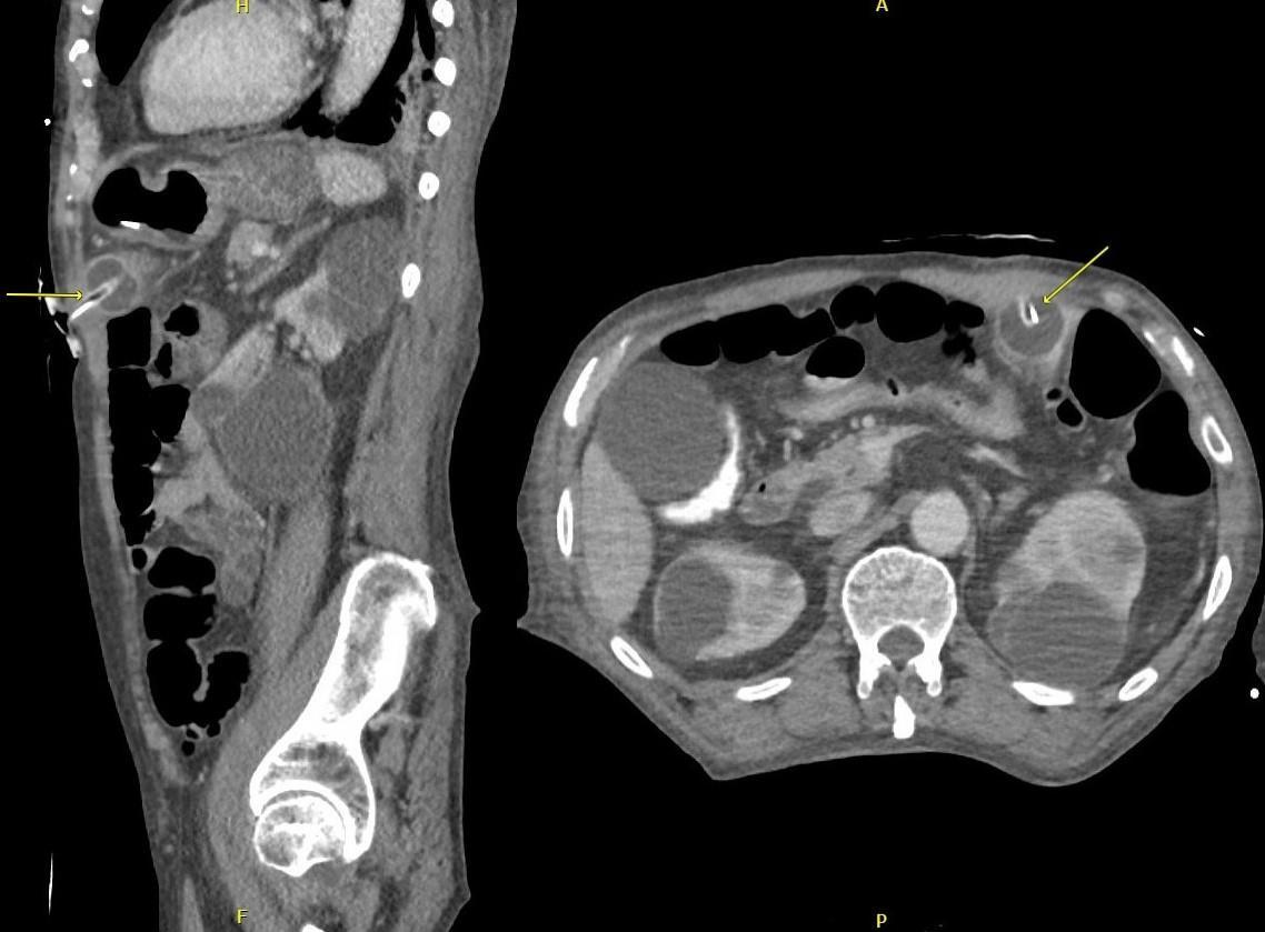
bowel on its way to the stomach.10 If the PEG tube bumper is in the gastric cavity at time of placement, patients are
generally asymptomatic until the tube either migrates into the colon from the gastric cavity or is dislodged in some manner. 3 Figure shows the mechanism of tube slippage secondary to balloon deflation. Replacement of the gastrostomy tube can expose the gastrocolocutaneous fistula if, during replacement, the tube does not follow the fistula all the way into the stomach and is inadvertently placed directly into the colon.11 Although most reports describe fistulas forming within the colon, there are reports of jejunocutaneous fistulas as well.12
This patient presented four months after initial tube placement and three months after a replacement had been done. The first replacement is suspected to have repenetrated the gastric cavity, but when the balloon bumper deflated at some point shortly before admission, the tube likely slipped back into the colon resulting in the drainage of stool-smelling liquid from the tube. The tube replacement in the ED only managed to enter back into the colon instead of the stomach. Presentation of patients with a gastrocolocutaneous fistula can be secondary to leakage of fecal contents through the PEG tube, as was the case for this patient. Patients may have the opposite problem and present with excessive diarrhea that is often correlated with PEG tube feedings. 6,8,11 Other rare presentations can be
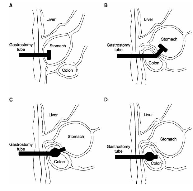
Figure. Hypothesized formation of gastrocolocutaneous fistula. (A) Normal percutaneous endoscopic gastrostomy (PEG) insertion (B) Formation of gastrocolocutaneous fistula due to errant PEG insertion. (C) Slippage of PEG tube into colon with deflation of balloon (D). Spontaneous closure of fistula opening in the stomach and PEG tube presence in the colon.15 Reproduced with author permission.
related to infection, peritonitis, and abscess formation, especially if the tube slips into or is reinserted into the peritoneal cavity and feeds are resumed.13 Long asymptomatic periods with a gastrocolocutaneous fistula have been noted, which is suspected to contribute to the lack of exposure to these malfunctions.5
Although some case reports describe unexplained migration or slippage of the PEG tube back into the colon, several report the discovery of the gastrocolocutaneous fistula to be shortly after replacement/exchange of the PEG tube8,11,12,14 This highlights the importance of correct replacement technique and procedure, especially with the risk of entering the peritoneal cavity and causing further harm. Replacement of the PEG tube is a delicate process as the tract formed with PEG is weaker than one from a surgical gastrostomy.13 While no formal guidelines for PEG replacement exists, good control into and along the tract, minimal force with insertion, and the confirmation of proper tube location are principles of a safe replacement.13 Confirmation can be done through techniques such as aspirating gastric fluid from the tube or listening for sounds when flushing air through the tube, which can be inconsistent. Radiographically confirming the location of the luminal end of the gastrostomy tube or a contrast study through the replacement tube after placement is a more accurate method that can be considered for patients with suspicious presentations, such as the feculent drainage seen in our patient.13
Due to the rarity of these cases in the literature, management has not been standardized. When a gastrocolocutaneous fistula is expected, endoscopy can be used to confirm diagnosis, but radiologic evidence using gastrograffin can be sufficient as well. Management is generally focused on removal of the PEG tube and supportive care for the patient while waiting for spontaneous closure of the fistula. There are, however, cases where endoscopic closure of the fistula was performed.15
Gastrocolocutaneous fistula should be considered in patients with feeding difficulties, unexpected PEG tube drainage, or diarrhea with feeds following tube replacement. Replacement of PEG tubes should be done with great care and if questionable, the placement should be confirmed radiologically. Endoscopic diagnosis and management can be considered for complex cases. Emergency physicians should have a high index of suspicion for a gastrocolocutaneous fistula in the event of a PEG tube with feculent-appearing drainage, difficult reinsertion or for a patient presenting with diarrhea following PEG placement.
Patient consent has been obtained and filed for the publication of this case report.
Feculent Drainage from PEG Tube
Address for Correspondence: Emmelyn J. Samones, BS, Loma Linda University Medical Center, Department of Emergency Medicine, 11234 Anderson St., Room A890A, Loma Linda, CA 92354. Email: esamones@llu.edu.
Conflicts of Interest: By the CPC-EM article submission agreement, all authors are required to disclose all affiliations, funding sources and financial or management relationships that could be perceived as potential sources of bias. The authors disclosed none.
Copyright: © 2024 Muchiutti et al. This is an open access article distributed in accordance with the terms of the Creative Commons Attribution (CC BY 4.0) License. See: http://creativecommons.org/ licenses/by/4.0/
1. Rahnemai-Azar AA, Rahnemaiazar AA, Naghshizadian R, et al. Percutaneous endoscopic gastrostomy: indications, technique, complications and management. World J Gastroenterol 2014;20(24):7739-51.
2. Pitsinis V and Roberts P. Gastrocolic fistula as a complication of percutaneous endoscopic gastrostomy. Eur J Clin Nutr 2003;57(7):876-8.
3. Patwardhan N, McHugh K, Drake D, et al. Gastroenteric fistula complicating percutaneous endoscopic gastrostomy. J Pediatr Surg. 2004;39(4):561-4.
4. Cheung SW. A silent and chronic complication of percutaneous endoscopic gastrostomy tube: small bowel enterocutaneous fistula. Case Rep Gastrointest Med. 2016;2016.
5. Kim HS, Bang CS, Kim YS, et al. Two cases of gastrocolocutaneous fistula with a long asymptomatic period after percutaneous endoscopic gastrostomy. intest Res. 2014;12(3):251.
6. Lee J, Kim J, Kim HI, et al. Gastrocolocutaneous fistula: an unusual case of gastrostomy tube malfunction with diarrhea. Clin Endosc 2018;51(2):196-200.
7. Vidal DV, Plaza FJ, David VV. Misplacement of the PEG tube through the transverse colon, an uncommon but possible complication. Rev Esp Enferm Dig. 2022;114(5):296-7.
8. Okutani D, Kotani K, Makihara S. A case of gastrocolocutaneous fistula as a complication of percutaneous endoscopic gastrostomy. Acta Medica Okayama. 2008;62(2):135-8.
9. Lenzen H, Weismüller T, Bredt M, et al. Gastrointestinal: PEG feeding tube migration into the colon; a late manifestation. J Gastroenterol Hepatol. 2012;27(7):1254.
10. Najafi K, Markowski H, Green D, et al. Managing a gastrocolocutaneous fistula with delayed presentation after PEG placement without surgery. J Case Rep. 2023;13(1):7-10.
11. Jamma S, Tippor S, Reddy JA. Colocutaneous fistula causing diarrhea: a complication of PEG tube replacement: 420. Am J Gastroenterol. 2007;102:S269.
12. Karhadkar AS, Schwartz HJ, Dutta SK. Jejunocutaneous fistula manifesting as chronic diarrhea after PEG tube replacement. J Clin Gastroenterol. 2006;40(6):560-1.
13. Lohsiriwat V. Percutaneous endoscopic gastrostomy tube replacement: a simple procedure? World J Gastrointest Endosc 2013;5(1):14-8.
14. Marcy P, Magne N, Lacroix J, et al. Late presentation of a gastrocolic fistula after percutaneous fluoroscopic gastrostomy. JBR BTR 2004;87(1):17-20.
15. Jung TY, Lee JR, Seok DK, et al. Endoscopic management for colocutaneous fistula as a complication of percutaneous endoscopic gastrostomy. Korean J Helicobacter Up Gastrointest Res. 2013;13(2):128-31.
Beth Kreutzer, MD
Blake Buehrer, PA
Andrew Pelikan, MD
Phillip Rohde, MD
Section Editor: Lev Libet, MD
University of Missouri, Department of Emergency Medicine, Columbia, Missouri
Submission history: Submitted March 25, 2024; Revision received August 1, 2024; Accepted July 25, 2024
Electronically published September 29, 2024
Full text available through open access at http://escholarship.org/uc/uciem_cpcem DOI: 10.5811/cpcem.20522
Introduction: Wernicke encephalopathy is a clinical diagnosis that requires a high degree of clinical suspicion to recognize. We report a case of a pregnant patient developing Wernicke encephalopathy in the setting of severe hyperemesis gravidarum.
Case Report: The patient was a 22-year-old female 13 weeks pregnant presenting to the emergency department (ED) with neurological deficits after several weeks of hyperemesis gravidarum requiring hospitalization. Exam and workup ultimately revealed the diagnosis of Wernicke encephalopathy. Her symptoms improved after administration of thiamine.
Conclusion: Wernicke encephalopathy is a consequence of thiamine deficiency, commonly seen in patients with alcohol use disorder but also with other causes of nutritional deficiency, such as hyperemesis gravidarum. Wernicke encephalopathy is a clinical diagnosis that requires a high degree of suspicion and is, therefore, often missed in the ED setting. Treatment is supplemental thiamine and management of the root cause for nutritional deficiency. [Clin Pract Cases Emerg Med. 2024;8(4):357–360.]
Keywords: Wernicke; hyperemesis; pregnancy, thiamine; case report.
The emergency department (ED) manages a large subset of patients who are malnourished to some degree. Critical dietary deficiencies that may be contributing to the presentation pose a diagnostic challenge. Wernicke encephalopathy, caused by thiamine deficiency, is one of those clinical diagnoses that is difficult to diagnose without a high degree of clinical suspicion Classically, Wernicke encephalopathy is associated with alcohol use disorder; however, any person who is malnourished is at risk for developing this deficiency. Early diagnosis and treatment are imperative to prevent Korsakoff syndrome. Here we discuss a patient who was hospitalized with severe hyperemesis gravidarum and after discharge returned to the ED with neurological symptoms consistent with Wernicke encephalopathy.
A 22-year-old female, gravida 1 para 0 at 13 weeks estimated gestational age, presented to the ED complaining
of weakness and confusion. She had fallen in the shower and was unable to ambulate independently. She had blurry vision, difficulty focusing, bulging eyes, headaches, and dizziness. She was intermittently confused and making nonsensical statements. Two weeks prior she had been admitted to the obstetrics service with a urinary tract infection and hyperemesis gravidarum. She was treated with ceftriaxone, intravenous (IV) fluids, and antiemetics and discharged on hospital day five with cefdinir and multiple antiemetics. She had noticed development of these symptoms while still hospitalized.
Prior to the pregnancy, she was healthy without any significant medical concerns. Medications at time of evaluation included cefdinir, doxylamine, ondansetron, pyridoxine, and loratadine. She had no surgical history.
On exam the patient was alert, interactive, and in no acute distress. Vital signs included temperature 36.2° Celsius, respiratory rate 20 breaths per minute, heart rate 109
beats per minute, blood pressure 109/77 millimeters of mercury, and pulse oximetry 100% on room air. She was alert and fully oriented but had repetitive questioning and made frequent nonsensical statements. She had full range of motion in all extremities with normal strength and sensation to upper and lower extremities bilaterally. Her gait was ataxic. She had restricted horizontal conjugate gaze bilaterally and decreased hearing to the left ear. She had no other cranial nerve deficits. Cardiac, pulmonary, and abdominal portions of the exam were unremarkable. She had no rashes, wounds, or lesions on skin exam.
On complete blood count the patient was mildly anemic with a hematocrit of 30.1% (reference range 36.0-48.0%), similar as compared to previous. Thyroid stimulating hormone level was 0.014 milliunits per liter (mU/L) (0.4-4.0 mU/L) with free T3 level of 1.28 picograms per milliliter (pg/mL) (1.2-2.7 pg/mL). Complete metabolic panel, magnesium, urinalysis, and drug screens were unremarkable. Computed tomography of the head and neck demonstrated no acute intracranial abnormality. A lumbar puncture was performed with an opening pressure of eight centimeters of water, with no cerebrospinal fluid abnormalities. Magnetic resonance imaging of the brain showed T2 FLAIR signal hyperintensity within the bilateral medial thalami, mammillary bodies, and periaqueductal gray matter, a pattern consistent with Wernicke encephalopathy (Images 1 and 2). Subsequent testing revealed a thiamine level of 29 nanomoles (nmol)/L (70-180 nmol/L).
The patient was admitted to the neurology team for high-dose IV thiamine therapy. She had overall improvement and was discharged on hospital day four with oral thiamine, vitamin D, and prenatal vitamin supplements. She has since given birth to a healthy baby girl and has made a full neurologic recovery.
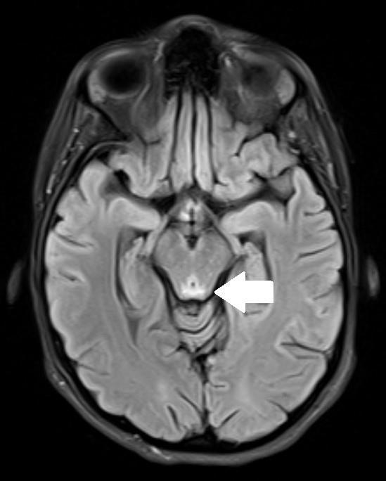
CPC-EM Capsule
What do we already know about this clinical entity?
Wernicke encephalopathy is a consequence of thiamine deficiency, usually related to alcohol use disorder and as a complication of gastric bypass surgery.
What makes this presentation of disease reportable?
Only one previous case report to our knowledge describes the emergency department presentation of Wernicke encephalopathy associated with hyperemesis gravidarum.
What is the major learning point?
Diagnosis of Wernicke encephalopathy requires a high degree of clinical suspicion and should be considered in any malnourished patient with new neurological symptoms.
How might this improve emergency medicine practice?
Early recognition and treatment initiation by emergency physicians is crucial to clinical improvement and prevention of sequelae such as Korsakoff syndrome.
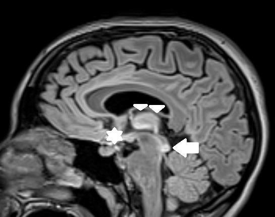
Wernicke encephalopathy is an acute manifestation of thiamine (vitamin B1) deficiency. Thiamine is a coenzyme
Kreutzer et al. Wernicke Encephalopathy Associated with Hyperemesis Gravidarum
essential to all cells, but it is particularly important for neurons.1,2 Carl Wernicke first described this encephalopathy in 1881, and in 1940 Campbell and Russell hypothesized thiamine deficiency as the cause.3,4 Development of brain lesions occurs in regions that have higher demands for thiamine: namely neurons in the thalami, mammillary bodies, tectal plate, and the periaqueductal region.1 Some patients who survive their acute encephalopathic episode without treatment go on to develop Korsakoff syndrome, a chronic form of thiamine deficiency that results in severe deficits in memory.4,5 Other forms of thiamine deficiency include dry and wet beriberi.
Wernicke encephalopathy is classically associated with people who are chronically malnourished secondary to excessive alcohol intake.2 However, any condition that leads to malnourishment increases the risk for development of vitamin deficiencies.4 Pregnancy is associated with increased demand for thiamine.6 There are numerous case reports describing Wernicke encephalopathy in pregnant patients, postulated to be caused by increased metabolic demand from the pregnancy coupled with inadequate intake due to hyperemesis gravidarum.7,8 However, the literature is sparse regarding diagnosis of Wernicke encephalopathy in the emergency setting, with only one other published case report.9
Subclinical thiamine deficiency presents with non-specific symptoms, including headaches, fatigue, abdominal discomfort, and weakness.4 Acute deficiency may lead to the classic triad of symptoms: mental status changes, ocular abnormalities (ophthalmoplegia, nystagmus, gaze palsy), and cerebellar abnormalities including ataxia. While only a minority of patients present with the classic triad, our patient presented with all three. Less common symptoms include hypothermia, seizures, hearing loss, and hallucinations.4 Late-stage symptoms include hyperthermia, spastic paresis, chorea, coma, and death.4
Diagnosis of thiamine deficiency requires a high degree of suspicion, as symptoms may be vague. Wernicke encephalopathy is a clinical diagnosis and should be suspected in any malnourished patient presenting with suggestive neurological symptoms. The Caine criteria have been proposed to help predict the likelihood of Wernicke encephalopathy, requiring at least two of the four following findings to support the diagnosis: dietary deficiencies, ocular signs, cerebellar dysfunction, and altered mentation or memory impairment.10,11 While serum thiamine levels may be measured, these measurements may not be reliable.12,13 Magnetic resonance is the imaging of choice for diagnosis of Wernicke encephalopathy, as it shows distinct patterns of alterations in the typical regions affected by the deficiency.14,15
Treatment for Wernicke encephalopathy is thiamine repletion. Standard prenatal vitamins have 1-2 milligrams thiamine, which would be insufficient in the pregnant patient who develops Wernicke encephalopathy. Therefore, pregnant patients who are struggling with hyperemesis should be treated with additional thiamine supplementation.
Wernicke encephalopathy is a consequence of acute thiamine deficiency, often associated with alcohol use disorder but can occur with any cause of nutritional deficiency. There are several case reports describing patients with hyperemesis gravidarum who developed Wernicke encephalopathy, but only one report regarding ED presentation and diagnosis. Wernicke encephalopathy is a clinical diagnosis that requires a high degree of suspicion; therefore, it should be suspected in any patient who is malnourished and displaying neurologic symptoms.
The authors attest that their institution requires neither Institutional Review Board approval, nor patient consent for publication of this case report. Documentation on file.
Address for Correspondence: Beth Kreutzer, MD, Department of Emergency Medicine, University of Missouri, 1 Hospital Drive, Columbia, MO 65212. Email: drkleinherr@gmail.com
Conflicts of Interest: By the CPC-EM article submission agreement, all authors are required to disclose all affiliations, funding sources and financial or management relationships that could be perceived as potential sources of bias. The authors disclosed none.
Copyright: © 2024 Kreutzer et al. This is an open access article distributed in accordance with the terms of the Creative Commons Attribution (CC BY 4.0) License. See: http://creativecommons.org/ licenses/by/4.0/
1. Manzo G, De Gennaro A, Cozzolino A, et al. MR imaging findings in alcoholic and nonalcoholic acute Wernicke’s encephalopathy: a review. Biomed Res Int. 2014;2014:503596.
2. Harper CG, Giles M, Finlay-Jones R. Clinical signs in the WernickeKorsakoff complex: a retrospective analysis of 131 cases diagnosed at necropsy. J Neurol Neurosurg Psychiatry. 1986;49(4):341-5.
3. Wernicke C. Die akute haemorrhagische polioencephalitis superior. In: Lehrbuch der Gehirnkrankheiten fur Aerzte und Studirende 1881;2:229-42.
4. Sechi GP and Serra A. Wernicke’s encephalopathy: new clinical settings and recent advances in diagnosis and management. Lancet Neurol. 2007;6(5):442-55.
5. Kopelman MD, Thomson AD, Guerrini I, et al. The Korsakoff syndrome: clinical aspects, psychology, and treatment. Alcohol Alcohol. 2009;44(2):148-54.
6. Frank LL. Thiamin in clinical practice. J Parenter Enteral Nutr. 2015;39(5):503-20.
7. Chiossi G, Neri I, Cavazzuti M, et al. Hyperemesis gravidarum complicated by Wernicke’s encephalopathy: background, case report
Wernicke Encephalopathy Associated with Hyperemesis Gravidarum
Kreutzer et al. and review of the literature. Obstet Gynecol Surv. 2006;61(4):255-68.
8. Rane MA, Boorugu HK, Ravishankar U, et al. Wernicke’s encephalopathy in women with hyperemesis gravidarum – case series and literature review. Trop Doct. 2022;52(1):98-100.
9. Meggs WJ, Lee SK, Parker-Cote JN. Wernicke encephalopathy associated with hyperemesis gravidarum. Am J Emerg Med 2020;38(3):690.e3-690.e5.
10. Galvin R, Brathen G, Ivashynka A, et al. EFNS guidelines for diagnosis, therapy, and prevention of Wernicke encephalopathy. Eur J Neurol. 2010;17(12):1408-18.
11. Caine D, Halliday GM, Kril JJ, et al. Operational criteria for the classification of chronic alcoholics: identification of Wernicke’s encephalopathy. J Neurol Neurosurg Psychiatry. 1997;62(1):51-60.
12. Victor M, Adams RD, Collins GH. The Wernicke-Korsakoff Syndrome, 2nd Ed; Philadelphia, PA: F.A. Davis Company, 1989.
13. Davies SB, Joshua FF, Zagami AS. Wernicke’s encephalopathy in a non-alcoholic patient with a normal blood thiamine level. Med J Aust. 2011;194(9):483-4.
14. Zuccoli G and Pipitone N. Neuroimaging findings in acute Wernicke’s encephalopathy: review of the literature. AJR Am J Roentgenol 2009;192(2):501–8.
15. Chung SP, Kim SW, Yoo IS, et al. Magnetic resonance imaging as a diagnostic adjunct to Wernicke’s encephalopathy in the ED. Am J Emerg Med. 2003;21(6):497-502.
Gavriel Rosenfeld-Barnhard, MD
Jessica R. Jackson, MD
Kendra A. Mendez, MD
Kraftin E. Schreyer, MD, MBA
Section Editor: Lev Libet, MD
Temple University Hospital, Department of Emergency Medicine, Philadelphia, Pennsylvania
Submission history: Submitted March 14, 2024; Revision received August 18, 2024; Accepted August 30, 2024
Electronically published November 9, 2024
Full text available through open access at http://escholarship.org/uc/uciem_cpcem
DOI: 10.5811/cpcem.20348
Introduction: Aortic dissection is a devastating clinical entity with a variety of presentations and requires prompt recognition and management. To our knowledge this is the first reported case of a patient who presented with a globus sensation and was diagnosed with an aortic dissection prior to clinical deterioration.
Case Report: The patient presented with an episode of near-syncope and globus sensation. Imaging studies revealed a type A aortic dissection with hemopericardium requiring emergent operative intervention. Unfortunately, the patient’s course was complicated by significant hemorrhage and periods of hypotension, and the family ultimately decided to pursue comfort care.
Conclusion: Aortic dissections can present with diverse and elusive symptoms, which can mimic other more common conditions, potentially leading to misdiagnosis and delayed intervention [Clin Pract Cases Emerg Med. 2024;8(4):361–364.]
Keywords: aortic dissection; globus sensation; case report.
Acute aortic dissection is one of the most devastating cardiovascular pathologies encountered in the emergency department (ED), with high levels of morbidity and mortality. Due to the potential rapid clinical deterioration, prompt recognition of this condition and appropriate timely diagnostic evaluation and treatment are crucial. The widely taught clinical presentation of aortic dissection is sharp, tearing chest pain that radiates to the back. In clinical practice, however, there is a wide range of signs and symptoms of aortic dissection including syncope, hypotension, and focal neurologic deficits. We describe an uncommon presentation of a type A aortic dissection in which the patient’s primary clinical symptom on initial presentation to the ED was a globus sensation.
A 60-year-old male presented to the ED after an episode
of near-syncope associated with diaphoresis while walking to church, with emergency medical services reporting low blood pressure at the scene. On arrival, he endorsed a tight sensation in his throat but otherwise denied chest pain, shortness of breath, back pain, nausea, or vomiting. He had a medical history of hyperlipidemia, well-controlled hypertension on amlodipine and remote history of prostate cancer status post radical prostatectomy but was otherwise unremarkable. He had a remote smoking history of 10 pack-years but otherwise no drug use.
On examination, the patient was alert and oriented and in no distress. Initial vital signs were temperature 36.7° Celsius, heart rate 72 beats per minute, blood pressure 105/72 millimeters of mercury (mm Hg) on the left arm, and respiratory rate 16 breaths per minute with an oxygen saturation of 98% on room air. Initial examination was notable for regular rate and rhythm with a normal S1 and S2, lungs were clear to auscultation bilaterally, and his abdomen
was benign. Radial and dorsalis pedis pulses were 2+ bilaterally. The neurologic exam was normal with symmetric strength in his bilateral upper and lower extremities without sensory deficits. The neck exam was notable for a midline trachea, no palpable masses. He also had an unremarkable oropharyngeal exam.
Diagnostic workup was notable for an initial electrocardiogram that was normal sinus rhythm with a normal rate, normal intervals, and no evidence of ischemic changes or arrythmias. A chest radiograph showed a mildly enlarged heart size and abnormal contour of the thoracic aorta. While awaiting laboratory studies, point-of-care ultrasound showed an enlarged left ventricular outflow tract (LVOT) measuring 4.4 centimeters (cm) (reference range: 1.6-2.4 cm)and evidence of a pericardial effusion without evidence of tamponade (Image 1).
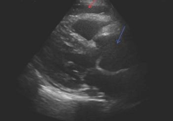
These ultrasound findings were highly suspicious for acute aortic pathology and prompted an emergent computed tomography angiography (CTA) of the chest, abdomen, and pelvis, which showed an intramural hematoma of the aortic root extending along the right pulmonary arterial wall and along the aorta to the aortic arch; dilation of the ascending aorta with a dissection flap with associated inflammatory changes of the mediastinum; and aneurysmal dilation of the infrarenal abdominal aorta, measuring 3.1 cm in the midportion and 3.4 cm just proximal to the bifurcation (Image 2).
While imaging was being obtained, blood tests began to result, which were notable for a white blood cell count of 13.7 x 10³ per cubic millimeter (K/mm3) (reference range: 4.0-11.0 K/mm3), hemoglobin 14.2 grams per deciliter (g/ dL) (14.0-17.5 g/dL), platelets 328 K/mm3 (150-450 K/ mm3), creatinine 1.44 milligrams (mg)/dL (mg/dL) (0.801.30 mg/dL) (last from one year prior 1.01 mg/dL), and high sensitivity troponin I 12 nanograms per liter (ng/L) (12-76 ng/L [males]). With concern for an acute aortic syndrome after the initial ultrasound was performed, a D-dimer was later obtained, which was elevated at 1,846 ng/mL (0-500 ng/mL). Otherwise, all other labs were within normal limits.
CPC-EM Capsule
What do we already know about this clinical entity?
Aortic dissection is a life-threatening condition with diverse symptoms, often requiring quick recognition and management to prevent deterioration.
What makes this presentation of disease reportable?
This is the first reported case of an acute aortic syndrome presenting with painless globus sensation, diagnosed before clinical deterioration.
What is the major learning point?
Atypical presentations like globus sensation in aortic dissection require high suspicion, as early diagnosis and management are crucial to improving outcomes.
How might this improve emergency medicine practice?
Clinicians must be vigilant in recognizing nontextbook presentations of critical conditions to improve early detection and treatment.
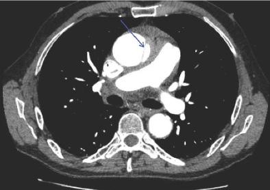
Given the CTA findings, the cardiothoracic surgery team was emergently consulted with concern for type A aortic dissection. While awaiting surgical recommendations, the patient subsequently developed sudden onset lower back pain associated with nausea, vomiting, and diaphoresis. At this time the patient was noted to have discrepant blood pressures. A right-sided radial arterial line was placed that showed a blood pressure of 64/58 mm Hg, and a left arm automated blood
Rosenfeld-Barnhard et
al. Painless Aortic Syndrome
pressure cuff read 154/105 mm Hg. The patient was given 4 mg intravenous (IV) ondansetron, 6 mg of IV morphine, and 800 micrograms (μg) of sublingual nitroglycerine. The patient was subsequently started on esmolol and nitroprusside at an initial rate of 50 μg per kilogram per minute (kg/min) and 0.5 μg/kg/min, respectively, for blood pressure and heart rate management. The patient then reported loss of sensation in his right lower extremity. Re-examination revealed total loss of motor function and no palpable femoral pulses.
A repeat point-of-care ultrasound showed an abdominal aortic flap concerning for propagation of the aortic dissection. The patient was taken emergently to the operating room and, intraoperatively, was noted to have a dissection extending 4 cm above the valve and coronaries. A transesophageal echocardiogram was also performed that showed a tricuspid aortic valve and redemonstrated the dissection extending from the ascending through descending aorta. After a prolonged surgery complicated by hemorrhagic shock, the family opted for comfort care measures on the following day.
An aortic dissection refers to the development of a focal tear in the intimal layer of the aortic wall that results in extension of the tear compromising blood flow to vital organs.The Stanford classification is a well-established system of describing aortic dissections: type A dissection refers to dissections that involve any part of the aorta proximal to the origin of the left subclavian artery and which may also include the descending aorta, while a type B dissection refers to dissections involving the descending aorta or the arch (distal to the left subclavian artery), without the involvement of the ascending aorta. Both entities are relatively rare diagnoses with an annual incidence of approximately 3.5 per 100,000 patients.1 Because of the significant morbidity and mortality associated with this condition, a high degree of clinical suspicion is required to make the diagnosis. Large systematic reviews have shown that over 80% of patients found to have an aortic dissection present with complaints either of chest or back pain, and about 10% are painless or may present with symptoms secondary to complications of the dissection.2,3 The International Registry of Acute Aortic Dissections reports that syncope was present in nearly 20% of type A aortic dissections and is often associated with a severe complication such as cardiac tamponade.4,5 Due to the difficulty of diagnosis, emergency physicians only suspected aortic dissection in 43% of confirmed cases.6
There are only a few case reports describing throat tightness as the initial presentation of patients found to have aortic dissections. In a 2004 case report by Liu and Ng, a patient presented to the ED with acute-onset throat pain and was subsequently discharged after symptomatic management. The patient returned to the ED later that day with worsening sore throat, severe chest pain, diaphoresis, and a syncopal episode and was found to have a type A aortic dissection.7 Another
case report by Cates et al describes a 58-year-old patient who initially presented with burning throat pain that eventually progressed to chest pain and right lower extremity cramping. The change in the patient’s symptoms prompted a CTA, which revealed the diagnosis of a type A aortic dissection.8
There is also a phenomenon in the literature called “dysphagia aortica,” which is defined as difficulty in swallowing caused by extrinsic compression of the esophagus due to an ectatic, tortuous, or aneurysmatic atherosclerotic thoracic aorta.9 This rare clinical entity is mostly described in case reports and small case studies. Most of the cases are descriptions of patients with weeks to months of dysphagia who, in the process of extensive outpatient workups, are found to have large thoracic aortic aneurysms.10,11 Although the pathophysiology is not well understood, some case reports suggest external esophageal compression from the aneurysm as the potential cause of the dysphagia. Dilated aortic aneurysms can compress the esophagus, recurrent laryngeal nerve, or superior cervical sympathetic ganglion that in turn can cause dysphagia, hoarseness, or Horner syndrome.12 Our case is unique in that the patient acutely developed symptoms suggestive of extrinsic compression of mediastinal structures related to his dissection and dilated aorta.
Our case underscores the critical need for emergency physicians to remain vigilant and open to atypical clinical presentations of aortic dissection. Aortic dissections can present with diverse and elusive symptoms, which can mimic other, more common conditions, potentially leading to misdiagnosis or delayed intervention. In our case as well as has been described in prior case reports, although a sore throat was the initial presenting symptom, advanced imaging diagnosing an aortic dissection was not obtained until after the patient’s clinical deterioration or development of other symptoms. To our knowledge this is the first reported case of a patient who presented with a globus sensation and was diagnosed with an aortic dissection prior to clinical deterioration. This case emphasizes the vital role of clinical acumen and the importance of maintaining a high index of suspicion, even when faced with non-textbook presentations. While the point-of-care echocardiogram was key to obtaining the diagnostic CTA, recognizing the subtle, unconventional signs of aortic dissection is paramount to ensure early diagnosis and timely management, which can significantly impact patient outcomes and save lives.
Due to the significant morbidity and mortality of aortic dissections, particularly of type A dissections, prompt recognition and management is essential. It is crucial to recognize the wide range of initial patient presentations to ensure early identification of this emergent pathology. This case underscores the importance of maintaining a high index of suspicion for aortic dissections and abnormal ultrasound findings, particularly in patients with known risk factors
Painless Aortic Syndrome Rosenfeld-Barnhard et al.
and non-specific symptom presentations that do not fit the “textbook” presentation.
At their institution, case studies are not classified as human subjects research, and therefore, IRB approval was not required for this report. Attestation documentation on file.
Address for Correspondence: Gavriel Rosenfeld-Barnhard, MD, Temple University Hospital, Department of Emergency Medicine, 401 N Broad Street, Philadelphia, Pennsylvania, 19140. Email: gavriel.rosenfeld-barnhard@tuhs.temple.edu.
Conflicts of Interest: By the CPC-EM article submission agreement, all authors are required to disclose all affiliations, funding sources and financial or management relationships that could be perceived as potential sources of bias. The authors disclosed none.
Copyright: © 2024 Rosenfeld-Barnhard et al. This is an open access article distributed in accordance with the terms of the Creative Commons Attribution (CC BY 4.0) License. See: http:// creativecommons.org/licenses/by/4.0/
1. Clouse WD, Hallett JW Jr, Schaff HV, et al. Acute aortic dissection: population-based incidence compared with degenerative aortic aneurysm rupture. Mayo Clin Proc. 2004; 79(2):176-80.
2. Spittell PC, Spittell JA, Joyce JW, et al. Clinical features and differential diagnosis of aortic dissection: experience with 236 cases
(1980 through 1990) Mayo Clinic Proc. 1993;68(7):642–51.
3. Khan IA, Wattanasauwan N, Ansari AW. Painless aortic dissection presenting as hoarseness of voice: cardiovocal syndrome: Ortner’s syndrome. Am J Emerg Med. 1999;17(4):361-3.
4. Evangelista A, Isselbacher EM, Bossone E, et al. Insights from the International Registry of Acute Aortic Dissection: a 20year experience of collaborative clinical research. Circulation 2018;137(17):1846-1860
5. Yuan X, Mitsis A, Nienaber CA. Current understanding of aortic dissection. Life. 2022;12(10):1606.
6. Sullivan PR, Wolfson AB, Leckey RD, et al. Diagnosis of acute thoracic aortic dissection in the emergency department. Am J Emerg Med. 2000;18(1):46–50
7. Liu WP and Ng KC. Acute thoracic aortic dissection presenting as sore throat: report of a case. Yale J Biol Med. 2004;77(3-4):53-8.
8. Cates AL, Leriotis T, Herres J. A case of burning throat pain. J Emerg Med. 2019 Sep;57(3):e69-e71.
9. Grimaldi S, Milito P, Lovece A, et al. Dysphagia aortica. Eur Surg 2022;54(5):228-239.
10. Badila E, Bartos D, Balahura C, et al. A rare cause of Dysphagia - dysphagia aortica - complicated with intravascular disseminated coagulopathy. Maedica (Bucur) 2014;9(1):83-7.
11. Wang WP, Yan XL, Ni YF, et al. An unusual cause of dysphagia: thoracic aorta aneurysm. J Thorac Dis. 2013;5(6):E224-6.
12. Ramanath VS, Oh JK, Sundt TM, et al. Acute aortic syndromes and thoracic aortic aneurysm. Mayo Clin Proc. 2009;84:465.
Jeffrey M. Kalczynski, DO*
John Douds, BS†
Michael E. Silverman, MD*
Section Editor: Shadi Lahham, MD
Sidney Kimmel Medical College, Philadelphia, Pennsylvania * †
Morristown Medical College, Department of Emergency Medicine, Morristown, New Jersey
Submission history: Submitted July 8, 2024; Revision received September 9, 2024; Accepted September 5, 2024
Electronically published: October 22, 2024
Full text available through open access at http://escholarship.org/uc/uciem_cpcem DOI: 10.5811/cpcem.21189
Introduction: We present a unique case of a patient who presented to the emergency department with stroke-like symptoms found to have a spontaneous, left-sided internal carotid artery dissection (ICAD).
Case Report: The patient was treated successfully with thrombectomy and subsequently developed contralateral symptoms caused by a right-sided ICAD. This was managed with a second contralateral thrombectomy. The patient’s course was complicated by persistent and mild hypotension, postulated to be secondary to bilateral carotid baroreceptor trauma from the dissections.
Conclusion: This case highlights the importance of close neurological monitoring for patients, preferably in a neurologic critical care setting, during and after invasive treatments such as systemic thrombolytic administration or mechanical thrombectomy. In this case, identifying the patient’s subsequent development of contralateral symptoms in a timely fashion was key to his positive outcome. An additional factor that had a positive impact on this outcome was the use of artificial intelligence software, which assists in determining whether thrombectomy may be indicated prior to receiving a formal radiologist read on computed tomography angiography/perfusion studies. Artificial intelligence technology such as this has great potential to augment and expedite patient care. [Clin Pract Cases Emerg Med. 2024;8(4):365–368.]
Keywords: stroke; carotid dissection; artificial intelligence; case report.
As a major cause of death in the United States, and a significant cause of disability, stroke management is critically important to the practice of emergency medicine. Ischemic stroke due to arterial dissection represents a subset of pathology that can cause large vessel occlusions, a devastating condition with major potential impacts to morbidity and mortality. The pathophysiology of internal carotid artery dissection (ICAD) involves a defect in the intima of the arterial wall, which can develop spontaneously as the result of trauma or due to multifactorial causes linked to cardiovascular risk factors.1 This defect allows blood products to enter the space between the intima and the media, expanding the intima into the vessel’s lumen, which reduces blood flow and can lead to thromboemboli.2 Internal carotid artery dissection
is a major cause of morbidity and mortality in young to middle-aged patients, accounting for approximately 25% of ischemic strokes in these populations.1 The incidence rates for spontaneous ICAD “have been previously reported to be 2.62.9 per 100,000.”3
We report the case of a patient who suffered an atraumatic, spontaneous ICAD, which was treated with thrombectomy, and who then subsequently developed contralateral symptoms. Upon reevaluation in the neurological intensive care unit (ICU), the patient was found to have a secondary contralateral, atraumatic, spontaneous ICAD. There are no reported cases like this in the literature to our knowledge. This case emphasizes the importance of close monitoring and reassessment of critically ill patients in the neurologic critical care setting, as well as the aggressive
nature of diagnosis and treatment that this subset of patients requires.1,4 Interestingly, our patient developed persistent mild hypotension after his bilateral dissections, presumably secondary to baroreceptor trauma from the dissection. There is a report of dysautonomia after bilateral dissections,5 and others with stroke-like symptoms and dysarthria,3,6-8 but none with isolated persistent hypotension.
This is a case of a 60-year-old male with a history of hyperlipidemia presenting to the emergency department complaining of right-sided paresthesias and clumsiness as well as progressive word-finding difficulties with an onset time of 37 minutes prior to evaluation. The patient’s wife provided the history as he was only able to say the word “yes” when questioned. Blood glucose was within normal limits, and an initial National Institutes of Health Stroke Scale was as follows: 8 Total (2: Loss of Consciousness questions, 2: Loss of Consciousness Commands, 1: Sensory, 2: Best Language, 1: Extinction and Inattention.) Code stroke was immediately activated, and a non-contrast computed tomography (CT) of the head was without signs of hemorrhage. A CT angiography
Table. Timeline of events from initial presentation to second thrombectomy.
Time Event
06:45 am Symptom onset
07:12 am Patient presentation to emergency department
07:22 am Code stroke activation
07:45 am Normal CT head prelim read by radiology
08:02 am Tenecteplase administration
08:10 am Perfusion abnormality noted by AI software
08:12 am Neurosurgery consulted
09:15-10:30 am Thrombectomy for left ICAD
12:30 pm Development of contralateral symptoms
12:30 pm Thrombectomy for right ICAD
3:00 pm Hypotension identified
3:15 pm Norepinephrine bitartrate support started
8:30 am (next day) Transitioned to midodrine
AI, artificial intelligence; CT, computed tomography; ICAD, internal carotid artery dissection.
CPC-EM Capsule
What do we already know about this clinical entity?
Internal carotid artery dissection (ICAD) can cause cerebral ischemia and neurological symptoms. It is frequently treated with mechanical thrombectomy.
What makes this presentation of disease reportable?
This patient suffered ICAD twice in one day on opposite sides of the body.
What is the major learning point?
Patients in the emergency department require close monitoring and reassessment, especially during and after interventions such as thrombolytic therapy and endovascular treatments.
How might this improve emergency medicine practice?
Artificial intelligence programs can be integrated with standard emergency care to improve patient outcomes.
of the head and neck with perfusion was concurrently performed and revealed a large, left-sided ischemic penumbra. There were no exclusions to thrombolytic therapy, and tenecteplase was administered. There was a significant improvement in neurological function five minutes after tenecteplase, but a repeat exam shortly after revealed return of symptoms. Artificial intelligence (AI) software (Rapid AI, San Mateo, CA) indicated that a mechanical thrombectomy could have been indicated; neurosurgery was consulted and agreed that thrombectomy was indicated. The patient was taken to the neurosurgery suite, where a left-sided mechanical thrombectomy was performed. He was found to have suffered a left-sided ICAD.
Shortly after arriving in the neurological ICU status postmechanical thrombectomy, the patient was noted to have developed new left-sided neurological symptoms, prompting repeat CT angiography with perfusion, which revealed a new right-sided perfusion deficit (Image 2). The patient was urgently returned to the thrombectomy suite for contralateral thrombectomy and was found to have suffered a spontaneous, right-sided ICAD.
On return to the neurological ICU, he was again with improved neurological function, but with persistent mild hypotension requiring vasopressor initiation. He was eventually transitioned to midodrine and was subsequently discharged to subacute rehab. It is theorized that the hypotension was secondary to bilateral carotid baroreceptor
Kalczynski et al. Internal Carotid Artery Dissection damage after the bilateral dissections.
The sequential, bilateral ICADs presented in this case represent a rare presentation. Unilateral headache and stroke-like symptoms have been reported before—once in a 43-year-old female found to have bilateral ICADs as a result of trauma3 and once in a 38-year-old female with spontaneous bilateral ICADs.6 Other presentations of ICAD including dysphagia, hoarseness, and dysarthria have been reported in middle- aged males.7,8
Uniquely, our patient developed persistent mild hypotension status post-bilateral mechanical thrombectomy of the carotid arteries, requiring vasopressor support. While dysautonomia involving episodic bradycardia with hypotension has been previously reported in a 49-year-old female with bilateral ICADs,5 there have been no previously reported cases of persistent dysautonomia as in this case we report. The prevailing theory is that damage to the carotid baroreceptors occurred bilaterally, either secondary to the dissections or iatrogenically by the mechanical thrombectomies, with resultant thromboemboli.
The carotid baroreceptors located in the carotid sinuses are responsible for sensing blood pressure via stretch receptors and afferently transmitting these signals along the glossopharyngeal nerve to the midbrain; then the midbrain sympathetically alters heart rate and contractility to adjust pressure accordingly, a process known as the carotid sinus baroreflex.9 Baroreceptors are also present in the aortic arch, which monitor pressure and transmit signals via the vagus nerve to the medulla, working in conjunction with the carotid sinuses to achieve homeostasis; these baroreceptors are thought to be more sensitive to increased pressure, while the carotid sinuses are sensitive to both increased and decreased pressure.10 Therefore, if the carotid sinuses were rendered inoperative, as proposed in this patient, the aortic arch baroreceptors may have been unable to initially sense the change and induce a compensatory response, resulting in persistent mild hypotension. In this case, the patient’s hypotension initially required pressor support with low-dose norepinephrine bitartrate, which was eventually titrated down, and the patient was switched to oral midodrine for further outpatient management.
The CT angiogram with perfusion studies obtained for this patient’s diagnostic evaluation were critical in the timely recognition and treatment of the pathology. The perfusion studies were particularly useful in identifying the brain tissue receiving reduced blood flow as a result of each individual ICAD. The initial perfusion study, as shown in Image 1, depicts 30 slices of the patient’s brain from the CT angiography on the left side, while the right side depicts the same slices with a green overlay depicting areas with reduced perfusion as identified by AI.11
The repeat CT angiography with perfusion study in Image 2 reflects the perfusion defects on the contralateral side at the time of the patient’s second spontaneous ICAD.
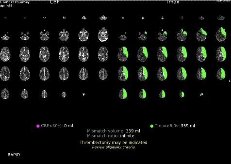
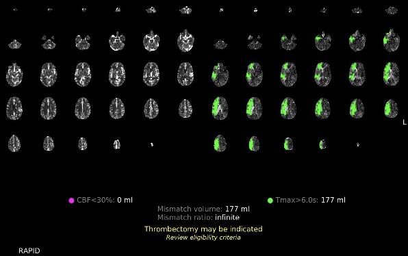
These AI-generated overlays assisted the emergency, neurological, and neurosurgical teams in early identification of a perfusion deficit of the regions experiencing reduced perfusion, and of the location of the perfusion deficit’s source. The integration of AI software into the evaluation of radiological imaging studies is becoming increasingly common.12 The development of AI with the ability to analyze CT imaging has been studied13 and has been deemed noninferior in its evaluation of brain ischemia.14 Furthermore, AI software integrated into imaging systems has been shown to improve the ability of non-experts to evaluate imaging.15 The RapidAI software we used with this patient analyzed the CT angiogram with perfusion study and determined whether thrombectomy was indicated. In Images 1 and 2, the decision made by the AI software can be seen in yellow lettering at the bottom of the studies, indicating that the software correctly identified the need for thrombectomy in both studies. This software allows the emergency physicians and neurosurgical teams to coordinate care with interventional neuroradiology in preparation for thrombectomy, even before radiology had
provided an official read of the images. The integration of AI software has the potential to decrease time to thrombectomy in patients with perfusion deficits and lead to improved patient outcomes.
This case is notable for the occurrence of bilateral, spontaneous ICADs with resulting persistent mild hypotension, emphasizing the need for close monitoring of patients in the neurological ICU. Patients with bilateral ICADs and/or bilateral thrombectomies should be monitored for signs of dysautonomia due to the potential for a deficient carotid baroreflex with insufficient aortic baroreceptor compensation. Computed tomography angiography with perfusion can be instrumental in the early identification and localization of perfusion deficits, especially when image evaluation is assisted by AI software. Further studies should evaluate the integration of such software into image evaluation to measure its ability to reduce time to treatment for patients with brain ischemia which could affect morbidity and mortality in this patient population.
The authors attest that their institution requires neither Institutional Review Board approval, nor patient consent for publication of this case report. Documentation on file.
Address for Correspondence: Jeffrey M. Kalczynski, DO, Morristown Medical Center, Department of Emergency Medicine, FAWM, 23 West Sand Dune Lane, Long Beach Township, NJ 08008. Email: jkalcz@gmail.com.
Conflicts of Interest: By the CPC-EM article submission agreement, all authors are required to disclose all affiliations, funding sources and financial or management relationships that could be perceived as potential sources of bias. The authors disclosed none.
Copyright: © 2024 Kalczynski et al. This is an open access article distributed in accordance with the terms of the Creative Commons Attribution (CC BY 4.0) License. See: http://creativecommons.org/ licenses/by/4.0/
1. Agarwala MK, Asad A, Gummadi N, et al. Bilateral spontaneous internal carotid artery dissection managed with endovascular stenting
– a case report. Indian Heart J. 2016;68(2):S69-71.
2. Robertson JJ and Koyfman A. Cervical artery dissections: a review. J Emerg Med. 2016;51(5):508-18.
3. Ewida A, Ahmed R, Luo A, et al. Spontaneous dissection of bilateral internal carotid and vertebral arteries. BMJ Case Rep. 2021;14(3):e241173.
4. Kurre W, Bansemir K, Aguilar Pérez M, et al. Endovascular treatment of acute internal carotid artery dissections: technical considerations, clinical and angiographic outcome. Neuroradiology. 2016;58(12):1167-79.
5. Carruthers R, Ropper A, Cohen AB. Dysautonomia from bilateral carotid artery dissection. Circ J. 2011;123(25):3009-11.
6. Isildak H, Karaman E, Ozdogan A, et al. Unusual manifestations of bilateral carotid artery dissection: dysphagia and hoarseness. Dysphagia. 2010;25(4):338-40.
7. Nguyen TT, Zhang H, Dziegielewski PT, et al. Vocal cord paralysis secondary to spontaneous internal carotid dissection: case report and systematic review of the literature. J Otolaryngol Head Neck Surg. 2013 May 13;42(1):34
8. Lee WW and Jensen ER. Bilateral internal carotid artery dissection due to trivial trauma. J Emerg Med. 2000;19(1):35-41.
9. Andani R and Khan YS. Anatomy, head and neck: carotid sinus. In: StatPearls [Internet]. July 24, 2023. Available at: https://www.ncbi. nlm.nih.gov/books/NBK554378/. Accessed July 1, 2024.
10. Costanzo LS. Chapter 3: Cardiovascular physiology – regulation of arterial pressure. In: Physiology 6th Edition. Wolter Kluwers Health; 2015. Available at: https://brs.lwwhealthlibrary.com/content.aspx?sectio nid=171958118&bookid=2240#171958119. Accessed June 24, 2024.
11. Wing SC and Markus HS. Interpreting CT perfusion in stroke. Pract Neurol. 2018;19(2):136-42.
12. Moawad AW, Fuentes DT, ElBanan MG, et al. Artificial intelligence in diagnostic radiology: Where do we stand, challenges, and opportunities. J Comput Assist Tomogr. 2022;46(1):78-90.
13. Chun M, Choi JH, Kim S, et al. Fully automated image quality evaluation on patient CT: multi-vendor and multi-reconstruction study. Jin M, ed. PLoS One. 2022;17(7):e0271724.
14. Kral J, Cabal M, Kasickova L, et al. Machine learning volumetry of ischemic brain lesions on CT after thrombectomy—prospective diagnostic accuracy study in ischemic stroke patients. Neuroradiology. 2020;62(10):1239-45.
15. Delio PR, Wong ML, Tsai JP, et al. Assistance from automated ASPECTS software improves reader performance. J Stroke Cerebrovasc Dis. 2021;30(7):105829.
Amit S Padaki, MD, MS*
Prabhdeep Uppal, DO †
Michael Perza, PharmD †
Section Editor: Steven Walsh, MD
Baylor College of Medicine, Department of Emergency Medicine, Houston, Texas Christiana Care Health System, Department of Emergency Medicine, Newark, Delaware
Submission history: Submitted May 31, 2024; Revision received September 2024; Accepted September 24, 2024
Electronically published: November 2, 2024
Full text available through open access at http://escholarship.org/uc/uciem_cpcem DOI: 10.5811/cpcem.21283
Introduction: Propofol is an anesthetic agent commonly used in emergency department (ED) procedural sedation. It is often preferred in orthopedic procedures because of its muscle-relaxing properties. Rarely, however, it can induce agitation and muscle hypertonicity.
Case Report: A 58-year-old man presented to the ED with a left ankle fracture-dislocation. Propofol was used to facilitate procedural sedation, but the patient became mildly agitated. Ketamine was used to achieve full induction, after which propofol was used again to facilitate muscle relaxation. Near the end of the procedure, the patient had opisthotonos and masseter spasm requiring bagvalve-mask ventilation and subsequent intubation. This reaction was ultimately attributed to adverse effects of the propofol.
Conclusion: While propofol is generally well tolerated, it can potentially cause agitation, hypertonicity, and other side effects such as muscle spasms and seizure-like activity. Acknowledging and preparing for these risks can potentially improve patient outcomes. [Clin Pract Cases Emerg Med. 2024;8(4):369–371.]
Keywords: propofol; adverse event; agitation; excitotoxicity; opisthotonos; case report.
Propofol sedation is commonly used in the emergency department (ED) for multiple indications, including fracture reduction and post-intubation sedation. Propofol’s predominant mechanism of action appears to be through gamma-aminobutyric acid (GABA) receptors in the central nervous system, although some additional effects may include inhibition of N-methyl-D-aspartate receptors1 and modulation of slow calcium channels.2 Propofol has both quick onset and offset, allowing it to be used for procedures of relatively short duration. It has the added benefit of muscle relaxation and is further used for its antiepileptic and neuroprotective properties. Propofol’s side effects include bradycardia, hypotension, and respiratory depression. While potentially serious, the risk of these adverse effects can be mitigated through thoughtful dose titration and appropriate monitoring.3 Unfortunately, propofol may also cause agitation and muscle
hypertonicity, directly opposite to its intended effects. We describe such a case below.
A 58-year-old man presented to the ED with a left ankle fracture-dislocation. The patient had tripped over his walker the prior evening. He then visited a medical aid unit the next morning, where radiographs revealed a comminuted and displaced distal fibular fracture with tibiotalar dislocation. He was transferred to the ED for additional evaluation.
In the ED, the patient had an obvious left ankle deformity with skin tenting to the medial malleolus and overlying necrosis. His foot was neurovascularly intact with a 2+ dorsalis pedis pulse, appropriate capillary refill, and the ability to flex, extend, and sense all his toes. He had no other injuries on exam. Vital signs were stable. The patient had a history of obesity and gait instability but otherwise no comorbidities. He
took no medications and denied any history of drug or alcohol use. Due to skin tenting and overlying necrosis, the team elected to perform an emergent reduction. The patient had a Mallampati Score of 3, American Society of Anesthesiologists score of 2, and weighed approximately 140 kilograms. He denied any history of complications from anesthesia.
The sedation was initiated with 50 milligrams (mg) of intravenous (IV) propofol. The patient became agitated and disoriented, picking at the air. An additional 50 mg of IV propofol was administered, but he then began sitting up and moving. Because the second dose of propofol appeared to paradoxically increase the patient’s agitation, the team chose to change to ketamine. The patient received a trial dose of 30 mg of IV ketamine and tolerated this well. He then received an additional 100 mg of IV ketamine. The patient was then fully induced but continued to have significant muscle spasms preventing reduction.
Given his muscle spasms, the team elected to return to propofol for its muscle-relaxing effects. The patient received smaller doses of IV propofol, ultimately adding to a full induction dose of 130 mg. He remained hemodynamically stable throughout this procedure. His ankle was able to be reduced but remained somewhat loose and would subluxate in and out of position. The decision was made to place a splint and to defer further reduction to the orthopedics service.
Five to six minutes after the final propofol dose, while holding the splint, the patient was noted to have whole body flushing, warmth, and muscle spasms, including clenching of the jaw and arching of the back. His oxygen saturation dropped to the low 70s. Jaw thrust was applied, bag-valvemask ventilation was initiated, and oxygen saturation improved to the high 80s. Despite 10 minutes of bagging, the patient did not have further improvement in mental status or oxygen saturation, and the decision was made to intubate. He was paralyzed with 150 mg of IV succinylcholine and intubated during the first attempt. After intubation, the patient’s oxygen saturation improved to 99%.
The orthopedist arrived at the bedside in the ED after intubation and reduced and re-splinted the patient’s ankle, which continued to re-dislocate. About 20 minutes after intubation, the patient’s muscle tension and spasms began to improve. He was admitted to the surgical intensive care unit and had an external fixation of the left ankle the following morning. The patient failed extubation on hospital day (HD) 1 but was subsequently extubated on HD 2 and discharged to a subacute rehab facility.
We believe our patient exhibited signs of propofol excitotoxicity, specifically opisthotonos, a tetanic condition in which the spine hyperextends into a backward-arched position. A review of the literature shows several similar case reports of propofol causing myoclonus,4 opisthotonos,5 neuroexcitation,6 and even seizure-like activity.7
CPC-EM Capsule
What do we already know about this clinical entity?
Propofol is an anesthetic agent used in procedural sedation; common side effects include hypotension and bradycardia.
What makes this presentation of disease reportable?
We report the unusual occurrence of agitation and masseter spasm in a patient who was administered propofol for an ankle fracture-dislocation.
What is the major learning point?
The mechanism underlying propofol causing agitation and muscle spasm is poorly understood. There is no specific treatment apart from supportive care.
How might this improve emergency medicine practice?
Awareness of these possible side effects can allow physicians to better recognize and respond to them.
The precise pathophysiology of propofol’s neuroexcitatory properties remains unclear. One possibility is that propofol’s inhibition of glycine, an inhibitory neurotransmitter, may lead to excitatory pathway activation and diffuse muscle contractions. A second possibility is that falling propofol concentrations near the end of procedures may lead to decreased GABA activation and rebound excitation. This is supported by opisthotonos often occurring near the end of procedures or even after their conclusion, as was witnessed in our case. Literature on risk factors is limited. Prior studies do suggest propofol neuroexcitation may be more common in female patients8 and those with epilepsy.7 Emergence agitation following any type of anesthesia may be more common in obese patients and those with histories of alcohol and substance use disorders.9
There is currently no standard treatment regimen for propofol excitotoxicity. Some physicians have noted resolution after cessation or reduction of the propofol administration.10 Others have attempted symptomatic treatment with benzodiazepines11 or with anticholinergics such as benztropine.12 Of note, while most cases are self-limited and can be treated conservatively, some cases of excitotoxicity have been noted to be refractory to benzodiazepines and other rescue medications.13 If prolonged, such excitation may require intubation for airway protection, as was seen in our case.14
We should note that similar excitotoxic effects have been seen with other anesthetic agents, including ketamine.15,16
It appears that most ketamine reactions appear to occur quickly after administration, as the ketamine peaks. The reaction in our case occurred significantly after the ketamine was administered, making ketamine excitation less likely. The combination of propofol and ketamine may also lead to increased emergence agitation, although not necessarily hypertonia.17
Propofol’s pharmacokinetics and risk profile have led to its increasing use in the ED setting. It is now used for procedural sedation and seizure management, as well as for indications as wide-ranging as alcohol withdrawal18 and migraine.19 Clinicians are classically taught that the risks of propofol include hypotension and bradycardia. Here, we describe a case of excitotoxicity, a nearly opposite adverse event. Awareness of this unusual phenomenon, including that it can occur even after procedures are concluded, could potentially improve patient safety and outcomes.
The authors attest that their institution requires neither Institutional Review Board approval, nor patient consent for publication of this case report. Documentation on file.
1. Kingston S, Mao L, Yang L, et al. Propofol inhibits phosphorylation of N-methyl-D-aspartate receptor NR1 subunits in neurons. J Am Soc Anesth. 2006;104(4):763-769.
2. Han L, Fuqua S, Li Q, et al. Propofol-induced inhibition of catecholamine release is reversed by maintaining calcium influx. Anesthesiology. 2016;124(4):878-884.
3. Muller B, Michalon A, Reuillard A, et al. Effect of propofol-based procedural sedation on risk of adverse events in a French emergency department: a retrospective analysis. Eur J Emerg Med. 2020;27(6):436-440.
4. Hughes NJ and Lyons J, B. Prolonged myoclonus and meningism following propofol. Can J Anaesth. 1995;42(4):744-746.
5. Saunders PRL and Harris MN. Opisthotonus and other unusual neurological sequelae after outpatient anaesthesia. Anaesthesia 1990;45(7):552-557.
6. Sneyd JR. Excitatory events associated with propofol anaesthesia: a review. J R Soc Med. 1992;85(5):288-291.
7. Walder B, Tramèr MR, Seeck M. Seizure-like phenomena and propofol: a systematic review. Neurology. 2002;58(9):1327-1332.
Address for Correspondence: Amit Padaki, MD, Baylor College of Medicine, Department of Emergency Medicine, 1504 Taub Loop, Houston, Texas, 77030. Email: amit.padaki@bcm.edu.
Conflicts of Interest: By the CPC-EM article submission agreement, all authors are required to disclose all affiliations, funding sources and financial or management relationships that could be perceived as potential sources of bias. The authors disclosed none.
Copyright: © 2024 Padaki et al. This is an open access article distributed in accordance with the terms of the Creative Commons Attribution (CC BY 4.0) License. See: http://creativecommons.org/ licenses/by/4.0/
8. Islander G and Vinge E. Severe neuroexcitatory symptoms after anesthesia—with focus on propofol anaesthesia. Acta Anaesthesiol Scand. 2000;44(2):144-149.
9. Lee SJ and Sung TY. Emergence agitation: current knowledge and unresolved questions. Korean J Anesthesiol. 2020;73(6):471-485.
10. Chao S, Khan R, Lieberman J, et al. Propofol-induced myoclonus during maintenance of anaesthesia. Anaesth Rep. 2023;11(2).
11. Ries CR, Soates PJ, Puil E. Opisthotonos following propofol: a nonepileptic perspective and treatment strategy. Can J Anaesth. 1994;41:414-419.
12. Schramm BM and Orser BA. Dystonic reaction to propofol attenuated by benztropine (Cogentin). Anesth Analg. 2002;94(5):1237-1240.
13. Kumar A, Kumar A, Kumar N, et al. Intraoperative refractory status epilepticus caused by propofol - a case report. Korean J Anesthesiol. 2021;74(1):70.
14. Pantelakis L, Alvarez V, Gex G, et al. Severe neuroexcitatory reaction: a rare and underrecognized life-threatening complication of propofol-induced anesthesia. Neurohospitalist. 2021;11(1):49-53.
15. Morgan MW and Dore E. A case report of opisthotonos associated with administration of intramuscular ketamine. Clin Pract Cases Emerg Med. 2021;5(4):429.
16. Heitz CR and Bence JR. Ketamine-induced catalepsy during adult sedation in the emergency department. J Emerg Med. 2013;44(2).
17. Miner JR, Moore JC, Austad EJ, et al. Randomized, double-blinded, clinical trial of propofol, 1 : 1 propofol/ketamine, and 4 : 1 propofol/ ketamine for deep procedural sedation in the emergency department. Ann Emerg Med. 2015;65(5):479-488.
18. Brotherton AL, Hamilton EP, Kloss HG, et al. Propofol for treatment of refractory alcohol withdrawal syndrome: a review of the literature. Pharmacotherapy. 2016;36(4):433-442.
19. Piatka C and Beckett R. Propofol for treatment of acute migraine in the emergency department: a systematic review. Acad Emerg Med. 2020;27(2):148-160.
Yosuke Maezawa,MD
Kazuya Nagasaki, MD, PhD*
Hiroyuki Kobayashi, MD, PhD*
Shunsuke Sakai, MD†
Toshiyuki Irie, MD, PhD‡
Section Editor: R. Gentry Wilkerson, MD
* † ‡ Mito Kyodo General Hospital, Department of Internal Medicine, University of Tsukuba, Mito, Japan
Mito Kyodo General Hospital, Department of Cardiology, University of Tsukuba, Mito, Japan
Mito Kyodo General Hospital, Department of Radiology, University of Tsukuba, Mito, Japan
Submission history: Submitted March 13, 2024; Revision received May 17, 2024; Accepted May 23, 2024
Electronically published [date]
Full text available through open access at http://escholarship.org/uc/uciem_cpcem DOI: 10.5811/cpcem.20340
Case presentation: An 86-year-old female presented to our emergency department with chest pain and orthopnea and was diagnosed with heart failure and ST-elevation myocardial infarction, prompting hospitalization. During hospitalization, she developed a fever. A chest and abdominal contrast-enhanced computed tomography (CT), conducted to investigate the cause of the fever, coincidentally revealed sedimentation of contrast agent in the descending aorta. To differentiate from aortic dissection, we conducted dynamic CT, and it was confirmed that the contrast agent within the aorta decreased over time. On the same day, an echocardiogram revealed a left ventricular ejection fraction of 36% with reduced contractile function, and a stagnant, hazy echo within the descending aorta.
Discussion: In aortic dissection, the retention of contrast agent in the false lumen of the aorta is a crucial finding for diagnosis. However, we experienced a case where contrast agent accumulated in the descending aorta, caused by low ejection fraction of the left ventricle. Differential diagnosis from aortic dissection may be possible due to the gradual decrease in contrast agent over time. This case is valuable to report given the limited number of previous reports on this phenomenon. [Clin Pract Cases Emerg Med. 2024;8(4):372–374.]
Keywords: heart failure; contrast agent pooling; aortic dissection.
An 86-year-old female with no remarkable medical history except for untreated and uncontrolled hypertension presented with bilateral lower leg edema for two weeks. She had palpitations starting a week prior. She complained of chest pain and orthopnea in the early morning, and she was subsequently transported to our hospital. She was diagnosed with heart failure and ST-elevation myocardial infarction involving the lateral wall, prompting hospitalization. Percutaneous coronary intervention (PCI) was considered but was initially declined by the patient. The decision was made to focus on heart failure management first and reconsider
PCI after heart failure improvement. Non-invasive positive pressure ventilation was initiated, along with nitroglycerin, hydralazine, and furosemide, improving her respiratory distress. On the fourth day of hospitalization, oxygen therapy was discontinued, and on the fifth day all treatments were transitioned from intravenous to oral administration.
On the eighth day of hospitalization, she developed a fever. A chest and abdominal contrast-enhanced computed tomography (CT), conducted to investigate the source of the fever, did not reveal any apparent focus of infection. During the portal venous phase of the contrast-enhanced CT, sedimentation of contrast agent (CA) in the descending aorta
was observed, suggesting a decrease in aortic flow velocity. To differentiate from aortic dissection, dynamic CT was performed. As the arterial, portal venous, and equilibrium phases were sequentially acquired, it was confirmed that the CA within the aorta decreased over time (Image).
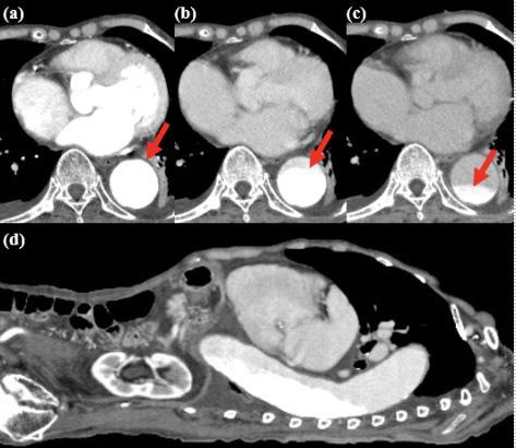
Image. Dynamic, contrast-enhanced computed tomography of the chest and abdomen,demonstrating lessening contrast in aorta over time (arrows): (a) axial image 30 seconds after contrast agent injection; (b) 90 seconds after injection; and (c) 240 seconds after injection; (d) sagittal image at 90 seconds.
On the same day as the CT, an echocardiogram was performed, revealing a left ventricular ejection fraction of 36% with reduced contractile function. Diffuse hypokinesis was noted, particularly with decreased wall motion in the inferolateral-inferior wall. There was dilation of the ascending aorta, and we observed moderate aortic and mitral valve regurgitation. No enlargement of the right heart chambers, thrombi, or vegetations were observed. The blood flow velocity in the descending aorta varied due to atrial fibrillation but was generally around 50-80 centimeters per second. Echography revealed a stagnant, hazy echo within the descending aorta.
Here we report CT images showing CA retention due to decreased aortic flow velocity in severe heart failure. In cases of cardiac arrest or cardiogenic shock, CA pooling in the venous system in CT images has been reported as “contrast agent pooling sign.”1,2 To our knowledge, there have been no reports of such findings of aortic involvement, particularly in those not requiring inotropic support or oxygen supplementation.
CPC-EM Capsule
What do we already know about this clinical entity?
Contrast agent pooling is linked to cardiogenic shock or cardiopulmonary arrest, but its association with low-output heart failure has not been reported.
What is the major impact of the image(s)?
Our case shows dynamic CT can detect changes in contrast agent pooling, ruling out aortic dissection.
How might this improve emergency medicine practice?
Dynamic CT is essential for diagnosing and differentiating between aortic dissection and contrast agent pooling conditions such as low-output heart failure.
We concluded that the pooling of CA was due to low cardiac output syndrome, resulting from the reduced ejection fraction and regurgitation. The retention of CA in the aorta on CT, particularly in patients with heart failure, strongly suggests reduced cardiac output. Although aortic dissection is a critical differential diagnosis that could cause such findings on CT, the gradual change in CA pooling observed in dynamic CT can help differentiate between these two conditions.
Documented patient informed consent and/or Institutional Review Board approval has been obtained and filed for publication of this case report.
Address for Correspondence: Kazuya Nagasaki, MD, PhD, Mito Kyodo General Hospital, Department of Internal Medicine, 3-2-7, Miyamachi, Mito, Ibaraki, 310-0015, Japan. Email: kazunagasaki@yahoo.co.jp
Conflicts of Interest: By the CPC-EM article submission agreement, all authors are required to disclose all affiliations, funding sources and financial or management relationships that could be perceived as potential sources of bias. The authors disclosed none.
Copyright: © 2024 Maezawa et al. This is an open access article distributed in accordance with the terms of the Creative Commons Attribution (CC BY 4.0) License. See: http://creativecommons.org/ licenses/by/4.0/
1. Lee YH, Chen J, Chen PA, et al. Contrast agent pooling (C.A.P.) sign and imminent cardiac arrest: a retrospective study. BMC Emerg Med. 2022;22(1):77.
2. Lin TC, Lin CS, Hsu HH, et al. Contrast pooling and layering in a patient with left main coronary artery occlusion and cardiogenic shock. Acta Cardiol Sin. 2015;31(5):461–3.
James DeChiara, MD*
Lisa Skinner, MD†
Section Editor: Melanie Heniff, MD, JD
* † Madigan Army Medical Center, Department of Emergency Medicine, Joint Base Lewis-McChord, Washington
Providence St. Peter Hospital, Olympia, Washington
Submission history: Submitted March 12, 2024; Revision received June 18, 2024; Accepted June 18, 2024
Electronically published August 26, 2024
Full text available through open access at http://escholarship.org/uc/uciem_cpcem DOI: 10.5811/cpcem.20330
Case Presentation: An 89-year-old male who had been holding dabigatran in the setting of transcarotid artery revascularization presented to the emergency department with sudden onset leg pain and weakness. Computed tomography angiography revealed acute aortic occlusion and thrombosis of the bilateral common iliac arteries. He underwent aortoiliac and femoral embolectomies and stenting of the bilateral common iliac arteries and returned to his baseline functional status.
Discussion: Acute aortic occlusion is a rare but often devastating vascular emergency characterized by obstruction of the aorta by an embolus or thrombosis. Diagnosis can be challenging as it may be mistaken for spinal pathology, which can lead to delays in diagnosis. Despite advances in diagnostic modalities and interventions, acute aortic occlusion often results in high rates of major morbidity and mortality. [Clin Pract Cases Emerg Med. 2024;8(4):375–376.]
Keywords: embolism; aortic occlusion; embolectomy; thrombosis.
CASE PRESENTATION
An 89-year-old male with a history of atrial fibrillation and left transcarotid artery revascularization (TCAR) four days prior presented to the emergency department with sudden onset pain and weakness in the bilateral lower extremities. His pain progressed to involve the lower back, and he began having extremity numbness and tingling. He was taking aspirin and clopidogrel but was holding his previously prescribed dabigatran in the setting of recent TCAR. His physical exam revealed abdominal tenderness, absent dorsalis pedis pulses bilaterally, and profound weakness and loss of sensation of bilateral lower extremities. Due to the rapidity of onset and lack of dorsalis pedis pulses, a vascular etiology was suspected.
Computed tomography angiography (CTA) chest abdomen and pelvis, and CTA abdomen-aorta with bilateral femoral runoff were ordered, which revealed acute aortic occlusion and thrombosis of the bilateral common iliac arteries (Images 1 and 2). The patient subsequently underwent aortoiliac and femoral embolectomies and stenting of the
bilateral common iliac arteries. He was transferred to the intensive care unit postoperatively, where he recovered and was extubated on day two. He was transferred to the acute care service on day four and discharged after achieving independent ambulation on day nine.
Acute aortic occlusion occurs due to thrombosis, embolus, or occluded grafts or stent, with in situ thrombosis accounting for approximately 64-72% of cases.1,2 Mortality rates are reported as 21-52%.1,3 Factors that increase the risk of aortic occlusion include peripheral arterial disease, smoking, and hypercoagulable states, among others. Physical exam findings can include lower extremity tenderness and neurologic deficits such as paralysis, weakness, and sensory deficits, which more frequently occur unilaterally.2 Abdominal pain and tenderness may suggest clot burden and occlusion above the level of the iliac bifurcation. Diminished or absent distal pulses, and the presence of cool or mottled skin can help differentiate aortic occlusion from neurologic pathology.
While ultrasonography may show an echogenicity in the vessel lumen and can be performed rapidly at the bedside,4 CTA is the test of choice for definitive diagnosis. Management of acute aortic occlusion includes anticoagulation and emergent revascularization.3 Most commonly, the surgery of choice is a thromboembolectomy, followed by thrombolysis and axillobifemoral or aortobifemoral bypass; however, this is dependent on the etiology of occlusion. While embolectomy is more common in patients with an underlying embolic occlusion, bypass surgery has been the procedure of choice for patients with in situ thrombosis.1,2
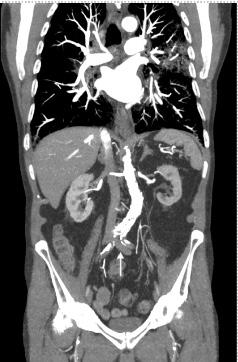

CPC-EM Capsule
What do we already know about this clinical entity?
Acute aortic occlusion typically presents with lower extremity motor and sensory deficits requiring emergent revascularization, yet still has high rates of mortality.
What is the major impact of the image(s)?
CT angiography abdomen-aorta with bilateral femoral runoff should be considered in patients who present with acute onset, lower extremity neurovascular deficits.
How might this improve emergency medicine practice?
Clinicians should consider vascular etiologies when developing a differential diagnosis for patients presenting with symptoms that raise concern for spinal pathology.
Address for Correspondence: James DeChiara, MD. Madigan Army Medical Center. Department of Emergency Medicine. 9040A Jackson Ave, Joint Base Lewis-McChord, WA 98431. Email: jrdechiara@gmail.com.
Conflicts of Interest: By the CPC-EM article submission agreement, all authors are required to disclose all affiliations, funding sources and financial or management relationships that could be perceived as potential sources of bias. The authors disclosed none. The views expressed here are those of the authors and do not reflect the official policy of the Department of the Army, the Department of Defense, or the U.S. Government.
Copyright: © 2024 DeChiara et al. This is an open access article distributed in accordance with the terms of the Creative Commons Attribution (CC BY 4.0) License. See: http://creativecommons.org/ licenses/by/4.0/
1. Grip O, Wanhainen A, Björck M. Acute aortic occlusion. Circulation. 2019;139(2):292–4.
2. Robinson WP, Patel RK, Columbo JA, et al. Contemporary management of acute aortic occlusion has evolved, but outcomes have not significantly improved. Ann Vasc Surg. 2016;34:178–86.
3. Crawford JD, Perrone KH, Wong VW, et al. A modern series of acute aortic occlusion. J Vasc Surg. 2014;59(4):1044–50.
4. Bloom B, Gibbons R, Brandis D, et al. Point-of-care ultrasound diagnosis of acute abdominal aortic occlusion. Clin Pract Cases Emerg Med. 2020;4(1):79–82.
John Coacci, DO
Peter Viccellio, MD
Section Editor: Austin Smith, MD
Submission history: Submitted May 2, 2024; Revision received June 25, 2024; Accepted June 25, 2024
Electronically published August 16, 2024
Full text available through open access at http://escholarship.org/uc/uciem_cpcem DOI: 10.5811/cpcem.20922
Case Presentation: A 19-year-old male presented for evaluation of breakthrough seizures after inability to refill his medication following recent immigration from Haiti. Previously, the patient had never received neuroimaging due to financial constraints and resource scarcity. Computed tomography and magnetic resonance imaging obtained in the emergency department was significant for large right frontoparietal open-lip schizencephaly with mass effect, a rare congenital neurologic disorder previously undiagnosed in this patient with intractable epilepsy.
Discussion: Schizencephaly is a rare congenital neurodevelopmental disorder, which has diverse presentations ranging from intractable epilepsy to variable degrees of neurocognitive dysfunction. Treatment is generally focused on seizure management and rehabilitation. Furthermore, emergency physicians must be cognizant of patients with social determinants of health, which may have formerly prevented thorough evaluation and aid in appropriate treatment of these patients. [Clin Pract Cases Emerg Med. 2024;8(4):377–378.]
Keywords: schizencephaly; epilepsy; seizure; neurology; neurosurgery.
A 19-year-old male presented for evaluation of breakthrough seizures after inability to refill his medication following recent immigration from Haiti. He had previously been diagnosed with an unspecified seizure disorder and prescribed diazepam daily. Neuroimaging was never obtained due to financial constraints and resource scarcity. Computed tomography revealed large right frontoparietal open-lip schizencephaly with right-to-left midline shift (Image A). Additional anomalies included absence of the septum pellucidum and communication of the lateral ventricles. Subsequent magnetic resonance imaging elucidated areas of gray-white matter heterotopia and polymicrogyria, and partial fusion of the fornix concerning for lobar holoprosencephaly (Image B).
The patient was admitted to the neurology service, where video electroencephalogram revealed bihemispheric dysfunction with epileptogenic potential from the left temporal region. The patient was started on an appropriate anti-epileptic regimen and given neurosurgery referral to discuss elective ventriculoperitoneal shunt placement.

Image. (A) Large right frontoparietal open-lip schizencephaly demonstrated on non-contrast computed tomography. (B) T1-weighted non-contrast magnetic resonance imaging demonstrates heterotopic gray matter (dashed arrow) and polymicrogyria (solid arrow).
Schizencephaly is a rare congenital disorder characterized by the presence of a cleft in the cerebral hemisphere lined with heterotrophic gray matter, extending
from the surface of the pia mater to the lateral ventricles. “Closed-lip” (type I) schizencephaly contains clefts that do not communicate with the ventricular system, while “open-lip” (type II) schizencephaly contains clefts that communicate with the ventricular system. The incidence is estimated at 1.54/100,000 live births.1-4 Patients may present with intractable epilepsy and varying degrees of neurocognitive dysfunction.1,3 Associated congenital anomalies may include agenesis of the corpus collosum or septum pellucidum.1,5 Treatment is targeted toward rehabilitation and seizure management. Surgery, including shunt placement, is indicated in cases of increased intracranial pressure secondary to hydrocephalus.4,5
For patients with schizencephaly, early diagnosis and treatment can aid in attaining better neurodevelopmental outcomes.4 Physicians must be cognizant of patients with social determinants of health, which may have impeded the ability to obtain thorough diagnostic evaluation, and aid in obtaining appropriate treatment resources.
The authors attest that their institution requires neither Institutional Review Board approval, nor patient consent for publication of this case report. Documentation on file.
Address for Correspondence:John Coacci, MD, Stony Brook Medicine, Department of Emergency Medicine, 101 Nicolls Rd, Stony Brook, NY 11794. Email: john.coacci@stonybrookmedicine.edu.
Conflicts of Interest: By the CPC-EM article submission agreement, all authors are required to disclose all affiliations, funding sources and financial or management relationships that could be perceived as potential sources of bias. The authors disclosed none.
Copyright: © 2024 Coacci et al. This is an open access article distributed in accordance with the terms of the Creative Commons Attribution (CC BY 4.0) License. See: http://creativecommons.org/ licenses/by/4.0/
CPC-EM Capsule
What do we already know about this clinical entity?
Schizencephaly is a rare neurodevelopmental disorder often presenting as intractable seizures and varying degrees of neurocognitive delay.
What is the major impact of the image(s)?
In this patient with an established seizure disorder who previously could not obtain neuroimaging, computed tomography revealed large, right open-lip schizencephaly.
How might this improve emergency medicine practice?
Emergency physicians must be cognizant of social determinants of health when evaluating patients, as some may have experienced significant barriers to proper care.
1. Braga VL, da Costa MDS, Riera R, et al. Schizencephaly: a review of 734 patients. Pediatr Neurol. Oct 2018;87:23-9.
2. Halabuda A, Klasa L, Kwiatkowski S, et al. Schizencephaly-diagnostics and clinical dilemmas. Childs Nerv Syst. 2015;31(4):551-6.
3. Hung PC, Wang HS, Chou ML, et al. Schizencephaly in children: a single medical center retrospective study. Pediatr Neonatol. 2018;59(6):573-80.
4. Kopyta I, Skrzypek M, Raczkiewicz D, et al. Epilepsy in paediatric patients with schizencephaly. Ann Agric Environ Med. 2020;27(2):279-83.
5. Packard AM, Miller VS, Delgado MR. Schizencephaly: correlations of clinical and radiologic features. Neurology. 1997;48:1427-34.
Matt Berger, MD
Jamal Hussain, MD
Marco Anshien, MD
Section Editor: Melanie Heniff, MD
Capital Health Emergency Medicine, Department of Emergency Medicine, Trenton, New Jersey
Submission history: Submitted May 10, 2024; Revision received July 24, 2024; Accepted August 1, 2024
Electronically published September 14, 2024
Full text available through open access at http://escholarship.org/uc/uciem_cpcem DOI: 10.5811/cpcem.20315
Case Presentation: A 63-year-old female presented to our emergency department with altered mental status and hypotension. She was transferred from the outpatient interventional radiology suite after becoming unresponsive during the removal of an inferior vena cava filter. The patient arrived somnolent with no other history available. Her physical exam was unremarkable. We used point-of-care-ultrasound to perform a rapid ultrasound for shock and hypotension (RUSH) exam. A large pericardial effusion along with signs of cardiac tamponade were identified. The cardiothoracic surgery team was notified, and the patient was taken to the operating room where pericardial blood and a large hematoma were evacuated. She recovered uneventfully and was discharged one week later.
Discussion: The above case describes a very unstable patient whose diagnosis was obtained using the RUSH exam. History and physical did not point to a clear etiology. Options were very limited. She was too unstable to go for computed tomography, and other tests such as electrocardiogram, chest radiograph, and lab work would have been non-diagnostic. It was only after the cardiac view of the RUSH exam was obtained that a pericardial effusion and developing tamponade were identified, facilitating timely management. The RUSH exam, like the extended focused assessment with sonography for trauma, is used to help determine pathologies that need immediate intervention. Incorporation in the evaluation of critically ill patients reduces the time to diagnosis. Our case is a unique example of how point-of-care ultrasound can be used to urgently identify a life-threatening pathology. [Clin Pract Cases Emerg Med. 2024;8(4):379–380.]
Keywords: point-of-care ultrasound; RUSH exam; cardiac tamponade.
A 63-year-old female with multiple comorbidities presented as a rapid response from the outpatient interventional radiology suite. During removal of an inferior vena cava (IVC) filter, she became hypotensive and unresponsive. She received reversal agents for her sedation, glucose, and a bolus of fluid with mild improvement. Upon arrival to the emergency department (ED), she remained severely hypotensive, and aggressive fluid resuscitation was initiated. Initial vital signs on arrival to the ED included blood pressure 77/59 millimeters of mercury, heart rate 76 beats per minute, respiratory rate 11 breaths per
minute, pulse oximetry 100% on 2 liters nasal canula, and temperature 36.7° Celsius. The patient was somnolent and confused but had normal work of breathing, and her abdomen was soft and nontender. She had weak peripheral pulses. Given her critical state, further physical exam was postponed in favor of a rapid point-of-care ultrasound. We performed a point-of-care rapid ultrasound for shock and hypotension (RUSH) exam (Video), which identified a large pericardial effusion with signs of cardiac tamponade and possible proximal IVC injury or thrombus. The cardiothoracic surgery team was immediately notified, and the patient was taken emergently to the
operating room where pericardial blood and a large hematoma were evacuated with immediate return of normal cardiac function.
The operative report confirmed an area of bruising to the right atrial appendage that was discovered after hematoma evacuation. This was identified as the site of right atrial perforation from the IVC filter removal wire that had caused the acute cardiac tamponade to develop. The patient was transferred to the intensive care unit. Postoperative computed tomography did not identify further injuries other than those mentioned in the operative report but did identify a small amount of hemoperitoneum, which potentially supported the possible IVC injury identified on RUSH exam. She recovered and was discharged to a rehabilitation facility on postoperative day seven.
The above case describes how a very unstable patient was diagnosed rapidly with point-of-care ultrasound using the RUSH exam. On arrival, the cause of the patient’s hypotension was unknown. History did not provide any further information, and physical exam was remarkable for only hypotension; muffled heart sounds or jugular veinous distention were not present. It was only with the cardiac view of the RUSH exam that a pericardial effusion and developing tamponade were identified. Options in this case were very limited. She was too unstable to send to radiology, and labs, electrocardiogram, and chest radiograph would have been non-diagnostic.
The RUSH exam, like the extended focused assessment with sonography for trauma, is used to identify pathology that requires immediate intervention.1-3 Because time is of the essence, each component of the RUSH exam is designed to answer a specific clinical question. This includes evaluation for reduced ejection fraction, signs of right heart strain, the state of the IVC and aorta, and the presence of pericardial effusion, free intraperitoneal fluid, pneumothorax, pleural effusion/hemothorax, and pulmonary edema.4 Despite the significant impact of point-of-care ultrasound on patient care, physicians who are further out from training have at times been reluctant to adopt its use; hence, education and training are still needed.5 Our case demonstrates how point-of-care ultrasound can rapidly identify a lifethreatening pathology.
Video Legend. Point-of-care rapid ultrasound for shock and hypotension exam videos showing parasternal long, parasternal short, apical four chamber, subxiphoid and inferior vena cava views, respectively.
RV, right ventricle: LV, left ventricle: LA, left atrium; RA, right atrium; IVC, inferior vena cava.
The authors attest that their institution requires neither Institutional Review Board approval, nor patient consent for publication of this case report. Documentation on file
CPC-EM Capsule
What do we already know about this clinical entity?
Point-of-care rapid ultrasound for shock and hypotension (RUSH) can identify dangerous pathology at the bedside.
What is the major impact of the image(s)?
This image shows the importance of RUSH in the undifferentiated hypotensive patient and the ability to rapidly diagnose cardiac tamponade.
How might this improve emergency medicine practice?
Routine use of RUSH can allow for faster diagnosis in critically ill patients.
Address for Correspondence: Matt Berger, MD, Capital Health, Department of Emergency Medicine, 750 Brunswick Ave, Trenton, NJ 08638. Email: mattberger@gmail.com.
Conflicts of Interest: By the CPC-EM article submission agreement, all authors are required to disclose all affiliations, funding sources and financial or management relationships that could be perceived as potential sources of bias. The authors disclosed none.
Copyright: © 2024 Berger et al. This is an open access article distributed in accordance with the terms of the Creative Commons Attribution (CC BY 4.0) License. See: http://creativecommons.org/ licenses/by/4.0/
1. Montoya J, Stawicki SP, Evans DC, et al. From FAST to E-FAST: an overview of the evolution of ultrasound-based traumatic injury assessment. Eur J Trauma Emerg Surg. 2016;42(2):119-26.
2. Moore CL, Rose GA, Tayal VS, et al. Determination of left ventricular function by emergency physician echocardiography of hypotensive patients [published correction appears in Acad Emerg Med 2002 Jun;9(6):642]. Acad Emerg Med. 2002;9(3):186-93.
3. Jones AE, Tayal VS, Sullivan DM, et al. Randomized, controlled trial of immediate versus delayed goal-directed ultrasound to identify the cause of nontraumatic hypotension in emergency department patients. Crit Care Med. 2004;32(8):1703-8.
4. Estoos E and Nakitende D. Diagnostic ultrasound use in undifferentiated hypotension. Available at: https://pubmed.ncbi.nlm.nih.gov/29763130/ Accessed March 9, 2023.
5. Kennedy SK, Duncan T, Herbert AG, et al. Teaching seasoned doctors new technology: an intervention to reduce barriers and improve comfort with clinical ultrasound. Cureus. 2021;13(8):e17248.
Erin N. Dankert Eggum, DO, MHA
Sara A. Hevesi, MD
Benjamin J. Sandefur, MD, MHPE
Section Editor: Joel Moll, MD
Mayo Clinic, Department of Emergency Medicine, Rochester, Minnesota
Submission history: Submitted February 21, 2024; Revision received July 30, 2024; Accepted July 31, 2024
Electronically published November 18, 2024
Full text available through open access at http://escholarship.org/uc/uciem_cpcem DOI: 10.5811/cpcem.19473
Case Presentation: We describe a case of an elderly female patient with a history of pseudogout who presented to the emergency department with atraumatic neck pain, fever, and malaise, who was found to have crowned dens syndrome on computed tomography imaging.
Discussion: It is important that emergency physicians consider crowned dens syndrome in elderly patients presenting with neck pain and signs of inflammation to ensure timely diagnosis, treatment, and to minimize unnecessary invasive testing. [Clin Pract Cases Emerg Med. 2024;8(4):381–383.]
Keywords: crowned dens syndrome; neck pain; pseudogout
CASE PRESENTATION
A 76-year-old female presented to the emergency department with five days of posterior neck pain and generalized weakness. The pain was sharp and began when she extended her arms to catch a ball. Movement exacerbated the pain, and it was unresponsive to over-the-counter analgesic medications. The pain radiated into both upper extremities. She reported three days of subjective fever and malaise. There was no numbness or weakness. Medical history was pertinent for cervical spine osteoarthritis and pseudogout.
On examination, the patient was well appearing. Vital signs included heart rate 90 beats per minute, blood pressure 166/62 millimeters of mercury, respiratory rate 19 breaths per minute, and temperature 37.2 degrees Celsius. She was sitting upright and tense in bed; however, she refused to move her neck due to pain. She had a normal neurologic exam, including normal strength and sensation, no meningismus, and no cervical spine tenderness to palpation. Laboratory studies revealed an elevated C-reactive protein at 158.4 milligrams per liter (mg/L) (reference range less than 5.0 mg/L), erythrocyte sedimentation rate at 105 millimeters per hour (mm/hr) (0-29 mm/hr), and a normal leukocyte count at 9.5x10⁹ per liter (L) (3.4-9.6x10⁹/L).
Computed tomography (CT) angiogram of the head and neck was obtained, and the initial report identified no acute findings Magnetic resonance imaging of the cervical spine was obtained and revealed no discitis, osteomyelitis, or epidural abscess.
The patient remained unable to move her neck despite intravenous (IV) analgesics. Upon further review of the CT images, calcification of the periodontoid ligaments was identified, which can be seen in crowned dens syndrome (Images 1 and 2). During hospitalization, the patient received IV and oral steroids, declined therapy with nonsteroidal anti-inflammatory drugs (NSAIDs), and demonstrated clinical improvement over a 12-hour period with conservative measures.
Crowned dens syndrome (CDS), first described by Bouvet et al in 1985, is characterized by a painful inflammatory condition resulting from calcium pyrophosphate dihydrate (CPPD) or hydroxyapatite crystalline deposition in the cervical spine ligaments.1 Despite its clinical significance, awareness of CDS among front-line physicians remains limited. Crowned dens syndrome may account for up to 1.9% of acute neck pain presentations in outpatient settings.1,2 It may mimic meningitis, giant cell arteritis, discitis, rheumatoid arthritis, polymyalgia rheumatica, and epidural abscess. Misdiagnosis can result in the patient undergoing invasive procedures such as lumbar puncture or temporal artery biopsy.2,3
Patients are typically female (60%) with an average age of 71 years. Crowned dens syndrome is more common in patients with pseudogout of peripheral joints.2 Symptoms include localized pain at the base of the skull resulting in neck
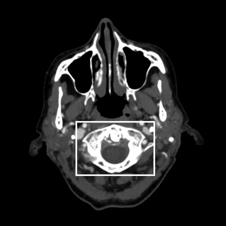
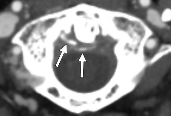
pyrophosphate dihydrate crystal deposition surrounding the odontoid process, creating a crown appearance.
stiffness. There is often systemic evidence of inflammation, including fever (80.4%) and elevated inflammatory markers (88.3%).2,3 The frequent occurrence of fever in CDS aligns with its classification as an inflammatory, crystalline deposition disease, similar to other conditions within this category.4 Non-enhanced CT is the gold standard for diagnosis.2,5 A crown-like appearance around the odontoid process on coronal views is observed, representing calcification from crystalline deposits of CPPD or hydroxyapatite.1,3 Magnetic resonance imaging is not sensitive in identifying calcification but is superior in excluding spinal cord compression.6 Treatment includes NSAIDs; however, oral colchicine or corticosteroids may be used if the patient has contraindications to NSAIDs.
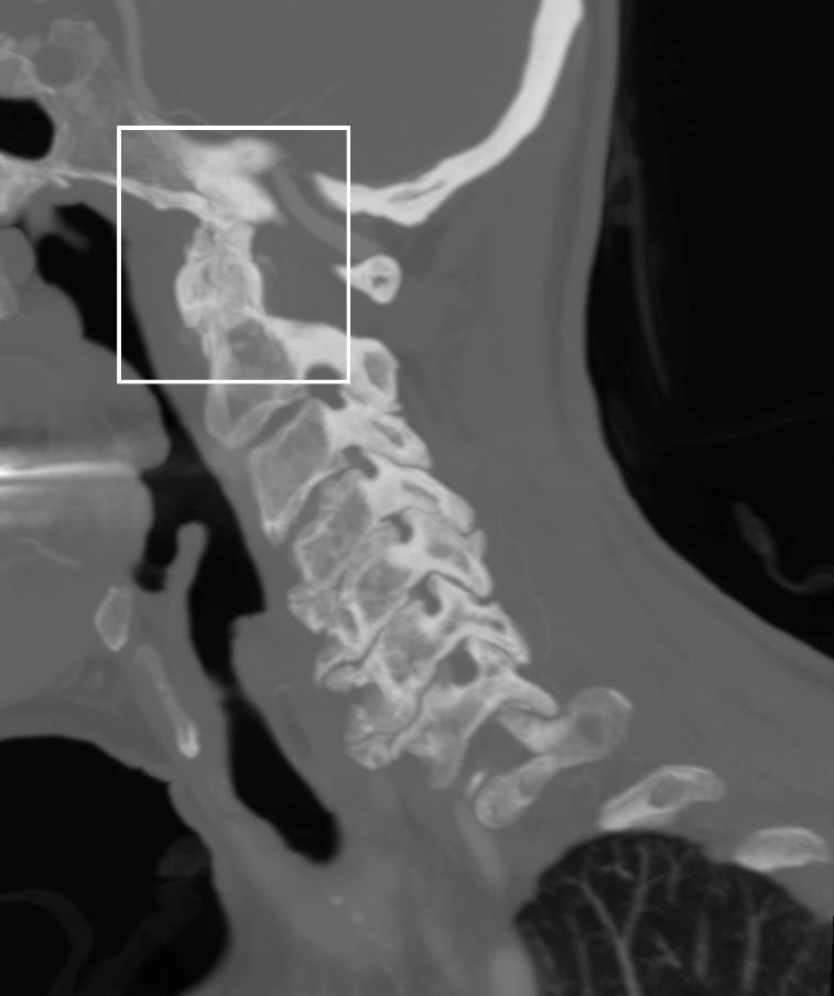
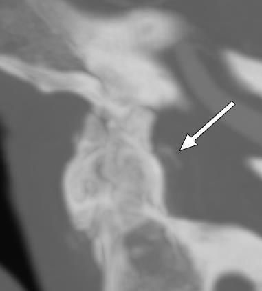
CPC-EM Capsule
What do we already know about this clinical entity?
Although information is sparse on crowned dens syndrome (CDS), we know it frequently presents in patients with a history of pseudogout.
What is the major impact of the image(s)?
The images will assist clinicians in considering this diagnosis for their patients.
How might this improve emergency medicine practice?
Emergency physicians should consider CDS in elderly patients presenting with neck pain and signs of inflammation to minimize unnecessary invasive testing.
It is important that emergency physicians consider CDS in elderly patients presenting with atraumatic neck pain, elevated inflammatory markers, and fever to ensure timely diagnosis and treatment, and to minimize unnecessary invasive testing.
We would like to thank Dr. Annie Sadosty, MD for her astute clinical acumen and identification of this diagnosis, leading to the submission of this case report. We would additionally like to thank Eric Sheahan for his assistance in image acquisition.
The Institutional Review Board approval has been documented and filed for publication of this images in emergency medicine.
Address for Correspondence: Erin Eggum, DO, MHA, Mayo Clinic, Department of Emergency Medicine, 1216 2nd St SW, Rochester, MN 55902. Email: eggum.erin@mayo.edu.
Conflicts of Interest: By the CPC-EM article submission agreement, all authors are required to disclose all affiliations, funding sources and financial or management relationships that could be perceived as potential sources of bias. The authors disclosed none.
Copyright: © 2024 Eggum et al. This is an open access article distributed in accordance with the terms of the Creative Commons Attribution (CC BY 4.0) License. See: http://creativecommons.org/ licenses/by/4.0/
Eggum et al. A Diagnosis Fit for a Queen: Crowned Dens Syndrome
1. Bouvet JP, le Parc JM, Michalski B, et al. Acute neck pain due to calcifications surrounding the odontoid process: the crowned dens syndrome. Arthritis Rheum. 1985;28(12):1417-20.
2. Oka A, Okazaki K, Takeno A, et al. Crowned dens syndrome: report of three cases and a review of literature. J Emerg Med. 2015;49(1):e9-13.
3. Huang P, Xu M, He X. Crowned dens syndrome: a case report and
literature review. Front Med (Laisanne). 2021;8:528663.
4. Masuda I and Ishikawa K. Clinical features of pseudogout attack. A survey of 50 cases. Clin Orthop Relat Res. 1988;229:173-81.
5. Scutellari PN, Galeotti R, Leprotti S, et al. The crowned dens syndrome. Evaluation with CT imaging. Radiol Med. 2007;112:195-207.
6. Balani P, Sarraf R, Sam M. Uneasy lies in the neck that wears the crown: the crowned dens syndrome: a rare case of acute neck pain. CHEST. 2023;164(4):A4080-1.
Pooja Dave, MD*
Annemarie Daecher, MD*†
Claire Abramoff, MD*
Section Editor: Jacqueline Le, MD
* † Albert Einstein Medical Center, Department of Emergency Medicine, Philadelphia, Pennsylvania
Georgetown University Medical Center, Division of Pulmonary and Critical Care Medicine, Washington, District of Columbia
Submission history: Submitted April 16, 2024; Revision received August 8, 2024; Accepted August 8, 2024
Electronically published November 4, 2024
Full text available through open access at http://escholarship.org/uc/uciem_cpcem
DOI: 10.5811/cpcem.20811
Case Presentation: We present a case of a 50-year-old patient who presented to the emergency department with palpitations, nausea, vomiting, and chest discomfort. She was found to have a reduced ejection fraction and basal wall hypokinesis on point-of-care ultrasound concerning for reverse takotsubo cardiomyopathy.
Discussion: Reverse takotsubo cardiomyopathy is a rare variant of takotsubo cardiomyopathy and involves basal ballooning instead of apical ballooning. Ultrasound findings concerning for reverse takotsubo cardiomyopathy are basal wall hypokinesis or akinesis. [Clin Pract Cases Emerg Med. 2024;8(4):384–385.]
Keywords: basal hypokinesis; catecholamine surge; reverse takotsubo cardiomyopathy.
A 50-year-old female with a past medical history of COVID-19-induced myocarditis, asthma, and a now-resolved left ventricular thrombus presented to the emergency department (ED) for two days of intermittent heart palpitations, chest discomfort, nausea, and vomiting. Her initial vital signs in the ED were a temperature of 36.5° Celsius, heart rate of 115 beats per minute, blood pressure of 145/64 millimeters of mercury, and oxygen saturation of 95% on room air. Pertinent physical exam findings included a regular heart rate and rhythm, with no murmurs. Her initial electrocardiogram (ECG) was significant for new ST-segment depressions laterally, which resolved on a repeat ECG four hours later. Troponin was initially 1.13 nanograms per milliliter (ng/mL) (normal range 0.00-0.03 ng/mL) and trended to 0.66 ng/mL four hours later and 0.22 ng/mL two days later. A point-of-care ultrasound was performed, which revealed decreased wall motion at the heart base with a decreased ejection fraction (Supplemental Videos 1-3).
Computed tomography of the chest, abdomen, and pelvis with contrast did not show signs of aortic pathology. Given
concern for non-ST-segment elevation myocardial infarction and reverse takotsubo cardiomyopathy, the patient was admitted to telemetry for a formal echocardiogram and monitoring for three days. She received aspirin 81 milligrams (mg), ondansetron, and morphine in the ED. Her formal echocardiogram showed a mildly enlarged left ventricle and moderately reduced left ventricular systolic function with an ejection fraction 35-40% (normal ejection fraction > 55%), as well as grade 1 diastolic dysfunction. The basal to mid anteroseptal, basal to mid inferoseptal, basal anterior, and basal to mid inferior and basal inferolateral walls were found to be akinetic, concerning for reverse takotsubo cardiomyopathy. The patient was ultimately discharged from the hospital with metoprolol extended-release 25 mg daily and losartan 50 mg daily. At the time of discharge, she had overall improvement of her chest pain and nausea but stated that it intermittently came back. Of note, she did endorse being anxious and stressed due to a family member requiring frequent hospitalizations. At a cardiology appointment one month after her presentation, her symptoms had resolved, and a cardiac magnetic resonance imaging was recommended.
Takotsubo cardiomyopathy is thought to be a form of left ventricular dysfunction that is often triggered by emotional or physical stress. While the exact pathophysiology of takotsubo cardiomyopathy is unknown, it is secondary to a catecholamine surge.2 Multiple variants of takotsubo cardiomyopathy exist including reverse takotsubo cardiomyopathy, which is characterized by basal ballooning, instead of the more typical apical ballooning. Reverse takotsubo cardiomyopathy is relatively rare and is thought to make up 1-23% of takotsubo cardiomyopathy cases.1 It is more commonly seen in a younger population and had a higher prevalence of preceding emotional or physical stress compared to the other types of takotsubo cardiomyopathy.3,4 Patients with reverse takotsubo were also less likely to present with severe heart failure symptoms such as dyspnea and cardiogenic shock.4 Including reverse takotsubo in our differential and performing point-of-care ultrasound at the bedside can help guide further treatment and provide insight about the disease course and recovery.
The authors attest that their institution requires neither Institutional Review Board approval, nor patient consent for publication of this case report. Documentation on file.
Address for Correspondence: Pooja Dave, MD, Albert Einstein Medical Center, Department of Emergency Medicine, 5501 Old York Rd, Korman B-9, Philadelphia, PA 19141, USA. Email: pooja. dave@jefferson.edu.
Conflicts of Interest: By the CPC-EM article submission agreement, all authors are required to disclose all affiliations, funding sources and financial or management relationships that could be perceived as potential sources of bias. The authors disclosed none.
Copyright: © 2024 Dave et al. This is an open access article distributed in accordance with the terms of the Creative Commons Attribution (CC BY 4.0) License. See: http://creativecommons.org/ licenses/by/4.0/
CPC-EM Capsule
What do we already know about this clinical entity?
Reverse takotsubo is a rare type of cardiomyopathy causing basal ballooning of the left ventricle. It is thought to be triggered by emotional or physical stress.
What is the major impact of the image(s)?
Reverse takotsubo classically presents with basal hypokinesis. The videos here depict basal hypokinesis in the subxiphoid and apical 4-chamber view.
How might this improve emergency medicine practice?
Visualizing examples of basal hypokinesis can improve a practitioner’s ability to recognize reverse takotsubo and thereby guide further treatment decisions.
1. Awad HH, McNeal AR, Goyal H. Reverse takotsubo cardiomyopathy: a comprehensive review. Ann Transl Med 2018;6(23):460.
2. Rawish E, Stiermaier T, Santoro F, et al. Current knowledge and future challenges in takotsubo syndrome: Part 1—pathophysiology and diagnosis. J Clin Med. 2021;10(3):479.
3. Ramaraj R and Movahed MR. Reverse or inverted takotsubo cardiomyopathy (reverse left ventricular apical ballooning syndrome) presents at a younger age compared with the mid or apical variant and is always associated with triggering stress. Congest Heart Fail 2010;16(6):284-6.
4. Song BG, Chun WJ, Park YH, et al. The clinical characteristics, laboratory parameters, electrocardiographic, and echocardiographic findings of reverse or inverted takotsubo cardiomyopathy: comparison with mid or apical variant. Clin Cardiol. 2011;34(11):693-9.
Justin Anderson, MD*
Ryan Grinnell, BS*
Kristina Domanski, MD*†
Jamie Baydoun, MD*†
Section Editor: Jacqueline Le, MD
* † University of Nevada, Las Vegas, Kirk Kerkorian School of Medicine, Las Vegas, Nevada University Medical Center of Southern Nevada, Emergency Department, Las Vegas, Nevada
Submission history: Submitted July 27, 2024; Revision received September 14, 2024; Accepted September 17, 2024. Electronically published: November 2, 2024
Full text available through open access at http://escholarship.org/uc/uciem_cpcem DOI: 10.5811/cpcem.25351
Case Presentation: A 32-year-old male with a history of left eye keratoconus presented to the emergency department with left eye pain and blurry vision for two days. Out of concern for corneal hydrops, ophthalmology was consulted, and the diagnosis was confirmed. Per ophthalmology recommendations, the patient was started on hypertonic saline and prednisolone eye drops and referred to a corneal specialist.
Discussion: Corneal hydrops is characterized by stromal edema caused by leakage of aqueous humor due to rupture of Descemet membrane. This case describes a patient with a keratoconus deformity who developed corneal hydrops. [Clin Pract Cases Emerg Med. 2024;8(4):386–387.]
Keywords: corneal hydrops; keratoconus; ocular ultrasound.
A 32-year-old male with a history of left eye keratoconus secondary to remote trauma presented to the emergency department (ED) with left eye pain and blurry vision for two days. Visual acuity was 20/40 in the right eye and 20/200 in the left eye, and 20/25 bilaterally with baseline corrective lenses. Examination showed central left eye corneal opacification overlying the pupil and keratoconus deformity (Image 1). Fluorescein exam revealed no uptake over the pupil (Image 2). Ocular ultrasound showed a deformed cornea (Image 3).

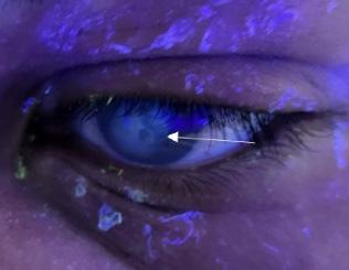
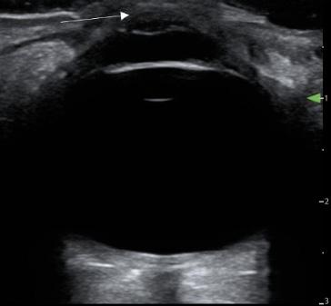
Corneal hydrops is characterized by stromal edema caused by leakage of aqueous humor due to rupture of Descemet membrane.1 It is a rare complication of keratoconus, likely due to a combination of corneal thinning and ectasia and trivial trauma to the eye.2 Risk factors include atopy, Down syndrome, keratoconus, and eye rubbing, which may incur the highest risk. Acute corneal hydrops can cause vision-debilitating scarring of the cornea.3 The ED workup of suspected corneal hydrops should rule out infectious causes of corneal edema such as keratitis and uveitis, as well as include a fluorescein exam to rule out corneal ulcer. Treatment is usually conservative, and most cases resolve within two to four months.
Given our patient’s history of keratoconus, with new onset opacification of the cornea and without fluorescein uptake, the presentation was concerning for corneal hydrops. An ocular ultrasound was performed, which revealed the keratonoconus deformity, but it was otherwise unremarkable, with normal optic nerve sheath, lens, iris, and without abnormal retinal contour. Ophthalmology was consulted, and the diagnosis was confirmed. The patient was started on 5% sodium chloride eye drops and prednisolone eye drops per ophthalmology recommendations to decrease edema, and he was referred for urgent outpatient follow-up with a corneal specialist.
Initial outpatient management of corneal hydrops centers around decreasing the edema and includes options such as antibiotics to prevent secondary infection, hypertonic saline drops to cause an osmotic gradient to reduce edema, cycloplegics for pain control, and topical nonsteroidal antiinflammatory drugs/steroids to decrease inflammation and pain.1 Surgery is sometimes indicated and can improve visual acuity and delay corneal transplantation.4 When ophthalmology is not on call, medical management is as discussed, and transfer to a facility with ophthalmology should be considered, as the patient will require urgent outpatient follow-up with ophthalmology as surgical intervention may be required.4
The authors attest that their institution does not Institutional Review Board approval for publication of this case report. Documentation on file. Patient consent has been obtained and filed for the publication of this case report.
1. Maharana PK, Sharma N, Vajpayee RB. Acute corneal hydrops in
CPC-EM Capsule
What do we already know about this clinical entity?
Corneal hydrops is a rare entity that can cause severe corneal damage and permanent blindness without intervention.
What is the major impact of the image(s)? Clinically it is similar in appearance to corneal abrasions or ulcerations, but it has no fluorescein uptake on staining.
How might this improve emergency medicine practice?
Corneal hydrops must be identified early and managed appropriately to avoid risk of significant morbidity.
Address for Correspondence: Justin Anderson, MD, University of Nevada, Las Vegas, Kirk Kerkorian School of Medicine, 6661 Silverstream Ave Apt 2019, Las Vegas, NV 89107. Email: justin. anderson@unlv.edu.
Conflicts of Interest: By the CPC-EM article submission agreement, all authors are required to disclose all affiliations, funding sources and financial or management relationships that could be perceived as potential sources of bias. The authors disclosed none.
Copyright: © 2024 Anderson et al. This is an open access article distributed in accordance with the terms of the Creative Commons Attribution (CC BY 4.0) License. See: http://creativecommons.org/ licenses/by/4.0/ keratoconus. Indian J Ophthalmol. 2013;61(8):461-464.
2. Barsam A, Petrushkin H, Brennan N, et al. Acute corneal hydrops in keratoconus: a national prospective study of incidence and management. Eye (Lond). 2015;29(4):469-474. Epub 2015 Jan 16.
3. Santodomingo-Rubido J, Carracedo G, Suzaki A, et al. Keratoconus: an updated review. Cont Lens Anterior Eye. 2022;45(3):101559. Epub 2022 Jan 4.
4. Fan Gaskin JC, Patel DV, McGhee CN. Acute corneal hydrops in keratoconus - new perspectives. Am J Ophthalmol. 2014;157(5):921-928.
Joshua Berko, DO
Christine Raps, MD
Quinlan Cacic, DO
Robert Stephen, MD
Megan Fix, MD
Allison M. Beaulieu, MD, MAEd
Section Editor: R. Gentry Wilkerson, MD
University of Utah, Department of Emergency Medicine, Salt Lake City, Utah
Submission history: Submitted June 13, 2024; Revision received October 4, 2024; Accepted October 5, 2024
Electronically published November 5, 2024
Full text available through open access at http://escholarship.org/uc/uciem_cpcem DOI: 10.5811/cpcem.24854
Case Presentation: A female patient with a known history of pustular psoriasis presented with sub-acute development of diffuse erythema and scaling of the skin with areas of exfoliation consistent with erythroderma. She was ill appearing and required admission and aggressive treatment with steroid-impregnated wet dressings, topical emollients, analgesics, and systemic immunosuppressants.
Discussion: Erythroderma is a dermatologic emergency characterized by diffuse erythema and scaling spanning greater than 90% of skin surfaces and is associated with a mortality rate as high as 64%. It is initially a clinical diagnosis and needs to be recognized and aggressively treated expeditiously to improve chances of a good outcome. [Clin Pract Cases Emerg Med. 2024;8(4):388–390.]
Keywords: dermatology; pustular psoriasis; erythroderma.
A 45-year-old female with a history of pustular psoriasis, type II diabetes, thyroid cancer, and endometrial cancer presented to the emergency department (ED) with a diffuse rash. Five weeks prior to presentation she was treated with intramuscular dexamethasone and nirmatrelvir/ritonavir for COVID-19. In the time since her medication administration, she developed a pruritic rash on her abdomen that gradually spread outward toward her extremities. The week prior to presentation, she developed pustules and sloughing of her skin with associated blurred vision, sore throat, chills, and vaginal pain.
On physical examination, the patient had a diffuse, erythematous, pustular rash with sloughing (Images 1-3). Nikolsky sign was negative; however, there was evidence of mucosal erythema and sloughing of the genital region, conjunctivae, and oropharynx. Ophthalmology was consulted given concern for toxic epidermal necrolysis and found

moderate ocular involvement. They recommended artificial tears and erythromycin ointment. Dermatology was also consulted and performed a biopsy, which later confirmed erythroderma secondary to general pustular psoriasis triggered in the setting of dexamethasone use and COVID-19. The patient was subsequently admitted to the hospital for further treatment, which included infliximab, triamcinolone wet wraps, secukinumab, cephalexin, and gabapentin.
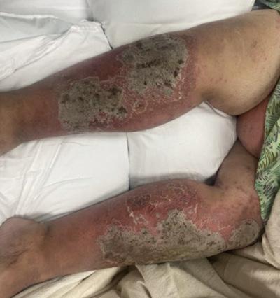
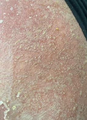
This case highlights an example of pustular psoriasis presenting to the ED with secondary severe erythroderma. Pustular psoriasis is a rare, immune-mediated chronic disease that presents in episodic flares.1 Erythroderma is a dermatologic emergency that is characterized by diffuse erythema and scaling involving greater than 90% of skin
CPC-EM Capsule
What do we already know about this clinical entity?
Erythroderma presents as diffuse erythema, scaling, and progressive exfoliation of over 90% of skin surface with fever, malaise, tachycardia, and peripheral edema.
What is the major impact of the image(s)?
Diffuse erythema with scaling and sloughing of skin in the setting of a known history dermatoses such as pustular psoriasis aids in the diagnosis.
How might this improve emergency medicine practice?
Awareness of this rare disease will aid in the early, correct diagnosis and treatment, which may decrease mortality and morbidity.
surfaces and is associated with a mortality rate as high as 64%.2 The most common causes are exacerbation of preexisting dermatosis and drug reactions, but it can also be seen with infections and other systemic diseases.2-4 Symptoms include diffuse skin involvement, notably pruritic, scaling, crusting, erythematous patches with progressive exfoliation and may be associated with systemic symptoms such as shivering, hypothermia, fever, malaise, peripheral edema, and tachycardia.4
Erythroderma can have a variable onset and is initially diagnosed clinically4; therefore, history should focus on identifying triggers or genetic factors by asking about prior skin conditions, medications, and family history. Physical exam should include a complete skin and mucosal exam to identify extent of involvement and evaluate for secondary infection. Laboratory testing should include a complete blood count, which commonly reveals leukocytosis. A comprehensive metabolic panel is useful in assessing electrolyte status, glucose and albumin levels, lactate dehydrogenase, and kidney and liver function.4 If systemic or superimposed infection is suspected, bacterial and fungal cultures, and a viral polymerase chain reaction for herpes simplex virus or varicella zoster virus may be warranted. Initial treatment for all causes of erythroderma include hemodynamic management, fluid and electrolyte replacement, wound care, temperature management, nutritional support, treatment of superimposed infections, and symptomatic management with wet dressings, emollients, oral antihistamines (eg, hydroxyzine hydrochloride), and low to medium dose systemic prednisone (0.5-1 milligram
Severe Erythroderma in a Patient with Pustular Psoriasis
per kilogram per day with taper) or topical steroids (clobetasol 0.05% or triamcinolone twice daily for 2-4 weeks).2-4 Emergency physicians need to be able to recognize erythroderma early to rapidly provide lifesaving, stabilizing treatment and admission.
The authors attest that their institution requires neither Institutional Review Board approval, nor patient consent for publication of this case report. Documentation on file.
1. Benjegerdes KE, Hyde K, Kivelevitch D, et al. Pustular psoriasis: pathophysiology and current treatment perspectives. Psoriasis (Auckl). 2016;6:131-144.
2. Sehgal VN, Srivastava G, Sardana K. Erythroderma/exfoliative dermatitis: a synopsis. Int J Dermatol. 2004;43(1):39-47.
Berko et al.
Address for Correspondence: Allison Beaulieu, MD, MAEd, University of Utah, Department of Emergency Medicine, Helix Building 5050, 30 N Mario Capecchi Drive, Level 2 South, Salt Lake City, UT 84112. Email: Allison.beaulieu@hsc.utah.edu.
Conflicts of Interest: By the CPC-EM article submission agreement, all authors are required to disclose all affiliations, funding sources and financial or management relationships that could be perceived as potential sources of bias. The authors disclosed none.
Copyright: © 2024 Berko et al. This is an open access article distributed in accordance with the terms of the Creative Commons Attribution (CC BY 4.0) License. See: http://creativecommons.org/ licenses/by/4.0/
3. Pal S and Haroon TS. Erythroderma: a clinico-etiologic study of 90 cases. Int J Dermatol. 1998;37(2):104-107.
4. Rothe MJ, Bialy TL, Grant-Kels JM. Erythroderma. Dermatol Clin. 2000;18(3):405-415.
Abdullah Khan, MD
Section Editor: Melanie Heniff, MD
Submission history: Submitted May 10, 2024; Revision received August 6, 2024; Accepted August 6, 2024
Electronically published November 9, 2024
Full text available through open access at http://escholarship.org/uc/uciem_cpcem DOI: 10.5811/cpcem.21173
Case Presentation: A 13-month-old child with past medical history of congenital adrenal insufficiency presented to the emergency department with vomiting and diarrhea. Initially the child was noticed to have bradycardia with normal blood pressure. An electrocardiogram (ECG) showed tall T waves, broad QRS complex, and widened PR interval suggestive of severe hyperkalemia. The initial blood gas showed potassium of 10.7 millimoles per liter. The patient was started on calcium gluconate with immediate resolution of ECG changes. Further management with insulin, dextrose, and sodium polystyrene sulfonate led to normal potassium levels.
Discussion: Hyperkalemia is a life-threatening condition in children, especially in those with congenital adrenal insufficiency. The ECG showed different changes as the levels of serum potassium levels increased ranging from tall T waves, wide QRS complex, increased PR interval to arrythmias. Immediate treatment with calcium gluconate in such cases has significant cardioprotective effect. It is important to recognize the ECG changes manifested by changes in serum potassium levels. Our patient had classic ECG changes manifested in severe hyperkalemia. [Clin Pract Cases Emerg Med. 2024;8(4):391–393.]
Keywords: hyperkalemia; congenital adrenal insufficiency; ECG changes in hyperkalemia.
A 13-month-old male child with a previous history of congenital adrenal insufficiency presented to the emergency department with multiple episodes of vomiting (non-bloody and non-bilious) and diarrhea (non-bloody and nonmucoid). The patient was not able to take his regular hydrocortisone doses at home. Initially the child did not look well and had bradycardia (heart rate 52-59 beats per minute) with normal blood pressure. The patient was attached to a cardiac monitor and a wide QRS-complex rhythm was noticed.
An electrocardiogram (ECG) was obtained. The ECG showed sinus bradycardia with regular rhythm. The QRS axis was rightward (106 degrees), which is normal in this age group. There were tall, peaked upright T waves in all precordial and limb leads, except for V1 where the T wave was deeply inverted. The QRS complexes were wide at 196
milliseconds (ms) (normal range 54-88 ms). The PR interval was prolonged at 226 ms (86-151 ms). There was rSr’ pattern in the V1 lead with widening of the S wave in lead I and aVF, suggesting right bundle branch block (Image 1).
The initial venous blood gas results showed potassium of 10.7 millmoles per liter (mmol/L) (reference range 3.5-5.2 mmol/L) and sodium of 122 mmol/L (135-145 mmol/L). The initial complete metabolic panel showed a blood urea nitrogen of 18.1 mmol/L (3.2 to 7.9 mmol/L) and creatinine of 132 mmol/L (13-35 mmol/L). With ECG findings suggestive of severe hyperkalemia, albuterol and calcium gluconate (100 milligrams per kilogram [mg/kg]) were started. The patient developed an episode of ventricular tachycardia (heart rate 200-215) with normal blood pressure. Albuterol was stopped, and calcium gluconate was continued. The patient successfully reverted to normal sinus rhythm with calcium gluconate (Image 2). A stress dose of hydrocortisone (25 mg) was given,

Image1. Electrocardiogram suggesting signs of hyperkalemia. Lead V1 shows rSr’ (green arrow), Precordial and limb leads show wide QRS complex (red arrows) and tall T waves (blue arrows).
and insulin (0.1 units/kg/hour) with dextrose 10% infusion was started. A 20 millimeter (mL) per kg bolus of normal saline was also given due to acute kidney injury. A small dose of furosemide (0.5 mg/kg) and rectal sodium polystyrene sulfonate (1 gram/kg) were also administered. The potassium levels were corrected over a period of five hours. The patient was observed in pediatric intensive care unit for 24 hours and discharged without any complications.
The ECG demonstrated typical changes of severe hyperkalemia. Hyperkalemia is a life-threatening condition that if not treated can lead to arrhythmias and asystole. Hyperkalemia produces a variety of dose-dependent changes on ECG that include tall peaked T waves (5.5-6.5 mmol/L); prolongation of the PR interval and QRS widening (6.5-7.5 mmol/L); ST-segment changes, QTc prolongation, and decrease in amplitude of P wave (7-8 mmol/L); and marked widening of QRS and sine wave pattern and arrhythmias.1 In children the abnormal values of different waves and segments of ECG are age and sex dependent, and upper limits of normal values are described as the 98th percentile.
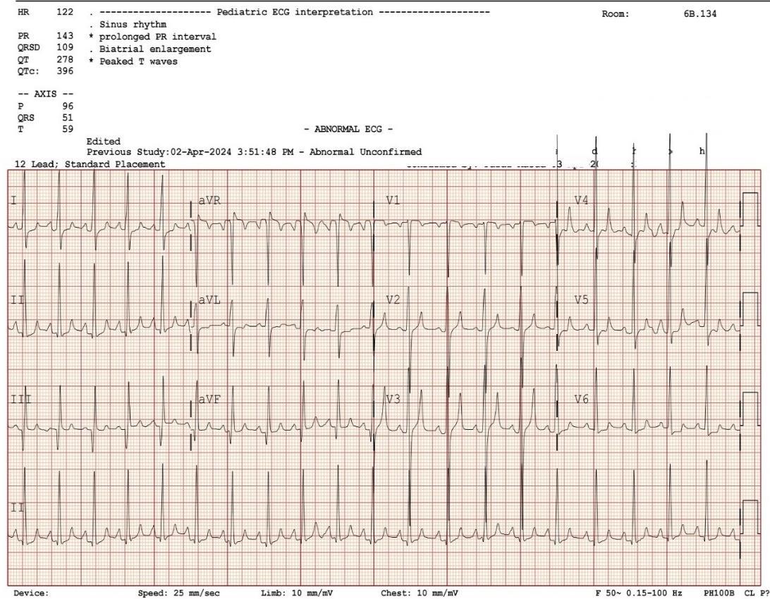
Image 2. Normal sinus rhythm after administration of calcium gluconate in a pediatric patient with hyperkalemia.
CPC-EM Capsule
What do we already know about this clinical entity?
Severe hyperkalemia can be by acute illness in children with congenital adrenal insufficiency.
What is the major impact of the image(s)?
It is important to identify electrocardiographic changes associated with hyperkalemia.
How might this improve emergency medicine practice?
In children with congenital adrenal insufficiency presenting with hyperkalemia triggered by acute illness, stress doses of hydrocortisone must be administered.
The 98th percentile values for male children between 1-3 years of age for PR-interval QRS duration and QTc-interval are 151 ms, 88 ms, and 455 ms, respectively.2 The ECG in our patient demonstrated prolongation of all these segments.
Hyperkalemia in children is usually caused by impaired excretion of potassium (adrenal or renal insufficiency), and movement of intracellular potassium to the extracellular space (acidosis, diabetes, tumor lysis syndrome, rhabdomyolysis), as well as drugs and toxins (spironolactone, angiotensinconverting enzyme inhibitors, beta blockers and calcium channel blockers).3 If ECG changes are present, the immediate step of administering cardiac membrane-stabilizing agent such as calcium gluconate 10% followed by potassium lowering agents, such as albuterol, insulin and dextrose, furosemide and sodium polystyrene sulfonate, is warranted.1,3 In our patient, hyperkalemia was caused by the underlying adrenal insufficiency exacerbated by missed doses of hydrocortisone due to vomiting and acute illness.
Adrenal insufficiency is caused by dysfunction at any level of hypothalamic-pituitary-adrenal axis. Hence, it can be broadly classified as primary (dysfunction of adrenal gland), secondary (decreased release of adrenocorticotropic hormone from the pituitary gland), and tertiary (decreased release of corticotropin hormone from the hypothalamus).4 Adrenal insufficiency leads to decreased production of glucocorticoid (cortisol) and mineralocorticoid (aldosterone). Aldosterone regulates sodium absorption and potassium excretion at the level of distal tubules of nephrons; hence, its deficiency leads to hyponatremia and
hyperkalemia. Cortisol regulates serum glucose levels and blood pressure in periods of stress. Patients with adrenal insufficiency develop adrenal crises in acute illness. Therefore, in addition to management of hyperkalemia it is important to administer stress doses of hydrocortisone (25 mg for ages 0-3 years) to achieve both glucocorticoid and mineralocorticoid effect in cases of congenital adrenal insufficiency.5
The author attests that their institution requires neither Institutional Review Board approval, nor patient consent for publication of this case report. Documentation on file.
1. Daly K and Farrington E. Hypokalemia and hyperkalemia in infants and children: pathophysiology and treatment. J Pediatr Health Care 2013;27(6):486-96; quiz 497-8.
2. Rijnbeek PR, Witsenburg M, Schrama E, et al. New normal limits for the paediatric electrocardiogram. Eur Heart J. 2001;22(8):702-11.
Severe Hyperkalemia in a Child with Vomiting and Diarrhea
Address for Correspondence: Abdullah Khan, MD, Sidra Medicine, Department of Emergency Medicine, Al Rayyan Road, Doha, Al Rayyan, Qatar. Email: abdullahkhan120@gmail.com.
Conflicts of Interest: By the CPC-EM article submission agreement, all authors are required to disclose all affiliations, funding sources and financial or management relationships that could be perceived as potential sources of bias. The authors disclosed none.
Copyright: © 2024 Khan. This is an open access article distributed in accordance with the terms of the Creative Commons Attribution (CC BY 4.0) License. See: http://creativecommons.org/licenses/ by/4.0/
3. Lehnhardt A and Kemper MJ. Pathogenesis, diagnosis and management of hyperkalemia. Pediatr Nephrol. 2011;26(3):377-84.
4. Bowden SA and Henry R. Pediatric adrenal insufficiency: diagnosis, management, and new therapies. Int J Pediatr. 2018;2018:1739831.
5. Nisticò D, Bossini B, Benvenuto S, et al. Pediatric adrenal insufficiency: challenges and solutions. Ther Clin Risk Manag. 2022;18:47-60.


