Clinical Practice and Cases in Emergency Medicine
CALIFORNIA ACEP’S ANNUAL CONFERENCE 2021 Education is targeted to Medical Students and Residents, but all are welcome to attend.
PAGES 276-368
Friday, September 10, 2021 Westin San Diego Gaslamp Quar ter
DATE
VOLUME 5 ISSUE 3 August 2021
SAVE
the
Volume V, Number 3, August 2021
Open Access at www.cpcem.org
Clinical Practice and Cases in Emergency Medicine
ISSN: 2474-252X
In Collaboration with the Western Journal of Emergency Medicine Clinicopathological Cases from the University of Maryland 276 19-year-old Woman with Intermittent Weakness Cavaliere GA, Murali N, Bontempo LJ, ZDW Dezman Medial Legal Case Reports 283 Three Cases of Emergency Department Medical Malpractice Involving “Consultations”: How Is Liability Legally Determined? Aldalati A, Bellamkonda VR, Moore GP, Finch AS Case Series 289 Nebulized Tranexamic Acid in Secondary Post-Tonsillectomy Hemorrhage: Case Series and Review of the Literature Dermendjieva M, Gopalsami A,Torbati S Case Reports 296 Euglycemic Diabetic Ketoacidosis in Type 1 Diabetes on Insulin Pump, with Acute Appendicitis: A Case Report Thompson BD, Kitchen A 299
An Anomalous Cause of Deep Venous Thrombosis: A Case Report Florian J, Duong HA, Sieber R, Roh JS
303
Altered Mental Status in the Emergency Department – When to Consider Anti-LGI-1 Encephalitis: Case Report Miljkovic SS, Koenig BW
307
Anaphylaxis Caused by Swimming: A Case Report of Cold-induced Urticaria in the Emergency Department McManus NM, Zehrung RJ, Armstrong TC, Offman RP
312
Under the Radar: A Case Report of a Missed Aortoenteric Fistula Briggs B, Manthey D Contents continued on page iii
A Peer-Reviewed, International Professional Journal
N ! I M O J AE /A L CA
Championing individual physician rights and workplace fairness BENEFITS
ACOEP stands with all emergency physicians and providers on the front line. We thank you for your tireless work and effort.
www.acoep.org
-
Western Journal of Emergency Medicine Subscription CAL/AAEM News Service email updates Discounted AAEM pre-conference fees And more!
CAL/AAEM NEWS SERVICE -
Healthcare industry news Public policy Government issues Legal cases and court decisions
In collaboration with our official journal
FACEBOOK.COM/CALAAEM
FOLLOW US @CALAAEM
HTTP://WWW.CALAAEM.ORG
Clinical Practice and Cases in Emergency Medicine Indexed in PubMed and full text in PubMed Central
Rick A. McPheeters, DO, Editor-in-Chief Kern Medical/UCLA- Bakersfield, California
Christopher Sampson, MD, Decision Editor University of Missouri- Columbia, Missouri Joel Moll, MD, Decision Editor Virginia Commonwealth University School of Medicine- Richmond, Virginia
Mark I. Langdorf, MD, MHPE, Senior Associate Editor University of California, Irvine School of Medicine- Irvine, California
Scott Goldstein, DO, Decision Editor Sidney Kimmel College of Medicine- Philadelphia, Pennsylvania
Shahram Lotfipour, MD, MPH, Senior Associate Editor University of California, Irvine School of Medicine- Irvine, California
Steven Walsh, MD, Decision Editor Einstein Medical Center Philadelphia-Philadelphia, Pennsylvania
Shadi Lahham, MD, MS, Associate Editor University of California, Irvine School of Medicine- Irvine, California
Melanie Heniff, MD, Decision Editor University of Indiana School of Medicine- Indianapolis, Indiana
Manish Amin, DO, Associate Editor Kern Medical/UCLA- Bakersfield, California
Gentry Wilkerson, MD, Decision Editor University of Maryland School of Medicine
John Ashurst, DO, Decision Editor/ ACOEP Guest Editor Kingman Regional Health Network, Arizona
Rachel A. Lindor, MD, JD, Decision Editor Mayo Clinic College of Medicine and Science
Anna McFarlin, MD, Decision Editor Louisiana State University Health Science Center- New Orleans, Louisiana
Jacqueline K. Le, MD, Decision Editor Desert Regional Medical Center
Austin Smith, MD, Decision Editor Vanderbilt University Medical Center-Nashville, Tennessee Amin A. Kazzi, MD, MAAEM The American University of Beirut, Beirut, Lebanon Anwar Al-Awadhi, MD Mubarak Al-Kabeer Hospital, Jabriya, Kuwait Arif A. Cevik, MD United Arab Emirates University College of Medicine and Health Sciences, Al Ain, United Arab Emirates Abhinandan A.Desai, MD University of Bombay Grant Medical College, Bombay, India Bandr Mzahim, MD King Fahad Medical City, Riyadh, Saudi Arabia Barry E. Brenner, MD, MPH Case Western Reserve University Brent King, MD, MMM University of Texas, Houston Daniel J. Dire, MD University of Texas Health Sciences Center San Antonio David F.M. Brown, MD Massachusetts General Hospital/Harvard Medical School
Christopher San Miguel, MD, Decision Editor Ohio State Univesity Wexner Medical Center
Editorial Board Khrongwong Musikatavorn, MD King Chulalongkorn Memorial Hospital, Chulalongkorn University, Bangkok, Thailand
Edward Michelson, MD Texas Tech University Edward Panacek, MD, MPH University of South Alabama Erik D. Barton, MD, MBA Icahn School of Medicine, Mount Sinai, New York Francesco Dellacorte, MD Azienda Ospedaliera Universitaria “Maggiore della Carità,” Novara, Italy
Gayle Galleta, MD Sørlandet Sykehus HF, Akershus Universitetssykehus, Lorenskog, Norway Hjalti Björnsson, MD Icelandic Society of Emergency Medicine
Rosidah Ibrahim, MD Hospital Serdang, Selangor, Malaysia
Linda S. Murphy, MLIS University of California, Irvine School of Medicine Librarian
Samuel J. Stratton, MD, MPH Orange County, CA, EMS Agency
Elena Lopez-Gusman, JD California ACEP American College of Emergency Physicians Adam Levy, BS American College of Osteopathic Emergency Physicians John B. Christensen, MD California Chapter Division of AAEM Lori Winston, MD California ACEP American College of Emergency Physicians Kaweah Delta Healthcare District Mark I. Langdorf, MD, MHPE UC Irvine Health School of Medicine
Scott Rudkin, MD, MBA University of California, Irvine Scott Zeller, MD University of California, Riverside
Niels K. Rathlev, MD Tufts University School of Medicine
Steven Gabaeff, MD Clinical Forensic Medicine
Pablo Aguilera Fuenzalida, MD Pontificia Universidad Catolica de Chile, Región Metropolitana, Chile
Steven H. Lim, MD Changi General Hospital, Simei, Singapore
Jacob (Kobi) Peleg, PhD, MPH Tel-Aviv University, Tel-Aviv, Israel
Peter A. Bell, DO, MBA Liberty University College of Osteopathic Medicine
Jonathan Olshaker, MD Boston University
Peter Sokolove, MD University of California, San Francisco
Katsuhiro Kanemaru, MD University of Miyazaki Hospital, Miyazaki, Japan
Robert M. Rodriguez, MD University of California, San Francisco
Advisory Board
Amal Khalil, MBA UC Irvine Health School of Medicine
Robert W. Derlet, MD University of California, Davis
Leslie Zun, MD, MBA Chicago Medical School
Nadeem Qureshi, MD St. Louis University, USA Emirates Society of Emergency Medicine, United Arab Emirates
Francis Counselman, MD Eastern Virginia Medical School
Robert Suter, DO, MHA UT Southwestern Medical Center
Terry Mulligan, DO, MPH, FIFEM ACEP Ambassador to the Netherlands Society of Emergency Physicians Vijay Gautam, MBBS University of London, London, England Wirachin Hoonpongsimanont, MD, MSBATS Siriraj Hospital, Mahidol University, Bangkok, Thailand
Editorial Staff
Jorge Fernandez, MD California ACEP American College of Emergency Physicians University of California, San Diego Peter A. Bell, DO, MBA American College of Osteopathic Emergency Physicians Liberty University, College of Osteopathic Medicine Robert Suter, DO, MHA American College of Osteopathic Emergency Physicians UT Southwestern Medical Center Shahram Lotfipour, MD, MPH UC Irvine Health School of Medicine Brian Potts, MD, MBA California Chapter Division of AAEM Alta Bates Summit-Berkeley Campus
Isabelle Nepomuceno, BS Executive Editorial Director
Cassandra Saucedo, BS Executive Publishing Director
Shreya Gupta, BS WestJEM Editorial Director Associate Marketing Director
Nathan Do, BS WestJEM Publishing Director
Sohrab Kharabaf, BS CPC-EM Editorial Director Associate Marketing Director
Trina Nguyen, BS CPC-EM Publishing Director
Stephanie Burmeister, MLIS WestJEM Staff Liaison
Paul Abdo, BS WestJEM Associate Publishing Director Associate Marketing Director
June Casey, BA Copy Editor
Official Journal of the California Chapter of the American College of Emergency Physicians, the America College of Osteopathic Emergency Physicians, and the California Chapter of the American Academy of Emergency Medicine
Available in MEDLINE, PubMed, PubMed Central, Google Scholar, eScholarship, DOAJ, and OASPA. Editorial and Publishing Office: WestJEM/Depatment of Emergency Medicine, UC Irvine Health, 333 City Blvd, West, Rt 128-01, Orange, CA 92866, USA Office: 1-714-456-6389; Email: Editor@westjem.org
Volume V, no. 3: August 2021
i
Clinical Practice and Cases in Emergency Medicine
Clinical Practice and Cases in Emergency Medicine Indexed in PubMed and full text in PubMed Central
This open access publication would not be possible without the generous and continual financial support of our society sponsors, department and chapter subscribers.
Professional Society Sponsors American College of Osteopathic Emergency Physicians California ACEP
California Chapter Division of AmericanAcademy of Emergency Medicine
Academic Department of Emergency Medicine Subscribers Albany Medical College Albany, NY
INTEGRIS Health Oklahoma City, OK
Robert Wood Johnson University Hospital New Brunswick, NJ
University of Florida, Jacksonville Jacksonville, FL
American University of Beirut Beirut, Lebanon
Kaweah Delta Health Care District Visalia, CA
Rush University Medical Center Chicago, IL
University of Illinois at Chicago Chicago, IL
Arrowhead Regional Medical Center Colton, CA
Kennedy University Hospitals Turnersville, NJ
Southern Illinois University Carbondale, IL
Augusta University Augusta GA
Kern Medical Bakersfield, CA
St. Luke’s University Health Network Bethlehem, PA
University of Illinois College of Medicine Peoria, IL
Baystate Medical Center Springfield, MA
Lakeland HealthCare St. Joseph, MI
Beaumont Hospital Royal Oak, MI
Lehigh Valley Hospital and Health Network Allentown, PA
Stanford/Kaiser Emergency Medicine Residency Program Stanford, CA
Beth Israel Deaconess Medical Center Boston, MA
Loma Linda University Medical Center Loma Linda, CA
Boston Medical Center Boston, MA
Louisiana State University Health Sciences Center New Orleans, LA
Brigham and Women’s Hospital Boston, MA
Madigan Army Medical Center Tacoma, WA
Brown University Providence, RI
Maimonides Medical Center Brooklyn, NY
Carl R. Darnall Army Medical Center Fort Hood, TX
Maricopa Medical Center Phoenix, AZ
Conemaugh Memorial Medical Center Johnstown, PA
Massachusetts General Hospital Boston, MA
Desert Regional Medical Center Palm Springs, CA
Mayo Clinic College of Medicine Rochester, MN
Doctors Hospital/Ohio Health Columbus, OH
Mt. Sinai Medical Center Miami Beach, FL
Eastern Virginia Medical School Norfolk, VA
North Shore University Hospital Manhasset, NY
Einstein Healthcare Network Philadelphia, PA
Northwestern Medical Group Chicago, IL
Emory University Atlanta, GA
Ohio State University Medical Center Columbus, OH
Genesys Regional Medical Center Grand Blanc, Michigan
Ohio Valley Medical Center Wheeling, WV
Hartford Hospital Hartford, CT
Oregon Health and Science University Portland, OR
Hennepin County Medical Center Minneapolis, MN
Penn State Milton S. Hershey Medical Center Hershey, PA
Henry Ford Hospital Detroit, MI
Presence Resurrection Medical Center Chicago, IL
University of Iowa Iowa City, IA University of Louisville Louisville, KY
Staten Island University Hospital Staten Island, NY
University of Maryland Baltimore, MD
SUNY Upstate Medical University Syracuse, NY
University of Michigan Ann Arbor, MI
Temple University Philadelphia, PA
University of Missouri, Columbia Columbia, MO
Texas Tech University Health Sciences Center El Paso, TX
University of Nebraska Medical Center Omaha, NE
University of Alabama, Birmingham Birmingham, AL
University of South Alabama Mobile, AL
University of Arkansas for Medical Sciences Little Rock, AR University of California, Davis Medical Center Sacramento, CA University of California Irvine Orange, CA University of California, Los Angeles Los Angeles, CA University of California, San Diego La Jolla, CA University of California, San Francisco San Francisco, CA UCSF Fresno Center Fresno, CA
University of Southern California/Keck School of Medicine Los Angeles, CA University of Tennessee, Memphis Memphis, TN University of Texas, Houston Houston, TX University of Texas Health San Antonio, TX University of Warwick Library Coventry, United Kingdom University of Washington Seattle, WA
University of Chicago, Chicago, IL
University of Wisconsin Hospitals and Clinics Madison, WI
University of Colorado, Denver Denver, CO
Wake Forest University Winston-Salem, NC
University of Florida Gainesville, FL
Wright State University Dayton, OH
State Chapter Subscribers Arizona Chapter Division of the American Academy of Emergency Medicine California Chapter Division of the American Academy of Emergency Medicine Florida Chapter Division of the American Academy of Emergency Medicine
Great Lakes Chapter Division of the AmericanAcademyofEmergencyMedicine Tennessee Chapter Division of the AmericanAcademyofEmergencyMedicine
Uniformed Services Chapter Division of the American Academy of Emergency Medicine Virginia Chapter Division of the American Academy of Emergency Medicine
International Society Partners Emergency Medicine Association of Turkey Lebanese Academy of Emergency Medicine MediterraneanAcademyofEmergencyMedicine
Norwegian Society for Emergency Medicine Sociedad Argentina de Emergencias
Sociedad Chileno Medicina Urgencia ThaiAssociationforEmergencyMedicine
To become a WestJEM departmental sponsor, waive article processing fee, receive print and copies for all faculty and electronic for faculty/residents, and free CME and faculty/fellow position advertisement space, please go to http://westjem.com/subscribe or contact: Alissa Fiorentino WestJEM Staff Liaison Phone: 1-800-884-2236 Email: sales@westjem.org
Clinical Practice and Cases in Emergency Medicine
ii
Volume V, no. 3: August 2021
Clinical Practice and Cases in Emergency Medicine Indexed in PubMed and full text in PubMed Central JOURNAL FOCUS Clinical Practice and Cases in Emergency Medicine (CPC-EM) is a MEDLINE-indexed internationally recognized journal affiliated with the Western Journal of Emergency Medicine (WestJEM). It offers the latest in patient care case reports, images in the field of emergency medicine and state of the art clinicopathological and medicolegal cases. CPC-EM is fully open-access, peer reviewed, well indexed and available anywhere with an internet connection. CPC-EM encourages submissions from junior authors, established faculty, and residents of established and developing emergency medicine programs throughout the world.
Table of Contents continued 316
A Baffling Bump: A Case Report of an Unusual Chest Wall Mass in a Pediatric Patient H Vertelney, M Lin-Martore
320
The Case of the Lime-green Stool: A Case Report and Review of Occult Blood Testing in the Emergency Department JD Salisbury, JG Goodrich, NM McManus, RP Offman
325
Stroke or No Stroke: A Case Report of Bilingual Aphasia M Gray, J Ernst, S Ashworth, R Patel, K Couperus
328
Anchoring on COVID-19: A Case Report of Human Granulocytic Anaplasmosis Masquerading as COVID-19 MJ Stice CA Bruen, KJH Grall
332
Case Report: Diagnosis of Late Spontaneous Intraocular Lens Dislocation on Point-of-care Ultrasound A Pizarro, A Kehrl
335
Anterior Lung Evisceration Following an Assault with Knife: A Case Report M Ferreira-Pozzi, PJ Erramouspe, JC Folonier, MP Perez, DG González, EG Laurin
341
A Holmes, B Allen
Case Report: An Intracranial Complication of COVID-19 Nasopharyngeal Swab
345
Cryptococcus gattii Meningitis in a Previously Healthy Young Woman: A Case Report
350
Acute Hemiballismus as the Initial Manifestation of Ischemic Stroke: A Case Report H Huang, SH Goh
353
Erector Spinae Plane Block in the Emergency Department for Upper Extremity: A Case Report
S Maciey, C Santa Maria, S Oshima, JA Newberry
DH Lee, ML Martel, RF Reardon
Images in Emergency Medicine 357 Acute Thromboembolism from Trauma in a Patient with Abdominal Aortic Aneurysm S Sebt, C Kim, W Hoonpongsimanont, E Leroux
Policies for peer review, author instructions, conflicts of interest and human and animal subjects protections can be found online at www.cpcem.org. Volume V, no. 3: August 2021
iii
Clinical Practice and Cases in Emergency Medicine
Clinical Practice and Cases in Emergency Medicine Indexed in PubMed and full text in PubMed Central
Table of Contents continued 360
Staghorn Calculus: A Stone out of Proportion to Pain
J Malone, R Gebner, J Weyand
362
NM Glover, R Roten
Gastric Perforation During MRI After Ingestion of Ferromagnetic Foreign Bodies
365
Y Miyamatsu, R Tanizaki
367
H Spungen, D Weingrow
Serotonin Syndrome Triggered by Increasing the Dose of Quetiapine
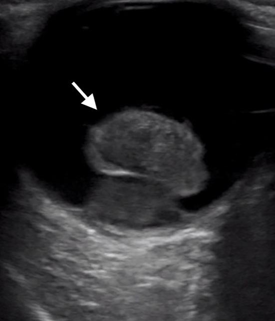
Uveal Melanoma Identified as Ocular Mass on Point-of-care Ultrasound
Clinical Practice and Cases in Emergency Medicine (CPC-EM) is a open-access Medline-indexed internationally recognized journal affiliated with the Western Journal of Emergency Medicine (WestJEM). CPC-EM is distributed electronically to 19,000 emergency medicine scholars. This includes our sponsors California ACEP, the American College of Osteopathic Emergency Physicians, California Chapter of AAEM, and over 83 academic department of emergency medicine subscribers and 8 AAEM State Chapters. Volume V, no. 3: August 2021
iv
Clinical Practice and Cases in Emergency Medicine
CALL FOR SUBMISSIONS
CPC-EM encourages submissions from junior authors and countries with developing emergency medicine programs
CALL FOR CPC-EM SECTION EDITORS! • Medical-Legal Case Reports • Case Reports/Images in Emergency Medicine Send your CV and letter of interest to editor@cpcem.org
Clinicopathological Cases from the University of Maryland
19-year-old Woman with Intermittent Weakness Garrett A. Cavaliere, DO, NRP* Neeraja Murali, DO, MPH† Laura J. Bontempo, MD, MEd† Zachary D.W. Dezman, MD, MS, MS†‡
*University of Maryland Medical Center, Department of Emergency Medicine, Baltimore, Maryland † University of Maryland School of Medicine, Department of Emergency Medicine, Baltimore, Maryland ‡ University of Maryland School of Medicine, Department of Epidemiology and Public Health, Baltimore, Maryland
Section Editor: Rick A. McPheeters, DO Submission history: Submitted: February 1, 2021; Revision received: March 20, 2021; Accepted: April 1, 2021 Electronically published July 27, 2021 Full text available through open access at http://escholarship.org/uc/uciem_cpcem DOI: 10.5811/cpcem.2021.4.52011
Introduction: Systemic weakness is a common chief complaint of patients presenting to the emergency department (ED). A well thought out approach to the assessment and workup of these patients is key to diagnostic accuracy and definitive therapy. Case Presentation: In this case, a 19-year-old female presented to the ED with generalized weakness and near syncope. She had global weakness in her extremities and multiple electrolyte abnormalities. Discussion: This case takes the reader through the differential diagnosis and evaluation of a patient with weakness and profound electrolyte derangements. It includes a discussion of the diagnostic studies and calculations that ultimately led to the patient’s diagnosis. [Clin Pract Cases Emerg Med. 2021;5(3):276–282.] Keywords: Clinicopathological cases; neurology; renal tubular acidosis.
CASE PRESENTATION (DR. CAVALIERE) A 19-year-old female was brought to the emergency department (ED) by emergency medical services (EMS) with complaints of generalized weakness, an inability to move her extremities, and near syncope. The patient stated that she began feeling generalized weakness that morning, which she initially attributed to her “sleeping position.” Over the day the weakness worsened, culminating in difficulty or inability to move her extremities and a near syncopal episode. The patient stated she had attempted to stand up from a seated position when she “felt like [she] was going to pass out.” The patient called 911 for assistance. On further discussion, the patient revealed she had experienced one similar episode of weakness earlier in the year, but this had resolved spontaneously and was not as severe. She does not have a primary care physician and she had never sought care for this complaint. The patient said she noticed generalized abdominal pain, nausea, and constipation, associated with each of these episodes of weakness and lightheadedness. She denied any recent illnesses. She stated she treats her bipolar disorder with daily cannabis and consumes alcohol daily as well.
Clinical Practice and Cases in Emergency Medicine
The patient had a history of anxiety, depression, migraines, and normocytic anemia. Surgical history included an adenoidectomy and tonsillectomy as a child. She had no pertinent family history. Her social history included daily alcohol use, drinking a total of 1.75 liters of vodka over a two-week period. She started smoking when she was 16 years old, smoking a pack per day, but quit a year prior to presentation. The patient smoked cannabis daily. Her only medication was ferrous sulfate 325 milligrams (mg) daily. She had no known drug or environmental allergies. On physical exam, the patient was alert and in no acute distress but appeared tired. She was able to stand unassisted. At the time of triage, she was afebrile (36.6o Celsius), her heart rate was 40 beats per minute, she was breathing 20 times per minute, her blood pressure was 115/90 millimeters of mercury, and she had an oxygen saturation of 98% on room air. She weighed 77.3 kilograms and was 1.65 meters tall (body mass index = 28.3). She was well developed, well nourished and speaking in complete sentences without accessory muscle use. She was oriented as to person, place
276
Volume V, no. 3: August 2021
19-year-old Woman with Intermittent Weakness
Cavaliere et al. and time. She was without sensory deficits and had normal muscle tone. Her strength was 4/5 with elbow flexion and extension, hand grip, knee flexion and extension, and ankle dorsi- and plantar-flexion bilaterally. Deep tendon reflexes were 2+ for the bilateral brachioradialis and patellar reflexes. No clonus could be provoked. She did not have any cranial nerve (II-XII) defects, and she had a normal gait and station. She had normal range of motion of all four extremities, and she did not have any edema. Her lower extremity compartments were soft in both the thighs and the lower legs bilaterally. She exhibited tenderness around her bilateral shoulders and shins. Her head was normocephalic and without signs of injury. Her oropharynx was clear and moist, and her pupils were equal, round, and reactive to light. Her conjunctiva and extraocular motions were normal. Her neck was supple and had a full range of motion, without jugular venous distention or adenopathy. On cardiovascular exam the patient was bradycardic with a regular rhythm, and she had a normal S1/S2 without gallops, friction rubs, or murmurs. On auscultation her breath sounds were clear without wheezes, rales or rhonchi. Her abdomen was non-distended, soft and non-tender throughout with normal bowel sounds. Her skin was warm and dry, and her capillary refill was less than two seconds. She did not have any rashes. Her mood, affect, and behavior were normal. The patient’s electrocardiogram (ECG) is shown (Image). The results of the patient’s initial laboratory evaluation are shown in Table 1. A test was ordered, and a diagnosis was made. CASE DISCUSSION (DR. MURALI) This case involved a young woman with episodic weakness. She reported near syncope, transient extremity paralysis, and generalized weakness. She reported associated nausea, abdominal pain, and constipation. She also reported regular substance use in the form of marijuana and alcohol. Her review of systems was otherwise unremarkable, and notably it was negative for recent illness or gastrointestinal (GI) distress outside of this episode.
Image. Electrocardiogram of a 19-year-old female with weakness.
Volume V, no. 3: August 2021
With this in mind, I began to formulate a differential diagnosis. Episodic weakness, particularly extremity paralysis, suggests metabolic and electrolyte derangements such as hypokalemic periodic paralysis. Weakness may also suggest a primary neurologic condition, including Guillain-Barré syndrome, multiple sclerosis, and other demyelinating disorders. The patient’s near syncope may be due to orthostatic hypotension or neurocardiogenic causes. Her GI symptoms could be due to a broad array of abdominal conditions, but her substance use suggests these symptoms may be related to an ingestion. The patient’s bradycardia could be due to disseminated Lyme disease, myocarditis, or other etiologies of heart block. More information is required. I used the information provided by her physical exam to further refine my differential diagnosis. Her physical exam was notable for a tired-appearing female with bradycardia. Pertinent negative findings included that the compartments of the legs were noted to be soft, clinically excluding a compartment syndrome. Additionally, the patient had no focal neurologic deficits based on the documented neurologic exam. Several findings, including cerebellar signs were not documented, but the patient was noted to have normal gait and station. The mention of normal compartments and the normal neurologic exam suggests that a neurologic cause is unlikely. Further, the case did not provide any imaging studies – notably, there was no neuroimaging included. The patient’s ECG showed a sinus bradycardia with sinus arrhythmia, short QTc with T-wave inversions in aVR and V1, and U waves, but it did not show a heart block. I then reviewed the patient’s laboratory findings. She was noted to have a mild anemia, elevated creatine kinase with myoglobinuria, hematuria, proteinuria, and urinary findings consistent with a urinary tract infection. Additionally, she has multiple electrolyte derangements, including hypokalemia, hyperchloremia with acidosis, hypermagnesemia and hypophosphatemia. She had an elevated creatinine and a mild transaminitis. These laboratory findings suggest her symptoms are due to a metabolic derangement. This patient had a non-anion gap metabolic acidosis. The differential diagnosis for non-anion gap metabolic acidosis includes diarrhea, intestinal fistulae, renal tubular acidosis (RTA), ureteroileostomy, ureterosigmoidostomy, toluene use, ketoacidosis, D-lactic acidosis, and administration of chloriderich solutions.1 After cross-referencing this with the case details, some of these diagnoses can be eliminated based on the history, exam, and review of systems. Specifically, the patient reported constipation, thereby eliminating diarrhea as a cause. She also had no surgical history, hence eliminating ureteroileostomy and ureterosigmoidostomy as causes. Although her diet is not mentioned, there is no reported history of abnormal ingestion of food or fluids; so I reasonably eliminated chloride-rich solution ingestion as a cause. This left proximal and distal RTA, toluene use, ketoacidosis, and D-lactic acidosis as diagnoses under consideration. When
277
Clinical Practice and Cases in Emergency Medicine
19-year-old Woman with Intermittent Weakness
Cavaliere et al.
Table 1. Initial laboratory results of a 19-year-old female with weakness. Laboratory test Patient value Complete blood count
Normal value
White blood cells
7.7 K/mcL
3.4 - 10.8 K/mcL
Hematocrit
31.9%
34.0 - 46.6%
Platelets
239 K/mcL
150 – 450 K/mcL
Serum chemistries
Sodium
139 mmol/L
134 – 144 mmol/L
Potassium
1.3 mmol/L
3.5 - 5.2 mmol/L
Chloride
116 mmol/L
96 – 106 mmol/L
Carbon dioxide
13 mmol/L
20 – 29 mmol/L
Blood urea nitrogen
17 mg/dL
5 – 18 mg/dL
Creatinine
1.16 mg/dL
0.49 - 0.90 mg/dL
Glucose
88 mg/dL
65 – 99 mg/dL
Calcium
10.4 mg/dL
8.9 - 10.4 mg/dL
Magnesium
2.7 mg/dL
1.6 - 2.6 mg/dL
Phosphorus
2.3 mg/dL
2.5 - 4.5 mmol/dL
Total protein
2.3 g/dL
6.3 - 8.2 g/dL
Albumin
4.6 g/dL
3.7 - 5.6 g/dL
Aspartate aminotransferase
65 u/L
14 – 36 u/L
Alanine aminotransferase
31 u/L
0 – 34 u/L
Total bilirubin
0.3 mg/dL
0.2 - 1.3 mg/dL
Alkaline phosphatase
115 u/L
38 – 126 u/L
Creatinine kinase
1117 u/L
30 – 135 u/L
Myoglobin
599 ng/mL
≤ 62 ng/mL
Thyroid stimulating hormone
0.850 mcIU/L
0.450 - 5.330 mcIU/mL
Human chorionic gonadotropin
Negative
Negative
Urine toxicology screen
Cannabis present
Negative
Additional labs
Urinalysis
Color
Straw
Appearance
Slightly cloudy
Clear
Specific gravity
1.004
1.002 - 1.030
pH
7.0
5.0 - 8.0
Glucose
Negative
Negative
Bilirubin
Negative
Negative
Urobilinogen
Negative
Negative
Ketones
Negative
Negative
Blood
2+
Negative
Protein
1+
Negative
Nitrite
Positive
Negative
Leukocyte esterase
1+
Negative
White blood cells
11-25 cells/hpf
0 – 5 cells/hpf
Red blood cells
0-2 cells/hpf
0 – 2 cells/hpf
Squamous epithelial cells
0-2 cells/hpf
0 – 2 cells/hpf
Bacteria
Small
Negative
cells/hpf, cells per high powered field; g/dL, grams per deciliter; K/mcL, thousand cells per microliter; mcIU/mL, micro-international units per milliliter; mg/dL, milligram per deciliter; mmol/L, millimole per liter; ng/mL, nanogram per milliliter; u/L, units per liter.
Clinical Practice and Cases in Emergency Medicine
278
Volume V, no. 3: August 2021
19-year-old Woman with Intermittent Weakness
Cavaliere et al. cross-referencing these with the case details and laboratory findings once again, some options were not consistent with the presentation. Specifically, there was no ketonuria making ketoacidosis unlikely. Lactic acidosis is a result of a hypoperfusion state, and the clinical case did not provide any evidence of hypoperfusion making this unlikely as well. There were some additional laboratory findings outside of the metabolic panel that needed to be considered. Namely, the patient’s hemoglobin and hematocrit were slightly abnormal (although this may be due to her known anemia). Also, she had an elevated creatine kinase and myoglobin as well as slight elevation in her aspartate transaminase. Her urine also showed some hematuria, pyuria, and proteinuria as well as findings of nitrites and leukocyte esterase. When these labs are considered in conjunction with the metabolic abnormalities, my differential diagnosis now included hypokalemic periodic paralysis, rhabdomyolysis, adrenal insufficiency, proximal and distal RTA, inflammatory myopathy, and poisoning (including toluene). Adrenal insufficiency can cause metabolic derangements and presents with symptoms including fatigue, weight loss, GI complaints, and myalgias, and may also include psychiatric symptoms. In primary adrenal insufficiency, the potassium is high and sodium is low, which is not consistent with this case. In secondary or tertiary adrenal insufficiency, potassium is normal or low, sodium can be high or low, and chloride is normal with a low glucose.2 These are not consistent with the findings in this case either; so I eliminated adrenal insufficiency from my differential diagnosis. Inflammatory myopathies present with muscle weakness, cardiac involvement, and laboratory findings including elevated serum creatinine kinase and elevated myoglobin levels in both urine and serum. These patients usually present with acute onset of “antisynthetase syndrome,” constitutional symptoms, Raynaud’s phenomenon, and a nonerosive arthritis.3 While the laboratory findings here were consistent with a possible myopathy, the clinical presentation was not classic, making this a less likely possibility. Another consideration was rhabdomyolysis potentially resulting from compartment syndrome. Compartment syndrome occurs from increased fascial compartment pressure with subsequent tissue hypoperfusion, which can lead to muscle necrosis and rhabdomyolysis. The classic triad of findings in rhabdomyolysis is muscle pain, weakness, and dark urine. Patients with rhabdomyolysis usually have some combination of highly elevated creatine kinase, myoglobinuria, hyperkalemia, hyperphosphatemia, acute kidney injury, hypocalcemia, and metabolic acidosis with or without an anion gap.4 In this patient’s case, there was no clear inciting event, and her symptoms were episodic with spontaneous resolution. Additionally, she did not complain of focal pain or weakness as would be expected in compartment syndrome. Although she did have an elevated creatine kinase, the elevation was not significant and the expected laboratory findings of hyperkalemia
Volume V, no. 3: August 2021
and hyperphosphatemia were not present. I felt that compartment syndrome and rhabdomyolysis were unlikely. In this young adult patient with episodic weakness and hypokalemia, hypokalemic periodic paralysis was immediately considered as part of the differential diagnosis. This condition is characterized by attacks of weakness with a normal neurologic exam in between, as seen in this patient. Primary hypokalemic periodic paralysis follows an autosomal dominant inheritance pattern, and notably this patient had no known family history of the same. Bulbar and respiratory functions are preserved and between attacks, patients will also present with normal plasma potassium. Triggers include stress, exercise, and carbohydrates. The condition also presents with arrhythmias.5 There are, however, other conditions that can cause non-familial hypokalemic paralysis, including RTA.6 All three subtypes of RTA are characterized by an inability to acidify the urine. As a result of this, RTAs present with an increased urine anion gap, but this information was not provided in the case history. In distal or type 1 RTA, there is impaired hydrogen ion secretion in the distal tubule of the nephron. In proximal or type 2 RTA, there is impaired bicarbonate reabsorption in the proximal tubule of the nephron. In type 4 RTA, there is decreased aldosterone secretion or aldosterone resistance.7 As a result of this, type 4 RTA is associated with serum hyperkalemia while the other two types of RTA result in hypokalemia.8 Due to the serum potassium levels, which were not suggestive of aldosterone resistance, I eliminated type 4 RTA from my list of possibilities. The types of hypokalemic RTAs are differentiated by examining the potential of hydrogen (pH) of the patient’s urine. In type 2 (proximal) RTA, urine pH is initially high, then decreases to < 5.5. The urine pH remains above 5.5 in type 1 (distal) RTA.8 This patient had a urine pH of 7.0, suggesting either a type 1 (distal) RTA or an early type 2 (proximal) RTA. Type 1 (distal) RTA can be hereditary or be caused by autoimmune diseases such as Sjögren’s syndrome, or as a complication of chemotherapy or toluene use. The causes of type 2 (proximal) RTA include genetic abnormalities, Fanconi syndrome, monoclonal gammopathy, and carbonic anhydrase inhibitor use.7 There was no mention of chemotherapy or carbonic anhydrase inhibitors with the patient’s presentation. The patient had no family history of similar issues, and it would stand to reason that a genetic abnormality would have come to light before age 19 years. As such, I feel type 1 (distal) RTA is more likely than type 2 (proximal) RTA. The Agency for Toxic Substances and Disease Registry notes that toluene is a solvent found in paints, nail polish, paint thinners, and adhesives, among other substances. It can have toxic effects if ingested or inhaled.9 The findings of acute toluene use include a hypokalemic paralysis and a metabolic acidosis. Patients are also often found to have liver injury and
279
Clinical Practice and Cases in Emergency Medicine
19-year-old Woman with Intermittent Weakness
Cavaliere et al.
rhabdomyolysis, and may present with altered mentation, renal failure, and acidemia.10 This patient’s presentation is most consistent with type 1 (distal) RTA due to toluene use. She denied any illicit drug use but did admit to a history of alcohol ingestion and marijuana use, raising the question of whether there could be toxic alcohols or other coingestions. Unfortunately, there is no diagnostic test for toluene use. However, proximal and distal RTA can be differentiated by calculating the urinary ammonium ion concentration from the measured urine anion gap and osmolar gap. Therefore, my test of choice would be a urine electrolyte panel to calculate the anion gap and osmolar gap. Additionally, I would consult nephrology to assist in management of this patient. CASE OUTCOME (DR. CAVALIERE) The patient developed bigeminy while in the ED but remained hemodynamically stable and did not have a change in her mental status. Her electrolytes were replaced with oral and intravenous potassium, with improvement of her arrythmia and symptoms. She declined central line placement for more rapid replacement. The patient was admitted to the pediatric intensive care unit (PICU) for further management and evaluation. Random urine electrolytes were obtained after the patient was admitted to the PICU (Table 2). Nephrology was consulted because these results demonstrated an elevated urine anion gap suggestive of RTA. The patient’s symptoms completely resolved once her electrolytes were repleted. She was found to have positive antinuclear (ANA-Abs) and anti–Sjögren’s-syndrome-related antigen A (SS-A/Ro) autoantibodies, leading to the diagnosis of Sjögren’s syndrome, despite a lack of phenotypic features. The patient’s urine anion gap was indeterminate for the etiology of her non-anion gap metabolic acidosis; however, her urine osmolar gap of less than 150 milliosmoles per kilogram (mOsm/kg) suggested type 1 or type 4 RTA as the etiology. This coupled with laboratory findings suggestive of autoimmune disease led to the diagnosis of type 1 RTA. Her RTA was treated with potassium supplementation and alkali therapy to achieve a normal serum bicarbonate concentration. Unfortunately, the patient has not been compliant with her home therapy and has required multiple hospitalizations since her original presentation. Her presentation and urine anion gap strongly suggest toluene toxicity, but the patient repeatedly denied insufflating glue and there is no diagnostic test for toluene. RESIDENT DISCUSION (DR. CAVALIERE) Hyperchloremic, hypokalemic metabolic acidosis is a condition all emergency providers should be prepared to diagnose and manage. In this case, the patient presented with a cardiac arrhythmia due to severe hypokalemia. The underlying
Clinical Practice and Cases in Emergency Medicine
etiology of the hypokalemia should be sought while simultaneously treating the condition. The initial ED evaluation includes obtaining a basic metabolic panel and a urinalysis. Once it is determined that the patient does not have a serum anion gap, the clinician should consider three broad categories of non-anion gap acidosis and their etiologies: increased acid production; loss of bicarbonate; and decreased renal excretion of acid (Table 3).11 Type 1 or distal RTA is a primary problem of urine acidification due to impaired hydrogen ion secretion in the distal convoluted tubules. The underlying etiology in adults is usually autoimmune diseases such as Sjögren’s syndrome or rheumatoid arthritis.12 In pediatrics, the cause is usually a hereditary gene mutation for either the basolateral chloridebicarbonate exchanger (SLC4A1) or the apical hydrogenadenosine triphosphatase (ATP6V0A4 and ATP6V1B1) gene.13,14 Lastly, a distal RTA can be iatrogenic due to ifosfamide, a chemotherapeutic analog of cyclophosphamide.15 Type 2 or proximal RTA is a primary problem of impaired bicarbonate reabsorption leading to increased bicarbonate loss.1,7 In adults, the underlying etiology is most commonly proximal tubular toxicity from increased exertion of monoclonal immunoglobulin light chains as seen in multiple myeloma.16 Type 2 RTAs are seen in Fanconi syndrome (loss of bicarbonate with impaired reabsorption of phosphate, glucose, uric acid and amino acids), and in patients prescribed carbonic anhydrase inhibitors (acetazolamide or topiramate).17 In pediatric patients, type 2 RTAs are usually idiopathic, but they can be due to a complication from chemotherapy, cystinosis (genetic disorder causing an accumulation of cystine leading to crystal formation), or inherited mutations in the KCNJ15 (autosomal dominant) and SLC4A4 (autosomal recessive) genes.16,18,19 The term “type 3 RTA” is rarely used as it is now considered a combination of types 1 and 2. Type 4 (hyperkalemic) RTA is beyond the scope of this discussion. The test of choice when evaluating for a RTA is urine electrolytes so that the clinician can calculate how much Table 2. Urine electrolytes of a 19-year-old female with weakness. Urine electrolyte
Patient value
Normal value
Chloride
25 mmol/L
32 - 290 mmol/L
Creatinine
24.6 mg/dL
20 –275 mg/dL
Potassium
11.5 mmol/L
12-129 mmol/L
Sodium
31 mmol/L
28 - 272 mmol/L
Urea
109 mg/dL
---
Glucose
0 mg/dL
0 mg/dL
Osmolality
259 mOsm/kg
50 – 1200 mOsm/kg
Urine anion gap
17.5 mmol/L
0 mmol/L
Urine osmolar gap 146.57 mOsm/kg 80-100 mOsm/kg mg/dL, milligram per deciliter; mmol/L, millimole per liter; mOsm/ kg, milliosmoles per kilogram.
280
Volume V, no. 3: August 2021
19-year-old Woman with Intermittent Weakness
Cavaliere et al. ammonium is being excreted.18 Ammonium excretion will be decreased in a true RTA and normal/increased if the acidosis is due to toluene use or chronic diarrhea. Unfortunately, ammonium excretion is rarely measured directly. Urine ammonium excretion can be estimated by using the urine anion gap or urine osmolar gap.20 The urine anion gap is calculated using the equation: Urine anion gap = [Sodium]urine + [Potassium]urine – [Chloride]urine A urine anion gap of 20-90 millimoles per liter (mmol/L) signifies impaired renal ammonium excretion and is diagnostic of a distal RTA. A value of -20 to –50 mmol/L is diagnostic of diarrhea and ingestions, as it indicates increased ammonium excretion. Values between -20 and 20 mmol/L are considered indeterminate. Urine osmolar gap, on the other hand, is more difficult to calculate. The urine osmolar gap is the difference between the measured and the calculated urine osmolarity. Urine osmolarity is calculated using the equation: Urine osmolarity (milliosmoles/kg) = 2([Sodium]urine + [Potassium] urine) + [Urea]urine + [Glucose]urine Values less than 150 mOsm/kg are consistent with a type 1 or 4 RTA, while values greater than 400 mOsm/kg are consistent with either diarrhea or toluene use.20 Patients with acute presentations of type 1 or 2 RTA should have their electrolytes replaced emergently to prevent cardiac arrythmias. Central line insertion and high concentration potassium replacement should be considered in severe, symptomatic hypokalemia. Chronic type 1 RTA is treated with alkali therapy to achieve a normal serum bicarbonate
Table 3. Categories and etiologies of non-anion gap metabolic acidosis 1,7,11,12 Category
Etiology
Increased acid production
Lactic acidosis Ketoacidosis (starvation, alcoholic and diabetes ketoacidosis)
concentration (22-24 mmol/L).21 The long-term treatment of type 2 RTA depends upon the underlying etiology.22 FINAL DIAGNOSIS Type 1 (distal) renal tubular acidosis with positive SS-A/ Ro antibodies. KEY TEACHING POINTS • Type 1 (distal) RTA is an uncommon cause of a non-anion gap metabolic acidosis. • A type 1 RTA can be diagnosed using a basic metabolic panel, the urine pH from a urinalysis, and by measuring urine electrolytes. • An elevated urine anion gap or decreased urine osmolar gap will clinch the diagnosis of a type 1 RTA.
The authors attest that their institution requires neither Institutional Review Board approval, nor patient consent for publication of this case report. Documentation on file.
Address for Correspondence: Laura Bontempo, MD, MEd, University of Maryland, Department of Emergency Medicine, 110 S Paca Street, 6th floor, Suite 200, Baltimore, MD 21201. Email: Lbontempo@som.umaryland.edu Conflicts of Interest: By the CPC-EM article submission agreement, all authors are required to disclose all affiliations, funding sources and financial or management relationships that could be perceived as potential sources of bias. The authors disclosed none. Copyright: © 2021 Cavaliere et al. This is an open access article distributed in accordance with the terms of the Creative Commons Attribution (CC BY 4.0) License. See: http://creativecommons.org/ licenses/by/4.0/ REFERENCES 1. Kraut J and Madias N. Differential diagnosis of nongap metabolic acidosis: value of a systematic approach. Clin J Am Soc of Nephro.
Ingestions (methanol, ethylene glycol, aspirin, toluene, diethylene glycol, propylene glycol, D-lactic acidosis) Loss of bicarbonate
2012;7(4):671-79. 2. Phipps A. Adrenal Crisis in the ED. emDOCs.net - Emergency Medicine Education. 2015. http://www.emdocs.net/adrenal-crisis-in-
Diarrhea
the-ed/. Accessed July 10, 2020. 3. Schmidt J. Current classification and management of inflammatory
Type 2 (proximal) RTA
myopathies. J Neuromuscul Dis. 2018;5(2):109-29.
Ketoacidosis recovery Carbonic anhydrase inhibitor use
4. Huerta-Alardín AL, Varon J, Marik PE. Bench-to-bedside review:
Ureteral diversion (ureteroileostomy, ureterosigmoidostomy)
5. Statland JM, Fontaine B, Hanna MG, et al. Review of the diagnosis
rhabdomyolysis – an overview for clinicians. Crit Care. 2004;9(2):158. and treatment of periodic paralysis. Muscle Nerve. 2018;57(4):522-30.
Decreased renal Type 1 (distal) RTA, Type 4 RTA excretion of acid RTA, renal tubular acidosis.
Volume V, no. 3: August 2021
6. Phuyal P and Nagalli S. (2021). Hypokalemic periodic paralysis. In: StatPearls [Internet]. Treasure Island, FL: StatPearls Publishing.
281
Clinical Practice and Cases in Emergency Medicine
19-year-old Woman with Intermittent Weakness
Cavaliere et al.
7. Dey A. Renal tubular acidosis (RTA). 2017. Available at: https://
14. Jobst-Schwan T, Klämbt V, Tarsio M, et al. Whole exome sequencing
medsphere.wordpress.com/2017/08/07/rta/. Accessed July 19, 2020.
identified ATP6V1C2 as a novel candidate gene for recessive distal
8. Santos F, Gil-Peña H, Alvarez-Alvarez S. Renal tubular acidosis. Curr Opin Pediatr. 2017;29(2):206-10.
renal tubular acidosis. Kidney Int. 2020 Mar;97(3):567-579. 15. Skinner R, Pearson AD, Price L, et al. Nephrotoxicity after ifosfamide.
9. Taylor J, Fay M, Williams R, et al. U.S. Department of Health and
Arch Dis Child. 1990;65(7):732-8.
Human Services Public Health Service Agency for Toxic Substances
16. Maldonado JE, Velosa JA, Kyle RA, et al. Fanconi syndrome in
and Disease Registry. 2017. Available at: https://www.atsdr.cdc.gov/
adults. A manifestation of a latent form of myeloma. Am J Med.
toxprofiles/tp56.pdf. Accessed January 15, 2021. 10. Camara-Lemarroy CR, Rodríguez-Gutiérrez R, Monreal-Robles R,
1975;58(3):354-64. 17. Mirza N, Marson AG, Pirmohamed M. Effect of topiramate on acid-
et al. Acute toluene intoxication--clinical presentation, management
base balance: extent, mechanism and effects. Br J Clin Pharmacol.
and prognosis: a prospective observational study. BMC Emerg Med. 2015;15(1):19.
2009;68(5):655-61. 18. Palmer BF and Clegg DJ. Hyperchloremic normal gap metabolic
11. Rose BD and Post TW. (2001). Metabolic Acidosis. In Wonsciewics M, McCullough K & David K (Eds.), Clinical Physiology of Acid-
acidosis. Minerva Endocrinol. 2019;44(4):363-77. 19. Gahl WA, Thoene JG, Schneider JA, et al. Cystinosis: progress in a
Base and Electrolyte Disorders. 5th ed. (578-646). New York, NY: McGraw-Hill.
prototypic disease. Ann Intern Med. 1988;109(7):557-69. 20. Kim GH, Han JS, Kim YS, et al. Evaluation of urine acidification by
12. Cohen EP, Bastani B, Cohen MR, et al. Absence of H(+)-ATPase in
urine anion gap and urine osmolar gap in chronic metabolic acidosis.
cortical collecting tubules of a patient with Sjogren’s syndrome and distal renal tubular acidosis. J Am Soc Nephrol. 1992;3(2):264-71.
Am J Kidney Dis. 1996;27(1):42-7. 21. Morris RC Jr and Sebastian A. Alkali therapy in renal tubular acidosis:
13. Jarolim P, Shayakul C, Prabakaran D, et al. Autosomal dominant distal renal tubular acidosis is associated in three families with
Who needs it? J Am Soc Nephrol. 2002;13(8):2186-8. 22. Lopez-Garcia SC, Emma F, Walsh SB, et al. Treatment and long-
heterozygosity for the R589H mutation in the AE1 (band 3) Cl-/
term outcome in primary distal renal tubular acidosis. Nephrol Dial
HCO3- exchanger. J Biol Chem. 1998 Mar 13;273(11):6380-8.
Transplant. 2019;34(6):981-91.
Clinical Practice and Cases in Emergency Medicine
282
Volume V, no. 3: August 2021
Medical Legal Case Report
Three Cases of Emergency Department Medical Malpractice Involving “Consultations”: How Is Liability Legally Determined? Alaa Aldalati, MBBS* Venkatesh R. Bellamkonda, MD† Gregory P. Moore, MD, JD† Alexander S. Finch, MD†
*Mayo Clinic College of Medicine and Science, Mayo Clinic School of Graduate Medical Education, Rochester, Minnesota † Mayo Clinic College of Medicine and Science, Department of Emergency Medicine, Rochester, Minnesota
Section Editor: Melanie Heniff, MD, JD Submission history: Submitted April 17, 2021; Revision received July1, 2021; Accepted July 2, 2021 Electronically published August 4, 2021 Full text available through open access at http://escholarship.org/uc/uciem_cpcem DOI: 10.5811/cpcem.2021.7.52680
This article presents three successfully litigated medical malpractice cases involving emergency physicians and consultants. We discuss the respective case medical diagnoses, as well as established legal principles that determine in a court proceeding which provider will be liable. Specifically, we explain the legal principles of “patient physician relationship” and “affirmative act.” [Clin Pract Cases Emerg Med. 2021;5(3):283–288.]
INTRODUCTION Emergency physicians (EP) often consult other specialty services to assist in optimal patient care. Here we present three cases that involved consultation and ultimately resulted in successfully litigated malpractice cases. Many EPs and consultants are unaware of the legal principles that govern their liability if a lawsuit is pursued. Specifically, we clarify what defines a “patient physician relationship” and an “affirmative act” as well as the pathophysiology and risk management of the diagnosis in each case. Case 1: Sozomentou v Arfaras – Florida A 66-year-old man presented to an emergency department (ED) with chest pain. After examination by the EP he was evaluated with a chest radiograph. The EP interpreted the study as “within normal limits” and reviewed the radiologist’s interpretation of “top normal size heart with tortuous aorta.” The patient was subsequently admitted to the hospital with a diagnosis of chest pain and rule out myocardial infarction. That evening, he died from an aortic dissection (AD) with associated pericardial tamponade. A lawsuit brought against the EP and the radiologist claimed that the radiologist failed to recognize and suggest to the EP the possibility of an AD and the need for immediate computed tomography (CT) of the chest. A second claim alleged that the EP failed to include AD in the differential diagnosis and order a CT of the chest. The radiologist maintained that they did not Volume V, no. 3: August 2021
exclude the diagnosis and it is the EP’s duty to clinically provide appropriate care and orders. After deliberation, a jury awarded the plaintiffs $6.4 million; they assigned 2% of the amount to the radiologist, 80% to the EP, and 18% to the inpatient provider.1 Case 2: Estate of Fischel v Mujic M – New Jersey A 39-year-old man presented to the ED via ambulance complaining of difficulty breathing. An electrocardiogram (ECG) was done. The cardiologist who over-read the tracing reported findings consistent with myocardial infarction and communicated this with the EP. The EP discharged the patient with a diagnosis of bronchitis. The patient’s wife had delivered a son two days prior and was discharged from the hospital at the same time. The next day the man collapsed on the floor and cardiopulmonary resuscitation was initiated. An ambulance was called, but nevertheless he died. A lawsuit was brought claiming that the EP should not have discharged the patient, especially with the cardiologist’s interpretation of the ECG obtained in the ED. The cardiologist claimed the entire fault rested with the EP. The EP’s defense was that his care was reasonable and he was not informed by the cardiologist of his reading. The case was settled by the EP for $2 million.2 Case 3: Anonymous N.P. v Anonymous Physician Parents brought their three-year-old son to the ED complaining that he had put a watch battery in his nose. The 283
Clinical Practice and Cases in Emergency Medicine
Three Cases of ED Medical Malpractice Involving “Consultations” provider did radiographs of the chest and abdomen and did not see a foreign body. On nasal examination they were unable to visualize the battery. An otolaryngologist (ENT) was called; the emergency provider was instructed to have the parents call the ENT office at eight in the morning for their son to be seen, and to have the child not take any oral liquids or solids after midnight and wait until he was seen by the ENT physician. The parents asked if their child could be taken to a nearby children’s hospital, but they were reassured that it was not necessary and were also instructed to return if there was any respiratory distress. About six hours later the child began having discharge from his nose. His parents took him to another hospital where a specialist removed the watch battery from his nose. By that time there was extensive tissue necrosis resulting in a perforated nasal septum requiring surgery. A lawsuit for malpractice was filed with the court for delay in care. The lawsuit was filed against the emergency provider and the ENT specialist. The ENT physician asked for the case to be dismissed with respect to his care, claiming he did not have a physician-patient relationship. He also claimed that the plan was a general one in the event he did see the patient the next day and that it was not specific care. A physician-patient relationship is required as the first element of a malpractice action. The court stated: “there is no physician patient relationship if the physician does not see, treat, or in any way participate in the care and diagnosis.” The court also said “the physician must perform some affirmative act.” The court examined the facts and declared that the ENT physician had placed an order for “nothing per mouth” status and directed when follow-up care was required, demonstrating a participation in care that constituted an affirmative act.3 DISCUSSION Aortic Dissection and Risk Management Acute AD is rare, extremely dangerous, and presents variably, resulting in a diagnostically challenging and highrisk situation for EPs. Aortic dissection has an estimated incidence of about 0.006% per year.4 Despite the rarity of AD, mortality may reach as high as 50% at 48 hours, 90% at one month if not operated upon, and even 30% at one month if operated upon.5,6 As a possible diagnosis, AD may occur secondary to predisposing factors including hypertension and smoking, as well as genetic conditions such as Marfan syndrome, congenital bicuspid aortic valve disease, and vasculitides. Aortic dissection may be acquired secondary to trauma or iatrogenically from healthcare procedures.7 While chest pain is the most frequent symptom reported in patients with acute AD, back pain, abdominal pain, syncope, neurologic symptoms, hypoperfusion syndromes, and other symptoms have been reported as well.8 Ordering the appropriate testing to evaluate for dissection is critical. Intravenous contrast-enhanced CT has sensitivity Clinical Practice and Cases in Emergency Medicine
Aldalati et al.
and specificity values between 95-98% and is widely available compared with transesophageal echocardiography and magnetic resonance imaging, making CT the mainstay of evaluation in EDs nationally.8 Evaluation of chest pain, which is the presenting complaint for about 10 million ED visits annually,9 costs the healthcare system about $10 billion each year in the United States.10 Indiscriminate use of CT may pose a risk from radiation to individual patients, as well as unnecessary cost. To assist the clinician in making a determnation, many have incorporated the use of D-dimer to exclude the diagnosis, citing its very high sensitivity values.11 However, a clinical policy statement from the American College of Emergency Physicians (ACEP) discourages reliance on D-dimer as a singular tool to exclude the diagnosis because the quality of studies supporting this practice are felt to be inadequate (February 2018). The European Society of Cardiology recommends using D-dimer to shift the degree of suspicion up and down rather than to make or exclude the diagnosis.4 When examining litigation of AD cases, a review by Elefteriades et al included 23 patients who had acute ADs, 22 of them fatal. The most common category of malpractice alleged was failure to diagnose or delayed diagnosis. This review showed that lawsuits were brought against a variety of physicians including EPs, radiologists, cardiothoracic surgeons, and many others including obstetriciangynecologists. In about two thirds of the cases, the medical care was felt to be suboptimal. The authors recommended simply including AD in the differential diagnosis, performing appropriate testing to assess the likelihood of this disease, and interpreting the results of the testing correctly. These three steps will both enhance diagnosis and decrease the likelihood that patients and families will perceive that they were treated suboptimally, reducing the chance of litigation.12 Electrocardiograms and the Diagnosis of Acute Myocardial Infarction The American Heart Association estimates that a myocardial infarction will occur approximately every 40 seconds in the US, making it a common emergency faced by healthcare teams. Unfortunately, heart disease is also the leading cause of death, representing just over 840,000 deaths domestically in 2016.13 Given the significant morbidity and mortality, this disease process is a focus of emergency medicine (EM) practice. Screening ECGs are commonly performed and read for findings associated with acute myocardial infarction (AMI). Acute coronary syndrome (ACS), as a spectrum of disease, is commonly associated with chest pain; however, dyspnea, diaphoresis, and jaw and arm pain are also presenting symptoms.14 Electrocardiogram changes can raise concern for AMI and ischemia. Furthermore, in many settings ECG findings in conjunction with a concerning history are an indication for percutaneous intervention (PCI) or thrombolytics with
284
Volume V, no. 3: August 2021
Aldalati et al.
Three Cases of ED Medical Malpractice Involving “Consultations”
a goal of restoring myocardial perfusion. Well established benchmarks of door-to-intervention times for both thrombolytics and PCI highlight the principle that any delay in diagnosis or treatment of AMI is deleterious to the patient.15 This principle was underscored by ACEP in its updated 2017 clinical policy statement “ED Management of Patients Needing Reperfusion Therapy for Acute ST-Segment Elevation Myocardial Infarction,” where there was a Level B recommendation that fibrinolytics be administered to patients when door-to-balloon time is anticipated to exceed 120 minutes.16 These well delineated benchmarks outline an expected standard of care for a high-risk disease process, which can also factor into the medicolegal risk involved. Patients with missed AMI on their index visit are at increased risk for ensuing cardiac events.17 These adverse outcomes increase morbidity and mortality for this patient population.18 Thus, careful interpretation of a patient’s ECG by the EP in conjunction with a detailed history and physical exam are necessary in all cases of patients presenting with symptoms that may be associated with AMI. In assessing the literature evaluating the medicolegal consequences of missed myocardial infarction, two clear themes emerge: 1) ACS represents a high-risk disease entity that is associated with more malpractice dollars recovered than any other condition19; and 2) it is a common source of litigation in the ED, representing up to 20% of EM-associated settlement funds.17,20 A 2010 review of ED malpractice claims found that AMI was the second most common disease process associated with a claim (5% of claims).21,22 Recognition of AMI based on ECG findings can be difficult and prone to error, making these misinterpretations among the costliest mistakes in terms of malpractice dollars.23 Actual rates of missed AMI are unknown, but studies estimate this to be about 2% in the US, which is quite high for such a high-stakes diagnosis.18,19 A more recent article affirmed that AMI accounts for the second most common malpractice claim in ED and urgent care settings.24 Translating an ECG into an AMI diagnosis requires both accurate interpretation of the ECG and correct extrapolation that the visualized pattern represents disease and not a mimic of disease. Studies looking specifically at ECG interpretations have found that misdiagnosis rates and accuracy can be variable among EPs. One study that focused on ECGs with ST-segment elevation found there was misinterpretation as to the underlying cause of the elevation in 5.9% of cases.25 Another study found that accuracy in identifying ST-segment elevation myocardial infarction (STEMI) representing acute coronary occlusion based on ECG was only 69.1%.26 Finally, these themes exist not only in the US but are seen internationally as well. A 2017 study of closed malpractice claims associated with AMI in Taiwan found that misdiagnosis was the most common dispute associated with claims.27 In summary, ACS represents a disease process with increased morbidity and mortality to the patient if the diagnosis is missed. It is a relatively common source of Volume V, no. 3: August 2021
litigation with high malpractice recovery and therefore represents a significant medicolegal risk. Given this risk, great care must be taken to consider ACS in the differential and to evaluate it thoroughly. Nasal Button Battery Foreign Body While most EPs are aware of the morbidity associated with gastrointestinal ingestion of button batteries, many may not be familiar with the significant risk when they are located in the nose. Patients are often asymptomatic, and many cases are unanticipated discoveries. Nasal foreign bodies (FB) are usually lodged in the floor of the nasal passage, below the inferior turbinate, or in the superior fossa anterior to the middle turbinate. Patients typically present with one-sided, foulsmelling nasal discharge. Having patients simply blow their nose while closing the alternate side may remove the FB. If it is not visualized, then referral to a specialist is indicated. The vast majority of patients in these cases are less than eight.years old. Button batteries are FBs that release toxic substances (eg, silver, zinc, mercury, or lithium), and cause local electrical burns. An electrical circuit is completed when lodged, and injury usually happens on the anode side of the battery. Subsequent electrolyte leakage results in a caustic injury. Pressure necrosis caused by an impacted FB is a third mechanism of pathology. The complications in the nasal cavity are based on length of time between placement and removal, the orientation of the impacted battery, and the site of contact of the negative pole (anode). If the anode is in contact with the septum or turbinate, then ulceration can occur in just 3-6 hours. Ulceration of the inferior meatus, saddle deformities, chondritis, rhinitis, and alar collapse can eventually result as well. The EP must obtain emergency removal when encountering a button battery in a patient’s nose.28,29 Legal Concepts when Determining Liability Regarding ED Consultants To successfully litigate a malpractice claim, four legal elements must be met: 1) a duty was owed to the patient; 2) the duty was breached (standard of care not met); 3) injury occurred to the patient; and 4) the injury was caused by the breach of duty (causation). To prevail in a malpractice lawsuit against a physician, the first element of a patient-physician relationship (duty) must be established. While an in-person evaluation may satisfy this element, it is clear in the case law that the relationship actually hinges on whether an affirmative act was performed by the physician on behalf of the patient. As in our reported pediatric case involving a retained battery demonstrates, the affirmative act can occur even via a telephone call. Courts have further delineated duty. In Walters v Rinker, the court defined an affirmative act by the physician as an action for the benefit of the patient. For example, this would involve participation in diagnosis or treatment of the patient.30 In contrast, merely having physical contact with a patient
285
Clinical Practice and Cases in Emergency Medicine
Three Cases of ED Medical Malpractice Involving “Consultations” does not establish a physician-patient relationship. In Giles v Anonymous Physician I, a hospitalist presented to the bedside of a patient and checked her medical chart. The hospitalist quickly realized that the patient would not be appropriate for their service. The court found that the hospitalist did not perform any affirmative act by presenting to the bedside or reviewing the medical record and, therefore, there was no physician-patient relationship.31 Emergency physicians commonly discuss patients with specialists by telephone. In the event of an adverse patient outcome resulting in a malpractice suit, consultants may be liable only if they establish a physician-patient relationship with the plaintiff. Courts have found that this requirement exceeds the scope of many “curbside” conversations but is established only when the specialist takes an affirmative action toward the patient. This often entails participating in diagnosis or treatment, which would be an action for the benefit of the patient. Establishing a patient-physician relationship sets up the basis to create medical liability on the physician’s part. Two essential parts confirm this relationship: 1) the patient requests, either directly or via representatives, that a physician provide care; and 2) the physician in return performs an “affirmative act” or shows “intent of care.” It is important to keep in mind that the affirmative act is not only applicable after a direct physician-patient contact. For example, if a test was interpreted by a specialist and advice was given based on it, a patientphysician relationship is established.32 In Walters v Rinker, examining a pathological specimen was enough to establish a patient-physician relationship as the recommendations were used to plan further treatment. Despite an initial pathology report stating that the finding appeared to be benign, two years later the patient was diagnosed with large cell lymphoma in the setting of declining physical health. The court in this case decided that since the treatment and interventions were based on this test result, the pathologist performed an affirmative act to contribute to this patient’s care.30 Medicine is a collegial profession. Often, EPs “curbside” other consulting services as “informal” consults regarding test interpretation or further guidance on treatment. For a consultant to be held legally liable usually a patient-physician relationship must be established.32 Generally, physicians who are not on duty are not obligated to treat patients; therefore, they mostly are not held accountable.33 Moreover, an on-call consultant giving general advice over the phone does not necessarily establish a relationship with the patient regardless of whether the caller was another physician or the patient. The relationship is established by providing patient- specific advice, or giving direct recommendations to the patient directly or through another party such as the EP.34 In addition, documenting in the patient’s chart or billing the patient for a consult, even if there was no physical interaction, strongly implies that a patient relationship exists via an affirmative act. Generally, unless one of the aforementioned circumstances are met, courts tend to be Clinical Practice and Cases in Emergency Medicine
Aldalati et al.
hesitant to hold consultants strictly liable. The courts generally have the opinion that physicians hold a unique set of skills and knowledge that can highly impact society and save lives. Holding the physician accountable for every phone call or professional conversation could negatively impact the willingness of medical specialtists to provide others with general recommendations. The court’s hesitation to hold consultants liable was illustrated in the case of Bessenyei v Raiti. The patient suffered an injury after paint thinner was injected into his thumb. Initially he was cared for by Dr. Raiti in the ED. Dr. Raiti recommended that the patient seek care at a specialized center immediately. However, the patient refused and was subsequently discharged home with antibiotics and a followup on the following Monday. Dr. Raiti had called Dr. Birely, a plastic surgeon, who agreed with Dr. Raiti’s plan. The patient’s condition deteriorated resulting in an amputation to the tip of his thumb. The patient sued both Dr. Raiti and Dr. Birely. The court ruled that Dr. Birely was not liable as no patientphysician relationship had been established for the following two reasons: 1) Dr. Birely was not on-call that evening, and although he agreed with Dr. Raiti, he did not provide specific advice for the patient; 2) Dr. Raiti was the immediate care provider who independently had the ability and control to either accept or reject Dr. Birely’s recommendations.33 A contrasting example is in Diggs v Arizona Cardiologists, Ltd. Dr. Valdez, a cardiologist, was determined to hold a duty of care toward Mrs. Diggs during what appeared to be an “informal consult.” Mrs. Diggs presented to the ED with chest pain, where she was originally being cared for by Dr. Johnson, an EP. Dr. Johnson thought that the most likely diagnosis was pericarditis but was also considering the diagnosis of myocardial infarction. Therefore, he sought expert advice from Dr. Valdez, who happened to be passing by the ED that day. Although he was not the on-call cardiologist, Dr. Valdez reviewed Dr. Johnson’s history, physical examination, and the patient’s ECG. He agreed with Dr. Johnson that it was most likely pericarditis. Mrs. Diggs was discharged from the ED. Three hours later, she suffered a cardiac arrest and died. Her family brought suit against both the cardiologist and the EP. Dr. Valdez claimed that he did not have a patient-physician relationship, and thus no liability. The court determined that Dr. Valdez reviewed the specific information available on Mrs. Diggs and gave advice and recommendations in his role as an expert that impacted the care Dr. Johnson provided. The court thus held Dr. Valdes was a liable party. Although he was not the on-call cardiologist, the court deemed that a patient-physician relationship had been established as he had given advice specific to Mrs. Diggs’s condition. Additionally, the court ruled that Dr. Johnson lacked the necessary skills and knowledge to interpret the ECG as he did not have admitting privileges. The court held that Dr. Johnson had relied on the expertise of Dr. Valdez when he made his decision to discharge the patient.35 286
Volume V, no. 3: August 2021
Aldalati et al.
Three Cases of ED Medical Malpractice Involving “Consultations”
CONCLUSION The reader should now clearly understand the rationale that led to the legal outcomes in these three reported cases. The first case represents a typical scenario in which all parties were held accountable for medical negligence; each of them had patientphysician relationship and performed an affirmative act. In the second case, the court ruled that there was no affirmative act made by the consultant; therefore the consultant was not held liable. Finally, in the third case, the court clearly stated why they made their decision. The consultant made an affirmative act that impacted the care delivered in the ED. In the three cases presented there were poor patient outcomes after interactions between EPs and consultants. Courts have defined when consultant/EP co-participation in cases results in specific liability. It is important for the EP to understand when they are solely responsible for a patient, and when consultants will share the liability. If the EP desires consultant’s shared liability, the EP should take the initiative to establish the physician-patient relationship between the consultant and the patient. The simplest way to ensure that consultants share liability is to solicit an “affirmative act.” This could be accomplished by asking the consultant to see the patient or alternatively discuss the patient’s specific details including history, physical examination, and test results.
Address for Correspondence: Alaa Aldalati, MBBS, Mayo Clinic, Department of Emergency Medicine, 200 First St SW, Rochester, Minnesota 55905. Email: aldalati.alaa@mayo.edu. Conflicts of Interest: By the CPC-EM article submission agreement, all authors are required to disclose all affiliations, funding sources and financial or management relationships that could be perceived as potential sources of bias. The authors disclosed none. Copyright: © 2021 Aldalati et al. This is an open access article distributed in accordance with the terms of the Creative Commons Attribution (CC BY 4.0) License. See: http://creativecommons.org/ licenses/by/4.0/
REFERENCES 1. Estate of Kyriacos Sozomentou v Artaras, et al.: Broward County (FL) Circuit Court p. 4. 2. Estate of Joshua A. Fischel v Lejla Mujic MD et al.: Essex County (NJ) Superior Court. p. 5. 3. Anonymous N.P versus Anonymous Physician Court of Appeals 4. Erbel R, Aboyans V, Boileau C, et al. 2014 ESC Guidelines on the diagnosis and treatment of aortic diseases: document covering acute and chronic aortic diseases of the thoracic and abdominal aorta of the adult. The Task Force for the Diagnosis and Treatment of Aortic
Take-home Points 1. The accuracy of identifying STEMI on ECG is less than 70%. It is critical to recognize ECG abnormalities to avoid liability and poor patient outcome. 2. Missed myocardial infarction is extremely significant as it accounts for 5% of EP malpractice claims and represents up to 20% of EP dollars paid out in those claims. 3. Aortic dissection is rare, and the most common lawsuit is for failure to diagnose. 4. The ability to intervene and improve mortality hinges upon being able to make the diagnosis accurately and timely and requires the following: 5. Consideration of AD within a differential diagnosis for an individual patient a) Using the appropriate testing b) Correctly interpreting the results of the testing. c) For a consultant to be held liable, the consultant must have a patient-physician relationship that is established by an “affirmative act.” Otherwise, the EP may solely hold the liability. 6. An affirmative act is elicited by seeing the patient or providing patient-specific advice based on a specific patient’s information. 7. An affirmative act is not obtained by answering a general question that is not patient specific.
Diseases of the European Society of Cardiology (ESC). Eur Heart J. 2014;35(41):2873-926. 5. Chiappini B, Schepens M, Tan E, et al. Early and late outcomes of acute type A aortic dissection: analysis of risk factors in 487 consecutive patients. Eur Heart J. 2005;26(2):180-6. 6. Trimarchi S, Nienaber CA, Rampoldi V, et al. Contemporary results of surgery in acute type A aortic dissection: The International Registry of Acute Aortic Dissection experience. J Thorac Cardiovasc Surg. 2005;129(1):112-22. 7. Erbel R, Aboyans V, Boileau C, et al. 2014 ESC guidelines on the diagnosis and treatment of aortic diseases: document covering acute and chronic aortic diseases of the thoracic and abdominal aorta of the adult. The Task Force for the Diagnosis and Treatment of Aortic Diseases of the European Society of Cardiology (ESC). Eur Heart J. 2014;35(41):2873-926. 8. Baliga RR, Nienaber CA, Bossone E, et al. The role of imaging in aortic dissection and related syndromes. JACC Cardiovasc Imaging. 2014;7(4):406-24. 9. Tomaszewski CA, Nestler D, Shah KH, et al. Clinical policy: Critical Issues in the Evaluation and Management of Emergency Department Patients with Suspected non-ST-Elevation Acute Coronary Syndromes. Ann Emerg Med. 2018;72(5):e65-e106. 10. Riley RF, Miller CD, Russell GB, et al. Cost analysis of the History, ECG, Age, Risk factors, and initial Troponin (HEART) Pathway randomized control trial. Am J Emerg Med. 2017;35(1):77-81.
The authors attest that their institution requires neither Institutional Review Board approval, nor patient consent for publication of this case report. Documentation on file.
Volume V, no. 3: August 2021
11. Elefteriades JA. Physicians should be legally liable for missing an atypical aortic dissection: CON. Cardiol Clin. 2010;28(2):245-52. 12. Elefteriades JA, Barrett PW, and Kopf GS. Litigation in nontraumatic
287
Clinical Practice and Cases in Emergency Medicine
Three Cases of ED Medical Malpractice Involving “Consultations” aortic diseases: a tempest in the malpractice maelstrom. Cardiology.
20. Herren KR, and Mackway-Jones K. Emergency management of cardiac
2008;109(4):263-72. 13. Benjamin EJ, Muntner P, Alonso A, et al. Heart Disease and Stroke
chest pain: a review. Emerg Med J. 2001;18(1):6-10. 21. Brown TW, McCarthy ML, Kelen GD, et al. An epidemiologic study of
Statistics-2019 Update: A report From the American Heart Association.
closed emergency department malpractice claims in a national database
Circulation. 2019;139(10):e56-e528. 14. Anderson JL, Adams CD, Antman EM, et al. ACC/AHA 2007 Guidelines
of physician malpractice insurers. Acad Emerg Med. 2010;17(5):553-60. 22. Vukmir RB. Medical malpractice: managing the risk. Med Law.
for the Management of Patients with Unstable Angina/Non-ST-Elevation Myocardial Infarction: a report of the American College of Cardiology/
2004;23(3):495-513. 23. Mele PF. The ECG dilemma: guidelines on improving interpretation. J
American Heart Association Task Force on Practice Guidelines (Writing Committee to Revise the 2002 Guidelines for the Management
Healthc Risk Manag. 2008;28(2):27-31. 24. Wong KE, Parikh PD, Miller KC, et al. Emergency department and
of Patients With Unstable Angina/Non-ST-Elevation Myocardial
urgent care medical malpractice claims 2001-15. West J Emerg Med.
Infarction) developed in collaboration with the American College of Emergency Physicians, the Society for Cardiovascular Angiography and
2021;22(2):333-8. 25. Brady WJ, Perron A, and Ullman E. Errors in emergency physician
Interventions, and the Society of Thoracic Surgeons endorsed by the
interpretation of ST-segment elevation in emergency department chest
American Association of Cardiovascular and Pulmonary Rehabilitation and the Society for Academic Emergency Medicine. Circulation.
pain patients. Acad Emerg Med. 2000;7(11):1256-60. 26. Veronese G, Germini F, Ingrassia S, et al. Emergency physician
2007;116(7):E148-E304.
accuracy in interpreting electrocardiograms with potential ST-segment
15. Antman EM, Anbe DT, Armstrong PW, et al. ACC/AHA guidelines
elevation myocardial infarction: Is it enough? Acute Card Care.
for the management of patients with ST-elevation myocardial infarction--executive summary: a report of the American College
2016;18(1):7-10. 27. Wu KH, Yen YL, Wu CH, et al. Learning from an analysis of closed
of Cardiology/American Heart Association Task Force on Practice
malpractice litigation involving myocardial infarction. J Forensic Leg
Guidelines (Writing Committee to Revise the 1999 Guidelines for the Management of Patients with Acute Myocardial Infarction). Circulation.
Med. 2017;48:41-5. 28. P. Sebastian G, Kaliyan R, and Devadason P. Button battery foreign
2004;110(5):588-636.
body among children: a case series. Int Otorhinolaryngol Head Neck
16. Promes SB, Glauser JM, Smith MD, et al. Clinical policy: emergency department management of patients needing reperfusion therapy for
Surg. 2020;6(5):5. 29. Heim SW, Maughan KL. Foreign bodies in the ear, nose, and throat. Am
acute ST-segment elevation myocardial infarction. Ann Emerg Med. 2017;70(5):724-39. 17. Collinson PO, Premachandram S, Hashemi K. Prospective audit of
Fam Physician. 2007;76(8):1185-9. 30. Walters v Rinker. Ind. Ct. App; 1988. 31. Giles v. Anonymous Physician I. Court of Appeals of Indiana; July,
incidence of prognostically important myocardial damage in patients discharged from emergency department. BMJ. 2000;320(7251):1702-4.
21, 2014. 32. Moore JJ, and Matlock AG. Shared liability? Consultants, pharmacists,
18. Pope JH, Aufderheide TP, Ruthazer R, et al. Missed diagnoses of
and the emergency physician: legal cases and caveats. J Emerg Med.
acute cardiac ischemia in the emergency department. N Engl J Med. 2000;342(16):1163-70.
Aldalati et al.
2014;46(5):612-6. 33. Bessenyei v Raiti. Maryland District Court. No. JFM-01–1029, June
19. Schull MJ. The risk of missed diagnosis of acute myocardial infarction
9, 2003.
associated with emergency department volume: In reply. Ann Emerg
34. Curbside consultations. Psychiatry (Edgmont). 2010;7(5):51-3.
Med. 2007;50(2):203-4.
35. Diggs v. Arizona Cardiologists Ltd. Ariz. Ct. App; 2008. p. 386.
Clinical Practice and Cases in Emergency Medicine
288
Volume V, no. 3: August 2021
Case Series
Nebulized Tranexamic Acid in Secondary Post-Tonsillectomy Hemorrhage: Case Series and Review of the Literature Mira Dermendjieva, PharmD, BCCCP* Anand Gopalsami, MD, MBA† Nicole Glennon, PA-C, MPAP† Sam Torbati, MD†
*Cedars Sinai Medical Center, Department of Pharmacy Los Angeles, California † Cedars Sinai Medical Center, Department of Emergency Medicine, Los Angeles, California
Section Editor: Scott Goldstein, MD Submission history: Submitted March 22, 2021; Revision received May 24, 2021; Accepted May 28, 2021 Electronically published July 27, 2021 Full text available through open access at http://escholarship.org/uc/uciem_cpcem DOI: 10.5811/cpcem.2021.5.52549
Introduction: Post-tonsillectomy hemorrhage is a serious postoperative complication, and its acute management can present a challenge for the emergency provider. Although various strategies have been proposed, guidance on the best approach for management of this condition in the emergency department (ED) setting remains limited. Anecdotal reports of the use of nebulized tranexamic acid (TXA) for management of tonsillar bleeding have emerged over the past two years. Two recently published case reports describe the successful use of nebulized TXA for stabilization of posttonsillectomy hemorrhage in an adult and a pediatric patient. Case Series: Eight patients who presented to our ED with secondary post-tonsillectomy hemorrhage received nebulized TXA for hemostatic management. The most common TXA dose used was 500 milligrams, and all but one patient received a single dose of the medication in the ED. Hemostatic benefit was observed in six patients, with complete bleeding cessation observed in five cases. Interventions prior to nebulized TXA administration were attempted in three of the six patients and included ice water gargle, direct pressure with TXA-soaked gauze, and nebulized racemic epinephrine. All but one of the patients were taken to the operating room for definitive management after initial stabilization in the ED. Conclusion: Nebulized TXA may offer a hemostatic benefit and aid in stabilization of tonsillectomy hemorrhage in the acute care setting, prior to definitive surgical intervention. Consideration of general principles of nebulization and aerosol particle size may be an important factor for drug delivery to the target tissue site. [Clin Pract Cases Emerg Med. 2021;5(3):289–295.] Keywords: tonsillectomy; hemorrhage; nebulized tranexamic acid.
INTRODUCTION Post-tonsillectomy hemorrhage is the most common serious complication of tonsillectomy, generally thought to occur at a rate of 0.1–3%.1,2 The majority of patients who encounter this complication experience secondary posttonsillectomy hemorrhage, which is defined as bleeding beyond 24 hours post-surgery. It typically occurs between postoperative days four and 10, as tonsillar eschar sloughs off.1 Guidance is limited on the best approach for management of post-tonsillectomy bleeding in the emergency department Volume V, no. 3: August 2021
(ED) setting. Strategies deployed for management of this condition in the ED include cold fluids, direct pressure, clot suction, silver nitrate, topical epinephrine, thrombin powder and – more recently – nebulized tranexamic acid (TXA).3,4,5 Published literature on the use of inhaled TXA for this indication is currently limited to two case reports: one pediatric and one adult.4,5 Despite the absence of controlled trials, extrapolation from correlate data has been deemed reasonable by some providers.6-12 Based on existing data of the use of inhaled TXA in management of hemoptysis, the 289
Clinical Practice and Cases in Emergency Medicine
Nebulized TXA in Secondary Post-Tonsillectomy Hemorrhage drug appears to have an excellent safety profile when given via nebulization. It is also easy to use, relatively quick to administer, low in cost, and requires no airway manipulation for delivery. This case series aims to add to existing case report literature on the use of nebulized TXA for management of post-tonsillectomy bleeding in the ED, as well as to review the basis of TXA use for this indication. CASE SERIES Case 1 A 43-year-old female with past medical history significant for obstructive sleep apnea, Ehlers-Danlos syndrome and obesity presented to the ED for oropharyngeal bleeding. She was six days status post tonsillectomy, uvulopalatopharyngoplasty, revision septoplasty, and bilateral inferior turbinate resection. Examination of her airway revealed postoperative granulation tissue, bright red blood, and a large clot in her posterior oropharynx, which was occluding most of her airway. Control of bleeding was first attempted with direct pressure using packing gauze soaked in TXA 500 milligrams (mg) per five milliliters (mL) solution. This was followed by nebulized racemic epinephrine 2.25% 0.25 mL, immediately followed by nebulized TXA 1000 mg / 10 mL. No active bleeding was noted upon arrival of the patient’s ear, nose and throat (ENT) surgeon within 30 minutes of the interventions. She was taken to the operating room (OR) where a blood clot from the right inferior tonsillar pole was evacuated with subsequent successful cauterization of the bleeding site. Case 2 A 13-year-old female with past medical history significant for obstructive sleep apnea secondary to adenotonsillar hypertrophy, recurrent epistaxis, and menometrorrhagia presented to the ED for hematemesis. She was seven days status post bilateral adenotonsillectomy. On examination of her airway, minimal oozing from the posterior oropharynx was noted. Tranexamic acid 500 mg / 5 mL diluted with 5 mL of normal saline was administered via nebulizer. On arrival of the ENT surgeon, about 40 minutes post-TXA administration, the patient was still noted to have bleeding from the mouth and a clot on the right superior tonsillar pole. She was taken to the OR where a large blood clot, partially obstructing her larynx, was suctioned and a bleeding point in the right lower tonsil was successfully cauterized. The patient completed an outpatient hematology workup, which did not identify a hematologic abnormality. Case 3 A 29-year-old male with past medical history significant for chronic tonsillitis presented to the ED following an episode of hemoptysis, four days status post bilateral tonsillectomy. Initial examination of the airway revealed displacement of the left tonsillar eschar by a large blood clot. The patient Clinical Practice and Cases in Emergency Medicine
Dermendjieva et al.
CPC-EM Capsule What do we already know about this clinical entity? Post-tonsillectomy hemorrhage is a serious postoperative complication, and guidance on management in the emergency department (ED) remains limited. What makes this presentation of disease reportable? This case series describes the use of nebulized tranexamic acid (TXA) in the ED to achieve hemostasis in post-tonsillectomy hemorrhage. What is the major learning point? Nebulized TXA may offer a hemostatic benefit and aid in stabilization of tonsillectomy hemorrhage in the ED setting prior to definitive surgical intervention. How might this improve emergency medicine practice? Given the limited therapeutic options, nebulized TXA may be a safe, non-invasive option for short-term stabilization pending definitive surgical intervention.
continued to experience small volume hemoptysis in the ED and was taken to the OR where hemostasis was attained via cauterization. He returned to the ED five days later, on postoperative day nine, for recurrent tonsillar bleeding. On examination of the airway, a left tonsillar clot was again visualized. Nebulized TXA 500 mg / 5 mL was administered, and the patient was admitted to the hospital for observation. No further bleeding was reported. Nebulized TXA 500 mg / 5 mL was continued for two additional doses, administered 12 hours apart during his short inpatient stay. Given the recurrent bleeding episodes, hematologic studies were subsequently performed with no overt hematologic abnormality identified. Case 4 A 17-year-old male with past medical history significant for recurrent sinus infection, tonsillar hypertrophy, and chronic tonsillitis presented to the ED with complaint of oropharyngeal bleeding. He was five days status post bilateral adenotonsillectomy and nasal septoplasty. Examination of the airway confirmed the presence of active bleeding from his posterior oropharynx. Nebulized TXA 500 mg / 5 mL was administered, followed by an ice water gargle. Each Volume V, no. 3: August 2021
Dermendjieva et al.
Nebulized TXA in Secondary Post-Tonsillectomy Hemorrhage
of the two interventions appeared to aid in decreasing the amount of bleeding; however, significant bleeding from his posterior oropharynx soon resumed. The patient was taken to the OR where an area of arterial bleeding was identified and successfully cauterized.
and repeat examination of the airway revealed hemostasis had been achieved. She was subsequently taken to the OR where a clot over the right tonsillar pole was observed along with recurrence of bleeding in the same region. The area was cauterized and microfiber collagen hemostat powder applied over the right tonsillar fossa.
Case 5 A 23-year-old male with no pertinent past medical history presented to the ED with a complaint of oropharyngeal bleeding, which had been unrelieved by application of ice chips at home. He was 13 days status post bilateral tonsillectomy and had undergone outpatient cauterization of the left tonsillar base for recurrent bleeding earlier that day. Examination of the airway revealed a clot and active bleeding at the left palatine tonsillar fossa. Ice water gargle was first attempted, followed by suctioning of some of the clot and a dose of nebulized TXA 500 mg / 5 mL. These interventions did not completely abate the bleeding and were followed by an additional dose of TXA 500 mg / 5 mL sprayed directly onto the bleeding site with a mucosal atomizer device. Hemostasis was only temporarily achieved. Next, oxymetazoline and lidocaine 4% solution were applied topically to anesthetize the area and allow local injection of lidocaine 1% plus epinephrine. Direct pressure was then applied for five minutes. Hemostasis was briefly attained, but oozing soon resumed. The patient was taken to the OR for definitive control of bleeding via cauterization of the tonsillar beds. Case 6 A six-year-old male with past medical history significant for tonsillar hypertrophy was brought to the ED for oropharyngeal bleeding and an episode of hematemesis 20 minutes prior to arrival. He was six days status post bilateral tonsillectomy and adenoidectomy. On examination of his airway, active posterior oropharyngeal bleeding was noted without a specific source identified. The patient was instructed to perform an ice water gargle, which was followed by a dose of nebulized TXA 500 mg / 5 mL. Some oozing was still notable immediately following the nebulization; however, on arrival of the ENT surgeon approximately 30 minutes later no active bleeding was observed. The patient was taken to the operating room where a large clot on the right tonsillar pole was suctioned, revealing a few punctate areas of oozing that were successfully cauterized. Case 7 A 13-year-old female with past medical history significant for tonsillar adenoid hypertrophy and chronic tonsillitis presented to the ED with complaints of hemoptysis with clots. She was 12 days status post bilateral tonsillectomy and adenoidectomy. On examination of her airway, active bleeding from the posterior pharynx was observed. Nebulized TXA 500 mg / 5 mL diluted in 5 mL normal saline was administered. The patient reported feeling better following the nebulization, Volume V, no. 3: August 2021
Case 8 A 24-year-old male with past medical history significant for chronic tonsillitis presented to the ED with complaint of recurrent tonsillar bleeding 15 days status post bilateral tonsillectomy. This was the third episode of bleeding he had experienced postoperatively, with the first occurrence necessitating a return to the OR for left tonsillar cauterization on postoperative day nine. The second episode had been successfully managed in the ED with bedside silver nitrate cauterization of the right tonsil on postoperative day 14. Upon presentation of this third recurrence, examination of the patient’s airway revealed bilateral tonsillar bleeding. Nebulized TXA 1000 mg / 10 mL was administered. Cessation of bleeding and clot formation were noted on re-examination within 20 minutes of completion of the nebulization. The patient was taken to the OR where a right tonsillar clot was suctioned and the right middle tonsillar pole successfully cauterized. Six days later, the patient reported a brief episode of a small amount of recurrent bleeding, which self-resolved. No further bleeding was reported thereafter. A summary of the patients’ characteristics, interventions and outcomes is shown in the table below. Coagulation parameters and platelet counts were assessed in all patients and were found to be within normal limits. There was no history of blood dyscrasias or overt suspicion for bleeding disorder in any patient, although two patients subsequently underwent negative outpatient hematology workups. There was no reported use of anticoagulants or antiplatelet agents in any patient. No hemodynamic instability, airway compromise or need for blood transfusion related the tonsillar hemorrhage occurred in any patient within this cohort. DISCUSSION Since its discovery and introduction in the 1960s by Japanese scientists Utako and Shosuke Okamoto, TXA has been used to aid the management of bleeding in a wide range of medical, dental, and surgical settings and among diverse patient populations.13-15 Pharmacologically TXA is a synthetic antifibrinolytic agent that prevents the breakdown of the polymerized fibrin clot matrix. Fibrinolysis is part of the usual, complex processes that comprise vascular hemostasis. It involves activation of circulating plasminogen by tissue plasminogen activator into plasmin, which subsequently degrades existing fibrin clot.16 For its role, TXA competitively inhibits the lysinebinding site on plasminogen, preventing it and subsequently formed plasmin from interacting with lysine residues on the fibrin polymer, thereby subverting fibrinolysis and subsequent clot
291
Clinical Practice and Cases in Emergency Medicine
Nebulized TXA in Secondary Post-Tonsillectomy Hemorrhage
Dermendjieva et al.
Table. Characteristics, interventions, and outcomes of patients with post-tonsillectomy bleeding treated with nebulized tranexamic acid. Hemostasis achieved in ED?
Age/gender
POD
TXA dose
Dilution
Doses given in ED
1
43/F
6
1000 mg
none
1
Yes
Direct pressure with TXAsoaked gauze, nebulized racemic epinephrine
Yes
2
13/F
7
500 mg
1:1 normal saline
1
No
Ice water gargle
Yes
3
28/M
9
500 mg
none
1
Yes
None
No*
4
17/M
5
500 mg
none
1
Temporarily
Ice water gargle
Yes
5
23/M
13
500 mg
none
1
No
Ice water gargle, clot suction, atomized TXA, topical oxymetazoline, topical lidocaine, lidocaine/epinephrine injection, direct pressure
Yes
6
6/M
6
500 mg
none
1
Yes
Ice water gargle
Yes
7
13/F
12
500 mg
1:1 normal saline
1
Yes
None
Yes
Case #
Additional interventions
Need for OR?
8 24/M 15 1000 mg none 1 Yes None Yes *Patient had previously required return to the OR on POD four due to postoperative bleeding. POD, postoperative day; TXA, tranexamic acid; F, female; M, male; mg, milligram; ED, emergency department; OR, operating room.
degradation.17 The degree of inhibition of fibrinolysis by TXA has been described as being concentration dependent.18,19 While TXA has traditionally been used via the oral, intaravenous (IV) and topical route, the past decade has seen the exploration of aerosolized application of TXA to anatomically sequestered areas such as the lungs and posterior nasal cavity.10-26 It has been hypothesized that TXA may be particularly effective for management of oropharyngeal bleeding because of the relatively high concentration of plasminogen and low concentration of intrinsic plasminogen inhibitors found in saliva.4,22 Aerosolization of the drug may also provide key advantages including the ability to reach anatomically sequestered sites and perhaps allow for better delivery of sufficient drug concentrations to the active site of bleeding when compared to other routes of administration. Although pharmacokinetic studies of nebulized TXA are not yet available, correlate data from other experiments provides some useful insights. Topical application of 5% TXA solution via two-minute 10 mL mouth rinse was found to achieve average salival concentration three times higher than those achieved following a one gram IV infusion of TXA (200 micrograms per milliliter [mcg/mL] vs 66 mcg/ mL, respectively).28,29 Notably, the salival drug concentration remained at a therapeutic level, typically described to be in the range of 10-15 mcg/mL, for at least two hours following the rinse.30 In contrast, TXA levels in saliva following administration of an oral dose were found to be undetectable.29 The concept that topical application of TXA produces superior and sustained oral tissue drug concentration inclines us to reason that nebulized application of TXA Clinical Practice and Cases in Emergency Medicine
has the potential to yield significant oropharyngeal drug concentrations as well. Solomonov and colleagues reported the first case series of nebulized TXA, at a dose of 500 mg, in four patients with hemoptysis. All four patients experienced cessation of bleeding following initial dose administration.20 Additional case reports and case series followed, using TXA for management of hemoptysis at doses ranging from 500 mg to 1000 mg, all reporting successful control of bleeding.21-23 The duration of therapy generally varied from single-dose administration to scheduled dosing for two to seven days. Scheduled dosing administration intervals ranged from every six hours to every 12 hours. There was considerable variability in the concentration of TXA solution used, ranging from 10-100 mg/mL. The first blinded, randomized controlled trial to formally evaluate the use of nebulized TXA for treatment of hemoptysis was published in 2018 by Wand and colleagues.31 Despite a relatively small sample size of 47 patients, the trial showed impressive results with control of bleeding achieved in 96% of patients in the TXA group, compared to 50% in the placebo arm. It allowed for administration of nebulized TXA 500 mg three times daily for up to five days with exact duration determined by the treating physician. The only adverse event attributed to the inhaled antifibrinolytic was a single report of bronchospasm, which was successfully managed with inhaled bronchodilators. In the pediatric population, Bafaqih and colleagues conducted a pilot study exploring the feasibility and efficacy of using nebulized TXA for diffuse alveolar hemorrhage in mechanically ventilated pediatric patients.32 A dose of TXA 250 mg (for weight less than 25 kilograms) to 500 mg Volume V, no. 3: August 2021
Dermendjieva et al.
Nebulized TXA in Secondary Post-Tonsillectomy Hemorrhage
(for weight greater than 25 kilograms) was administered every six hours. Of the 18 patients enrolled in the study, 10 (55.6%) responded to nebulized TXA therapy within 24 hours. More recently, O’Neil and colleagues reported a seven-year retrospective observational review of nebulized or endotracheally instilled TXA for pulmonary hemorrhage in 19 pediatric intensive care unit patients.33 The use, dosing, and frequency of administration of the nebulized TXA was determined by the treatment team in each case and varied between 250-500 mg every six to 24 hours, with the most frequent dosing interval being every eight hours. A single administration is reported to have been made via direct endotracheal installation and all but one of the patients in the study were mechanically ventilated. Improvement in pulmonary hemorrhage was reported in nearly all cases following the initial dose of TXA, and 18 patients (95%) achieved cessation of bleeding within 48 hours (primary outcome). Neither study reported occurrence of side effects related to the inhaled TXA. Successful use of aerosolized TXA has been reported in the setting of epistaxis as well. Booth and colleagues reported the use of nebulized TXA 500 mg in a patient with recent septoplasty who had presented with bilateral epistaxis and posterior oropharyngeal blood. The patient was instructed to breathe through her nose during the 15-minute TXA nebulization to maximize drug delivery into the nasal passages. Cessation of bleeding was observed within 15 minutes of administration of the full dose and was maintained until the patient’s discharge four hours later.26 Heymer and colleagues described an alternative method of local application of TXA for management of posterior nasal bleeding via mucosal atomizer device, in lieu of posterior nasal packing.24 They propose a dose of 200 mg (2 mL) of TXA applied into the affected nostril using a commercially available mucosal atomizer and suggest a repeat dose if bleeding persists after three minutes following the initial application. In the setting of tonsillectomy, various studies have evaluated the use of TXA in the pre-, peri- and immediate post-operative settings with mixed results on operative blood loss and subsequent bleeding rates.34 Of those that have evaluated postoperative bleeding rates, a distinction between primary and secondary events has not always been made.35 Importantly, all the studies that have involved the use of TXA in the postoperative period have aimed to assess its potential prophylactic effect on preventing post-tonsillectomy hemorrhage, which may have different outcomes when compared to the drug’s application for management of active bleeding. In 2012, Chan and colleagues performed the first systematic review and meta-analysis of the use of TXA in the setting of tonsillectomy, and their work has been cited as having concluded that there is no benefit of TXA on posttonsillectomy hemorrhage.35,36 However, six of the seven studies included in the meta-analysis did not use the drug
beyond the immediate postoperative period. Given TXA’s relatively short half-life of three hours and the reversible nature with which it inhibits fibrinolysis, it is unlikely that its effects would persist beyond a few to several hours after the last dose. Thus, it would be unsurprising that TXA administration prior to discharge offers no benefit on secondary bleeding rates. The remaining study did extend the use of TXA for four days postoperatively; however, it used the drug in its oral form. It has since been demonstrated that oral administration of TXA does not achieve detectable drug concentration in saliva and so would not be suitable for this application. Hinder and Tschopp evaluated the use of TXA 0.2% solution applied topically, via gargle or spray at the tonsillar fossa, five to six times daily on postoperative days 5-10.37 They found no difference in secondary bleeding rates between the 246 patients in the TXA group and a historic control cohort (19% vs 22%, respectively). A tendency toward lower rates of bleeding requiring surgical intervention was noted in the treatment arm (8.9% vs 11.3%). This study provides the most convincing evidence for a lack of prophylactic benefit on secondary bleeding rates, although the finding may be somewhat unsurprising given that secondary tonsillectomy bleeding events typically result from mechanical displacement or sloughing of tonsillar eschar. The authors’ findings do, however, suggest that there may be a small benefit of prophylactic topical TXA on severity of bleeding and need for repeat surgical intervention in patients older than 12 years. The reproducibility of this finding and its clinical implications merit further evaluation via prospective randomized controlled trial. No publication to date has directly studied the effect of TXA, in any formulation, on management of acute secondary post-tonsillectomy bleeding. No specific method for nebulization has been previously proposed in the context of TXA use for tonsillar bleeding. Without available evidence from controlled trials it is impossible to deduce the optimal dose, dilution, and administration method. However, there are some general considerations that could be applied to optimize drug delivery to the tonsillar region in this setting. The amount of nebulized solution deposited within the respiratory system is influenced by the size of the aerosolized particles. Aerosol particles larger than 15 micrometers (μm) generally deposit in the mouth and nose. Particles in the range of 10-15 μm reach the upper airways, and those smaller than 10 μm and 5 μm reach the large bronchi and lower airways, respectively.38 Thus, to achieve higher drug concentration at the tonsillar region and minimize drug delivery to the lower airways, programming nebulizer settings to deliver aerosol particles in the range of 10-15 μm would be desired. This is, of course, in contrast to the approach applied to nebulized bronchodilator medications where the goal is to generate aerosol particles smaller than 5μm to reach the large bronchi and alveoli of the lower respiratory system. Specific settings
Volume V, no. 3: August 2021
293
Clinical Practice and Cases in Emergency Medicine
Nebulized TXA in Secondary Post-Tonsillectomy Hemorrhage vary between nebulizer brands and types, but larger aerosol particles are generally achieved using a lower gas-flow rate or lower pressure. Whenever possible, the use of a mouthpiece would be preferred over the use of a facemask as the former decreases the amount of aerosol deposited onto the nose, eyes, and face.38 The “dead space” of the nebulizer should also be taken into consideration, as liquid occupying it would not be nebulized. Available data suggest that increasing nebulizer fill volume decreases the amount of drug that remains trapped in the delivery system.38,39 Thus, we suggest that TXA doses be diluted to at least the recommended fill volume (typically 4-5 mL) of the nebulizer chamber. This may be particularly important with smaller doses, which may be used in younger patients. CONCLUSION A hemostatic benefit in tonsillar bleeding following nebulized TXA administration was observed in 75% of the patients with active hemorrhage in this small case series. Given the limited therapeutic options for managing tonsillar bleeding in the ED, the use of nebulized TXA may be a reasonable, safe, non-invasive option for shortterm stabilization pending definitive surgical intervention. Nebulization of the drug offers the advantage of simple and fast delivery of the antifibrinolytic to the targeted tissue site while requiring minimal patient cooperation. Future randomized controlled trials would be valuable to better assess the effectiveness of TXA in patients with post-tonsillectomy hemorrhage and provide insight into remaining questions including optimal dosing and dilution method, optimal duration of administration, the role of repeat or scheduled dosing, and the potential for synergy with other agents (eg, racemic epinephrine).
Dermendjieva et al. REFERENCES 1. Mitchell, RB, Archer SM, Ishman, SL, et al. Clinical practice guideline: tonsillectomy in children (update)-executive summary. Otolaryngol Head Neck Surg. 2019;160(2):187-205. 2. Liu JH, Anderson KE, Willging JP, et al. Posttonsillectomy hemorrhage: What is it and what should be recorded? Arch Otolaryngol Head Neck Surg. 2001;127(10):1271-5. 3. Clark CM, Schubart JR, Carr MM. Trends in the management of secondary post-tonsillectomy hemorrhage in children. Int J Pediatr Otorhinolaryngol. 2018;108:196-201. 4. Schwarz W, Ruttan T, Bundick K. Nebulized tranexamic acid use in pediatric secondary post-tonsillectomy hemorrhage. Ann Emerg Med. 2019;73(3):269-71. 5. Poppe M and Grimaldo F. A case report of nebulized tranexamic acid for post-tonsillectomy hemorrhage in an adult. Clin Pract Cases Emerg Med. 2020;4(3):443-5. 6. Rezaie SR. Tranexamic acid (TXA) for everything that bleeds? R.E.B.E.L EM - Emergency Medicine Blog. R.E.B.E.L EM - Emergency Medicine Blog. 2019. Available at: http://rebelem.com/tranexamic-acidtxa-for-everything-that-bleeds/. Accessed January 28, 2021. 7. @jennrep1. TXA is the Swiss Army knife of bleeding says @Rick_ Pescatore, use it for: epistaxis|dental bleeding|GI bleeding|vaginal bleeding|or whenever there is so much blood #aaem19 [Image]. Posted March 12, 2019. Available at: https://twitter.com/jennrep1/ status/1105534967038140416. Accessed February 17, 2021. 8. @ACEPNow. Although this single case report of secondary post-tonsillectomy hemorrhage can’t determine causation from correlation, the pharmacokinetics of tranexamic acid support its potential use in this setting. [Image]. Full case report including outcome here: https://bit.ly/2CejR8r. Posted March 18, 2018. Available at: https://twitter.com/ACEPNow/ status/1107711268717248513. Accessed February 17, 2021. 9. @jeremyfaust. Per @srrezaie: There’s a case report from 2019 on the scary-ass condition POST-TONSILLECTOMY BLEEDING
The Institutional Review Board approval has been documented and filed for publication of this case series.
controlled with nebulized TXA. Pour it in the chamber...have patient breathe it in. #EMupdates. 2019. Available at: https:// twitter.com/jeremyfaust/status/1110334085036019713. Accessed February 17, 2021.
Address for Correspondence: Mira Dermendjieva, Pharm.D.,BCCCP, Cedars Sinai Medical Center, Department of Pharmacy, 8700 Beverly Blvd, MOT W, Ste 1165m Los Angeles, CA 90048. Email: lubomira.dermendjieva@cshs.org.
10. @GreenvilleSCEM. Post tonsillectomy bleeding can quickly
Conflicts of Interest: By the CPC-EM article submission agreement, all authors are required to disclose all affiliations, funding sources and financial or management relationships that could be perceived as potential sources of bias. The authors disclosed none.
twitter.com/GreenvilleSCEM/status/1150104081773539328.
Copyright: © 2021 Dermendjieva et al. This is an open access article distributed in accordance with the terms of the Creative Commons Attribution (CC BY 4.0) License. See: http:// creativecommons.org/licenses/by/4.0/
become something scary and dangerous! Dr. Jeremiah Smith, peds EM attending went through some great tricks, such as nebulized TXA and “swish and spit.” 2019. Available at: https:// Accessed February 17, 2021. 11. @Dallas_Holladay. Post tonsillectomy bleed? Consider nebulized TXA! #ACEP19. 2019. Available at: https://twitter.com/Dallas_Holladay/ status/1189579383020765186. Accessed February 17, 2021. 12. @resusjay. Post tonsillectomy bleeding nebulized TXA @andyglittle @ BoldCityEM #EMConf. 2020. Available at: https://twitter.com/resusjay/ status/1258418781812113408. Accessed February 17, 2021. 13. Cai J, Ribkoff J, Olson S, et al. The many roles of tranexamic acid: an overview of the clinical indications for TXA in medical and surgical
Clinical Practice and Cases in Emergency Medicine
Volume V, no. 3: August 2021
Nebulized TXA in Secondary Post-Tonsillectomy Hemorrhage
Dermendjieva et al. patients. Eur J Haematol. 2020;104(2):79-87.
administration in massive weight loss skin-reducing surgery. Plast
14. Taam J, Yang QJ, Pang KS, et al. Current evidence and future
Reconstr Surg. 2019;143(6):1169e-1178e.
directions of tranexamic acid use, efficacy, and dosing for major
29. Sindet-Pedersen S. Distribution of tranexamic acid to plasma and
surgical procedures. J Cardiothorac Vasc Anesth. 2020;34(3):782-90.
saliva after oral administration and mouth rinsing: a pharmacokinetic
15. Ambrogio RI and Levine MH. Tranexamic acid as a hemostatic adjunct
study. J Clin Pharmacol. 1987;27(12):1005-8.
in dentistry. Compend Contin Educ Dent. 2018;39(6):392-401.
30. Picetti R, Shakur-Still H, Medcalf RL, et al. What concentration
16. Weisel JW and Litvinov RI. (2014). Chapter 13: Mechanisms of
of tranexamic acid is needed to inhibit fibrinolysis? A systematic
fibrinolysis and basic principles of management. In: Saba HI, Roberts
review of pharmacodynamics studies. Blood Coagul Fibrinolysis.
HR, eds. Hemostasis and Thrombosis: Practical Guidelines in
2019;30(1):1-10.
Clinical Management (169-158). Chichester: Wiley Blackwell.
31. Wand O, Guber E, Guber A, et al. Inhaled tranexamic acid for
17. Pfizer. Cyklokapron® (tranexamic acid) injection [package
hemoptysis treatment: a randomized controlled trial. Chest.
insert]. U.S. Food and Drug Administration website. 2021.
2018;154(6):1379-84.
Available at: https://www.accessdata.fda.gov/drugsatfda_docs/
32. Bafaqih H, Chehab M, Almohaimeed S, et al. Pilot trial of a novel
label/2021/019281s047lbl.pdf. Accessed February 17, 2021.
two-step therapy protocol using nebulized tranexamic acid and
18. Wellington K and Wagstaff AJ. Tranexamic acid: a review of its use in
recombinant factor VIIa in children with intractable diffuse alveolar
the management of menorrhagia. Drugs. 2003;63(13):1417–33.
hemorrhage. Ann Saudi Med. 2015;35(3):231-9.
19. Hoylaerts M, Lijnen HR, Collen D. Studies on the mechanism of the
33. O’Neil ER, Schmees LR, Resendiz K, et al. Inhaled tranexamic
antifibrinolytic action of tranexamic acid. Biochem Biophys Acta.
acid as a novel treatment for pulmonary hemorrhage in critically
1981;673(1):75-85.
ill pediatric patients: an observational study. Crit Care Explor.
20. Solomonov A, Fruchter O, Zuckerman T, et al. Pulmonary
2020;2(1):e0075.
hemorrhage: a novel mode of therapy. Respir Med.
34. Fuzi J, Budiono GR, Meller C, et al. Tranexamic acid in
2009;103(8):1196-200.
otorhinolaryngology: a contemporary review. World J
21. Hankerson MJ, Raffetto B, Mallon WK, et al. Nebulized tranexamic
Otorhinolaryngol Head Neck Surg. 2020. In press.
acid as a noninvasive therapy for cancer-related hemoptysis. J Palliat
35. Chan CC, Chan YY, Tanweer F. Systematic review and meta-
Med. 2015;18(12):1060-2.
analysis of the use of tranexamic acid in tonsillectomy. Eur Arch
22. Segrelles CG, De Granda-Orive I, López PD. Inhaled tranexamic acid as an alternative for hemoptysis treatment. Chest. 2016;149(2):604.
Otorhinolaryngol. 2013;270(2):735-48. 36. Koizumi M, Ishimaru M, Matsui H, et al. Tranexamic acid and
23. Komura S, Rodriguez RM, Peabody CR. Hemoptysis? Try inhaled
post-tonsillectomy hemorrhage: propensity score and instrumental
tranexamic acid. J Emerg Med. 2018;54(5):e97-e99.
variable analyses. Eur Arch Otorhinolaryngol. 2019;276(1):249-54.
24. Heymer J, Schilling T, Räpple D. Use of mucosal atomization device
37. Hinder D and Tschopp K. Topische Anwendung von Tranexamsäure
for local application of tranexamic acid in epistaxis. Am J Emerg Med.
zur Prophylaxe von Nachblutungen nach Tonsillektomie [Topical
2018;36(12):2327.
application of tranexamic acid to prevent post-tonsillectomy
25. Sarkar D and Martinez J. Use of atomized intranasal tranexamic acid as an adjunctive therapy in difficult-to-treat epistaxis. J Spec Oper
haemorrhage]. Laryngorhinootologie. 2015;94(2):86-90. 38. Gardenhire DS, Burnett D, Strickland S, et al. A Guide to Aerosol
Med. 2019;19(2):23-8.
Delivery Devices for Respiratory Therapists. 4th ed. Irving, TX:
26. Booth DY, Cruz JE, Benson CL. Nebulized tranexamic acid for the
American Association for Respiratory Care; 2017. Available at:
use of epistaxis: a case report. J Emerg Med. 2020;58(1):110-2.
https://www.aarc.org/wp-content/uploads/2015/04/aerosol_guide_
27. Moody GH. Plasminogen in human saliva. Int J Oral Surg. 1982;11(2):110-4.
rt.pdf. Accessed December 23, 2020. 39. Hess D, Fisher D, Williams P, et al. Medication nebulizer
28. Ausen K, Pleym H, Liu J, et al. Serum concentrations and
performance. Effects of diluent volume, nebulizer flow, and nebulizer
pharmacokinetics of tranexamic acid after two means of topical
Volume V, no. 3: August 2021
brand. Chest. 1996;110(2):498-505.
295
Clinical Practice and Cases in Emergency Medicine
Case Report
Euglycemic Diabetic Ketoacidosis in Type 1 Diabetes on Insulin Pump, with Acute Appendicitis: A Case Report Brian D. Thompson, DO Anthony Kitchen, MD
University of Massachusetts Medical School - Baystate Health, Department of Emergency Medicine, Springfield, Massachusetts
Section Editor: Rick A. McPheeters, DO Submission history: Submitted June 29, 2020; Revision received December 28, 2020; Accepted January 11, 2021 Electronically published June 29, 2021 Full text available through open access at http://escholarship.org/uc/uciem_cpcem DOI: 10.5811/cpcem.2021.1.48905
Introduction: Recently, euglycemic diabetic ketoacidosis has been an increasing topic of discussion within emergency medicine literature. Euglycemic diabetic ketoacidosis can easily be missed, as a normal point-of-care glucose often mistakenly precludes the work-up of diabetic ketoacidosis. Case Report: A 16-year-old female with a past medical history of type 1 diabetes presented to the emergency department with altered mental status, vomiting, and abdominal pain. She was diagnosed with euglycemic diabetic ketoacidosis. Conclusion: Reported cases of euglycemic diabetic ketoacidosis are most frequently attributed to sodium glucose cotransporter-2 inhibitors, but other potential causes have been discussed in the literature. In this patient, a starvation state with continued insulin use in the setting of acute appendicitis led to her condition. [Clin Pract Cases Emerg Med. 2021;5(3):296–298.] Keywords: Euglycemic diabetic ketoacidosis; emergency medicine.
INTRODUCTION Euglycemic diabetic ketoacidosis (eDKA) is a clinical syndrome that can occur in patients with type 1 or type 2 diabetes. It is characterized by euglycemia, defined as blood glucose less than 250 milligrams per deciliter (mg/dL), in the presence of severe metabolic acidosis, defined as arterial pH less than 7.3 and serum bicarbonate less than 18 milliequivalents per liter (mEq/L) with ketonemia.1 This condition was first described in 1973. Recent case reports suggest an increase in presentations due to increased use of sodium glucose cotransporter-2 (SGLT2) inhibitors.2-11 Sodium glucose cotransporter-2 inhibitors work by blocking glucose reabsorption of filtered glucose in the proximal tubule and subsequently lowering serum glucose. The initial laboratory values of a patient presenting with eDKA may mislead clinicians, as over-reliance on point-of-care glucose testing, and the finding of a normal blood glucose, may cause clinicians to prematurely contract their list of differential diagnoses in an undifferentiated patient with altered mental status. Initial glucose of less than 250 mg/dL (reference range 80-140 mg/dL) may delay the diagnosis of eDKA or lead the clinician down a differential of elevated gap metabolic acidosis without
Clinical Practice and Cases in Emergency Medicine
considering DKA as the primary metabolic derangement. This may prompt unnecessary testing and treatments that could delay diagnosis and definitive treatment. Cases of eDKA reported in the literature are most often attributed to SGLT2 inhibitors, but occasional cases have been reported involving starvation states, pregnancy, cocaine abuse, prolonged vomiting, diarrhea, and insulin use.3 Emergency physicians should not only be aware of this diagnosis but also potential causes other than SGLT2 inhibitors, as delays in the diagnosis of DKA can cause substantial morbidity and mortality. CASE REPORT The patient was a 16-year-old female with a past medical history of type 1 diabetes on insulin pump therapy, previous episodes of DKA, and autoimmune hypothyroidism. She presented to the emergency department with altered mental status, severe abdominal pain, and multiple episodes of vomiting. According to her mother, all her symptoms started within the prior 24 hours. The patient was observed to be awake but disoriented with generalized abdominal tenderness to palpation. The insulin pump was taken off by the family
296
Volume V, no. 3: August 2021
Euglycemic DKA with Acute Appendicitis
Thompson et al. just prior to presenting to the hospital. She presented to the hospital via emergency medical services, and her vital signs were normal upon arrival. Initial physical examination revealed a Glasgow Coma Scale (GCS) of 12 (eyes 3, verbal 4, motor 5) and the patient was localizing to pain on palpation of the right lower quadrant, without any other significant exam findings. Monitoring and intravenous (IV) access were established, and blood for laboratory evaluation was collected. The patient was started on a lactated Ringer’s infusion. Her vital signs remained stable and upon re-evaluation, her GCS remained unchanged. With her clinical picture and previous history with DKA, initial concern was for DKA, although her initial point-of-care glucose was 109 mg/dL (reference 80-140 mg/dL) Subsequent point-of-care glucose readings showed decreasing levels less than 100 mg/dL (80-140 mg/dl) requiring glucose administration. Because of initial normal point-of-care glucose readings, the treating team pursued other causes of altered mental status. Initial labs were significant for a venous pH of 7.2 (7.35-7.45), bicarbonate of 8 mEq/L (21-28 mEq/L); anion gap of 33 millimoles per liter (mmol/L) (4-12 mmol/L); lactate of 2 mmol/L (0.5-1 mmol/L); sodium 133 mEq/L (135-145 mEq/L); potassium 4.9 mEq/L (3.6-5.2 mEq/L); blood urea nitrogen 19 mg/dL (7-20 mg/dL); creatinine of 0.8 mg/dL (0.84-1.21 mg/dL); beta-hydroxybutyrate of 3.26 mmol/L (0.4-0.5 mmol/L); initial plasma glucose of 108 mg/dL (80-140/mg/dL); and leukocytosis of 20,600 per microliter (4500-11000 per microliter). Urine toxicology, salicylate, and acetaminophen screen and pregnancy test were negative. A non-contrast computed tomography (CT) of the brain was unremarkable. She also had an IV contrast-enhanced CT of the abdomen, which was suspicious for possible early appendicitis. With her resulting labs, other causes of elevated gap metabolic acidosis were considered. An endocrine consult was obtained for possible eDKA. Based on the consult and test results, the patient was started on an insulin drip at 0.1units per kilogram per hour as well as an infusion of a dextrose 10% solution. Pediatric surgery was consulted, and after exam and review of the CT results recommended that the patient be taken to the operating room for an appendectomy. Surgical findings were noted to be equivocal and the appendix was removed without complication. The patient’s mental status improved post appendectomy. With fluid and insulin therapy her condition improved, laboratory abnormalities normalized within a few days, and she was subsequently discharged home. The consulting teams agreed that the final diagnosis was eDKA likely caused by appendicitis. DISCUSSION A literature review of this topic reveals mostly case reports. In the published reports, most patients with eDKA had been taking SGLT2 inhibitors, which were thought to have precipitated the eDKA. Very few case reports recognized other causes of eDKA. Approximately 2.6-3.2% of DKA admissions are euglycemic, although cases of eDKA may be under-reported.1,14,15 Volume V, no. 3: August 2021
CPC-EM Capsule What do we already know about this clinical entity? Euglycemic diabetic ketoacidosis (DKA) is a clinical syndrome first described in 1973. There has been an increase in presentations due to the increased use of sodium-glucose cotransporter-2 (SGLT2) inhibitors. What makes this presentation of disease reportable? Reported euglycemic DKA cases are most often attributed to SGLT2 inhibitors, but occasional cases have involved starvation states, as this one. What is the major learning point? Emergency physicians should be aware of this diagnosis and potential causes other than SGLT2 inhibitors, as delays can lead to morbidity and mortality. How might this improve emergency medicine practice? Clinicians should be able to recognize and treat Euglycemic DKA and be avoid the pitfall of using normal blood glucose to rule out DKA.
The exact mechanism of eDKA is not entirely known. It is proposed that insulin-dependent diabetics in a fasting state can develop severe ketoacidosis without pronounced hyperglycemia as a consequence of a carbohydrate deficit. This results in decreased serum insulin and excess counter-regulatory hormones such as glucagon, epinephrine, and cortisol. The increased glucagon/ insulin ratio leads to increased lipolysis, increased free fatty acids, and ketoacidosis. This state leads to decreased gluconeogenesis and enhanced cellular utilization of the limited available glucose.2 The liver, in theory, will enter into a state of glycogen depletion and due to the fasting state, lipolysis and fatty acid production will also occur, causing excessive ketone body production resulting in acidemia with euglycemia.2,12,13 This is in contrast to starvation ketoacidosis, where the clinical course is usually more insidious and the bicarbonate level is usually greater than18 mEq/L (21-28 mEq/L). Presumably, in our patient’s case, starvation and vomiting in the setting of early appendicitis served as a triggering event for eDKA. Her vomiting, fasting state, and continued use of insulin contributed to glycogen storage depletion causing euglycemia and even mild hypoglycemia.
297
Clinical Practice and Cases in Emergency Medicine
Euglycemic DKA with Acute Appendicitis
Thompson et al.
Although the patient was still using insulin, presumably the ratio between glucagon and insulin was altered because as her serum glucose remained low, her injected insulin remained at her basal rate. Her family then removed the insulin pump just prior to arrival, further decreasing her insulin levels. In this state lipolysis and fatty acid production were upregulated, which led to excessive ketone body production. And with her brief period of illness, she was still in a state of relative euvolemia. In terms of treatment, a patient may require more IV dextrose than hyperglycemic DKA patients in order to maintain a euglycemic state, since treatment in both DKA and eDKA requires administering fluids and continuous IV insulin.
REFERENCES 1. Modi A, Agrawal A, Morgan F. Euglycemic diabetic ketoacidosis: a review. Curr Diabetes Rev. 2017;13(3):315-21. 2. Munro JF, Campbell IW, McCuish AC, et al. Euglycemic diabetic ketoacidosis. BMJ. 1973;2(5866):578-80. 3. Peters AL, Buschur EO, Buse JB, et al. Euglycemic diabetic ketoacidosis: a potential complication of treatment with sodium-glucose cotransporter 2 inhibition. Diabetes Care. 2015;38(9):1687-93. 4. Andrews TJ, Cox RD, Parker C, et al. Euglycemic diabetic ketoacidosis with elevated acetone in a patient taking a sodium-glucose cotransporter-2 (SGLT2) inhibitor. J Emerg Med. 2017;52(2):223-6. 5. Bader N and Mirza L. Euglycemic diabetic ketoacidosis in a 27-yearold female patient with type-1 diabetes treated with sodium-glucose
CONCLUSION Diabetic ketoacidosis is commonly diagnosed and treated by emergency clinicians, but euglycemic diabetic ketoacidosis is less frequently encountered. Clinicians should be able to both recognize and treat eDKA, and be aware of the pitfalls of using a euglycemic blood glucose alone to rule out the possibility of DKA. In patients who potentially have diabetic ketoacidosis but have normal glucose levels, clinicians should consider continuing the work up for eDKA, particularly in patients with common potential risk factors such as treatment with SGLT2 inhibitors or a history of recent starvation or vomiting.
cotransporter-2 (SGLT2) inhibitor canagliflozin. Pak J Med Sci. 2016;32(3):786-8. 6. Brown F and McColl T. Euglycemic diabetic ketoacidosis secondary to dapagliflozin use: a case report. J Emerg Med. 2018;54(1):109-11. 7. Chou YM, Seak CJ, Goh ZNL, et al. Euglycemic diabetic ketoacidosis caused by dapagliflozin: a case report. Medicine (Baltimore). 2018;97(25):e11056. 8. St Hilaire R and Costello H. Prescriber beware: report of adverse effect of sodium-glucose cotransporter 2 inhibitor use in a patient with contraindication. Am J Emerg Med. 2014:S0735-6757(14)00694-9. 9. Pereyra A M, Ramírez C, Román R. Cetosis normoglicémica en adolescente con diabetes tipo 1 recibiendo insulina y dapaglifozina: Reporte de un caso [Euglycemic ketosis in an adolescent with type 1
The authors attest that their institution requires neither Institutional Review Board approval, nor patient consent for publication of this case report. Documentation on file.
diabetes on insulin and dapaglifozin: case report]. Rev Chil Pediatr. 2017;88(3):404-10. Spanish. 10. Rawla P, Vellipuram AR, Bandaru SS, et al. Euglycemic diabetic ketoacidosis: a diagnostic and therapeutic dilemma. Endocrinol Diabetes Metab Case Rep. 2017;2017:17-0081. 11. Wang AY, Hou SK, Li SJ, et al. Euglycemic diabetic ketoacidosis in type
Address for Correspondence: Brian D. Thompson, DO, University of Massachusetts Medical School - Baystate Health, Department of Emergency Medicine, 759 Chestnut St, Springfield, MA 01199. Email: Bthompson0034@gmail.com.
2 diabetes with sodium glucose cotransporter 2 inhibitors. Am J Emerg Med. 2017;35(2):379.e5-6. 12. Thawabi M and Studyvin S. Euglycemic diabetic ketoacidosis, a misleading presentation of diabetic ketoacidosis. N Am J Med Sci.
Conflicts of Interest: By the CPC-EM article submission agreement, all authors are required to disclose all affiliations, funding sources and financial or management relationships that could be perceived as potential sources of bias. The authors disclosed none.
2015;7(6):291-4. 13. Burge MR, Hardy KJ, Schade DS. Short-term fasting is a mechanism for the development of euglycemic ketoacidosis during periods of insulin deficiency. J Clin Endocrinol Metab. 1993;76(5):1192-8.
Copyright: © 2021 Thompson et al. This is an open access article distributed in accordance with the terms of the Creative Commons Attribution (CC BY 4.0) License. See: http://creativecommons.org/ licenses/by/4.0/
Clinical Practice and Cases in Emergency Medicine
14. Yu X, Zhang S, Zhang L. Newer perspectives of mechanisms for euglycemic diabetic ketoacidosis. Int J Endocrinol. 2018;2018:7074868. 15. Jenkins D, Close CF, Krentz AJ, et al. Euglycaemic diabetic
298
ketoacidosis: Does it exist? Acta Diabetol. 1993;30(4):251-3.
Volume V, no. 3: August 2021
Case Report
An Anomalous Cause of Deep Venous Thrombosis: A Case Report Jana Florian, MD* Huy A. Duong, BS† Jennifer S. Roh, MD*
*University of California, Irvine, Department of Emergency Medicine, Orange, California † University of California, Irvine School of Medicine, Irvine, California
Section Editor: Melanie Heniff, MD Submission history: Submitted January 7, 2021; Revision received April 4, 2021; Accepted April 1, 2021 Electronically published June 1, 2021 Full text available through open access at http://escholarship.org/uc/uciem_cpcem DOI: 10.5811/cpcem.2021.4.51517
Introduction: Lower extremity deep venous thrombosis (DVT) is a common diagnosis in the emergency department (ED). Deep venous thromboses can be the result of anatomical variation in the vasculature that predisposes the patient to thrombosis. May-Thurner syndrome (MTS) is one such anatomic variant defined by extrinsic compression of the left common iliac vein between the right common iliac artery and lumbar vertebrae. Case Report: We report such a case of a 39-year-old woman with no risk factors for thromboembolic disease who presented to the ED with extensive unilateral leg swelling and was ultimately diagnosed with MTS. Conclusion: This diagnosis is an important consideration particularly in patients who are young, female, have scoliosis or spinal abnormalities, or are at low risk for DVT yet who present with extensive lower extremity swelling and are found to have proximal thrombus burden. Often further imaging, anticoagulation, angioplasty, or thrombectomy are indicated to prevent morbidity and postthrombotic syndrome in these patients. [Clin Pract Cases Emerg Med. 2021;5(3):299–302.] Keywords: May-Thurner syndrome; deep venous thrombosis; case report.
INTRODUCTION Lower extremity deep venous thrombosis (DVT) is frequently diagnosed in the emergency department (ED) setting. Risk factors include malignancy, recent major surgery, trauma, obesity, pregnancy, prolonged immobilization, and hormone therapy. In addition, anatomical abnormalities can also predispose patients to DVT. One such frequently recognized anatomical variant is May-Thurner syndrome (MTS), defined by extrinsic compression of the left common iliac vein between the right common iliac artery and lumbar vertebrae. This compression can lead to venous congestion and, ultimately, left iliofemoral DVT. The incidence of MTS ranges between 18-49% among patients with left lower extremity DVT.1 It is three times more common in women than men, and typically presents in patients between ages 30-40.2 Most cases of MTS are asymptomatic and do not require treatment. Symptomatic
Volume V, no. 3: August 2021
MTS, however, frequently presents as DVT and requires intervention beyond medical management with anticoagulation. Angioplasty, stenting, and/or thrombectomy prevent complications and minimize morbidity and mortality. Failure to identify MTS as a cause of DVT results in suboptimal treatment and often recurrent thrombosis. MayThurner syndrome is an essential consideration for the emergency physician in the differential diagnosis of unilateral leg swelling. CASE REPORT A 39-year-old woman with history of alcohol use disorder presented to our ED with two days of atraumatic left leg swelling and pain. She had no risk factors for DVT and no personal or family history of thromboembolic disease or hypercoagulability. Physical exam revealed significant swelling of the left lower extremity with positive Homans’ sign and severe pitting edema
299
Clinical Practice and Cases in Emergency Medicine
Anomalous Cause of DVT
Florian et al.
extending from the foot to the groin. The extremity was well perfused, hyperemic, with palpable distal pulses. We initially considered uncomplicated DVT, cellulitis, lymphedema, thrombophilic disease, and early phlegmasia cerulea dolens. Doppler ultrasonography revealed a large occlusive thrombus in the left common femoral vein extending inferiorly to the great saphenous vein. Given the severity of the patient’s symptoms, lack of significant DVT risk factors, and extensive thrombus found on ultrasound, a computed tomography (CT) venogram was obtained to evaluate for underlying structural pathology. Computed tomography showed a fully occlusive filling defect in the left common iliac and femoral veins, extending into the distal inferior vena cava, with evidence of compression of the left iliac vein by the overlying artery, suggestive of May-Thurner syndrome (MTS). A heparin drip was initiated, and the patient was admitted to the hospital for further management. Given the severity of her symptoms and risk for postthrombotic syndrome, interventional radiology (IR) was consulted during her admission. The patient elected to proceed with IR-guided thrombectomy, angioplasty, and stenting the following day. The procedure showed a stenotic region of the common iliac vein, confirming a diagnosis of MTS. A 14-millimeter venous stent was placed in this area after pharmacomechanical thrombectomy. She had an uneventful postoperative course, with near-resolution of her symptoms, and was discharged three days later with rivaroxaban and life-long clopidogrel therapy. DISCUSSION Anatomic variants are important but often overlooked risk factors for DVT. May-Thurner syndrome is the most common structural anomaly that predisposes patients to left lower extremity DVT (Figure).
Figure. Area of compression (black arrows) in May-Thurner syndrome, as viewed in the coronal (top left) and sagittal planes (bottom left) (medical Illustration design by Dylan Ma). IVC, inferior vena cava; AA, abdominal aorta; RCIA, right common iliac artery; LCIV, left common iliac vein.
Clinical Practice and Cases in Emergency Medicine
CPC-EM Capsule What do we already know about this clinical entity? Lower extremity deep venous thrombosis (DVT) can be caused by anatomic variants of the vasculature. What makes this presentation of disease reportable? This patient was at low risk for venous thromboembolism yet with significant thrombus burden due to anatomic compression of the iliac vein: May-Thurner syndrome (MTS). What is the major learning point? Patients with DVT due to MTS benefit from treatment with a combination of chemical and mechanical intervention, as opposed to sole anticoagulation. How might this improve emergency medicine practice? Providers should assess for anatomic variants as the etiology of DVT, which requires mechanical intervention to prevent long-term disability.
The proposed pathogenesis behind symptomatic MTS is the development of a venous spur. The arterial pulsations of the right iliac artery damage the connective tissue in the iliac vein, leading to the development of a DVT.1,3 Risk factors for MTS include female gender, scoliosis, pregnancy or the postpartum period, and hormone therapy. Importantly, it is also prudent for the emergency physician to have a high index of suspicion for MTS in patients who have no risk factors for hypercoagulability and yet present with significant left leg swelling, particularly proximal swelling, and/or recurrent DVT.4 The presence of significant symptoms and thrombus burden with a lack of predisposing factors for thromboembolism can be a clue to underlying structural or anatomical variation as the etiology. In terms of evaluation, ultrasound is the mainstay of initial diagnostic imaging in cases where DVT is suspected. Unfortunately, ultrasound cannot visualize DVT proximal to the groin or external compression of the iliac vein. Thus, further imaging using computed tomography (CT) or magnetic resonance (MR) venography is necessary to diagnose MTS. The use of CT venography has been shown to provide rapid and reliable detection of compression syndromes and is the gold standard for diagnosis.5 Overall, certain patient factors and exam findings should raise the clinical suspicion for MTS
300
Volume V, no. 3: August 2021
Anomalous Cause of DVT
Florian et al. and prompt further imaging with CT venography in the ED where DVT is suspected or confirmed. These include female gender, scoliosis or spinal abnormalities, extensive left-sided or recurrent DVT, or new diagnosis of DVT in an otherwise low-risk patient. There are several unique considerations in the management of DVT caused by MTS. Importantly, treatment with anticoagulation alone results in suboptimal outcomes as it does not address the underlying structural pathology of this condition. This can lead to re-thrombosis, recurrent symptoms, iliac vein rupture, and chronic venous stasis. Thus, the first line treatment of MTS is mechanical management with endovascular treatment, namely angioplasty and stenting. This approach reduces the risk for recurrent DVT and long-term negative sequelae such as post-thrombotic syndrome.6 Post-thrombotic syndrome is a common complication of DVT and is thought to be caused by venous hypertension from outflow obstruction due to a thrombus. Symptoms are similar to other causes of venous insufficiency, ranging from minor leg swelling to significant leg pain with ulceration.7 In one study comparing the use of anticoagulation alone vs stent implantation plus anticoagulation after thrombectomy, 72% of patients had postoperative re-thrombosis as opposed to 13% of patients who underwent stent placement.6 Furthermore, there have been a number of other case reports and reviews that advocate for the successful use of endovascular stent placement in long-term treatment of DVT secondary to MTS.8,9 Overall, angioplasty and stent placement significantly improve quality of life for patients and continue to be the evidence-based method of treatment. Notably, there is no consensus on the optimal antithrombotic regimen for MTS following venous stenting. Some have advocated for an aggressive approach with both anticoagulation and antiplatelet therapy to prevent rethrombosis and stent failure.10 This was the treatment approach for the patient presented here. Others have suggested sole anticoagulation for at least six months after stent placement. Unfortunately, there is a paucity of literature on the optimal anticoagulation regimen and the exact duration of treatment. Studies report use of a variety of agents, including warfarin, enoxaparin, and direct oral anticoagulants such as rivaroxaban and apixaban. To date, no randomized controlled trials have been conducted to establish efficacy of one anticoagulation regimen over another. However, most data supports the need for some form of post-procedural anticoagulation.11 In addition, studies have shown better outcomes in terms of preventing stent thrombosis with longer duration of anticoagulation therapy. One such systematic review showed that six months of warfarin anticoagulation yielded a 78% 12-month stent patency rate, compared to 89% when warfarin anticoagulation extended beyond six months.12 Overall, further investigation is required to determine the optimal antithrombotic regimen for MTS patients after endovascular treatment. Volume V, no. 3: August 2021
CONCLUSION It is essential for emergency physicians to consider anatomic variants such as MTS in the evaluation of patients with unilateral leg swelling. In this case report, we highlight MTS as an important anatomical cause of DVT, especially in young women with left lower extremity swelling and patients without other significant risk factors for thromboembolic disease. Cases are often missed, as the diagnosis of MTS in the ED setting requires a high index of suspicion and CT or MR venography, which is not part of the standard workup for DVT. Prompt recognition of this condition and referral for advanced mechanical or thrombolytic intervention can significantly decrease morbidity and ischemic complications. ACKNOWLEDGMENTS We thank Dylan Ma for his assistance in designing our medical illustrations.
The authors attest that their institution requires neither Institutional Review Board approval, nor patient consent for publication of this case report. Documentation on file.
Address for Correspondence: Jana Florian MD, University of California, Irvine, Department of Emergency Medicine, 333 City Boulevard West, Suite 640, Orange CA, 92868. Email: janaf@uci.edu. Conflicts of Interest: By the CPC-EM article submission agreement, all authors are required to disclose all affiliations, funding sources and financial or management relationships that could be perceived as potential sources of bias. The authors disclosed none. Copyright: © 2021 Florian et al. This is an open access article distributed in accordance with the terms of the Creative Commons Attribution (CC BY 4.0) License. See: http://creativecommons.org/ licenses/by/4.0/
REFERENCES
301
1. Al-Nouri O and Milner R. May-Thurner syndrome. Vascular disease management. 2011. Available at: https://www. vasculardiseasemanagement.com/content/may-thurner-syndrome. Accessed March 4, 2021. 2. Rehman ZU. May-Thurner and Paget-Schroetter syndromes: a review. Ann Vasc Dis. 2020;13(2):132-6. 3. Ahmed HK and Hagspiel KD. Intravascular ultrasonographic findings in May-Thurner syndrome (iliac vein compression syndrome). J Ultrasound Med. 2001;20(3):251-6. 4. Kibbe MR, Ujiki M, Goodwin AL, et al. Iliac vein compression in an asymptomatic patient population. J Vasc Surg. 2004;39(5):937-43.
Clinical Practice and Cases in Emergency Medicine
Anomalous Cause of DVT
Florian et al.
5. Zucker EJ, Ganguli S, Ghoshhajra BB, et al. Imaging of venous
Vasc Interv Radiol. 2000;11(7):823-36.
compression syndromes. Cardiovasc Diagn Ther. 2016;6(6):519-32.
9. Ye K, Lu X, Li W, et al. Long-term outcomes of stent placement for
6. Mickley V, Schwagierek R, Rilinger N, et al. Left iliac venous
symptomatic nonthrombotic iliac vein compression lesions in chronic
thrombosis caused by venous spur: treatment with thrombectomy
venous disease. J Vasc Interv Radiol. 2012;23(4):497-502.
and stent implantation. J Vasc Surg. 1998;28(3):492-7.
10. Birn J and Vedantham S. May-Thurner syndrome and other
7. Kahn SR, Comerota AJ, Cushman M, et al. The postthrombotic
obstructive iliac vein lesions: meaning, myth, and mystery. Vasc Med.
syndrome: evidence-based prevention, diagnosis, and treatment
2015;20(1):74-83.
strategies: a scientific statement from the American Heart Association
11. Xiao N and Desai KR. Antithrombotic therapy after venous stent
[published correction appears in Circulation. 2015;131(8):e359]. Circulation. 2014;130(18):1636-61.
placement. Vascular and Endovascular Review. 2020;3. 12. J Padrnos L and Garcia D. May-Thurner syndrome and thrombosis:
8. O’Sullivan GJ, Semba CP, Bittner CA, et al. Endovascular
a systematic review of antithrombotic use after endovascular stent
management of iliac vein compression (May-Thurner) syndrome. J
Clinical Practice and Cases in Emergency Medicine
placement. Res Pract Thromb Haemost. 2018;3(1):70-8.
302
Volume V, no. 3: August 2021
Case Report
Altered Mental Status in the Emergency Department – When to Consider Anti-LGI-1 Encephalitis: Case Report Stephanie S. Miljkovic, MS, BS, BA B. Witkind Koenig, DO, MS, MPH
Creighton University School of Medicine, Department of Emergency Medicine, Phoenix, Arizona
Section Editor: R. Gentry Wilkerson, MD Submission history: Submitted January 8, 2021; Revision received March 21, 2021; Accepted April 1, 2021 Electronically published July 27, 2021 Full text available through open access at http://escholarship.org/uc/uciem_cpcem DOI: 10.5811/cpcem.2021.4.51535
Introduction: Anti-leucine-rich glioma inactivated-1 (LGI-1) is one of few antibodies implicated in limbic encephalitis, a syndrome that can result in permanent neurological symptoms if left untreated. Case Report: We present a patient with dystonic seizures, progressive cognitive decline, psychiatric symptoms and short-term memory loss, and mild hyponatremia diagnosed with anti-LGI-1 antibody limbic encephalitis. Conclusion: There are few reports in the emergency medicine community describing anti-LGI-1 antibody limbic encephalitis. Delay in diagnosis can risk irreversible limbic damage. Therefore, it is important for the emergency physician to be aware of anti-LGI-1 antibody limbic encephalitis when presented with adult-onset seizures and altered mental status of unknown etiology. [Clin Pract Cases Emerg Med. 2021;5(3):303–306.] Keywords: Autoimmune encephalitis; altered mental status; LGI-1 antibody; limbic encephalitis.
INTRODUCTION Limbic encephalitis (LE), a subtype of autoimmune encephalitis, is characterized by an antibody-mediated inflammation of the limbic region. The limbic system is comprised of the hippocampus, hypothalamus, cingulate gyrus, amygdala, and limbic cortex. Together, these structures play a crucial role in one’s emotions, learning, memory, and motivation. In essence, the limbic system significantly contributes to a person’s temperament. Limbic encephalitis is classically recognized as a paraneoplastic disease, associated most often with small-cell carcinoma of the lung, various testicular tumors, thymoma, breast cancer, and Hodgkin lymphoma. Nevertheless, it is now understood that LE can also manifest secondary to an autoimmune or post-infectious process. Patients typically present with dystonic seizures, altered mental status, and other neurologic deficits related to limbic function. While LE is potentially reversible with early immunotherapy, failure to recognize the condition can lead to irreversible disease progression and poor neurological outcomes. We discuss a patient who presented to the
Volume V, no. 3: August 2021
emergency department (ED) with progressive cognitive decline, seizures, memory loss, and emotional instability who was ultimately diagnosed with anti-LGI-1 antibody LE. CASE REPORT A 70-year-old, Spanish-speaking female with a two-month history of newly diagnosed Parkinson’s disease, anxiety, and complex partial seizures, who was recently started on valproic acid, presented to our ED in the company of her daughter for evaluation of increasing seizure episodes and confusion in the prior 24 hours. The patient denied any infectious symptoms, headache, or back pain. Her daughter noted that the patient had been high functioning, serving as the primary caregiver for her own mother until two months earlier, when she abruptly developed progressive short-term memory deficits, decreased appetite, insomnia, severe paranoia, hallucinations, and episodes of stiffening and jerking movements of the face and arms that had worsened, despite initiation of valproic acid. Her past medical history was significant for polyps on her last colonoscopy. Her surgical, social, and family histories were
303
Clinical Practice and Cases in Emergency Medicine
When to Consider Anti-LGI-1 Encephalitis
Miljkovic et al.
negative for neurological diseases besides the aforementioned recent diagnoses. On examination, her vital signs were unremarkable. Despite her confusion, she was alert and oriented with clear speech in Spanish when questioned, but she did not participate in history taking. Her pupils were normal. The patient’s right face tensed for approximately 10 seconds followed by mild confusion, but she could follow commands appropriately afterward without an obvious postictal period. No other focal neurological deficits were appreciated on neurological exam. Her basic metabolic panel was significant for mild hyponatremia (133 millimoles per liter [mmol/L] (reference range 136-145 mmol/L). Her complete blood count was within normal limits. Despite abnormal urinalysis, her urine cultures were negative for growth. Her cerebrospinal fluid (CSF) protein (280.4 milligrams per deciliter [mg/dL] (reference range 15-60 mg/dL) and CSF red blood cell count (260,000/ microliter, normal = 0) were moderately elevated but more likely attributed to a traumatic lumbar puncture. Her CSF Gram stain was negative. Non-contrast head computed tomography (CT) and magnetic resonance imaging (MRI) showed multiple abnormalities. Her CT was significant for scattered calcifications in the right caudate, right insular region, and right frontal lobe, which were reported as non-specific findings, possibly representing calcified emboli. Her MRI, which was obtained inpatient, demonstrated right amygdala and anterior temporal lobe edema and hyperintense thickening on the fluidattenuated inversion recovery (FLAIR) sequence (Image). Once autoimmune encephalitis was suspected, serum specimens were sent and resulted positive in less than one month. The inpatient encephalopathy, autoimmune
Image. Fluid-attenuated inversion recovery magnetic resonance imaging demonstrating asymmetric swelling and edema within the right hippocampus (white arrows) suggestive of limbic encephalitis.
Clinical Practice and Cases in Emergency Medicine
CPC-EM Capsule What do we already know about this clinical entity? Autoimmune limbic encephalitis is a rare cause of adult-onset seizures and altered mental status with associated memory loss, cognitive decline and psychiatric manifestations. What makes this presentation of disease reportable? Our patient had anti-LGI-1 antibody encephalitis characterized by a constellation of faciobrachial dystonic seizures, memory loss, emotional lability, and psychiatric disturbances. What is the major learning point? In the setting of acute adult onset seizures, cognitive decline, and psychiatric manifestations, emergency physicians should have a high degree of suspicion for an autoimmune encephalitis. How might this improve emergency medicine practice? Early recognition and appropriate treatment of autoimmune encephalitis in the emergency department has the opportunity to reverse existing limbic damage with complete resolution of symptoms.
evaluation, and spinal fluid 2 assay confirmed the antiLGI-1 antibody in her CSF. This finding, along with hyponatremia, faciobrachial dystonic seizures, and hyperintense limbic thickening on MRI (Image) were all consistent with LGI-1 antibody encephalitis. The patient began immunotherapy treatment with rituximab and a prednisone taper. On follow-up, she has reportedly demonstrated a robust response with near-resolution of symptoms. The presence of occult malignancy is under investigation at this time, with a chest radiograph without evidence of malignancy, and a pending colonoscopy. DISCUSSION Autoimmune encephalitis is an uncommon cause of acute altered mental status and new-onset seizures in the ED. While there is an exhaustive list of antibody targets implicated in autoimmune encephalitis as a whole (ie, N-methyl-D-aspartate receptor, α-amino-3-hydroxy-5-methyl-4-isoxazolepropionic acid receptor), only a few proteins have been identified that are specifically targeted in the limbic system, namely leucinerich glioma inactivated-1 (LGI-1) and contactin-associated
304
Volume V, no. 3: August 2021
When to Consider Anti-LGI-1 Encephalitis
Miljkovic et al. protein-like 2 (CASPR2) membrane protein.1 These two proteins are functional components of the voltage-gated potassium channels (VGKC) in limbic neurons.2 While the pathophysiology of LE is not completely understood, it is thought that the antibodies targeting LGI-1 and CASPR2 disrupt the function of VGKCs, leading to hyperexcitability of neurons found in the limbic system.1 While LE as a cause of autoimmune encephalitis is uncommon, anti-LGI-1 antibody subtype of LE is rarer still, with an estimated annual incidence of .83 cases/million.3 Antibody production in LE was once thought to arise solely in the context of paraneoplastic disease; however, individual case reports in the last decade demonstrate anti-LGI-1 antibodies in the CSF without an underlying malignancy. Anti-LGI-1 antibody LE is exceedingly rare. Thus, LE can arise from both paraneoplastic and independent autoimmune or post-infectious processes, the latter now clinically presenting more frequently in current literature.1 Given our patient’s recent colonoscopy demonstrating polyps, it would not be unreasonable to consider further investigation for an underlying neoplastic process to explain a potential paraneoplastic etiology of her LE. Limbic encephalitis is commonly preceded by episodic seizures known as faciobrachial dystonic seizures (FBDS).4 Emergency physicians may recognize these uncommon seizures as brief (ie, seconds), unilateral facial grimacing and arm posturing, as were seen in our patient on physical exam.5 The presentation of FBDS are characteristically adult-onset, refractory to anti-epileptic drugs, and immunosuppressivetherapy responsive.6 Although highly associated with the LGI-1 antibody, it is unclear whether presence of FBDS is rather a symptom than a cause of the development of LE.5 Nevertheless, emergency physicians may need to consider anti-LGI-1 antibody LE should these rare but classic seizures be observed. Additionally, limbic involvement in autoimmune encephalitis should present with one or more of the following psychiatric features: emotional instability; psychosis; memory loss; cognitive decline; or sleep-related disorders. Our patient struggled with all of these symptoms at various times in her illness. The delay in diagnosis of LE can be reasonably attributed to the overlap of psychiatric findings found in more prevalent geriatric conditions, including dementia, substance use, delirium, and other primary mood disorders. Lastly, imaging and CSF analysis play a supportive role to further delineate a diagnosis of anti-LGI-1 antibody LE.6 Patients suspected of the disease will typically present with either an additional finding of a neoplastic mass, a confirmed anti-LGI-1 antibody on CSF encephalopathy-autoimmune evaluation, and/or an increased FLAIR hyperintense signaling in the medial temporal lobes on MRI (unilaterally or bilaterally).7 In a case such as the one we report here, it is unlikely that an emergency physician would include MRI in the Volume V, no. 3: August 2021
initial workup; rather, a head CT would more likely be performed. However, CT would probably only demonstrate calcifications that are largely non-specific and more representative of chronic changes common to our patient’s age group, raising little suspicion for further investigation. Unfortunately, if left untreated, most patients progress to permanent hippocampal atrophy with irreversible cognitive dysfunction.8 Additionally, hyponatremia has been found to be a characteristic finding of LE, although non-specific and associated with many seizure disorders.9 One month prior to her visit to the ED, our patient was initially misdiagnosed with parkinsonism, and generalized anxiety. Nevertheless, her facial and upper extremity seizures were refractory to valproic acid, and her hallucinations and cognitive decline worsened despite the increased dosing of haloperidol and quetiapine. The delay in diagnosis is typical of patients with autoimmune encephalitis. Although autoimmune encephalitis of any kind is ultimately not an ED diagnosis, it is crucial for the emergency physician to recognize this constellation of FDBS, hyponatremia, and worsening cognitive decline as being highly suspicious of anti-LGI-1 antibody LE. The physician should obtain CSF studies and MRI to evaluate the limbic region if the diagnosis is possible, as early diagnosis and treatment of LE can prevent or reverse existing hippocampal damage and cognitive decline.9 The first-line treatment of LE includes corticosteroids, intravenous immunoglobulins, and plasmapheresis. These treatment options have shown clinical improvement in up to 80% of patients.10 The second-line treatments include the addition of immunosuppressive therapy, such as mycophenolate mofetil or rituximab, although the benefits of these options are still not clear.11 If the diagnosis is being considered, neurology consultation will help guide treatment choice. CONCLUSION The adult patient with acutely altered mental status and newly diagnosed seizures in the absence of clear etiology presenting to the ED should be considered for workup of autoimmune encephalitis. Emergency physicians encountering the additional findings of faciobrachial dystonic seizures, hyponatremia, and MRI abnormalities in the limbic region should have high clinical suspicion for anti-LGI-1 antibody limbic encephalitis. Confirmatory tests include anti-LGI-1 antibodies in the cerebrospinal fluid. Early diagnosis and treatment with first-line agents have a high likelihood of reversal of cognitive decline and permanent limbic damage.
The authors attest that their institution requires neither Institutional Review Board approval, nor patient consent for publication of this case report. Documentation on file.
305
Clinical Practice and Cases in Emergency Medicine
When to Consider Anti-LGI-1 Encephalitis
Miljkovic et al.
Address for Correspondence: B. Witkind Koenig, DO, MS, MPH, Creighton University School of Medicine, Department of Emergency Medicine, 7332 E. Butherus Dr. Hangar 1, Scottsdale, AZ 85260. Email: Babette_Koenig@dmgaz.org.
4. Andrade DM, Tai P, Dalmau J, et al. Tonic seizures: a diagnostic clue of anti-LGI1 encephalitis? Neurology. 2011;76(15):1355-7. 5. Irani SR, Michell AW, Lang B, et al. Faciobrachial dystonic seizures precede Lgi1 antibody limbic encephalitis. Ann Neurol. 2011;69(5):892-900.
Conflicts of Interest: By the CPC-EM article submission agreement, all authors are required to disclose all affiliations, funding sources and financial or management relationships that could be perceived as potential sources of bias. The authors disclosed none.
6. Thompson J, Bi M, Murchison AG, et al. The importance of early immunotherapy in patients with faciobrachial dystonic seizures. Brain. 2018;141(2):348-56. 7. Navarro V, Kas A, Apartis E, et al. Motor cortex and hippocampus
Copyright: © 2021 Miljkovic et al. This is an open access article distributed in accordance with the terms of the Creative Commons Attribution (CC BY 4.0) License. See: http://creativecommons.org/ licenses/by/4.0/
are the two main cortical targets in LGI1-antibody encephalitis. Brain. 2016;139(Pt 4):1079-93. 8. Heine J, Prüss H, Kopp UA, et al. Beyond the limbic system: disruption and functional compensation of large-scale brain networks in patients with anti-LGI1 encephalitis. J Neurol Neurosurg Psychiatry. 2018;89(11):1191-9.
REFERENCES
9. Broadley J, Seneviratne U, Beech P, et al. Prognosis in autoimmune
1. Wang M, Cao X, Liu Q, et al. Clinical features of limbic encephalitis with LGI1 antibody. Neuropsychiatr Dis Treat. 2017;13:1589-96.
encephalitis: Database. Data Brief. 2018;21:2694-703. 10. Wong SH, Saunders MD, Larner AJ, et al. An effective
2. Vincent A, Buckley C, Schott JM, et al. Potassium channel antibody-
immunotherapy regimen for VGKC antibody-positive limbic
associated encephalopathy: a potentially immunotherapy-responsive form of limbic encephalitis. Brain. 2004;127(Pt 3):701-12.
encephalitis. J Neurol Neurosurg Psychiatry. 2010;81(10):1167-9. 11. Titulaer MJ, McCracken L, Gabilondo I, et al. Treatment and
3. van Sonderen A, Thijs RD, Coenders EC, et al. Anti-LGI1
prognostic factors for long-term outcome in patients with anti-NMDA
encephalitis: clinical syndrome and long-term follow-up. Neurology.
receptor encephalitis: an observational cohort study. Lancet Neurol.
2016;87(14):1449-56.
2013;12(2):157-65.
Clinical Practice and Cases in Emergency Medicine
306
Volume V, no. 3: August 2021
Case Report
Anaphylaxis Caused by Swimming: A Case Report of Coldinduced Urticaria in the Emergency Department Nicholas M. McManus, DO Robert J. Zehrung, DO Trevor C. Armstrong, DO Ryan P. Offman, DO
Mercy Health Hospital, Department of Emergency Medicine, Muskegon, Michigan
Section Editor: Austin Smith, MD Submission history: Submitted December 14, 2020; Revision received March 29, 2021; Accepted April 6, 2021 Electronically published July 25, 2021 Full text available through open access at http://escholarship.org/uc/uciem_cpcem DOI: 10.5811/cpcem.2021.4.51164
Introduction: Cold-induced urticaria is a subset of physical urticaria that presents as wheals or angioedema in response to cold exposure. While most cases are idiopathic, secondary associations with infections, medications, and certain cancers have been described. Case Report: We discuss the case of a 50-year-old male with recent episodes of urticaria from cold air exposure following a flu-like illness six months prior, who presented with symptoms of anaphylaxis upon jumping into a lake. Conclusion: While the majority of patients develop localized symptoms, understanding this disease entity is imperative as up to one-third of patients can develop severe symptoms including anaphylaxis, particularly from water submersion during activities such as swimming. [Clin Pract Cases Emerg Med. 2021;5(3):307–311.] Keywords: Case report; physical urticaria; cold-induced urticaria; anaphylaxis; angioedema.
INTRODUCTION Physical urticaria is the term given to a spectrum of conditions where urticaria or angioedema develops in response to a physical stimulus.1,2 This may occur following exposure to heat (heat-induced urticaria), cold (cold-induced urticaria), water of any temperature (aquagenic urticaria), sunlight (solar urticaria), vibrating machinery (vibratory urticaria), elevated body temperature (cholinergic urticaria), firm stroking or scratching of the skin (dermographism), or in a delayed form up to 12 hours after application of pressure (delayed pressure urticaria).1,3,4 Obtaining a detailed history in regard to exposure to the various triggers seen in physical urticarias is essential to establish a diagnosis. A summary of the various physical urticarias and their triggers are summarized in Figure 1. The majority of patients with cold-induced urticaria (CIU) develop localized urticarial wheals or cutaneous angioedema within minutes of cold exposure.5 However, awareness of this Volume V, no. 3: August 2021
disease process should be of particular interest to emergency physicians as nearly 37% of individuals may experience systemic symptoms ranging from generalized urticaria to anaphylaxis.4,5,6,7,8,9 We report the case of a patient presenting to the emergency department (ED) with symptoms suggestive of anaphylaxis immediately upon jumping into a lake in the middle of summer. CASE REPORT A 50-year-old male presented to the ED with complaints of reoccurring syncope, diffuse urticaria, shortness of breath, vomiting, and reported hypotension by palpation after jumping into a lake. On the day of presentation to the ED, the ambient air temperature was 85° Fahrenheit, while the surface temperature of the lake into which the patient jumped was 68°F. Immediately upon jumping into the lake, bystanders observed the patient to have altered mentation, respiratory distress, and inability to swim back to the boat,
307
Clinical Practice and Cases in Emergency Medicine
Anaphylaxis Caused by Swimming
McManus et al.
although his head remained above water. After being pulled onto the boat, he was described as hyperemic and was noted to be dyspneic. The patient had multiple episodes of syncope along with nausea and vomiting during transport to the ED. He was administered diphenhydramine prior to arrival. Upon arrival to the ED, a diffuse urticarial rash was appreciated. However, further systemic symptoms had improved as he no longer demonstrated respiratory distress, nausea, vomiting, lightheadedness, or syncope. His lungs were clear to auscultation, heart rate was mildly tachycardic at 108 beats per minute and his reported hypotension was improving with a triage blood pressure of 95/67 millimeters of mercury. Epinephrine was deferred due to his rapidly improving symptoms following diphenhydramine administration and removal of the suspected trigger. Further questioning revealed the patient to have no chronic medical conditions. He did not carry a formal diagnosis of CIU. However, he described symptoms consistent with CIU as he reported that he had several episodes of an unexplained skin eruption recurring on skin exposed to cold air for the prior six months (following a flu-like illness the previous winter). His symptoms would resolve with over-the-counter diphenhydramine, and therefore he never sought medical attention. Further, since the warmer months, he had not had any ongoing episodes until ED presentation. His ED course was uncomplicated. After complete resolution of his symptoms, the patient was provided education about cold urticaria and told to avoid water submersion. An epinephrine autoinjector was prescribed along with daily antihistamines and he was referred to an allergist for follow-up. DISCUSSION Cold-induced urticaria is the development of wheals or angioedema in response to cold air, liquid, or solid objects.7,10
Educational Merit Capsule What do we already know about this clinical entity? Most patients with cold-induced urticaria develop localized urticarial wheals or cutaneous angioedema within minutes of cold exposure. What makes this presentation of disease reportable? Awareness of possible triggers are important as 37% of these patients may experience systemic symptoms, including anaphylaxis after an inconspicuous exposure. What is the major learning point? While management is not dissimilar from other causes of anaphylaxis, recognition of a physical trigger is imperative as recurrent exposure could be lethal. How might this improve emergency medicine practice? Obtaining a detailed history in regard to exposure to the various triggers seen in physical urticarias is essential to establish a diagnosis.
According to international guidelines from the 2016 consensus recommendations of the European Academy of Allergology and Clinical Immunology/ the Global Allergy and Asthma
Figure 1. Clinical features and triggers of physical urticarias.1,3,4
Clinical Practice and Cases in Emergency Medicine
308
Volume V, no. 3: August 2021
Anaphylaxis Caused by Swimming
McManus et al. European Network/ the European Dermatology Forum/the Urticaria Network e.V., CIU falls under a subset of chronic inducible urticarias.3 While considered rare, with a reported prevalence of 0.05%, CIU makes up 5-34% of physical urticaria subtypes, second only to dermographism.1,2,7,9 The acquired onset of CIU is most common in early adult life with a mean age onset of 22 years. Up to 25% of cases occur in childhood.1,5,6,11 Some studies suggest a higher prevalence in females as well as a higher incidence in geographic areas with cooler climates.2,5,11 The majority of CIU cases never have an identifiable cause. Secondary causes have been associated with a wide range of possible etiologies. These include infections (Lyme disease, human immunodeficiency virus, Helicobacter pylori colonization, syphilis, mononucleosis, rubeola, toxoplasmosis, varicella, hepatitis); medications (oral contraceptives, penicillin, angiotensin-converting enzyme inhibitors); cryoglobulinemia; Hymenoptera stings; hematologic malignancies; and immunotherapy.2,7,12 A familial variant with autosomaldominant transmission has also been identified and tends to portray a higher morbidity.7,9 See Figure 2 for a summary of secondary causes of chronic urticaria. The exact pathogenesis of CIU has not been well established. Current hypotheses include the presence of immunoglobulin (Ig) E autoantibodies that react against specific skin antigens only at low temperatures, leading to activation and degranulation of skin mast cells and the resultant release of histamine and other proinflammatory
mediators.1,6,7,10 While the long-term prognosis of acquired forms of CIU is generally good, the condition can lead to a diminished quality of life for a large number of patients. Some studies suggest that a self-limited remission can be achieved in up to 50% of patients with acquired disease within 5-6 years, while others are less positive reporting that only 25% have resolution of symptoms by 10 years.2,7,9,11 In patients who achieve remission, recurrence is unlikely; however, the majority of patients with familial variants will have symptoms lasting a lifetime.7,9 Most patients develop localized urticarial wheals or cutaneous angioedema within minutes of exposure to cold air, surfaces, or water.5 However, nearly 37% of patients may experience systemic symptoms including generalized urticaria, headache, fatigue, respiratory distress, or anaphylaxis with cardiovascular compromise, most commonly following swimming.4,5,6,7,8,9 A distinct subtype of CIU (delayed type) in which patients may not develop symptoms for 24-72 hours after cold exposure also deserves recognition in the emergency medicine literature.7,12 For those attempting to establish a diagnosis in the ED, guidelines from the 2016 international consensus recommendations suggest appropriate diagnostic testing be done through cold provocation testing using either the ice cube test or measurements using TempTest (Professor Marcus Maurer, Charité – Universitätsmedizin Berlin, Berlin,Germany).1,3 With the ice cube test, the clinician places an ice cube on the forearm for 3-5 minutes and then
Figure 2. Secondary causes of chronic inducible urticarias.2,7,12,15 Ig, Immunoglobulin.
Volume V, no. 3: August 2021
309
Clinical Practice and Cases in Emergency Medicine
Anaphylaxis Caused by Swimming
McManus et al.
it is removed for 10 minutes. The clinician then observes for development of wheals after rewarming, indicating a positive test with sensitivity of 83-90% and a specificity of 100%.3,7,10 Alternatively, cold provocation can be done with a device such as the TempTest, where the patient is exposed to a variety of temperatures yielding similar diagnostic accuracy to the ice cube test (sensitivity of 93% and specificity of 100%).7 While unlikely to be available in the ED, devices like the TempTest enable clinicians to both establish a diagnosis and determine the threshold temperature at which symptoms occurs for a given patient.7,10,13 Cold provocation testing using ice packs or cold-water baths is not recommended due to the risk of systemic response with the increased percentage of surface area affected.12,13 It is worth mentioning that a subset of patients will have a negative cold provocation test, which does not exclude the diagnosis, as atypical variants are possible. Acute treatment of patients presenting to the ED is with antihistamines and, if symptoms are suggestive of anaphylaxis, epinephrine.2 Intravenous fluids should be warmed, as some patients’ thresholds for histamine release may be above that of room temperature fluids.8 Patients should be counseled on avoiding over-exposure in cold weather and to avoid cold water submersion.7,14,15 If patients do not know their personal temperature threshold, counseling can include avoiding water temperatures less than 77°F (25°C), as temperatures above this threshold are generally considered safe for most patients.8 Further, patients
should be advised that cold food and beverages should be avoided as they could induce oropharyngeal angioedema or anaphylaxis.11 Patients who present with systemic or atypical symptoms should be prescribed an epinephrine autoinjector and referred to an allergy/immunology specialist for diagnostic testing and chronic management.7,14 A short (less than 10 days) course of corticosteroids may be considered for other types of chronic urticaria; however, the literature has not shown this to be beneficial in preventing the histamine release seen in the cold-induced urticaria subset of these patients and is therefore not routinely recommended.7,15 Addition of a histamine receptor (H2)-antihistamine blocker, such as famotidine or cimetidine, may be helpful in patients refractory to H1-antihistamine monotherapy.15,16 Beyond the acute management of CIU, chronic treatment is largely aimed at avoidance of cold and prevention of symptoms with prophylactic dosing of second-generation H1-antihistamine blockers (eg, cetirizine, loratadine, desloratadine, ketotifen, bilastine).2,3,10,12,15 In 2014, a joint task force representing the American Academy of Allergy, Asthma & Immunology; the American College of Allergy, Asthma & Immunology; and the Joint Council of Allergy, Asthma & Immunology published a practice parameter update describing a stepwise approach to the general management of chronic urticaria15 (Figure 3). H1-antihistamine dosing should begin at standard dosing and titrated upward as needed. High dose regimens (up to four times the daily recommended dose) are often needed; studies have not shown an increase
Figure 3. Stepwise management of chronic urticaria.15 H1, histamine-1 receptor, H2, histamine-2 receptor.
Clinical Practice and Cases in Emergency Medicine
310
Volume V, no. 3: August 2021
Anaphylaxis Caused by Swimming
McManus et al. in adverse events with this regimen.10,15 For patients with symptoms refractory to high-dose antihistamines, the addition of another second-generation antihistamine, an H2-antagonist (ie, cimetidine), a leukotriene receptor antagonist (ie, montelukast, zafirlukast) or the addition of a first-generation antihistamine (ie, diphenhydramine) taken at bedtime can be considered. For patients refractory to these measures, dose advancements of potent antihistamines (ie, cyproheptadine, hydroxyzine or doxepin) could be considered followed by the addition of an IgE antagonist (omalizumab) or immunosuppressant (ie, cyclosporine), but should only be started in conjunction with an allergy clinician.3,7,10,12,15 While systemic corticosteroids are frequently used, no controlled studies have shown efficacy for chronic urticarias, and their long-term use is not recommended.15
2018;44(1):135. 2. Stepaniuk P, Vostretsova K, Kanani A. Review of cold-induced urticaria characteristics, diagnosis and management in a Western Canadian allergy practice. Allergy Asthma Clin Immunol. 2018;14:85. 3. Magerl M, Altrichter S, Borzova E, et al. The definition, diagnostic testing, and management of chronic inducible urticarias: the EAACI/ GA(2) LEN/EDF/UNEV consensus recommendations 2016 update and revision. Allergy. 2016;71(6):780-802. 4. Deza G, Brasileiro A, Bertolín-Colilla M, et al. Acquired cold urticaria: clinical features, particular phenotypes, and disease course in a tertiary care center cohort. J Am Acad Dermatol. 2016;75(5):918-924. e2. 5. Jain SV and Mullins RJ. Cold urticaria: a 20-year follow-up study. J Eur Acad Dermatol Venereol. 2016;30(12):2066-71. 6. Deza G, Mensa-Vilaró A, March-Rodriguez A, et al. Acquired cold
CONCLUSION Cold-induced urticaria is a rare disease process with the potential to carry significant morbidity and even mortality if it is not properly identified upon initial presentation to the ED. When patients present to the ED with concerns for allergic reaction, it is important for the clinician to assess for the presence of physical triggers when obtaining a detailed history, particularly when the underlying etiology is not apparent. While the emergent management of CIU is similar to other causes of allergic response or anaphylaxis, recognition of cold exposure as an etiology is imperative as recurrent exposure, particularly during swimming, could be lethal.
urticaria vs. autoinflammatory diseases, genetic and clinical profile and differential diagnosis: study of a cohort of patients in a tertiary reference centre. Acta Derm Venereol. 2019;99(12):1071-7. 7. Singleton R and Halverstam CP. Diagnosis and management of cold urticaria. Cutis. 2016;97(1):59-62. 8. Alangari AA, Twarog FJ, Shih MC, et al. Clinical features and anaphylaxis in children with cold urticaria. Pediatrics. 2004;113(4):e313-7. 9. Siebenhaar F, Weller K, Mlynek A, et al. Acquired cold urticaria: clinical picture and update on diagnosis and treatment. Clin Exp Dermatol. 2007;32(3):241-5. 10. Kulthanan K, Hunnangkul S, Tuchinda P, et al. Treatments of cold urticaria: a systematic review. J Allergy Clin Immunol.
The authors attest that their institution requires neither Institutional Review Board approval, nor patient consent for publication of this case report. Documentation on file.
2019;143(4):1311-31. 11. Katsarou-Katsari A, Makris M, Lagogianni E, et al. Clinical features and natural history of acquired cold urticaria in a tertiary referral hospital: a 10-year prospective study. J Eur Acad Dermatol Venereol. 2008;22(12):1405-11.
Address for Correspondence: Nicholas M. McManus, DO, Mercy Health Hospital, Department of Emergency Medicine, 1675 Leahy Street, Suite 315A, Muskegon, MI 49442. Email: nicholas.m.mcmanus@mercyhealth.com.
12. Wanderer AA and Hoffman HM. The spectrum of acquired and familial cold-induced urticaria/urticaria-like syndromes. Immunol Allergy Clin North Am. 2004;24(2):259-86, vii. 13. Zuberbier T, Bindslev-Jensen C, Canonica W, et al. EAACI/GA2LEN/
Conflicts of Interest: By the CPC-EM article submission agreement, all authors are required to disclose all affiliations, funding sources and financial or management relationships that could be perceived as potential sources of bias. The authors disclosed none.
EDF guideline: definition, classification and diagnosis of urticaria. Allergy. 2006;61(3):316-20. 14. Siebenhaar F, Degener F, Zuberbier T, et al. High-dose desloratadine decreases wheal volume and improves cold provocation thresholds
Copyright: © 2021 McManus et al. This is an open access article distributed in accordance with the terms of the Creative Commons Attribution (CC BY 4.0) License. See: http://creativecommons.org/ licenses/by/4.0/
compared with standard-dose treatment in patients with acquired cold urticaria: a randomized, placebo-controlled, crossover study. J Allergy Clin Immunol. 2009;123(3):672-9. 15. Bernstein J, Lang D, Khan D, et al. The diagnosis and management of acute and chronic urticaria: 2014 update. J Allergy Clin Immunol. 2014;133(5):1270-7.
REFERENCES
16. Duc J and Pécoud A. Successful treatment of idiopathic cold urticaria
1. Benelli E, Longo G, Barbi E, et al. Anaphylaxis in atypical cold
with the association of H1 and H2 antagonists: a case report. Ann
urticaria: case report and review of literature. Ital J Pediatr.
Volume V, no. 3: August 2021
Allergy. 1986;56(4):355-7.
311
Clinical Practice and Cases in Emergency Medicine
Case Report
Under the Radar: A Case Report of a Missed Aortoenteric Fistula Blake Briggs, MD* David Manthey, MD†
*University of South Alabama, Department of Emergency Medicine, Mobile, Alabama † Wake Forest University, Department of Emergency Medicine, Winston-Salem, North Carolina
Section Editor: Scott Goldstein, MD Submission history: Submitted January 18, 2021; Revision received March 28, 2021; Accepted April 6, 2021 Electronically published July 27, 2021 Full text available through open access at http://escholarship.org/uc/uciem_cpcem DOI: 10.5811/cpcem.2021.4.51791
Introduction: An aortoenteric fistula (AEF) is an abnormal connection between the aorta and the gastrointestinal tract that develops due to a pathologic cause. It is a rare, but life-threatening, cause of gastrointestinal (GI) bleeding. Although no single imaging modality exists that definitively diagnoses AEF, computed tomography angiography (CTA) of the abdomen and pelvis is the preferred initial test due to widespread availability and efficiency. Case Report: Many deaths occur before the diagnosis is made or prior to surgical intervention. We describe a case of a patient with a history of aortic graft repair who presented with active GI bleeding. Conclusion: Although CTA can make the diagnosis of AEF, it cannot adequately rule it out. In patients with significant GI bleeding and prior history of aortic surgery, vascular surgery should be consulted early on, even if CTA is equivocal. [Clin Pract Cases Emerg Med. 2021;5(3):312–315.] Keywords: Vascular surgery; aortoenteric fistula; radiology; gastrointestinal bleeding.
INTRODUCTION An aortoenteric fistula (AEF) is an abnormal connection that forms between the aorta and the gastrointestinal tract due to pathologic cause. It is a rare but life-threatening condition with an annual incidence of 0.007 per million. Primary causes are due to compression of an abdominal aortic aneurysm (AAA) against gastrointestinal (GI) structures. In these cases, there is usually some type of inflammation affecting the aorta, whether it be septic aortitis from bacteremia, cancer, inflammatory bowel disease, peptic ulcer, radiation, perforating biliary stones, or autoimmune disease.1 Primary AEFs have an incidence of 0.04-0.07%. The most affected portion of bowel in AEF is between the infrarenal aorta and the third and fourth portion of the duodenum.2 Secondary etiology is due to erosion of an aortic prosthetic graft after an open repair into the surrounding GI structures.3 The most frequently affected portion of the bowel is the third portion of the duodenum, likely due to its retroperitoneal fixation and proximity to aorta.4 Secondary aortoenteric fistulas (SAEF) are far more common than
Clinical Practice and Cases in Emergency Medicine
primary.5 The overall incidence of SAEF has been measured at 0.36-1.6%. They are exceedingly rare after endovascular aneurysm repair. Gastrointestinal bleeding is the most common initial presentation, occurring in 90% of patients. Abdominal pain is only present in 28%, and fever is present in up to 25%.4 The classically taught triad of GI bleeding, abdominal pain, and a palpable mass, however, is seen in only 6-12% of patients. One study demonstrated that only 29% of patients arrived with massive hemorrhage.6 Consequently, diagnosis is not easy due to its rarity and varied presentation, requiring astute clinical judgment. Early diagnosis of SAEF often relies upon recognition of typical “herald bleed,” which is an episode of self-limited bleeding that precedes catastrophic hemorrhage. Although no single imaging modality definitively diagnoses SAEF, computed tomography angiography (CTA) of the abdomen and pelvis is the preferred initial test due to widespread availability and efficiency. However, CTA has been found to range from 40-90% sensitive and 33-100% specific.7 This is likely due to the aspects surrounding AEF. Inflammation often
312
Volume V, no. 3: August 2021
Missed Aortoenteric Fistula
Briggs et al. precludes proper viewing of the affected area, the graft bleeding can often be a small site, and the bleeding might be intermittent and not occurring at the time of radiographic imaging.8-10 We discuss a case where the abdominal CTA was equivocal for suspected SAEF, leading to delayed diagnosis and mortality. This case report emphasizes the importance of maintaining high clinical suspicion for a rare but deadly etiology of GI bleeding, and to understand the limitations of studies used to identify it. Without treatment, AEF is universally fatal.
CPC-EM Capsule What do we already know about this clinical entity? An aortoenteric fistula (AEF) is a rare cause of gastrointestinal (GI) bleeding most often diagnosed with computed tomography angiography (CTA).
CASE REPORT A 74-year-old female presented from her skilled nursing facility after development of dark tarry stool from her ostomy and one episode of dark red hematemesis about two hours prior. She denied any abdominal pain. On arrival she was found to be hypotensive with mean arterial pressure of 50 millimeters of mercury and heart rate in the 120s. The patient had a complicated past medical history, including autoimmune hepatitis, AAA mesh repair in 2015 complicated by pulseless electrical activity arrest, fourth thoracic vertebrae infarct resulting in paraplegia, and neurogenic bladder requiring a chronic indwelling catheter. She also had a left hemicolectomy secondary to ischemic colitis, requiring an ostomy. The patient took 81 milligrams aspirin daily, and otherwise was not on any blood thinners. She had no history of peptic ulcer disease and was not on chronic nonsteroidal anti-inflammatory medications. Immediately on arrival the patient was given one liter crystalloid with no improvement; therefore, one unit whole blood was given, followed by multiple units of emergency release packed red blood cells. While in the emergency department (ED), the patient’s ostomy bag was changed multiple times due to it being filled with dark red bloody stool. A CT abdomen and pelvis with intravenous (IV) contrast was performed but did not identify any obvious source of acute GI bleeding or acute pathology. Clear identification of the aortic graft occurred but showed no contrast extravasation or fistula (Image 1 and 2).
Image 1. Identification of the aortic graft above (circled red), demonstrating no contrast extravasation or fistula.
Volume V, no. 3: August 2021
What makes this presentation of disease reportable? We describe a case of a patient with a history of aortic graft repair presenting with active GI bleeding whose CTA was equivocal, leading to delayed diagnosis. What is the major learning point? In patients with significant GI bleeding and prior history of aortic surgery, vascular surgery should be consulted early on, even if CTA is equivocal. How might this improve emergency medicine practice? Early consultation of vascular surgery is critical as mortality approaches 100% if AEF is not treated in a timely fashion.
After consultation with gastroenterology, the patient was admitted and rapidly transferred to the medical intensive care unit (ICU). By that time, she continued to have intermittent output from her ostomy. Shortly after admission she was administered vasopressor; her mental status declined, and she was intubated. The patient became too unstable for esophagogastroduoden-oscopy (EGD), and so CTA was performed. The CTA showed no active extravasation, but there was contrast within the distal colon and within parastomal hernia from the original contrasted study on pre-contrast images of the CTA (Image 3). Interventional radiology was consulted, but they deferred arteriography given lack of active extravasation on CTA. Hours later, the patient had increased pressor requirements, and there was renewed concern for active bleeding. While preparing for EGD, the patient’s metabolic acidosis worsened. Lactate increased to 12.5 millimoles per liter (mmol/L) (reference range 2-4 mmol/L), white blood cell count 33 (4.5-11 x 109/L), and arterial blood pH 7.24. Vascular surgery consult voiced concerned for AEF. By this time the patient had rapidly declined in her course of illness and surgery was no longer a viable option. Due to decompensation with multisystem organ failure, further discussions were held
313
Clinical Practice and Cases in Emergency Medicine
Missed Aortoenteric Fistula
Briggs et al.
Image 2. Computed tomography with intravenous contrast of the abdomen and pelvis. No active extravasation was found, and in the circled area above in the parastomal hernia (large red circle).
Image 3. Computed tomography (CT) angiography of the abdomen and pelvis. This scan was performed hours after the CT (Image 2). It was found that inside the parastomal hernia (large red circle), contrast had appeared in this hernia, which was not previously visible on prior CT (circled yellow).
with the patient’s two daughters, and the decision was made to move her to comfort care. DISCUSSION This case highlights two important concepts germane to emergency physicians: The first is to maintain a high level of suspicion for life-threatening diseases, especially rare ones; and the second is to understand the difference between the timing of a CT with contrast and one timed as an aortogram. Secondary AEF is difficult to diagnose for several reasons. It is a rare disease that can occur with or without aortic aneurysmal repair. It may not present as GI bleeding, but instead with hypotension and a fever mimicking septic shock. Myriad causes may be blamed for the apparent GI bleeding, especially if it is self-limited. We would argue that any patient with a history of an open aortic aneurysmal repair with GI bleeding should be considered an SAEF until proven otherwise. Given the often-vague presentation yet high propensity of morbidity and mortality in AEF, consulting vascular surgery early is the right decision. Clinical Practice and Cases in Emergency Medicine
In this case, there was never definitive radiographic proof (eg, active extravasation, periaortic edema, graft thrombosis, or thickening and close proximity of the graft to the bowel) that an AEF was present. The first CT ordered was with contrast. This study, in comparison to the gold standard of CT angiography, is not timed to follow the contrast through the vasculature, and thus may not identify active extravasation. Unfortunately, in this case, the CT with IV contrast added a false sense of security among the ED, ICU, GI, and interventional radiology teams. Computed tomography angiography of the abdomen and pelvis, with and without contrast, is the preferred initial test. Traditional CT with IV contrast does not have adequate sensitivity and specificity for detecting aortic injuries, and its inability to perform accurate arterial-phase scanning followed by detailed 1-millimeter (mm) collimation makes it an inferior first-line test when concerned about AEF. However, CTA is not without limitations. It has been found to range from 40-90% sensitivity and 33-100% specificity. Typical features on CTA that suggest AEF include ectopic gas adjacent to, or within, the aorta, focal bowel wall thickening, discontinuity of the aortic wall, and active extravasation of contrast into the bowel lumen. Additionally, the fat plane between the aorta and bowel is often obliterated along the affected segment. In most cases the site of the fistula is between the proximal suture line and the duodenum. It must be emphasized that CTA of the abdomen and pelvis must be performed properly to not limit its sensitivity and specificity.13 All aorta evaluations should occur on 16or 64-detector CT scanners. Initially, unenhanced scanning should occur, followed by arterial-phase CT performed with bolus contrast tracking and 1-mm collimation. Importantly, there must be an 80-second delay before imaging of the abdomen. This delayed imaging should be performed to detect endoluminal leakage when an aortic graft or stent is present. Oral contrast should never be used, as it can obscure subtle extravasation from the aorta into the bowel lumen.12 A positive, as well as an indeterminate or equivocal CTA in a patient with active or intermittent GI bleeding, should prompt emergent vascular surgery consultation. Traditional CT with IV contrast has very limited sensitivity and specificity and should not be relied upon to rule out AEF. The CTA in this case demonstrated contrast in the distal colon and parastomal hernia on pre-contrast images, which implied contrast from the prior CT of the abdomen and pelvis performed hours earlier had moved from the vasculature into the GI tract. This finding is expected in a patient with a GI bleed but does not indicate the origin of the contrast-containing blood. Deferring arteriography due to a lack of active extravasation may be argued from a standpoint of efficacy of the study. Vascular surgery was consulted very late in the patient’s disease course – too late to offer surgical intervention. Without surgical intervention, AEFs are virtually 100% fatal.12 When surgery was performed, one study found mortality to be 36%. Most deaths occurred before the diagnosis was made or prior
314
Volume V, no. 3: August 2021
Missed Aortoenteric Fistula
Briggs et al. to surgical intervention.12 The risk of mortality of this patient who presented in extremis with need for blood products, however, was very high, especially given her age and other underlying medical conditions. As is often the case in patients with GI bleeding, gastroenterology was primarily consulted instead of vascular surgery. However, endoscopy is not the preferred evaluation or management of AEFs. The sensitivity of endoscopy for SAEF is only 50%.13 Interestingly, peptic ulcers were found three times more often in association with AAA than in the general population.14 Even more concerning, a normal endoscopy or one positive for peptic ulcer disease without active bleeding does not definitively rule out AEF.15
REFERENCES 1. Keunen B, Houthoofd S, Daenens K, et al. A case of primary aortoenteric fistula: review of therapeutic challenges. Ann Vasc Surg. 2016;33:230.e5-230.e13. 2. Dossa CD, Pipinos II, Shepard AD, et al. Primary aortoenteric fistula: Part I. Ann Vasc Surg. 1994;8(1):113-20. 3. O’Mara C and Imbembo AL. Paraprosthetic-enteric fistula. Surgery. 1977;81(5):556-66. 4. Maltezos K, Bisdas T, Patelis N. Endovascular repair of aortoenteric fistulae as a definitive treatment strategy. Vasc Dis Manag. 2020;17(3):46-50. 5. Pipinos II, Carr JA, Haithcock BE, et al. Secondary aortoenteric fistula. Ann Vasc Surg. 2000;14(6):688-96. 6. Voorhoeve R, Moll FL, de Letter JA, et al. Primary aortoenteric
CONCLUSION Aortoenteric fistula remains a rare but deadly cause of GI bleeding. Without treatment, AEF is universally fatal. Any patient with a history of aortic surgery and GI bleeding should be considered to have an AEF until proven otherwise, and CTA should be used early on. Computed tomography angiography can rule in the diagnosis of AEF quite well, and is considered the first-line test, but it cannot adequately rule it out.8 In patients with GI bleeding and prior history of aortic surgery, vascular surgery should be consulted early on, even if CTA is equivocal.
fistula: report of eight new cases and review of the literature. Ann Vasc Surg. 1996;10(1):40-8. 7. Hughes FM, Kavanagh D, Barry M, et al. Aortoenteric fistula: a diagnostic dilemma. Abdom Imaging. 2007;32(3):398-402. 8. Yazdanpanah K and Minakari M. Intermittent herald bleeding: an alarm for prevention of the exsanguination of aortoenteric fistula before it arrives. Int J Prev Med. 2012;3(11):815-6. 9. Vallabh H and Kupec J. Secondary aortoenteric fistula causing intermittent gastrointestinal bleeding. Am J Gastroenterol. 2017;113:S1554. 10. Odemiş B, Başar O, Ertuğrul I, et al. Detection of an aortoenteric
The authors attest that their institution requires neither Institutional Review Board approval, nor patient consent for publication of this case report. Documentation on file.
fistula in a patient with intermittent bleeding. Nat Clin Pract Gastroenterol Hepatol. 2008;5(4):226-30. Erratum in: Nat Clin Pract Gastroenterol Hepatol. 2008;5(5):289. 11. Wilson WR, Bower TC, Creager MA, et al. Vascular graft infections, mycotic aneurysms, and endovascular infections: a scientific
Address for Correspondence: Blake Briggs, MD, University of South Alabama, Department of Emergency Medicine, 2451 University Hospital Dr., 10th Floor, Suite L, Mobile, Alabama 36617-2293. Email: brigbc271@gmail.com.
statement from the American Heart Association. Circulation. 2016;134(20):e412-60. 12. Chopra A, Cieciura L, Modrall JG, et al. Twenty-year experience with aorto-enteric fistula repair: gastrointestinal complications predict mortality. J Am Coll Surg. 2017;225(1):9-18.
Conflicts of Interest: By the CPC-EM article submission agreement, all authors are required to disclose all affiliations, funding sources and financial or management relationships that could be perceived as potential sources of bias. The authors disclosed none.
13. Deijen CL, Smulders YM, Coveliers HME, et al. The importance of early diagnosis and treatment of patients with aortoenteric fistulas presenting with herald bleeds. Ann Vasc Surg. 2016;36:28-34.
Copyright: © 2021 Briggs et al. This is an open access article distributed in accordance with the terms of the Creative Commons Attribution (CC BY 4.0) License. See: http://creativecommons.org/ licenses/by/4.0/
Volume V, no. 3: August 2021
14. Jones AW, Kirk RS, Bloor K. The association between aneurysm of the abdominal aorta and peptic ulceration. Gut. 1970;11(8):679-84. 15. Saers SJ and Scheltinga MR. Primary aortoenteric fistula. Br J
315
Surg. 2005;92(2):143-52.
Clinical Practice and Cases in Emergency Medicine
Case Report
A Baffling Bump: A Case Report of an Unusual Chest Wall Mass in a Pediatric Patient Haley Vertelney, MD Margaret Lin-Martore, MD
University of California, San Francisco, Department of Emergency Medicine, San Francisco, California
Section Editor: Melanie Heniff, MD Submission history: Submitted January 28, 2021; Revision received April 8, 2021; Accepted March 15, 2021 Electronically published July 27, 2021 Full text available through open access at http://escholarship.org/uc/uciem_cpcem DOI: 10.5811/cpcem.2021.3.51958
Introduction: Chest wall masses are rare in children, but the differential diagnosis is broad and can include traumatic injury, neoplasm, and inflammatory or infectious causes. We report a novel case of an eight-year-old, previously healthy female who presented to the emergency department (ED) with one month of cough, fevers, weight loss, and an anterior chest wall mass. Case Report: The patient’s ultimate diagnosis was necrotizing pneumonia with pneumatocele extending into the chest wall. This case is notable for the severity of the patient’s pulmonary disease given its extension through the chest wall, and for the unique speciation of her infection. Conclusion: Although necrotizing pneumonia is a rare complication of community-acquired pneumonia, it is important for the emergency physician to recognize it promptly as it indicates severe progression of pulmonary disease even in children with normal and stable vital signs, as in this case. The emergency physician should consider complications of pneumonia including pneumatocele and empyema necessitans when presented with an anterior chest wall mass in a pediatric patient. Additionally, point-of-care ultrasound was used in the ED to facilitate the diagnosis of this illness and was particularly useful in determining the continuity of the patient’s lung infection with her extrathoracic chest wall mass. [Clin Pract Cases Emerg Med. 2021;5(3):316–319.] Keywords: Necrotizing pneumonia; empyema necessitans; infectious disease; ultrasound; pediatric; case report.
INTRODUCTION The emergency physician must consider a wide differential diagnosis for a pediatric patient with an acquired chest wall mass. An incomplete list of some of the most common etiologies includes trauma, neoplasm (most commonly lymphoma, germ cell or neurogenic tumors, sarcoma, lipoma), and inflammatory or infectious causes (abscess, granuloma, osteomyelitis, cellulitis).4 We present a case of an anterior chest wall mass in an eight-yearold, previously healthy female patient that was ultimately determined to be an extrathoracic extension of a necrotizing pneumonia of the right lung. We found no other reports in the literature describing a previously healthy pediatric patient with a chest wall mass arising from a necrotizing pneumonia
Clinical Practice and Cases in Emergency Medicine
communicating through the chest wall. We present this case to encourage the consideration of necrotizing pneumonia in a pediatric patient with a chest wall mass. CASE REPORT An eight-year-old, previously healthy female presented to the emergency department (ED) with one month of weight loss, fatigue, dry cough, and low-grade fevers. She had been seen one month prior to presentation for cough and fatigue at a clinic and was given return precautions for a presumed viral upper respiratory infection. Over the course of the subsequent month, her symptoms worsened to include weight loss, fatigue, and persistent low-grade fevers. She presented again to the clinic and was found to have labs significant for hemoglobin of 6.5 grams
316
Volume V, no. 3: August 2021
Unusual Chest Wall Mass in a Pediatric Patient
Vertelney et al. per deciliter (g/dL) (reference range: 11.6-15.5 g/dL), hematocrit of 19% (35.0-45.0%), white blood cell count of 12.5x10⁹/ liter (L) (4.5-15.5 x10⁹/L), and platelets of 544 x109/L (140-450 x109/L). The patient was then referred to the ED for further evaluation. Initial vital signs included temperature of 36.9°C, heart rate of 145 beats per minute, blood pressure of 107/72 millimeters of mercury, respiratory rate of 34 breaths per minute, and oxygen saturation of 96% on room air at rest. Initial exam showed an ill-appearing, thin child in no apparent distress. She had multiple dental caries without tonsillar exudate or erythema, and no abscess noted around the teeth or in the soft tissues of the mouth, with midline uvula. Auscultation of the lungs revealed rhonchi with decreased air movement on the right. She had a 4-centimeter (cm) soft mass on her right anterior chest wall superior to the nipple in the mid-clavicular line without induration or erythema. Her abdomen was flat, soft, and nontender. Neurological examination was non-focal. Her skin was pale with scattered areas of hyperpigmentation on all extremities, and she had pale sclera. The patient’s initial ED bloodwork was significant for a hemoglobin of 5.9 g/dL with hematocrit of 20.2%, white blood cell count of 9.4 x10⁹/L (reference range: 4.5-15.5x10⁹/L), and platelet count of 412 x109/L (140-450x109/L). C-reactive protein was 92.4 milligrams (mg)/L (mg/L) (0.1-1 mg/L). Her bloodwork was otherwise unremarkable including electrolytes within normal limits. Point-of-care ultrasound (POCUS) in the ED was performed to investigate the anterior chest wall mass using a linear transducer (Image 1). Heterogeneous hypoechoic fluid with scattered areas of echogenic air artifact was identified in the patient’s right chest. An extrathoracic small anterior chest
Image 1. Point-of-care ultrasound demonstrating complex, extrathoracic, avascular heterogeneous fluid collection (E) that communicates with fluid with similar echogenic features in the patient’s intrathoracic space (A). C: Pleural line. B and D: Ribs.
Volume V, no. 3: August 2021
CPC-EM Capsule What do we already know about this clinical entity? Necrotizing pneumonia is a rare but serious complication of community-acquired pneumonia in children. What makes this presentation of disease reportable? We found no previously reported cases of necrotizing pneumonia in a pediatric patient that progressed through the chest wall to an extrathoracic pneumatocele. What is the major learning point? Community-acquired pneumonia can progress to necrotizing pneumonia and pleural infections involving the chest wall. How might this improve emergency medicine practice? Emergency physicians should consider advanced complications of pneumonia when presented with an acquired chest wall mass in a pediatric patient.
wall collection demonstrated similar echogenic features with foci of gas and no evidence of internal vascularity. It appeared to communicate with the patient’s right lung. Computed tomography of the chest (Images 2 and 3) showed massive right hemithorax 12.7 x 7.7 x 13.8 cm multiloculated fluid- and air-containing collection with an extrathoracic, rim-enhancing fistulous extension to the right anterior chest wall, measuring 3.3 x 2.0 cm. There was evidence of leftward mediastinal shift and occlusion of the right mainstem bronchus with significant mass effect onto a patent superior vena cava, with severe narrowing of the right main pulmonary artery and venous branches with arterial branches visualized distally. The patient remained hemodynamically stable throughout her time in the ED and maintained adequate oxygenation with two liters of nasal cannula placed for comfort. Pediatric hematology/oncology was consulted from the ED for concern for possible malignancy. The patient was transfused three units of 5 milliliters (mL) per kilogram packed red blood cells and transferred to the pediatric intensive care unit (PICU). The patient’s hospital course is summarized as follows: The right chest was aspirated, with approximately 400 mL of purulent material and blood drained in the operating room. A chest tube was placed to suction. The pleural cultures grew Streptococcus pneumoniae, Staphylococcus aureus,
317
Clinical Practice and Cases in Emergency Medicine
Unusual Chest Wall Mass in a Pediatric Patient
Vertelney et al.
Image 2. Computed tomography of the chest, axial view, demonstrating massive right hemithorax of multiloculated fluid (B) with extrathoracic fluid and air collection (A) at right anterior chest wall.
Image 3. Computed tomography of the chest, coronal view. Massive hemithorax measuring 12.7 x 7.7 x 13.8 centimeters, (B) with leftward mediastinal shift (D) and occlusion of the right mainstem bronchus (A) with significant mass effect onto a patent superior vena cava (C).
Fusobacterium nucleatum, and Streptococcus viridans. The patient completed a course of vancomycin and ampicillin and sulbactam before transitioning to amoxicillin clavulanate for three weeks of total antibiotic therapy. Pleural fluid cultures and imaging studies were as described previously. The patient intermittently required up to 10 L of high-flow nasal cannula during her PICU stay and elevated head of bed due to desaturations with activity. She was moved from the PICU on hospital day six to the floor on room air and was discharged to home after 19 total days hospitalized with a Heimlich valve in place with regular dressing changes and follow-up. Clinical Practice and Cases in Emergency Medicine
DISCUSSION Necrotizing pneumonia is a severe complication of community-acquired pneumonia (CAP) in children. It is characterized by a progressive bacterial pneumonia, often in a previously healthy and young (commonly less than five years of age) host. 5 Although it is relatively uncommon and occurs in less than 4% of children with CAP by some estimates,5,6 serious complications such as septic shock, respiratory failure, pyopneumothorax, and empyema have been reported.1,2 The most common pathogens are Streptococcus pneumoniae and Staphylococcus aureus. 5 Computed tomography (CT) is the most sensitive diagnostic tool, with blood and pleural fluid cultures supporting speciation. 5 The mechanism underlying the progression of bacterial pneumonia to necrotizing pneumonia is poorly understood but likely relates to both host susceptibility and bacterial virulence factors.5 Necrotizing pneumonia is often resistant to adequate antibiotic therapy and may require surgical treatment or drainage of pleural fluid and gas that cause mass effect in the chest.2,5,7 Despite its severity, long-term sequelae and death are uncommon, and patients typically achieve recovery 5-6 months following diagnosis.5 In contrast, empyema necessitans is a rare complication of pleural space infections and occurs when the infected fluid dissects from the pleural space spontaneously into the chest wall. These cases most often result from inadequate treatment of an empyema but can occur after a necrotizing pneumonia or pulmonary abscess.8,9 Actinomyces and S pneumoniae are the most common pathogens, and less typically can include S aureus, Streptococcus milleri, Fusobacterium nucleatum, Mycobacterium avium, Mycobacterium intracellulare, Burkholderia cepacia, and Nocardia asteroides.10 This case is noteworthy because the patient had microbiologic features of both necrotizing pneumonia as well as a pleural infection arising from several different bacterial species. Her pleural viridans streptococci and Fusobacterium were likely oral flora originating from her poor dentition and are more characteristic of a pleural infection. The extrathoracic fluid collection was initially postulated to be empyema necessitans arising from this pleural infection; however, subsequent imaging evidenced that her disease process was more consistent with a necrotizing right upper lobe pneumonia with a pneumatocele extending through the chest wall. Her imaging did not show enhancing thickened pleura or split pleural sign that would suggest empyema extending to empyema necessitans. Secondly, we propose that POCUS was a key diagnostic component in the initial ED workup. This imaging modality demonstrated complex avascular heterogeneous hypoechoic fluid with scattered echogenic air artifact both in the patient’s lung and small anterior chest wall collection. The movement of the substance visible on POCUS suggested that the contents of the patient’s lung infection communicated with the extrathoracic fluid collection, a finding that was later corroborated by CT of the chest.
318
Volume V, no. 3: August 2021
Unusual Chest Wall Mass in a Pediatric Patient
Vertelney et al. CONCLUSION We present a case of necrotizing pneumonia that is unique for its microbiologic profile as well as its severity and extension through the patient’s chest wall. Point-of-care ultrasound in the ED facilitated visualization of the communication between the patient’s pulmonary infection and the anterior chest mass, which was a key component in diagnosis of necrotizing pneumonia extending to the chest wall. Emergency physicians should consider necrotizing pneumonia in patients with persistent cough, fevers, fatigue, and weight loss and should consider extension of a pulmonary infection when presented with an acquired chest wall mass in a pediatric patient.
REFERENCES 1. Masters IB, Isles AF, Grimwood K. Necrotizing pneumonia: an emerging problem in children? Pneumonia (Nathan). 2017;9:11. 2. Tsai YF and Ku YH. Necrotizing pneumonia: a rare complication of pneumonia requiring special consideration. Curr Opin Pulm Med. 2012;18(3):246-52. 3. Ramgopal S, Ivan Y, Medsinge A, et al. Pediatric necrotizing pneumonia: a case report and review of the literature. Pediatr Emerg Care. 2017;33(2):112-5. 4. Moskowitz SM, Shailam R, Mark EJ. Case Records of the Massachusetts General Hospital. Case 25-2015. An 8-year-old girl with a chest-wall mass and a pleural effusion. N Engl J Med. 2015;373(7):657-67. 5. Krenke K, Sanocki M, Urbankowska E, et al. Necrotizing
The authors attest that their institution requires neither Institutional Review Board approval, nor patient consent for publication of this case report. Documentation on file.
pneumonia and its complications in children. Adv Exp Med Biol. 2015;857:9-17. 6. Gross I, Gordon O, Cohen-Cymberknoh M, et al. Giant lung cysts following necrotizing pneumonia: resolution with conservative
Address for Correspondence: Haley Vertelney, MD, University of California, San Francisco, Department of Emergency Medicine, 505 Parnassus Avenue, M24, San Francisco, CA 94143. Email: haley.vertelney@ucsf.edu.
treatment. Pediatr Pulmonol. 2019;54(6):901-6. 7. Bolaños-Morales FV, Gómez-Portugal EP, Aguilar-Mena ME, et al. Lung necrosectomy in pediatric patients with necrotizing pneumonia. Gen Thorac Cardiovasc Surg. 2018;66(3):155-60. 8. Ahmed SI, Gripaldo RE, Alao OA. Empyema necessitans in the
Conflicts of Interest: By the CPC-EM article submission agreement, all authors are required to disclose all affiliations, funding sources and financial or management relationships that could be perceived as potential sources of bias. The authors disclosed none.
setting of pneumonia and parapneumonic effusion. Am J Med Sci. 2007;333(2):106-8. 9. Elyashiv D, Alpert EA, Granat N. Empyema necessitans diagnosed
Copyright: © 2021 Vertelney et al. This is an open access article distributed in accordance with the terms of the Creative Commons Attribution (CC BY 4.0) License. See: http://creativecommons.org/ licenses/by/4.0/
Volume V, no. 3: August 2021
by point-of-care ultrasound. J Emerg Med. 2020;59(6):e221-3. 10. Mizell KN, Patterson KV, Carter JE. Empyema necessitatis due
319
to methicillin-resistant Staphylococcus aureus: case report and review of the literature. J Clin Microbiol. 2008;46(10):3534-6.
Clinical Practice and Cases in Emergency Medicine
Case Report
The Case of the Lime-green Stool: A Case Report and Review of Occult Blood Testing in the Emergency Department James D. Salisbury, DO Jennifer G. Goodrich, DO Nicholas M. McManus, DO Ryan P. Offman, DO
Mercy Health, Department of Emergency Medicine, Muskegon, Michigan
Section Editor: Joel Moll, MD Submission history: Submitted January 13, 2021; Revision received March 29, 2021; Accepted April 6, 2021 Electronically published May 25, 2021 Full text available through open access at http://escholarship.org/uc/uciem_cpcem DOI: 10.5811/cpcem.2021.4.51656
Introduction: Food dyes mimicking gastrointestinal (GI) hemorrhage have been described in literature. However, reports of food additives causing melanotic stools and falsely positive fecal occult blood tests (FOBT) are uncommon in literature. Case Report: We present a case of a 93-year-old with FOBT positive melanotic stool, felt to be falsely positive due to food additives. Conclusion: Evaluation for GI bleeding accounts for 0.3% of yearly visits to the emergency department (ED).1 While FOBT is commonly used, its clinical validity in the ED is not supported by guidelines. We showcase the limitations of the FOBT and review the causes of false positive FOBT. [Clin Pract Cases Emerg Med. 2021;5(3):320–324.] Keywords: Melena; food coloring; fecal occult blood test; gastrointestinal hemorrhage; GlasgowBlatchford Score.
INTRODUCTION Naturally derived food dyes using annatto extract, betacarotene, beet powder, caramel color, paprika, saffron, turmeric, and fruit and vegetable juices have been used in food preparation for centuries. Synthetic color additives were first discovered in 1856 and gained popularity in food and cosmetic production in the early 1900s.2 While color additives are approved for human consumption by the US Food and Drug Administration due to a lack of scientific evidence of direct harm to consumers, the secondary effects of food dyes are still in question.2 Since the explosion of fruit-flavored children’s cereals in the 1970s, cases of red-colored stool prompting evaluation of gastrointestinal (GI) bleeding have been documented in the medical literature.3 Further, certain foods such as those with animal-derived heme, green vegetables, broccoli, cauliflower, cantaloupe, carrots, squash, figs, horseradish, grapefruit, melons, tomatoes, pumpkins, and gourds have all been documented to cause falsely positive chemical fecal occult blood tests (FOBT).4
Clinical Practice and Cases in Emergency Medicine
Concern for stool color abnormalities is frequently encountered by the emergency physician. While the true clinical consequences of falsely positive FOBT are unknown, the downstream consequences of over-diagnosis should be considered. We present a case of an under-reported scenario with potential clinical implications that highlights both the limitations of the FOBT and the importance of a detailed history to assist in the diagnosis of pathology and to protect our patients from the risks of over-testing. CASE REPORT A 93-year-old female presented to the ED with complaints of two episodes of black, tarry stool over the previous 24 hours that turned lime-green in color when it contacted the water in the toilet bowl. She expressed personal concern for GI hemorrhage, as she had a similar episode of melena one year prior and was subsequently found to have a gastric ulcer thought to be secondary to non-steroidal anti-inflammatory
320
Volume V, no. 3: August 2021
Occult Blood Testing in the ED
Salisbury et al. drug (NSAID) use. The ulcer was treated with endoscopic ablation and the patient had been symptom free since that time. Further past medical history included hypertension and stage III chronic kidney disease. Her only home medications included a daily multivitamin and an omega-3 polyunsaturated fatty acid supplement. A detailed history obtained in the ED revealed that she had eaten a fast-food hamburger and frozen beverage the evening prior to the onset of her perceived melanotic stool. Further questioning revealed that the hamburger bun and beverage were both dyed black in color as a promotional event by the fast-food restaurant in celebration of Halloween. She denied ongoing NSAID use that had seemingly contributed to her previously mild upper GI bleed (UGIB). The patient was hemodynamically stable with a blood pressure of 132/79 millimeters of mercury, a heart rate of 74 beats per minute, a respiratory rate of 16 breaths per minute, and an oxygen saturation of 96% on room air. She denied abdominal discomfort. Serology testing showed a chronic and otherwise stable hemoglobin of 11.0 grams per deciliter (g/dL) (reference range 11.5-15.5 g/dL), a blood urea nitrogen (BUN) of 17 milligrams per deciliter (mg/dL) (8-28 mg/dL), a creatinine of 1.8 mg/dL (0.5-1.5 mg/dL), and a BUN to creatinine ratio of 9.4. Digital rectal exam done in the ED revealed a bright, lime-green stool without obvious evidence of melena or hematochezia. A guaiac FOBT (gFOBT) was performed. Despite the grossly bright, lime-green appearance of the specimen, this turned dark green to black in color with a blue hue when developer was introduced. Appearance of occult blood in stool specimens is considered when a blue coloration is appreciated on the indicator card. However, given the duration of the patient’s symptoms, the absence of acute anemia, the lack of observed melena on digital rectal exam, and the history of recent ingestion of artificially dyed food, a false positive gFOBT was considered. A shared decision-making discussion on inpatient observation vs close outpatient follow-up was done with the gastroenterologist, the emergency physician, and the patient. Given the absence of melena on digital rectal exam, a stable hemoglobin level near her baseline, and patient preference, she was felt to be reasonable for close outpatient follow-up. Further, she had a calculated Glasgow-Blatchford Bleeding Score of 1, which would suggest a predicted survivability without intervention at a sensitivity of 98.6%.6 Following a four-hour observation period, she remained asymptomatic and without observed melena. The patient was discharged home with close outpatient follow-up the next day. A repeat hemoglobin level obtained 12 hours after ED discharge was unchanged at 11.2 g/dL. A one-year follow-up of the patient’s medical record from her primary physician showed no return visits to the ED, and no diagnostic or therapeutic colonoscopy had been performed. Her hemoglobin levels remained stable within her baseline range of 11.0 to 12.3 g/dL in the one-year period since ED discharge. Volume V, no. 3: August 2021
CPC-EM Capsule What do we already know about this clinical entity? Evaluation for gastrointestinal bleeding accounts for 0.3% of yearly visits to the emergency department (ED), and fecal occult blood testing (FOBT) is frequently used as a screening tool. What makes this presentation of disease reportable? Our case highlights the potential for food dyes to cause false positive FOBT results and recommends against its use as a screening tool. What is the major learning point? While FOBT is validated for its use in colorectal cancer screening, it has limited utility as a screening tool in the ED. How might this improve emergency medicine practice? Emergency clinicians will better understand the limitation of FOBT and the lack of evidence of its superiority to a thorough history and physical exam.
DISCUSSION Our patient presented with the subjective complaint of melanotic stools, which alone carries a likelihood ratio (LR) of 5.1-5.9 for UGIB.5 Further clinical findings to support a suspicion of acute UGIB including melena on examination (LR of 25) and a ratio of BUN to creatinine greater than 30 (LR of 7.5), were not present in our patient.5 The three most common causes of UGIB are peptic ulcer disease, esophagogastric varices, and erosive esophagitis.5 A strong clinical history to include all medications, supplements, and ingested foods is paramount to assist with supporting or opposing a diagnosis of GI bleed, and patients should be risk stratified based on all available factors of the history and physical exam. In acute GI bleeding, hemoglobin levels may initially falsely appear at baseline values as it takes several hours to be reflected in laboratory specimens and should not be used as a sole predictor of bleeding severity.5 The GlasgowBlatchford Bleeding Score is a risk-stratification tool to assist clinicians in determining who is most likely to need intervention in the setting of UGIB.6 A score less than or equal to 1, as was present in our patient, yields a sensitivity and specificity of 98.6% and 34.6%, respectively, and can be used as an adjunctive measurement of those patients who may be
321
Clinical Practice and Cases in Emergency Medicine
Occult Blood Testing in the ED
Salisbury et al.
reasonable for outpatient evaluation.6 However, it is important to understand that a risk-stratification score should not replace clinical evaluation and physician judgment.5 While it is recommended that all patients with high clinical concern for UGIB should undergo a diagnostic esophagoduodenoscopy, endoscopy is not a zero-risk procedure, and a risk-to-benefit analysis should be considered on an individual basis.5 Serious risks for upper GI endoscopy include bleeding, perforation, infection, and complications of anesthesia, and occur in as many as 0.5% of all cases.7 The addition of small-bowel enteroscopy carries a minor and major adverse event rate of 9.1% and 0.7%, respectively.7 Significant adverse events of lower endoscopy include bleeding, perforation, and death occurring in 0.02-1% of adult and 1.12.4% of pediatric colonoscopies.8 In a 2008 cost analysis of management strategies in patients with obscure GI bleeding, patients who did not receive diagnostic intervention were associated with a 59% bleeding cessation rate, while dualballoon enteroscopy was associated with a fourfold increase in total cost and an 86% bleeding cessation rate.9 The FOBT is endorsed by the US Preventive Services Task Force and the American College of Gastroenterology for colorectal cancer screening.10 However, its use as a diagnostic tool for suspected acute upper GI bleeding lacks validation in literature.10 Three types of fecal occult tests are available: chemical; immunochemical; and deoxyribonucleic acid testing. Deoxyribonucleic acid testing is less commonly available, and clinical utility is limited by its high cost.4 Fecal immunochemical tests (FIT) detect human globin, a protein component of hemoglobin and have the advantage of not falsely turning positive from medications, animal hemoglobin, fruit or vegetable compounds.4 However, FITs are limited in that they are only sensitive in detecting bleeding from the lower GI tract as globin from upper bleeds are rapidly degraded by proteolytic enzymes, and blood volumes < 100 mL have gone undetected in studies.4 In contrast, chemical tests such as the gFOBT are more widely used for bedside testing as they are easy to conduct and comparatively inexpensive. When blood-derived heme is subjected to hydrogen peroxide, oxidation of guaiac acid occurs, and the indicator paper turns blue.4 The gFOBT has the advantage of being able to detect small amounts of blood from the upper or lower GI tracts. However, the tests are severely limited by a high rate of false positives with a specificity of only 50% in some studies.4 False positive results have been described from ingestion of animal-derived heme, foods with high levels of peroxidase (green vegetables, broccoli, cauliflower, cantaloupe, carrots, squash, figs, horseradish, melons, and pumpkins and gourds), chlorophyll, methylene blue-containing tablets, and blue and blue-green colored tablets at high concentrations.4,12,13 Topical povidone-iodine, which is used as an antiseptic when inserting a Foley catheter, has also been shown to give false positive gFOBT results when the test card is Clinical Practice and Cases in Emergency Medicine
contaminated with this solution.11 Further, a false positive result can occur from extra-intestinal sources such as epistaxis, inflammatory conditions such as gastritis, or in the setting of clinically insignificant losses.10 Decreased sensitivity of guaiac results have been described with activated charcoal, dimethylaminoethanol, N-acetylcysteine, red chile, red Jell-O, orange juice, Pepto-Bismol, simethicone, spaghetti sauce, red wines, and vitamin C, which inhibits guaiac oxidation.10,13 False negatives are also possible as is seen with slow or intermittent bleeding.10 While iron supplementation has historically been shown to cause a falsely positive gFOBT, more recent in vivo studies have failed to confirm this finding, suggesting iron ingestion will not alter the clinical results of guaiac testing.14 Food dyes are well documented in the literature to alter stool color.3 However, the direct effect of synthetic food additives on the validity of the FOBT is not apparent in the available literature. When we reviewed the list of ingredients used to make the hamburger bun that had been dyed black and ingested by our patient, we found that the black coloration was achieved using a combination of artificial dyes, including the US Federal Food, Drug and Cosmetic Act-approved Yellow #6, Blue #1, and Red #40. To test our hypothesis that food additives can affect FOBT coloration, a combination of the afore-mentioned dyes was emulsified with a burger bun and water to give the appearance of melena. We then took a sample of this mixture, applied it to our institutional gFOBT, and then applied developer. To our surprise, a blue discoloration similar to the test indicator was
Image 1. Artifical food coloring emulsified with a burger bun and water (left). Sample of this mixture tested on a guaiac fecal occult blood test (right).
observed (Image 1). As a secondary assessment, a weak solution using just water and the three food dyes was tested on a gFOBT card. When the hydrogen peroxide developer was applied, the colors separated with a blue hue moving to the periphery, more similar to what was seen in our case patient (Image 2). It has been documented that 13% of attending physicians and 15% of resident physicians are unable to accurately interpret FOBT results due to a lack of understanding of result
322
Volume V, no. 3: August 2021
Occult Blood Testing in the ED
Salisbury et al.
Address for Correspondence: Nicholas M. McManus, DO, Mercy Health, Department of Emergency Medicine, 1675 Leahy Street, Suite 315A, Muskegon, MI 49442. Email: Nicholas.m.mcmanus@mercyhealth.com. Conflicts of Interest: By the CPC-EM article submission agreement, all authors are required to disclose all affiliations, funding sources and financial or management relationships that could be perceived as potential sources of bias. The authors disclosed none. Image 2. Weak solution of artifical food coloring emulsified with water (left). Sample of this mixture tested on a guaiac fecal occult blood test card (right).
Copyright: © 2021 Salisbury et al. This is an open access article distributed in accordance with the terms of the Creative Commons Attribution (CC BY 4.0) License. See: http://creativecommons.org/ licenses/by/4.0/
interpretation.10,13 One can see how this could significantly limit the test’s clinical utility, particularly if a similar patient presentation were to be encountered. In a 2017 publication the Society of Hospital Medicine, in conjunction with the Choosing Wisely Initiative, recommended against the use of FOBT for the diagnosis of UGIB due to lack of supporting benefit in current literature.10 Other authors mirror this sentiment and argue against the use of chemical and immunochemical FOBT in the ED altogether, citing a lack of evidence and suggesting inpatient management or outcomes are altered by results of occult blood samples.10,16 The majority of patients with suspected GI bleeds will undergo endoscopy regardless of FOBT results, and only one-third of patients admitted with a positive FOBT will end up with an endoscopic evaluation.10,17
REFERENCES 1. Rui P and Kang K. National Hospital Ambulatory Medical Care Survey: 2017 emergency department summary tables. 2017. Available at: https://www.cdc.gov/nchs/data/nhamcs/web_ tables/2017_ed_web_tables-508.pdf. Accessed August 23, 2020. 2. International Food Information Council and U.S. Food and Drug Administration. Overview of Food Ingredients, Additives & Colors. 2018. Available at: https://www.fda.gov/food/foodingredients-packaging/overview-food-ingredients-additivescolors. Accessed August 23, 2020. 3. Payne JV. Benign red pigmentation of stool resulting from food coloring in a new breakfast cereal (the Franken Berry stool). Pediatrics. 1972;49(2):293-4.
CONCLUSION While fecal occult blood testing has been validated for its use in colorectal cancer screening, it has limited utility as a screening tool for UGIB in the hospital.4,16,17 It is important for the emergency clinician to understand these limitations and to recognize that current guidelines do not support its routine use as a diagnostic tool for UGIB. Given the lack of evidence that the FOBT is superior to a strong history and physical exam, and the potential downstream risk of adverse effects from unnecessary endoscopy, we advise against its routine use as a screening tool in the ED in the absence of other clinical features consistent with GI bleeding. Instead, we stress the importance of a detailed history and highlight the importance of clinicians to review patient medications both prescribed and over the counter, in addition to supplements and recent food ingestions when evaluating patients. Making front-line clinicians aware of the implications of false-positive guaiac testing is imperative to advance this understanding. Further, our case points to the need for future research to include the direct effect of synthetic food color additives on results of fecal occult blood testing
4. Kościelniak-Merak B, Radosavljević B, Zając A, et al. Faecal occult blood point-of-care tests. J Gastrointest Cancer. 2018;49(4):402-5. 5. Kamboj AK, Hoversten P, Leggett CL. Upper gastrointestinal bleeding: etiologies and management. Mayo Clin Proc. 2019;94(4):697-703. 6. Stanley AJ, Laine L, Dalton HR, et al. Comparison of risk scoring systems for patients presenting with upper gastrointestinal bleeding: international multicentre prospective study. BMJ. 2017;356:i6432. 7. Ben-Menachem T, Decker GA, Early DS, et al. Adverse events of upper GI endoscopy. Gastrointest Endosc. 2012;76(4):707-18. 8. Kothari ST, Huang RJ, Shaukat A, et al. ASGE review of adverse events in colonoscopy. Gastrointest Endosc. 2019;90(6):863-76.e33. 9. Gerson L and Kamal A. Cost-effectiveness analysis of management strategies for obscure GI bleeding. Gastrointest Endosc. 2008;68(5):920-36. 10. Mathews B, Ratcliffe T, Sehgal R, et al. Fecal occult blood testing in hospitalized patients with upper gastrointestinal bleeding. J Hosp Med. 2017;12(7):567-9. 11. Blebea J and McPherson RA. False-positive guaiac testing with iodine. Arch Pathol Lab Med. 1985;109(5):437-40.
The authors attest that their institution requires neither Institutional Review Board approval, nor patient consent for publication of this case report. Documentation on file.
Volume V, no. 3: August 2021
12. de Klerk CM, Vendrig LM, Bossuyt PM, et al. Participant-related risk
323
factors for false-positive and false-negative fecal immunochemical tests in colorectal cancer screening: systematic review and meta-
Clinical Practice and Cases in Emergency Medicine
Occult Blood Testing in the ED
Salisbury et al.
analysis. Am J Gastroenterol. 2018;113(12):1778-87.
professionals to interpret fecal occult blood tests accurately. Am J
13. Gogel HK, Tandberg D, Strickland RG. Substances that interfere with guaiac card tests: implications for gastric aspirate testing. Am J
Med. 2003;114(1):64-7. 16. Mosadeghi S, Ren H, Catungal J, et al. Utilization of fecal occult
Emerg Med. 1989;7(5):474-80.
blood test in the acute hospital setting and its impact on clinical
14. Konrad G and Katz A. Are medication restrictions before FOBT necessary? Practical advice based on a systematic review of the
management and outcomes. J Postgrad Med. 2016;62(2):91-5. 17. Ip S, Sokoro AA, Kaita L, et al. Use of fecal occult blood testing
literature. Can Fam Physician. 2012;58(9):939-48.
in hospitalized patients: results of an audit. Can J Gastroenterol
15. Selinger RR, Norman S, Dominitz JA. Failure of health care
Clinical Practice and Cases in Emergency Medicine
Hepatol. 2014;28(9):489-94.
324
Volume V, no. 3: August 2021
Case Report
Stroke or No Stroke: A Case Report of Bilingual Aphasia Matthew Gray, MD Jacob Ernst, MD Simeon Ashworth, MD Ronak Patel, MD Kyle Couperus, MD
Madigan Army Medical Center, Department of Emergency Medicine, Joint Base Lewis-McChord, Washington
Section Editor: Manish Amin, DO Submission history: Submitted December 16, 2020; Revision received March 29, 2021; Accepted April 6, 2021 Electronically published June 21, 2021 Full text available through open access at http://escholarship.org/uc/uciem_cpcem DOI: 10.5811/cpcem.2021.4.51206
Introduction: Bilingual aphasia is an atypical stroke presentation in the multilingual patient where an isolated aphasia occurs in one language while the other remains unaffected. Case Report: A multilingual male presented to the emergency department with expressive aphasia to English but who was still able to speak fluently in French. Receptive English was preserved. While his National Institute of Health Stroke Scale score was technically zero, his pure aphasia component qualified him as an exception. He regained some repetitive English, so fibrinolyitic therapy was not initiated. Conclusion: Bilingual aphasia is an indication for fibrinolysis given the impact that a pure aphasic stroke has on quality of life. [Clin Pract Cases Emerg Med. 2021;5(3):325–327.] Keywords: Case report; bilingual aphasia; stroke.
INTRODUCTION Language processing is one of the most unique, vital, and complex functions of the human brain. This involves the planning of speech, the motor act of its articulation, the auditory reception of the response, as well as the comprehension of the meaning behind vibrations in the air. The brain’s ability to understand the world by giving image to thought and feeling through rhetoric is dependent upon language. In the same way it serves as the mortar that binds social groups together through its sharing. Disturbances in language can be devastating as they infantilize the brain, returning it to a time when it did not comprehend the world around it.1 CASE REPORT A 93-year-old male presented to the emergency department aphasic to English but still fluent in French. Two hours prior, the Creole-born Louisiana native felt suddenly lightheaded without more definite subjective symptoms. The multilingual patient understood English, confirmed through a French translator; however, when asked to repeat English words like “house” his
Volume V, no. 3: August 2021
response was the correct French word “maison.” He had no motor deficits. Per the translator, he was neither dysarthric nor disarticulate in his speech but was speaking with an “English” accent, the colloquial term used by French Canadians for their English-speaking countrymen. Most noteworthy, the patient was not speaking his native Creole but rather true French, a learned language, but one in which he was fluent. His friend noted a similar episode had occurred several months before and lasted about an hour, but they had not sought treatment as it resolved. With a National Institute of Health Stroke Scale (NIHSS) of zero, and his three-hour fibrinolytic window rapidly closing, the most pressing question was whether to administer fibrinolytic therapy for an isolated speech deficit. Vital signs were only notable for moderate hypertension, 158/72 millimeters of mercury; the remainder of his neurologic testing was normal with symmetric and appropriate reflexes, strength, sensation, and cerebellar testing. The patient underwent rapid computed tomography without any evidence of hemorrhage. His glucose and electrolytes were normal, and his electrocardiogram was unremarkable. He bore no evidence of acute trauma, and he
325
Clinical Practice and Cases in Emergency Medicine
Stroke or No Stroke: Bilingual Aphasia
Gray et al.
had no identifiable infection. Over the hour of his workup, the patient’s expressive English began to return, providing a relative contraindication to fibrinolytic therapy, as his clinical course was more suggestive of a transient ischemic attack. He retained his memory of the entire event, unsure of why it had occurred. Follow-up magnetic resonance imaging did not demonstrate an acute stroke, and the patient was discharged by the neurology service the following day on optimal medical therapy. DISCUSSION Similar presentations of bilingual aphasia have been found to be the result of stroke, focal seizure, migraine, concussion, and illness.2 Notably, patients undergoing a stroke have been found to revert to speaking only their learned language in a phenomenon known as bilingual aphasia, while some are left only with their native language.3 Theories exist as to why this occurs especially given the discrepancy among the various cases; however, no definitive explanation exists. Likely, hypoperfusion of Broca’s area results in selective processing and expression along the motor arm of speech.4,5 With Wernicke’s area unaffected, comprehension of speech, regardless of the language, is unaffected. Total loss of a language would suggest a different etiology such as hypoperfusion of the whole dominant hemisphere or even in the whole brain.6 Alternative case series describe a slightly different phenomenon known as foreign accent syndrome in which a patient suffers a stroke and thereafter speaks with what bystanders deem to be a foreign accent.7 This is actually a form of mild dysarthria giving only the appearance of accented speech rather than true transition to a new phonation. In this case, however, it was clarified with the Québec interpreter that by “English” he meant the colloquial term French Canadians use for their Englishspeaking countrymen, rather than a “British” accent. The patient spoke as expected for someone who had learned traditional French as a second rather than primary language. Stroke care is highly protocolized with screening questions to assess the indications and contraindications of fibrinolytic therapy. . Fibrinolysis is generally contraindicated in mild strokes classified by an NIHSS of less than six.8 The rationale is that the benefit of fibrinolysis is typically decreased with very small strokes, just as the risk is significantly increased in very large strokes with an NIHSS greater than 25.9 Aphasia is considered an exception to these limitations given the degree of impact it has on the patient’s quality of life.10,11 Not only is language vital to quality of life but isolated aphasia can portend more serious unmanifested ischemia.10 Thus, fibrinolysis is recommended in patients with isolated aphasia, especially in a language of daily use.11,12 CONCLUSION Although the patient in this case did not ultimately require fibrinolytic therapy, this presentation highlights an important exception to the common inclusion/exclusion criteria for fibrinolytic therapy in stroke. Although rare in their Clinical Practice and Cases in Emergency Medicine
CPC-EM Capsule What do we already know about this clinical entity? Bilingual aphasia is a unique stroke presentation in which a singular language, rather than all language function, is lost. What makes this presentation of disease reportable? Despite not meeting typical stroke scale criteria for thrombolysis, bilingual aphasia is considered a thrombolytic candidate. What is the major learning point? Bilingual aphasia should be evaluated and worked up as a stroke as this is a time-sensitive diagnosis for intervention. How might this improve emergency medicine practice? Rapid diagnosis of bilingual aphasia can lead to quicker activation of code stroke pathways and improve some patients’ long-term outcomes.
presentation, pure aphasic strokes can affect all or some of a patient’s ability to comprehend or express language. Thus, in an appropriate candidate, fibrinolytic therapy is recommended by the literature for these situations. The authors attest that their institution requires neither Institutional Review Board approval, nor patient consent for publication of this case report. Documentation on file.
Address for Correspondence: Matthew Gray, MD, Madigan Army Medical Center, Department of Emergency Medicine, 9040A Jackson Ave, Joint Base Lewis-McChord, WA 98431. Email: matthewgray444@gmail.com. Conflicts of Interest: By the CPC-EM article submission agreement, all authors are required to disclose all affiliations, funding sources and financial or management relationships that could be perceived as potential sources of bias. The authors disclosed none. The views expressed are those of the author(s) and do not reflect the official policy of the Department of the Army, the Department of Defense or the U.S. Government. Madigan Regulation 360-2, para 4.b. Copyright: © 2021 Gray et al. This is an open access article distributed in accordance with the terms of the Creative Commons Attribution (CC BY 4.0) License. See: http://creativecommons.org/ licenses/by/4.0/
326
Volume V, no. 3: August 2021
Stroke or No Stroke: Bilingual Aphasia
Gray et al. REFERENCES
syndrome: differential diagnosis between organic and functional cases. Dement Neuropsychol. 2020;14(3):329-32.
1. Perani D, Farsad M, Ballarini T, et al. The impact of bilingualism
8. Brown MD, Burton JH, Nazarian DJ, et al. Clinical Policy: Use of
on brain reserve and metabolic connectivity in Alzheimer’s
intravenous tissue plasminogen activator for the management of
dementia. Proc Natl Acad Sci U S A. 2017;114(7):1690-5.
acute ischemic stroke in the emergency department. Ann Emerg
2. Khachatryan E, Vanhoof G, Beyens H, et al. Language processing
Med. 2015;66(3):322-33.
in bilingual aphasia: a new insight into the problem. Wiley
9. Chapman SN, Mehndiratta P, Johansen MC, et al. Current
Interdiscip Rev Cogn Sci. 2016;7(3):180-96.
perspectives on the use of intravenous recombinant tissue
3. Lorenzen B and Murray LL. Bilingual aphasia: a theoretical and
plasminogen activator (tPA) for treatment of acute ischemic stroke.
clinical review. Am J Speech Lang Pathol. 2008;17(3):299-317.
Vasc Health Risk Manag. 2014;10:75-87.
4. Kuzmina E, Goral M, Norvik M, et al. What influences language impairment in bilingual aphasia? A meta-analytic review. Front
10. Nesi M, Lucente G, Nencini P, et al. Aphasia predicts unfavorable outcome in mild ischemic stroke patients and prompts thrombolytic
Psychol. 2019;10:445.
treatment. J Stroke Cerebrovasc Dis. 2014;23(2):204-8.
5. Morelli N, Rota E, Colombi D, et al. Functional MRI provides insights into language organization of bilingual aphasia.
11. Denier C, Chassin O, Vandendries C, et al. Thrombolysis in stroke patients with isolated aphasia. Cerebrovasc Dis. 2016;41(3-
Neurology. 2019;93(24):1073-4.
4):163-9.
6. Miller DJ, Simpson JR, Silver B. Safety of thrombolysis in acute ischemic stroke: a review of complications, risk factors, and newer
12. Menichelli A, Furlanis G, Sartori A, et al. Thrombolysis’ benefits on early post-stroke language recovery in aphasia patients. J Clin
technologies. Neurohospitalist. 2011;1(3):138-47.
Neurosci. 2019;70:92-95.
7. Barreto SDS and Ortiz KZ. Speech in the foreign accent
Volume V, no. 3: August 2021
327
Clinical Practice and Cases in Emergency Medicine
Case Report
Anchoring on COVID-19: A Case Report of Human Granulocytic Anaplasmosis Masquerading as COVID-19 Mark J. Stice, MD* Charles A. Bruen, MD*† Kristi J.H. Grall, MD*
*HealthPartners Institute/Regions Hospital, Department of Emergency Medicine, Saint Paul, Minnesota † HealthPartners Institute/Regions Hospital, Department of Critical Care, Saint Paul, Minnesota
Section Editor: Christopher Sampson, MD Submission history: Submitted January 31, 2021; Revision received March 30, 2021; Accepted April 6, 2021 Electronically published May 25, 2021 Full text available through open access at http://escholarship.org/uc/uciem_cpcem DOI: 10.5811/cpcem.2021.4.51970
Introduction: Human granulocytic anaplasmosis (HGA) is caused by Anaplasma phagocytophilum and transmitted through the deer tick. Most cases are mild and can be managed as an outpatient, but rare cases can produce severe symptoms. Case Report: A 43-year-old male presented with severe respiratory distress mimicking coronavirus disease 2019 (COVID-19). Labs and imaging were consistent with COVID-19; however, polymerase chain reaction was negative twice. Peripheral smear revealed inclusion bodies consistent with HGA. Conclusion: Human granulocytic anaplasmosis is an uncommon diagnosis and rarely causes severe disease. Recognition of unique presentations can aid in quicker diagnosis, especially when mimicking presentations frequently seen during the COVID-19 pandemic. [Clin Pract Cases Emerg Med. 2021;5(3):328–331.] Keywords: Human granulocytic anaplasmosis; COVID-19; critical care; case report.
INTRODUCTION Human granulocytic anaplasmosis (HGA) is a disease caused by Anaplasma phagocytophilum through the deer tick (Ixodes scapularis) as a vector.1 The majority of cases occur in the Midwest and Northeast United States,2 producing mild and nonspecific symptoms that can generally be managed as an outpatient. However, rare cases can cause severe illness necessitating inpatient and even intensive care unit (ICU) management.3,4 While most cases are contained to specific geographic regions, severe cases are uncommon. The nonspecific nature of symptoms can make diagnosis challenging, especially when presentations may mimic coronavirus disease 2019 (COVID-19) infection during a global pandemic.4 CASE REPORT A 43-year-old male arrived via emergency medical services as a transfer from a stand-alone emergency department (ED) with hypoxemia and severe respiratory distress. He provided a limited history secondary to his respiratory distress but noted
Clinical Practice and Cases in Emergency Medicine
he had experienced progressively worsening shortness of breath and chest pain over the prior 1-2 days. The patient had a COVID-19 exposure at an airport, approximately 7-10 days prior to arrival. Paramedics noted he had oxygen saturations in the low 80s on room air and improved to the mid 90s with 15 liters per minute (LPM) through a non-rebreather mask. He felt better with the supplemental oxygen but was still experiencing shortness of breath and speaking in two- to three-word sentences. He denied significant medical history other than tobacco abuse with recent cessation. The patient’s initial vitals were notable for a temperature of 101.2oF, respiratory rate of 51 breaths per minute, and heart rate of 138 beats per minute. On examination, he remained in respiratory distress with profound tachypnea, but auscultation revealed clear bilateral lung sounds without wheezes, rhonchi, or rales. Other notable exam findings were diffuse patches of capillary dilation with each collection originating from a single locus scattered throughout the distal extremities, tachycardia with regular rhythm, and mild scleral icterus.
328
Volume V, no. 3: August 2021
HGA Masquerading as COVID-19
Stice et al. After examination, the patient was transitioned to high-flow nasal cannula, and his work of breathing improved with a flow rate of 45 LPM and 100% oxygen. Consideration was given to initiating bilevel positive airway pressure (BiPAP); however, because his clinical presentation was suspicious for COVID-19 infection BiPAP was not started to reduce potential staff exposure. The patient intermittently self-proned with some subjective symptomatic improvement. The patient’s notable laboratory work-up is described in the Table. A chest radiograph obtained showed bilateral interstitial and airspace infiltrates. In addition, the patient’s COVID-19 polymerase chain reaction (PCR) resulted negative.
CPC-EM Capsule What do we already know about this clinical entity? Tick-borne infections are typically mild, but occasionally have severe manifestations. The rarity of these presentations can make diagnosis challenging. What makes this presentation of disease reportable? This rare presentation of human granulocytic anaplasmosis closely mimicked the commonly seen coronavirus disease 2019 during the global pandemic.
Table. Patient’s significant laboratory values with site’s reference ranges. Lab test (units)
Patient’s results
Reference range
9
150-450
0.3
1.0-4.8
Platelets (109/L) Lymphocytes (10⁹/L) Sodium (mmol/L)
121
136-145
Creatinine (mg/dL)
2.16
0.73-1.18
AST (U/L)
644
10-40
ALT (U/L)
144
0-55
D-dimer (ug/mL) C-reactive protein (mg/dL)
>20.00
<0.50
29.4
0.0-0.7
What is the major learning point? When an uncommon disease presents similarly to a common diagnosis, it is possible for anchoring bias to occur. How might this improve emergency medicine practice? Recognizing the possibility of anchoring bias can produce a faster, accurate diagnosis and lessen potential morbidity.
Procalcitonin (ng/mL) 16.67 <0.24 L, liter; mmol, millimoles; mg, milligrams, dL, deciliter; AST, aspartate transaminase; U, units; ALT, alanine transaminase; ug, micrograms; mL, milliliter; ng, nanograms.
Despite the negative COVID-19 polymerase chain reaction test (PCR), his clinical presentation and work-up appeared most consistent with severe COVID-19 pneumonia. Blood cultures were drawn, and the patient was started on cefepime and vancomycin in the event his presentation was a result of a bacterial pneumonia. A computed tomography (CT) angiography of the chest was obtained due to the elevated D-dimer and
respiratory distress. No pulmonary embolism was present, but the CT did reveal extensive bilateral infiltrates consistent with a COVID-19 pneumonia (Image). After results had been obtained, a report from the transferring ED contained similar lab results, including a negative COVID-19 PCR. The patient was then admitted to the ICU for ongoing care. After the patient transferred to the ICU, a third COVID-19 PCR test was obtained and resulted negative.
Image. Computed tomography angiography of the chest. Black arrows indicate ground glass opacities. (A) Axial image showing bilateral central and peripheral ground-glass opacities. (B) Coronal image demonstrating bilateral ground-glass opacities in upper and lower lung fields.
Volume V, no. 3: August 2021
329
Clinical Practice and Cases in Emergency Medicine
HGA Masquerading as COVID-19 The profound thrombocytopenia triggered a review of the patient’s hematopathology. His peripheral smear demonstrated neutrophil intracellular organismal inclusions consistent with HGA. Confirmatory anaplasmosis PCR testing was sent and would later result positive. The patient was empirically switched to doxycycline 100 milligrams (mg) twice a day antibiotic therapy for 14 days. Further information was later obtained from the patient’s wife, who noted the patient had been hunting approximately 1-2 weeks prior to presentation and had removed numerous ticks from himself afterward. Despite treatment with doxycycline, the patient’s respiratory status worsened and he required intubation for acute respiratory distress syndrome on hospital day (HD) one and subsequent proning. The patient required five days of mechanical ventilation; he was then extubated and transferred out of the ICU on HD six. He had a progressive, severe, nonoliguric acute kidney injury thought to be secondary to acute tubular necrosis with creatinine peaking at 7.31 mg per deciliter on HD four, but never requiring hemodialysis. The patient’s thrombocytopenia required a platelet transfusion on HD zero and again on HD three after platelets dropped to 9 x10⁹ per liter (109/L). However, platelets improved to 183x109/L by the time of ICU transfer and did not require further transfusions. The patient recovered and was discharged to home with home physical therapy after a 12-day hospitalization. DISCUSSION Human granulocytic anaplasmosis is a disease caused by the gram-negative bacterium, Anaplasma phagocytophilum.1 It is transmitted by the deer tick (Ixodes scapularis), which is also the vector for Lyme disease and babesiosis.2 Although it has long been considered in veterinary pathology, HGA was not known to infect humans until it was discovered in 1994.1 Highest incidence in the United States occurs in regions endemic for the deer tick, which include the upper Midwest and Northeast regions, with 10 states recording over 90% of reported cases (Minnesota, Wisconsin, Massachusetts, New York, Maine, Connecticut, New Jersey, Rhode Island, Vermont, and New Hampshire), making the amount of time spent in an endemic area the highest risk factor.2,3 Most infections occur seasonally, corresponding with tick activity.3 Most patients are initially diagnosed with a mild viral illness that readily resolves with supportive care.4 Symptoms may include fevers, myalgias, headache, and rigors. Rash is surprisingly uncommon.1,2 Although most patients relate tick exposure approximately 1-2 weeks prior to onset of symptoms, one quarter of patients do not recall a tick bite.4 Lab testing often reveals leukopenia with a left shift, thrombocytopenia, and mild to moderate elevation in liver function testing.4 Thirty six percent of patients may develop more severe illness and require hospitalization, and 17% of these end up in the ICU.4 Life-threatening complications develop in 3% and include acute respiratory distress syndrome, acute renal failure, and hemodynamic collapse.2 Poorer prognosis is seen in Clinical Practice and Cases in Emergency Medicine
Stice et al. elderly, immunocompromised patients, those with underlying malignancy, or in patients where there is a significant delay to diagnosis.3,4 While HGA can be quite serious, reported mortality rate is less than 1%.3,4 Diagnosis is via serologic testing for antibodies, although treatment with doxycycline may be started with presumptive diagnosis in patients who have fever, myalgias, and suggestive changes in laboratory values.2,3,4 Patients treated with doxycycline usually resolve symptoms within 48-72 hours.4 Coronavirus disease 2019, the viral illness that originated in Wuhan, China, and caused a global pandemic, has become readily recognized in EDs across the world.5,6 Typically patients present 4-8 days after exposure,5 describing fever, shortness of breath, cough, myalgias, fatigue, loss of taste and/or smell, or diarrhea.7 Several unusual phenomenon have been observed on examination, including the following: “happy hypoxemia” where patients are significantly hypoxemic without respiratory distress or perceived shortness of breath8; and “COVID toes” where patients exhibit purple discoloration in their distal toes, although some controversy exists as to whether this is specific to COVID-19 or generalizable to systemic inflammatory disease.9 Laboratory studies often show leukopenia, elevated liver transaminases, C-reactive protein, and D-dimer.5 Chest radiographs in COVID-19 patients frequently show bilateral involvement with mixed airspace and interstitial opacification.10 Chest computed tomography (CT) provides greater detail, and in severe disease ground-glass opacities can be observed in 100% of patients with a multilobular and bilateral distribution favoring posterior involvement.11 In our patient found to have HGA, his presentation mimicked numerous severe COVID-19 cases we have seen in our ED. Many aspects of his history and work-up were further consistent with our initial suspicion. The patient’s incubation period mirrored the duration described earlier, and his symptoms were consistent with those reported by the US Centers for Disease Control and Prevention. Although some controversy exists, the discoloration in his distal extremities appeared consistent with described “COVID toes.” Many laboratory results were suggestive of COVID-19 with elevated transaminases, C-reactive protein, D-dimer, and lymphocytopenia. Imaging studies matched those frequently seen in COVID-19 patients. Further confounding the diagnosis was his relief with self-proning, which has been a method used to reduce intubation and mechanical ventilation in COVID-19 patients by increasing oxygenation through recruitment of collapsed alveoli.12 While all of this was suggestive of COVID-19, his thrombocytopenia, hyponatremia, and two negative COVID-19 PCR tests were inconsistent with a COVID-19 infection. A single PCR test cannot completely rule out infection with sensitivities reported between 60-70%,13 and studies have shown CT findings can precede a positive PCR.14 However, after receiving the second negative COVID-19 PCR in the ED, our leading diagnosis needed to be reconsidered. 330
Volume V, no. 3: August 2021
HGA Masquerading as COVID-19
Stice et al. Although our ED is located in an endemic HGA area, his diagnosis was further challenging because other tick-borne illnesses are more prevalent, his presentation was outside of the typical infectious season, his illness severity was rare for HGA, and we did not have reports of tick exposure. Our failure to move beyond COVID-19 as the leading diagnosis represents anchoring bias, and it is commonly encountered in medicine.15 Bias arises from heuristics and mental shortcuts developed from experience to rapidly assess and treat patients; specifically, anchoring occurs when a physician locks on to a diagnosis despite the presentation of contrarian evidence.15 If a correct diagnosis had not been suggested by pathology assessing evidence while blinded to clinical context, the patient could have suffered significant morbidity or mortality by not receiving appropriate treatment.
Address for Correspondence: Mark Stice, MD, HealthPartners Institute/Regions Hospital, Department of Emergency Medicine, 640 Jackson St, Saint Paul, MN 55101. Email: Mark.j.stice@ healthpartners.com. Conflicts of Interest: By the CPC-EM article submission agreement, all authors are required to disclose all affiliations, funding sources and financial or management relationships that could be perceived as potential sources of bias. The authors disclosed none. Copyright: © 2021 Stice et al. This is an open access article distributed in accordance with the terms of the Creative Commons Attribution (CC BY 4.0) License. See: http://creativecommons.org/ licenses/by/4.0/
December 29, 2020. 4. Bakken JS and Dumler JS. Human granulocytic anaplasmosis. Infect Dis Clin North Am. 2015;29(2):341-55.
CONCLUSION Human granulocytic anaplasmosis rarely presents with severe disease manifestations, even in endemic areas. A difficult diagnosis can be confounded when presentation mimics one frequently observed during the COVID-19 pandemic. Anchoring bias arises from heuristics used in medicine, especially in a clinical setting, necessitating rapid assessment and treatment such as in emergency medicine. Despite the need to provide expedited care, when examination and testing do not fit a diagnosis the situation requires reassessment. The case described above illustrates the importance of keeping a broad differential and discarding a diagnosis when it is not supported by evidence. Providing timely diagnosis and treatment is vital, but just as important is recognizing when anchoring bias may be present and then having the humility to admit you were wrong.
5. Guan W, Ni Z, Hu Y, et al. Clinical characteristics of coronavirus disease in China. N Engl J Med. 2020;382:1708-20. 6. World Health Organization. WHO director-general’s opening remarks at the media briefing on COVID-19. Available at: https://www.who. int/director-general/speeches detail/who-director-general-s-openingremarks-at-the-media-briefing-on-covid-19---11-march-2020. Accessed January 4, 2021. 7. Centers for Disease Control and Prevention. Symptoms of coronavirus. Available at: https://www.cdc.gov/ coronavirus/2019-ncov/symptomstesting/symptoms.html. Accessed December 22, 2020. 8. Xie J, Tong Z, Guan X, et al. Critical care crisis and some recommendations during the COVID-19 epidemic in China. Intensive Care Med. 2020;46(5):837-40. 9. Baeck M and Herman A. COVID toes: Where do we stand with the current evidence? Int J Infect Dis. 2021;102:53-5. 10. Pakray A, Walker D, Figacz A, et al. Imaging evaluation of COVID-19 in the emergency department. Emerg Radiol. 2020;27(6):579-88.
The authors attest that their institution requires neither Institutional Review Board approval, nor patient consent for publication of this case report. Documentation on file.
11. Caruso D, Zerunian M, Polici M, et al. Chest CT features of COVID-19 in Rome, Italy. Radiology. 2020;296(2):E79-85. 12. Sun Q, Qiu H, Huang M, et al. Lower mortality of COVID-19 by early recognition and intervention: experience from Jiangsu Province. Ann Intensive Care. 2020;10(1):33.
REFERENCES
13. Kanne JP, Little BP, Chung JH, et al. Essentials for radiologists on COVID-19: an update-Radiology Scientific Expert Panel. Radiology.
1. Dumler JS, Choi KS, Garcia-Garcia JC, et al. Human granulocytic
2020;296(2):E113-4.
anaplasmosis and Anaplasma phagocytophilum. Emerg Infect Dis. 2005;11(12):1828-34.
14. Ai T, Yang Z, Hou H, et al. Correlation of chest CT and RT-PCR testing for coronavirus disease 2019 (COVID-19) in China: a report of 1014
2. Ismail N, Bloch KC, McBride JW. Human ehrlichiosis and
cases. Radiology. 2020;296(2):E32-40.
anaplasmosis. Clin Lab Med. 2010;30(1):261-92. 3. REBEL EM. Emerging tick-borne illnesses: not just Lyme disease.
15. Ogdie AR, Reilly JB, Pang WG, et al. Seen through their eyes:
Part 2 Anaplasmosis. Available at: https://rebelem.com/emerging-tick-
residents’ reflections on the cognitive and contextual components of
borne-illnesses-not-just-lyme-disease-part-2-anaplasmosis/ Accessed
diagnostic errors in medicine. Acad Med. 2012;87(10):1361-7.
Volume V, no. 3: August 2021
331
Clinical Practice and Cases in Emergency Medicine
Case Report
Case Report: Diagnosis of Late Spontaneous Intraocular Lens Dislocation on Point-of-care Ultrasound Alexandra Pizarro, DO Thompson Kehrl, MD
WellSpan York Hospital, Department of Emergency Medicine, York, Pennsylvania
Section Editor: Scott Goldstein, MD Submission history: Submitted February 19, 2021; Revision received May 17, 2021; Accepted March 28, 2021 Electronically published July 27, 2021 Full text available through open access at http://escholarship.org/uc/uciem_cpcem DOI: 10.5811/cpcem.2021.3.52208
Introduction: Spontaneous intraocular lens (IOL) dislocation is a rare, but serious, complication following cataract surgery. Case Report: We report a case of patient with a remote history of cataract surgery presenting to the emergency department with monocular blurred vision. Ocular point-of-care ultrasound (POCUS) facilitated diagnosis of a late spontaneous IOL dislocation. Discussion: Prosthetic IOL dislocations are being reported with increasing frequency. Prompt recognition of IOL dislocation is essential to prevent secondary complications, including acute angleclosure glaucoma and retinal detachment, which can result in permanent vision loss. Conclusion: Point-of-care ultrasound is a rapid, noninvasive imaging modality for early detection of IOL dislocation to help guide management, improve patient outcomes, and mitigate long-term sequelae. [Clin Pract Cases Emerg Med. 2021;5(3):332–334.] Keywords: Intraocular lens; spontaneous; dislocation; point-of-care ultrasound; and POCUS.
INTRODUCTION Between 2007–2010, the US Centers for Disease Control and Prevention reported an average of 2.4 million ocularrelated emergency department (ED) visits annually.1 Point-ofcare ultrasonography (POCUS) is a non-invasive diagnostic tool that provides direct visualization of ocular structures. The use of POCUS in the ED has evolved and expanded in recent years to aid in the diagnosis of time-sensitive ocular conditions, including retinal detachment, vitreous hemorrhage, and intraocular lens (IOL) dislocation.2 CASE REPORT A 63-year-old male with history of retinal detachment and remote history of bilateral cataract surgery presented to the ED with blurry vision in his right eye. He described suddenonset visual disturbance “like a curtain dropping” while walking earlier in the day. His vision transiently improved with bending over but would blur again when he stood upright. He denied any ocular trauma, eye pain or swelling,
Clinical Practice and Cases in Emergency Medicine
discharge, headache, or other focal neurological complaints. His past medical history included coronary artery disease, diabetes mellitus, hyperlipidemia, and hypertension. On physical exam, pupils were equal and reactive to light bilaterally. Extraocular movements were intact. Visual acuity was 20/100 in the right eye and 20/30 in the left eye. Slit lamp exam of the right eye revealed a deep, quiet anterior chamber, but a lack of pupillary reflection and inability to visualize the retina. Point-of-care ultrasound of the affected eye was performed in the sagittal and transverse anatomic planes using a high-frequency linear transducer. The prosthetic lens was visualized in the posterior chamber with the temporal side haptic still adherent to the lens capsule consistent with an IOL dislocation (Video, Image), whereas an appropriately positioned prosthetic lens would appear as a hyperechoic curvilinear structure within the lens capsule posterior to the iris. The patient was then asked to perform the six cardinal positions of gaze to maximize visualization of the ocular
332
Volume V, no. 3: August 2021
Pizarro et al.
Diagnosis of Late Spontaneous Intraocular Lens Dislocation on POCUS
CPC-EM Capsule What do we already know about this clinical entity? Point-of-care ultrasonography is a rapid, noninvasive imaging modality that can provide direct visualization of ocular structures to aid in the diagnosis of time-sensitive ophthalmologic conditions. What makes this presentation of disease reportable? Intraocular lens dislocation is a rare, but serious, complication following cataract surgery that is being reported with increased frequency. Image. Ultrasound image showing intraocular lens dislocation (arrow) into the posterior chamber.
What is the major learning point? Prompt recognition of intraocular lens dislocation is essential in preventing secondary complications, such as acute angle-closure glaucoma and retinal detachment, which can result in permanent vision loss.
structures. There was no evidence of retinal detachment, vitreous detachment, or vitreous hemorrhage. The case was discussed with his ophthalmologist, and the patient underwent urgent operative repair of the dislocated lens as an outpatient. DISCUSSION We report a unique case of a late spontaneous posterior IOL dislocation diagnosed by POCUS in an ED patient with remote history of cataract surgery. Cataract surgery involves removal of the natural cataractous lens and insertion of an artificial lens into the capsular bag. Intraocular lens dislocation is a rare complication of cataract surgery that often requires repeat surgical intervention.3-5 Intraocular lens dislocations occur in a bimodal distribution. Early IOL dislocation occurs within the initial three-month postoperative period, secondary to improper IOL fixation to the capsular bag or instability of the capsular bag resulting in zonular rupture. Late IOL dislocation occurs more than three months postoperatively, secondary to progressive zonular weakness and capsular bag contraction. Incidence of late IOL dislocations ranges between 0.053.0%.6 Rates of IOL dislocation increase over time, with a cumulative risk of 0.1% at five years, 0.2% at 15 years, and 1.7% at 25 years.7 The incidence of late IOL dislocations has been increasing and some studies suggest that this may be a result of an increasing life expectancy and longer life span of current lens materials.6 Other studies suggest that this trend is multifactorial, related to greater numbers of cataract surgeries performed annually, variations in surgical approach, lens material, and other risk factors such as zonular weakness.5, 8 Dislocation of the prosthetic lens through a defect or tear in the capsular bag is referred to as in-the-bag dislocation, Volume V, no. 3: August 2021
How might this improve emergency medicine practice? Point-of-care ultrasound is an excellent first-line imaging modality for ocular complaints that can help guide management, improve patient outcomes, and mitigate long-term sequelae.
whereas destabilization and migration of the entire capsular bag constitutes an out-of-the-bag dislocation. Severity ranges from partial dislocations causing mild phacodonesis (vibration/tremulousness of the lens with eye movement) to complete dislocations with migration of the lens into the anterior or posterior chambers.3, 9 Displacement of the lens into the anterior chamber can result in acute angle-closure glaucoma, and displacement into the posterior chamber can cause retinal detachment. Other serious complications associated with IOL dislocation include corneal decompensation, lens perforation or dislocation, vitreous detachment, and corneal or macular edema.7, 10 Patients with IOL dislocation typically present with visual changes, including blurred vision, double vision, or the ability to visualize the edge of lens implant. An IOL dislocation can be diagnosed using multiple imaging modalities, including ultrasonography, computed tomography, and magnetic resonance imaging. Point-of-care ultrasound is a rapid, noninvasive modality that provides direct visualization of the ocular structures and is an
333
Clinical Practice and Cases in Emergency Medicine
Diagnosis of Late Spontaneous Intraocular Lens Dislocation on POCUS excellent first-line choice for ocular complaints in the ED.11 Intraocular lens dislocation typically requires surgical repositioning or replacement of the prosthetic lens. Urgency of treatment largely depends on the type of IOL, the site of IOL dislocation, and coexisting ocular pathology.12
Pizarro et al.
REFERENCES 1. McCaig and Linda F. QuickStats: Average annual rate of eye-related emergency department visits for injuries and medical conditions,* by age group - United States, 2007–2010. 2013. Available at: www. cdc.gov/mmwr/preview/mmwrhtml/mm6218a9.htm. Accessed December 17, 2020.
CONCLUSION Complications following cataract surgery, such as IOL dislocation, are being reported with increased frequency. Prompt recognition of IOL dislocation is essential in preventing complications that can result in permanent loss of vision. Point-of-care ultrasound provides a rapid, noninvasive method for early detection of IOL dislocation to help guide management, improve patient outcomes, and mitigate longterm sequelae.
2. Blaivas M, Theodoro D, Sierzenski PR. A study of bedside ocular ultrasonography in the emergency department. Acad Emerg Med. 2002;9(8):791-9. 3. Ascaso FJ, Huerva V, Grzybowski A. Epidemiology, etiology, and prevention of late IOL-capsular bag complex dislocation: review of the literature. J Ophthalmol. 2015;2015:805706. 4. Dabrowska-Kloda K, Kloda T, Boudiaf S, et al. Incidence and risk factors of late in-the-bag intraocular lens dislocation: evaluation of 140 eyes between 1992 and 2012. J Cataract Refract Surg. 2015;41(7):1376-82. 5. Pueringer SL, Hodge DO, Erie JC. Risk of late intraocular lens
Video. Ultrasound clip showing intraocular lens dislocation (arrow) into the posterior chamber. The prosthetic lens appears as a hyperechoic curvilinear structure in the posterior chamber with the temporal side haptic still adherent to the lens capsule, whereas an appropriately positioned prosthetic lens would appear within the lens capsule posterior to the iris.
dislocation after cataract surgery, 1980-2009: a population-based study. Am J Ophthalmol. 2011;152(4):618-23. 6. Dabrowska-Kloda K, Kloda T, Boudiaf S, et al. Incidence and risk factors of late in-the-bag intraocular lens dislocation: evaluation of 140 eyes between 1992 and 2012. J Cataract Refract Surg. 2015;41(7):1376-82. 7. Fernández-Buenaga R, Alio JL, Pérez-Ardoy AL, et al. Late in-thebag intraocular lens dislocation requiring explantation: risk factors and
The authors attest that their institution requires neither Institutional Review Board approval, nor patient consent for publication of this case report. Documentation on file.
outcomes. Eye (Lond). 2013;27(7):795-801. 8. Gross JG, Kokame GT, Weinberg DV. In-the-bag intraocular lens dislocation. Am J Ophthalmol. 2004;137(4):630-5. 9. Soh YQ, Ting D, Wong E. Diagnosis and management of posteriorly dislocated lenses. 2017. Available at https://www.aao.org/eyenet/
Address for Correspondence: Alexandra Pizarro, DO, WellSpan York Hospital, Department of Emergency Medicine, 1001 S George Street, York, PA 17401. Email: apizarro@wellspan.org.
article/management-of-posteriorly-dislocated-lenses. Accessed December 17, 2020. 10. Krėpštė L, Kuzmienė L, Miliauskas A, et al. Possible predisposing factors for late intraocular lens dislocation after routine cataract surgery.
Conflicts of Interest: By the CPC-EM article submission agreement, all authors are required to disclose all affiliations, funding sources and financial or management relationships that could be perceived as potential sources of bias. The authors disclosed none.
Medicina (Kaunas). 2013;49(5):229-34. 11. Blaivas M, Theodoro D, Sierzenski PR. A study of bedside ocular ultrasonography in the emergency department. Acad Emerg Med. 2002;9(8):791-9.
Copyright: © 2021 Pizarro et al. This is an open access article distributed in accordance with the terms of the Creative Commons Attribution (CC BY 4.0) License. See: http://creativecommons.org/ licenses/by/4.0/
Clinical Practice and Cases in Emergency Medicine
12. Shah MA, Shah SM, Mehta R, et al. Spontaneous dislocation
334
of lens bag with acrylic lens after uneventful cataract surgery unusual complication of cataract surgery. GMS Ophthalmol Cases. 2015;5:Doc11.
Volume V, no. 3: August 2021
Case Report
Anterior Lung Evisceration Following an Assault with Knife: A Case Report Martín Ferreira-Pozzi, MD* Pablo Joaquin Erramouspe, MD, MPhil†‡ Juan Carlos Folonier, MD* Mauro Perdomo Perez, MD* Daniel González González, MD* Erik G. Laurin, MD‡
*University of the Republic, Maciel hospital, Department of Surgery, Clínica quirúrgica 3, Montevideo, Uruguay † Queensland University of Technology, Translational Research Institute, Brisbane, Queensland, Australia ‡ University of California Davis Health, Department of Emergency Medicine, Sacramento, California
Section Editor: Rick A McPheeters, DO Submission history: Submitted January 10, 2021; Revision received April 7, 2021; Accepted April 14, 2021 Electronically published July 27, 2021 Full text available through open access at http://escholarship.org/uc/uciem_cpcem DOI: 10.5811/cpcem.2021.4.51603
Introduction: Evisceration of the lung is a rare consequence of open chest trauma that can be fatal. Evisceration of the lung refers to the protrusion of lung parenchyma through a defect of the thoracic wall, without parietal pleural or skin coverage. Case report: A 20-year-old man was brought to the emergency department (ED) with left lung evisceration from stab wounds. The eviscerated lung was left in place, and the patient was not intubated in the ED. He was immediately taken to the operating room (OR) for intubation and surgical repair. Other significant injuries were ruled out, the eviscerated lung was retrieved, the chest wall defect was closed, and the patient recovered well. He was discharged after seven days in good condition. Conclusion: The initial management of patients with lung evisceration is critical to prevent rapid decompensation and death. Appropriate ED airway management, lung retrieval in the OR, and thoracic wall repair is recommended for patients with lung evisceration. [Clin Pract Cases Emerg Med. 2021;5(3):335–340]. Key Words: Penetrating injury; open trauma; chest trauma; lung hernias; lung evisceration.
INTRODUCTION Evisceration of the lung is an uncommon consequence of chest trauma that can lead to respiratory distress and death. This condition requires an accurate assessment of the patient’s respiratory condition and early definitive surgical repair. Without appropriate medical treatment patients with this medical and surgical pathology would not survive to the operating room for surgical treatment. Unfortunately, there are few articles with recommendations on the medical management of these patients.1-6 Moreover, there is often confusion regarding the terminology used to define evisceration. Among clinicians, evisceration and herniation of the lung are terms sometimes used interchangeably. However, it is important to highlight that these two entities are quite different pathologies. Evisceration of the lung refers to the protrusion of parenchyma through a defect of the thoracic wall Volume V, no. 3: August 2021
without parietal pleura or skin coverage.1 On the other hand, lung herniation is the protrusion of the lung parenchyma through the chest wall, which is covered by skin or pleura. Lung herniation has a more common and less dramatic presentation than evisceration.2,7 Lung hernias are classified based on the etiology and anatomy, and two etiologic groups are recognized: congenital and acquired, the latter further divided into traumatic or spontaneous. Congenital hernias are caused by attenuation of the endothoracic fascia, occurring either at the thoracic inlet or an intercostal space where weakness of the fascia is usually combined with congenital absence of intercostal muscles.8 On the other hand, acquired hernias are caused by weakness of the intercostal muscles in combination with a sudden increase of intrathoracic pressure, usually seen as complications after thoracotomies. This could be one of the reasons why the two entities are sometimes
335
Clinical Practice and Cases in Emergency Medicine
Anterior Lung Evisceration Following an Assault with Knife
Ferreira-Pozzi et al.
confused, as thoracotomies are used during the repair of lung eviscerations yet may lead to complications such as lung herniation.9 The most common cause of acquired lung hernia is high-energy trauma to the chest, either penetrating or blunt, particularly following a motor vehicle accident.10 Additionally, acquired hernias have been reported after traumatic cardiopulmonary resuscitation with multiple rib fractures.11 The mechanism responsible for evisceration of the lung also involves high-energy trauma but is typically associated with penetrating thoracic injuries.2 Stab wounds are a common mechanism for this injury since the defect in the chest wall can be large enough for lung to eviscerate.1,4,6,7 Blunt trauma may also be a mechanism, with displaced rib fractures of the chest wall and elevated intrathoracic pressure causing fractured ribs to pierce the parietal pleura and skin. Pneumothorax may or may not be present, and hemorrhage from the internal thoracic artery has been described.1,3 In contrast to lung eviscerations, the clinical diagnosis of lung hernias can be challenging because symptoms, in addition to pain, can be subtle such as a subcutaneous mass and breath-dependent expansion of the mass. In these scenarios, a radiograph can be suggestive, but an early computed tomography (CT) is recommended for correct diagnosis.12 The diagnosis of lung evisceration, however, is usually obvious but the presentation may vary. While some patients may present in stable condition, others may have respiratory distress or arterial hemorrhage. Hernias and evisceration also differ in management. While small hernias do not typically require advanced airway management, lung evisceration often requires endotracheal intubation and operative care. Finally, some hernias can be managed with a nonsurgical approach, but surgical repair is recommended for large hernias and eviscerations.13,14 CASE REPORT A 20-year-old man with no remarkable past medical history was brought to the emergency department (ED) of a resource-limited hospital by ambulance with bilateral, anterior thoracic stab-wound lacerations (Image 1). The patient was awake and responsive with a Glasgow Coma Scale of 15; he had a heart rate of 84 beats per minute, blood pressure of 90/60 millimeters of mercury, 22 breaths per minute, and oxygen saturation of 97% on room air. He complained of bilateral chest pain and shortness of breath. Physical exam primary survey revealed decreased breath sounds on the left, with bilateral large anterior chest wall lacerations. The left chest laceration had lung parenchyma protruding, and it expanded with each inspiration (Image 2). There were no other traumatic injuries or active hemorrhage on further primary survey, and secondary survey was unremarkable. Closer examination of the wounds showed the right chest wound at the ninth intercostal space (ICS) with no evidence of exposed lung, and the left chest wound also at the ninth ICS but clearly exposing lung parenchyma. Bedside focused Clinical Practice and Cases in Emergency Medicine
CPC-EM Capsule What do we already know about this clinical entity? Evisceration of the lung is a rare consequence of open chest trauma that can be fatal. What makes this presentation of disease reportable? This case demonstrates that basic airway management was enough to maintain proper levels of oxygenation on a stable patient. What is the major learning point? The initial management of patients with lung evisceration is critical, including standard trauma evaluation and resuscitation and appropriate airway management. How might this improve emergency medicine practice? Heightened awareness of this presentation is imperative to ensure that clinicians provide appropriate treatment.
assessment with sonography for trauma (FAST) did not demonstrate a pericardial effusion or intra-abdominal free fluid. The standard blood tests for trauma patients were collected in this patient and were unremarkable, including
Image 1. Penetrating wounds (A and B ) on the patient’s right and left anterior chest during primary assessment in the emergency department. ICS, intercostal space.
336
Volume V, no. 3: August 2021
Anterior Lung Evisceration Following an Assault with Knife
Ferreira-Pozzi et al.
Image 2. The lingula of the lung can be observed protruding beyond the penetrating injury of the thorax, expanding with each inspiration (arrows).
initial hemoglobin of 13.6 grams (g) per deciliter (dL) (reference range: 12.0 – 16.0 g/dL). Initial management included fluid resuscitation with one liter of normal saline (NS), analgesia with ketoprofen and tramadol intravenously, and oxygen facemask at six liters per minute. His blood pressure improved, and he did not require blood products or intubation in the ED. The eviscerated lung portion was left in place and covered with saline-saturated sterile towels. A portable radiograph machine was not immediately available, and CT was not performed because of concerns for clinical instability occurring while in CT. Instead the patient was assessed by the trauma team, which recommended immediate surgical intervention. The patient was taken to the operating room (OR), and under general anesthesia the chest was explored using a left anterolateral thoracotomy at the fifth ICS. Injury to the heart, great vessels, and diaphragm was ruled out; surgical exploration revealed only a small amount of hemothorax. The left lung had no signs of laceration, necrosis, or thrombosis. The eviscerated portion of the lung was determined to be the lingula. After confirming the integrity of the vasculature, the lingula of the lung was retrieved back into the left hemithorax, the left hemithorax was irrigated with two liters of NS, and the patient was ventilated by the anesthesia team to assess the anatomic integrity of the parenchyma. Once the lung showed no signs of air leakage during the insufflation test, the NS irrigation and secretions were suctioned, and closure of the left thoracotomy incision was performed. A thoracostomy tube was placed in the left hemithorax and attached to underwater seal drainage. After 48 hours, a chest radiograph was obtained for evaluation of re-expansion of the lung (Image 3). The patient was kept under observation for 12 hours in the postoperative care unit and spent the rest of his hospital stay in the general surgery ward, not requiring intermediate or intensive care unit admission. He received tramadol for analgesia, prophylactic antibiotics with ampicillin-sulbactam three grams intravenously every six hours for seven days as an in-patient, and then continued with amoxicillin-clavulanic acid 875/125 milligrams orally every 12 hours for three days upon Volume V, no. 3: August 2021
Image 3. Anterior-posterior chest radiograph taken in the postoperative period for confirmation of the lung expansion. The chest tube was still in place (arrow).
hospital discharge. He had an uneventful postoperative course, with a fully expanded lung on postoperative radiograph and steady functional and cosmetic improvement at 15- and 30-day follow-up outpatient visits. DISCUSSION Evisceration of the lung is a rare and life-threatening injury. Patients presenting with injuries this uncommon may encounter inappropriate treatment because clinicians have little to no experience with this injury; so increasing awareness of the presentation and treatment of lung evisceration is imperative. The mechanism of injury can be penetrating or blunt. Blunt trauma mechanisms were reported in three cases with high-energy (motorcycle accident) mechanisms and resulting penetrating injuries to the thorax, possibly from open rib fractures.2,3,5 Our patient, like four others in published case reports, had stab wounds. All these patients were in stable condition (Table), with presenting complaints of chest pain and shortness of breath. Although having a large chest wall defect may lead to open pneumothorax and respiratory distress, the presence of eviscerated lung can mechanically close the defect, preventing the passage of air and allowing the maintenance of normal ventilatory mechanics. A hemopneumothorax may be present, but the lung can still expand inside the hemithorax with each breath. Our patient’s presentation is concordant with the four previous reported cases that share
337
Clinical Practice and Cases in Emergency Medicine
Anterior Lung Evisceration Following an Assault with Knife
Ferreira-Pozzi et al.
the same mechanism of injury.1,4,6 Due to the limited number of cases, there are no universal guidelines regarding initial management of these patients. Therefore, the initial management upon patient presentation follows the standard trauma protocols with attention to oxygenation, perfusion, and hemorrhage control. In the majority of the cases where the patients were reported in stable condition, including this case, basic airway management was enough to maintain proper levels of oxygenation. However, if respiratory distress is suspected from a hemopneumothorax, tube thoracostomy should be performed. Furthermore, endotracheal intubation should be considered if relief of a tension pneumothorax does not improve the patient’s respiratory status, or if multi-system trauma or depressed mental status are present. Some patients, despite having large eviscerations, were in stable condition and definitive airway management occurred in the operating room.1,3,4,6 However, when respiratory distress was present,
endotracheal intubation was performed in the ED.2 An important point of discussion is the appropriate time for endotracheal intubation, specifically whether it should be performed in the ED or OR. For example, a stable patient not requiring intubation for other reasons and going directly from the ED to OR may be managed safely by delaying intubation until OR arrival. However, if the patient needs to go to CT or elsewhere prior to the OR, or if there are delays in OR availability, then intubation in the ED may be appropriate since these patients can deteriorate at any time. In addition, positive pressure ventilation of an injured lung may affect the seal created by the eviscerated lung parenchyma and potentially create a tension pneumothorax or hemothorax; so endotracheal intubation may be considered in conjunction with tube thoracostomy of the affected side. These factors have to be considered especially if the patient requires transport to another facility, such as a trauma center, or from a battlefield.
Table. Reported cases of lung evisceration in the literature. Case report
Mechanism of trauma
Presentation
Airway management
Site of injury
Bowely1
Penetrating stab wound
Stable
Intubation in the OR
Left fourth, ITA fifth and sixth intercostal joints
LangLazdunsi2
Blunt/ penetrating (MCA)
Respiratory distress
Intubation in the ED
Left clavicle, first, second and third ribs
Intercostal Transcostal 26 vessels vertical thoracotomy
One year good cosmetic result
de Leo3
Blunt/ penetrating (MCA)
Stable
Intubation in the OR
Sternum, right clavicle and first to fourth ribs
ITA
Anterior 8 thoracotomy
Two year good cosmetic result
García Toral4
Penetrating stab wound
Stable
Intubation in the OR
Left second to seventh costal cartilages
Not reported
Via stab
5
Six month good cosmetic result
David 5
Blunt/ penetrating (MCA)
Not reported
Not reported
Right clavicle, right second and third rib fracture
None
Via defect
10
Two month good cosmetic result
Suman Mewa Penetrating Kinoo 6 stab wound
Stable
Intubation in the OR
Left second, third and fourth costal cartilages
Intercostal Via stab vessels
6
One month good cosmetic result
Suman Mewa Penetrating Kinoo 6 stab wound
Stable
Intubation in the OR
Left second and third costal cartilages
Intercostal Via stab vessels
5
Lost to followup
Our report
Stable
Intubation in the OR
Left hemithorax, ninth ICS
None
Penetrating stab wound
Vessel injury
Surgical access
Hospital stay (days)
Anterior 10 lateral thoracotomy
Anterior 7 lateral thoracotomy
Outcome Lost to follow up
One month, good functional – cosmetic result
MCA, motorcycle accident; OR, operating room; ED, emergency department; ICS, intercostal space; ITA, internal thoracic artery.
Clinical Practice and Cases in Emergency Medicine
338
Volume V, no. 3: August 2021
Anterior Lung Evisceration Following an Assault with Knife
Ferreira-Pozzi et al. In addition to recommendations on airway management, we also support an existing practice style to maintain the eviscerated lung in its position, without disturbance, if respiratory function is adequate and no air leak through the thoracic wall is detected. The eviscerated lung portion can block the passage of air through the thoracic wall defect, protecting the patient from massive pneumothorax and respiratory distress.6 Furthermore, the eviscerated lung may tamponade hemorrhage from a lacerated internal thoracic artery.1,3 Therefore, in a stable patient, do not attempt to replace the eviscerated lung back into the thorax but instead just cover it with salinedampened sterile gauze or towels to keep the tissue viable. Leaving the eviscerated lung in place can be lifesaving, until it can be safely retracted into the chest in the OR under sterile and controlled conditions. Some of the studies to consider before transport to the OR include a radiograph to evaluate for hemopneumothorax and FAST exam to evaluate for concurrent cardiac or intraabdominal injuries. An E-FAST protocol (“E” stands for extended), which incorporates two views of the anterior thorax and allows the screening for pneumothorax with high sensitivity, may be preferred to FAST. In this case, the ultrasound operator was not trained in thoracic views, and so they were not performed to evaluate for pneumothorax. When the stability of the patient allows, a CT may be preferred to provide information about the size of the chest wall defect and associated injuries in the thorax and abdomen.1-6 However, these patients are at high risk of serious internal injuries and decompensation, and delay of definitive operative treatment can be detrimental. Patients with penetrating thoracic injuries are at high risk of death due to injury of vital organs and great vessels. Furthermore, when lower chest (below the level of the nipples) stab wounds occur there is an increased risk of diaphragmatic and intra-abdominal injury. Additionally, hemorrhage may be significant if the internal thoracic or intercostal arteries are injured, which has been associated with lung evisceration and has a mortality rate of 40%.15 In this case the patient sustained a penetrating injury to the chest at the ninth ICS, which is not only concerning for intrathoracic injuries but also intra-abdominal injuries. During the operative exploration, lung, cardiac, vessels, and diaphragmatic injuries were ruled out. Only a small amount of hemothorax was found, which was attributed to intercostal muscle lacerations that were tamponaded by the lingula evisceration. After the integrity of organs and vessels was confirmed, the trauma team proceeded with the evisceration repair and sutured the superficial wound on the right hemithorax. The patient recovered uneventfully. Most of the patients reported in the literature had good functional and aesthetical outcomes, as did our patient. The complications these patients may have include chronic chest pain and possible lung hernias.10 Herniation occurs as Volume V, no. 3: August 2021
a result of injured intercostal muscles and sudden or sustained increases in intrathoracic pressure. Anterior herniation is more common than lateral herniation due to lateral reinforcement of the thoracic wall by the serratus muscle.9,10 Therefore, these patients must seek medical care in the event of new chest pain, shortness of breath, or chest wall deformities. CONCLUSION The proper initial management of patients with lung evisceration is critical, including standard trauma evaluation and resuscitation, appropriate airway management, and not reducing the eviscerated lung. Prompt surgical exploration is important to retrieve and replace the lung back into the hemithorax, evaluate for associated injuries, and repair the chest wall defect.
Patient consent has been obtained and filed for the publication of this case report. The authors attest that their institution does not require Institutional Review Board approval. Documentation on file.
Address for Correspondence: Pablo Joaquin Erramouspe, MD, MPhil, Queensland University of Technology, 37 Kent Street, Brisbane, QLD 4102, Australia. Email: joaquinerramouspe@hotmail.com. Conflicts of Interest: By the CPC-EM article submission agreement, all authors are required to disclose all affiliations, funding sources and financial or management relationships that could be perceived as potential sources of bias. The authors disclosed none. Copyright: © 2021 Ferreira-Pozzi et al. This is an open access article distributed in accordance with the terms of the Creative Commons Attribution (CC BY 4.0) License. See: http:// creativecommons.org/licenses/by/4.0/
REFERENCES
339
1. Bowley DM and Boffard KD. Penetrating lung hernia with pulmonary evisceration: case report. J Trauma. 2001;50(3):560-61. 2. Lang-Lazdunski L, Bonnet PM, Pons F, et al. Traumatic extrathoracic lung herniation. Ann Thorac Surg. 2002;74(3):927-9. 3. de Leo S, Patriti A, Boselli, C. Traumatic evisceration of the lung without pneumothorax. Eur J Trauma. 2004;30(4):262–4. 4. García TR, Torrano ME, Medel LB. Successful delayed repair of lung evisceration secondary to stab wound. Rev Inst Nal Enf Resp Mex. 2008;21(1):35-7. 5. David JS, Tassin C, Maury JM. Post-traumatic pulmonary hernia. Thorax. 2013;68(10):982. 6. Mewa Kinoo S, Ismail SB, Naidoo R, et al. Lung evisceration following penetrating chest trauma. Indian J Thorac Cardiovasc Surg. 2017;33(4):347–50.
Clinical Practice and Cases in Emergency Medicine
Anterior Lung Evisceration Following an Assault with Knife
Ferreira-Pozzi et al. with hemothorax following cardiopulmonary resuscitation. Respir
7. Wani AS, Kalamkar P, Alhassan S, et al. Spontaneous intercostal lung
Care. 2011;56(7):1037-9.
herniation complicated by rib fractures: a therapeutic dilemma. Oxf Med Case Reports. 2015;2015(12):378-81.
12. Brock MV and Heitmiller RF. Spontaneous anterior thoracic lung hernias. J Thorac Cardiovasc Surg. 2000;119(5):1046-7.
8. Hiscoe DB and Digman GJ. Types and incidence of lung hernias. J Thorac Surg. 1955;30(3):335-42.
13. François B, Desachy A, Cornu E, et al. Traumatic pulmonary hernia: surgical versus conservative management. J Trauma.
9. Ishibashi H, Hirose M, Ohta S. Lung hernia after video-assisted
1998;44(1):217-9.
thoracoscopic lobectomy clearly visualized by three-dimensional computed tomography. Eur J Cardiothorac Surg. 2007;31(5):938.
14. Choe CH and Kahler JJ. Herniation of the lung: a case report. J Emerg Med. 2014;46(1):28-30.
10. Arslanian A, Oliaro A, Donati G, et al. Posttraumatic pulmonary hernia. J Thorac Cardiovasc Surg. 2001;122(3):619-21.
15. Ritter DC and Chang FC. Delayed hemothorax resulting from stab wounds to the internal mammary artery. J Trauma. 1995;39(3):586-9.
11. Emberger JS Jr, Racine L, Maheshwari V. Lung hernia associated
Clinical Practice and Cases in Emergency Medicine
340
Volume V, no. 3: August 2021
Case Report
Case Report: An Intracranial Complication of COVID-19 Nasopharyngeal Swab Alexandria Holmes, MD Bryant Allen, MD
Carolinas Medical Center, Department of Emergency Medicine, Charlotte, North Carolina
Section Editor: Scott Goldstein, MD Submission history: Submitted February 22, 2021; Revision received May 25, 2021; Accepted May 11, 2021 Electronically published August 4, 2021 Full text available through open access at http://escholarship.org/uc/uciem_cpcem DOI: 10.5811/cpcem.2021.5.52232
Introduction: Cerebrospinal fluid (CSF) leaks are often the result of trauma or recent surgical procedures; however, a subset can develop from non-traumatic etiologies. Cerebrospinal fluid leaks from congenital and spontaneous encephaloceles can be clinically occult and have devastating consequences if undetected for prolonged periods of time. This report highlights a unique case of meningitis after CSF leak caused by ruptured congenital meningocele during a routine nasopharyngeal swab. Case Report: A 54-year-old female with diagnosed CSF leak presented to the emergency department (ED) with acute onset of severe headache, and neck and back pain. Prior to this presentation, the patient had experienced two months of persistent headache and rhinorrhea since her coronavirus disease 2019 (COVID-19) nasopharyngeal swab. As part of her outpatient workup, an otolaryngology consultation with subsequent beta-2 transferrin testing and magnetic resonance imaging was performed and she was diagnosed with a CSF leak from ruptured congenital meningocele. On ED presentation, she was afebrile, but with mild tachycardia, leukocytosis, and meningismus. Lumbar puncture revealed acute streptococcal meningitis. This patient’s meningitis developed due to prolonged occult CSF leak after her COVID-19 nasopharyngeal swab ruptured a pre-existing congenital meningocele. Conclusion: Nasopharyngeal swabs are being performed much more frequently due to the COVID-19 pandemic. All front-line providers should be aware of the potential presence and rupture of congenital meningoceles in patients who have undergone recent nasopharyngeal swab when riskstratifying for potential CSF leak and meningitis. [Clin Pract Cases Emerg Med. 345;5(3):341–344.] Keywords: nasopharyngeal swab; meningitis; cerebrospinal fluid leak; meningocele; encephalocele.
INTRODUCTION Cerebrospinal fluid leaks (CSF-L) are rare but can occur in patients of all ages with potentially devastating consequences if not diagnosed and treated expeditiously. Most notably, the abnormal communication between the subarachnoid space and the extracranial space poses significant risk for meningitis. Meningitis, which often occurs within a year from onset of the leak, has been found to complicate almost 20% of patients with persistent leak with 10% mortality.1 Most CSF-Ls encountered in the emergency department (ED) are in the setting of trauma or recent surgical
Volume V, no. 3: August 2021
procedures; however, it is important to recognize that leaks can also occur secondary to nontraumatic causes, including radiation, tumors, infections, or from either congenital or spontaneous encephaloceles.2 Basal encephaloceles are lesions consisting of herniation of intracranial contents – meninges alone (meningoceles); brain tissue (meningoencephalocele); or a ventricle (hydroencephalomeningocele) – through a defect in the skull base. Congenital basal encephaloceles have an incidence of 1 in 35,000 live births (higher in Southeast Asia), but can also occur spontaneously.3 Encephaloceles almost invariably
341
Clinical Practice and Cases in Emergency Medicine
Intracranial Complication of COVID-19 Nasopharyngeal Swab
Holmes et al.
present as a CSF-L; they are difficult to diagnose and often clinically occult until they result in potentially fatal complications such as meningitis, brain abscess, and sepsis.2 Few cases of spontaneous or iatrogenic CSF-L and meningitis from encephaloceles have been described in the literature, and virtually none in the emergency medicine literature, especially pertaining to awareness of encephaloceles as a risk in development of CSF-L and meningitis.3–9 We describe a case of a patient who presented to the ED with acute bacterial meningitis as a complication of CSF-L caused by ruptured meningocele during a routine nasopharyngeal (NP) swab for coronavirus disease 2019 (COVID-19). CASE REPORT A 54-year-old obese female with a history of antiphospholipid syndrome on apixaban and recently diagnosed with CSF-L presented with acutely worsened headache and new-onset neck and back pain for one hour. She had photophobia, chills, and worsening of pain with any flexion of the spine. She denied a history of trauma, drug use, ill contacts, or recent travel. Two months prior, she had a NP swab of her left nare for COVID-19 during which she described a “popping sensation and intense pain.” Since the NP swab performance, the patient had experienced waxing and waning headache, left-sided rhinorrhea, post-nasal drip, cough, and intermittent fever. After failed therapies for pneumonia and allergies, she was referred to an otolaryngologist who performed a beta-2 transferrin test on her nasal discharge, confirming a diagnosis of CSF-L. After rigid nasal endoscopy was unrevealing for a source, magnetic resonance imaging (MRI) revealed small bilateral olfactory recess meningoceles (Image). She was scheduled for surgical repair and given appropriate return precautions that led her to present when she developed acutely worsened symptoms.
Image. Magnetic resonance imaging of the brain without intravenous contrast, coronal slice. Small bilateral protrusions of cerebrospinal fluid below the level of the cribiform plate in the olfactory recess area of the nasal cavities bilaterally just ventral to the superior turbinates, left (arrow) slightly larger than right.
Clinical Practice and Cases in Emergency Medicine
CPC-EM Capsule *Pending* What do we already know about this clinical entity? Cerebrospinal fluid (CSF) leaks are relatively rare but do carry the complicating risk of secondary bacterial meningitis with a high associated mortality. Though rare, presence of encephalocele may increase risk of CSF leak as complication to nasopharyngeal (NP) swab performance. What makes this presentation of disease reportable? Incidence of basal encephaloceles is low at 1 in 35K live births. However, due to the widespread use of NP swabbing in the coronavirus disease 2019 pandemic, the potential exposure of these lesions to potential harm is great. What is the major learning point? Nasopharyngeal swab performance is not without risk, so care should be used educate performers on the appropriate technique. Onset of severe pain with persistent rhinorrhea after performance should prompt evaluation for CSF leak. How might this improve emergency medicine practice? Increased caution when performing NP swab may lead to decrease in complications, or earlier intervention for complications if recognized early.
On ED presentation, the patient was afebrile, although mildly tachycardic. With the exception of meningismus, her exam, including neurologic exam, was normal. Her white blood cell count was 19 x 103 cells/microliters (uL) (reference range: 4.1-10.7 x 10³ cells/uL) with a neutrophilic predominance. She was given intravenous (IV) dexamethasone, cefepime and vancomycin, and lumbar puncture was performed. The laboratory analysis revealed cloudy fluid, a glucose level of less than 10 milligrams per deciliter (mg/dL) (reference range: 40-70 mg/dL), protein level of 732 mg/dL (reference range: 15-45 mg/dL), and nucleated cell count of 2764/uL (reference range: 0-5 cells/mL ) with 87% segmented neutrophils. The CSF Gram stain revealed Gram-positive cocci, and CSF culture grew Streptococcus salivarius. While awaiting admission, the patient developed a mild encephalopathy, which resolved over the subsequent 24 hours with continued antibiotic therapy. During her hospitalization, the patient underwent bilateral endoscopic ethmoid repair with lumbar drain placement by otolaryngology and neurosurgery. Her hospital course was complicated by post-repair re-leak and development of ventriculitis. She had a ventriculostomy placed for treatment 342
Volume V, no. 3: August 2021
Holmes et al.
Intracranial Complication of COVID-19 Nasopharyngeal Swab
with a short course of intrathecal antibiotics in addition to a prolonged course of IV antibiotics. Once her CSF studies cleared, she underwent ventriculo-peritoneal shunt placement. Her clinical status improved, and she was ultimately discharged home with minimal neurologic sequelae.
An additional takeaway from this case is the importance of recognizing the signs and symptoms of CSF-L to facilitate early diagnosis. The most common clinical symptoms of leak are headache, clear rhinorrhea or otorrhea, and salty postnasal drip.1 Patients are often mis-diagnosed with viral illness, the common cold, sinusitis, or allergic rhinitis.5 Rhinorrhea and otorrhea in CSF-L often show positional dependency and are worse with valsalva maneuvers. Providers can look for the classic target or halo sign on gross assessment, which usually occurs when the CSF is mixed with blood or nasal discharge. A glucose oxidase test can also be performed on a sample of discharge; however, the test can often have false positive results when CSF is mixed with blood, and false negative results if meningitis has already developed. The most sensitive and specific test for detection is the beta-2-transferrin, which is only present in CSF, perilymph, and vitreous humor.1 Only a 0.5-milliliter sample of discharge is required to detect the presence of beta-2-transferrin and confirm leak.2 Once a leak is confirmed, the imaging modality of choice to evaluate for dural or osseous defect is most often high-resolution computed tomography (CT), which has a reported sensitivity of 89%.1 That said, CT is less valuable in localizing cases of non-traumatic CSF-L, such as those associated with encephalocele, in which case magnetic resonance imaging may be more useful.2 Cerebrospinal fluid leak can be managed conservatively with strict bed rest, head elevation to 30 degrees, and avoidance of blowing the nose, coughing, yawning, or straining; however, we know that longer duration of leakage and delayed repair have higher risk for meningitis. Early surgery is indicated in cases of penetrating injury, intracranial hematoma, meningitis, large intracranial aerocele, encephalocele, and low probability of success with natural repair.1 There is controversy surrounding antibiotic prophylaxis while awaiting definitive management; however, to date there is little evidence to suggest that this provides any benefit.12
DISCUSSION This patient had pre-existing, undiagnosed congenital meningoceles, the larger of which was iatrogenically ruptured during her NP swab for COVID-19, resulting in a CSF-L that remained undetected for two months until diagnosed by otolaryngology. While awaiting surgical repair, the patient ultimately developed meningitis requiring a lengthy hospital stay and prolonged antibiotic administration. To our knowledge, this is the first reported case of a serious complication of a NP swab, despite performance instructions mentioning potential risk of injury. Nasopharyngeal swabs are routinely used for the collection of specimens from the surface of the respiratory mucosa to aid in diagnosis of infection, most often for influenza, respiratory syncytial virus, parainfluenza, and now COVID-19. There are no specific contraindications for performing a NP swab, other than exercising caution if the patient has undergone recent nasal trauma or surgery.10 During the COVID-19 pandemic, as many as one million COVID-19 NP swabs per day were performed in the United States.11 Relative to the one-in-35,000 incidence of congenital basal encephaloceles, plus additional spontaneous cases, providers must be aware of CSF-L as a potential consequence of such common testing, as the implications if undetected could be devastating. This case also highlights congenital and spontaneous encephaloceles as potential risk factors for CSF-L and meningitis. Most emergency providers are aware of traumatic skull fracture and encephalocele, subsequent CSF-L, and risk for meningitis. This case argues that it is prudent for emergency providers to also consider the presence of often clinically occult congenital and spontaneous encephalocele(s) when risk-stratifying a patient for CSF-Land meningitis. Congenital encephaloceles are due to a defect in the skull base usually resulting from defective ossification during development. Because of this, they are usually associated with other congenital anomalies. Providers should consider undiagnosed encephalocele in patients with a history of midline facial dysmorphisms, such as hypertelorism, broad nasal root, or cleft lip or palate, as well as optic disc anomalies.3 Spontaneous encephaloceles are thought to occur primarily as a result of intracranial hypertension and exertion of hydrostatic pressure at anatomically weakened sites within the skull base, which act as release valves for the high pressure.6 The presence of spontaneous encephaloceles should thus be considered in those patients at risk for increased intracranial hypertension, most commonly middle-aged, obese, hypertensive, multiparous women.7 Volume V, no. 3: August 2021
CONCLUSION This case demonstrates the presence of an uncommon congenital anomaly that suffered injury during testing for COVID-19, complicated further by a secondary infection resulting in bacterial meningitis. Although such anomalies are rare, the drastic increase in the performance of nasopharyngeal swabs during the COVID-19 pandemic created an increased risk of iatrogenic injury and associated complication. Emergency physicians and other front-line providers should be aware of symptoms concerning for a cerebrospinal fluid leak following NP swab, as early identification and surgical intervention may help avoid serious complications.
Patient consent has been obtained and filed for the publication of this case report.
343
Clinical Practice and Cases in Emergency Medicine
Intracranial Complication of COVID-19 Nasopharyngeal Swab
Holmes et al.
Address for Correspondence: Bryant Allen, MD, Carolinas Medical Center, Department of Emergency Medicine, 1000 Blythe Boulevard, MEB 3rd Floor, Charlotte, NC 28203. Email: bryant. allen@carolinashealthcare.org,
meningoceles with different outcomes: a case series. J Med Case Rep. 2017;11(1):359. 5. Sachdeva S. Recurrent meningitis with upper airway obstruction in a child: frontonasal encephalocele. A case report. J Clin Diagn Res. 2014;8(8):PD01-PD02.
Conflicts of Interest: By the CPC-EM article submission agreement, all authors are required to disclose all affiliations, funding sources and financial or management relationships that could be perceived as potential sources of bias. The authors disclosed none.
6. Bhat M. A case of intranasal meningocele. Mcgill J Med. 2006;9(1):31-3. 7. Darouassi Y, Mliha Touati M, Chihani M, et al. Spontaneous cerebrospinal fluid leak of the sphenoid sinus mimicking allergic rhinitis, and managed successfully by a ventriculoperitoneal shunt: a
Copyright: © 2021 Holmes et al. This is an open access article distributed in accordance with the terms of the Creative Commons Attribution (CC BY 4.0) License. See: http://creativecommons.org/ licenses/by/4.0/
case report. J Med Case Rep. 2016;10(1):308. 8. Patel K, Memon Z, Prince A, et al. Streptococcus oralis meningitis from right sphenoid meningoencephalocele and cerebrospinal fluid leak. BMC Infect Dis. 2019;19(1):960. 9. Radonjic A, Kassab AM, Moldovan ID, et al. Idiopathic intracranial hypertension presenting as bilateral spontaneous lateral
REFERENCES
intrasphenoidal and transethmoidal meningoceles: a case report and
1. Oh JW, Kim SH, Whang K. Traumatic cerebrospinal fluid leak: diagnosis and management. Korean J Neurotrauma. 2017;13(2):63-7.
review of the literature. J Med Case Rep. 2019;13(1):62. 10. Marty FM, Chen K, Verrill KA. How to obtain a nasopharyngeal swab
2. Vemuri NV, Karanam LSP, Manchikanti V, et al. Imaging review of cerebrospinal fluid leaks. Indian J Radiol Imaging. 2017;27(4):441-6.
specimen. N Engl J Med. 2020;382(22):e76. 11. Daily testing trends in the US - Johns Hopkins. Accessed October 19,
3. Sharma M, Mally R, Velho V, et al. Spheno-orbital encephalocele: a rare entity. A case report and review of literature. Asian J Neurosurg.
2020. https://coronavirus.jhu.edu/testing/individual-states 12. Ratilal BO, Costa J, Pappamikail L, et al. Antibiotic prophylaxis
2014;9(2):108-11.
for preventing meningitis in patients with basilar skull fractures.
4. Okano S, Tanaka R, Okayama A, et al. Congenital basal
Clinical Practice and Cases in Emergency Medicine
Cochrane Database Syst Rev. 2015;(4):CD004884.
344
Volume V, no. 3: August 2021
Case Report
Cryptococcus gattii Meningitis in a Previously Healthy Young Woman: A Case Report Sarabeth Maciey, MD* *Stanford School of Medicine, Stanford Health Care, Department of Emergency Chloe Santa Maria, MBBS† Medicine, Palo Alto, California † Sachie Oshima, MD* Stanford School of Medicine, Stanford Comprehensive Otolaryngology Clinic, Jennifer A. Newberry, MD, JD, MSc* Department of Otolaryngology, Stanford School of Medicine, Palo Alto, California Section Editor: Rick A McPheeters, DO Submission history: Submitted March 4, 2021; Revision received May 24, 2021; Accepted May 28, 2021 Electronically published August 4, 2021 Full text available through open access at http://escholarship.org/uc/uciem_cpcem DOI: 10.5811/cpcem.2021.5.52344
Introduction: Cryptococcus gattii (C. gatti) is a rare cause of meningitis in the United States. Outbreaks in new geographic distributions in the past few decades raise concern that climate change may be contributing to a broader distribution of this pathogen. We review a case of C. gattii in a 23-year-old woman in Northern California who was diagnosed via lumbar puncture after six weeks of headache, blurred vision, and tinnitus. Case Report: A 23-year-old previously healthy young woman presented to the emergency department (ED) after multiple visits to primary care, other EDs, and neurologists, for several weeks of headache, nausea, tinnitus, and blurred vision. On examination the patient was found to have a cranial nerve VI palsy (impaired abduction of the left eye) and bilateral papilledema on exam. Lumbar puncture had a significantly elevated opening pressure. Cerebrospinal fluid studies were positive for C. gattii. The patient was treated with serial lumbar punctures, followed by lumbar drain, as well as amphotericin and flucytosine. The patient had improvement in headache and neurologic symptoms and was discharged to another facility that specializes in management of this disease to undergo further treatment with immunomodulators and steroids. Conclusion: Fungal meningitis is uncommon in the US, particularly among immunocompetent patients. Due to climate change, C. gattii may be a new pathogen to consider. This finding raises important questions to the medical community about the way global climate change affects day to day medical care now, and how it may change in the future. [Clin Pract Cases Emerg Med. 2021;5(3):345– 349.] Keywords: Climate change; meningitis; Cryptococcus gattii; headache; emergency medicine.
INTRODUCTION Climate change is expanding the landscape of infectious disease and may impact the daily practice of emergency medicine. Cryptococcus gattii (C gattii) is an encapsulated yeast whose global distribution has begun changing, possibly due to climate change. This pathogen lives in the soil and in association with certain trees. It was first isolated from eucalyptus but has also been isolated in other tree species in tropical and subtropical geographic distributions, especially Australia and Papua, New Guinea, and to a lesser degree Volume V, no. 3: August 2021
Africa, Europe, Mexico, and South America.1 The host is usually infected via the respiratory system from the environment, causing a pneumonia-like illness. This can disseminate to skin and other organs, including the central nervous system. Symptoms of neurologic involvement include headache, fever, neck pain, nausea, vomiting, photophobia, and altered mental status. The incubation period for C gattii is not well documented but average symptom onset may be six to seven months from exposure. It is not contagious between people.1
345
Clinical Practice and Cases in Emergency Medicine
Cryptococcus gattii Meningitis
Maciey et al.
In the early 2000s, an outbreak was documented in the Pacific Northwest in primarily human immune-deficiency virus (HIV)-negative patients.2 From 1999 to 2007, British Columbia saw an increase in incidence of cases from five per year to 38 per year.3 Then from 2010-2012, cases were documented in states outside the Pacific Northwest.4 Cases were reported in the following states: Alabama (1 case); California (13); Florida (1); Georgia (5); Hawaii (1); Michigan (1); Montana (1); and New Mexico (2). These cases raised concern that C gattii has either adapted to a new climate niche or that climate change has opened new areas that C gattii can inhabit.5,6
CPC-EM Capsule What do we already know about this clinical entity? Cryptococcus gattii is a leading cause of meningitis in immunocompetent hosts in tropical/ subtropical regions and has a high mortality rate.
CASE REPORT A 23-year-old previously healthy woman presented to the emergency department (ED) with nausea and vomiting, weight loss, headache, tinnitus, diplopia, photophobia, dizziness, and paresthesia. Six weeks prior to presentation, the patient started having light-headedness, tinnitus and zigzags in her vision. She then developed nausea, vomiting, and headaches, and over several weeks had multiple visits with her primary care provider, a neurologist, and an outside hospital ED. Outpatient labs and a non-contrast computed tomography (CT) of the head were reportedly negative. She was prescribed a succession of antibiotics for presumed sinusitis, migraine prophylaxis and abortive medications, and received botulinum toxin injections to her forehead, temples, and neck. On arrival to our ED, the patient reported progressive headache and emotional lability, as well as new numbness and tingling in her left fourth and fifth fingers and right foot. She denied alcohol, smoking, or drug use. Her only reported travel was to Singapore and Taiwan approximately three months prior to symptoms onset. She denied any preceding illnesses. Intake vital signs were as follows: heart rate of 84 beats per minute, blood pressure 107/83 millimeters of mercury, respiratory rate of 20 breaths per minute, temperature of 36.8° Celsius, and pulse oximetry 99% on room air. Physical exam was notable for a supple neck with a full range of motion without pain. Her neurologic exam was notable for a sixth (VI) cranial nerve palsy of the left eye, and subjective decrease in sensation in her left fourth and fifth digits and in her right ankle and foot. While the patient was alert and oriented, her affect fluctuated between animated and flat. On fundoscopic exam, she had pale optic discs bilaterally and papilledema. Blood tests, including a complete blood count and comprehensive metabolic panel, were notable only for a mildly elevated ammonia (39 micromoles per liter [umol/L] [reference range 11-32 umol/L]). A non-contrast CT of the head was obtained, which was negative for any acute intracranial process (Image 1). A lumbar puncture demonstrated elevated opening pressures of greater than 55 centimeters of water (cm H20) (normal range 5-25 cm H2O). Cerebrospinal fluid studies showed 70 white blood cells (normal range 0-5 microliters [uL]); 75% lymphocytes (4080%); protein 62 milligrams per deciliter (mg/dL) (15-45 mg/ Clinical Practice and Cases in Emergency Medicine
What makes this presentation of disease reportable? The rarity and nonspecific presentation of this pathogen require a high level of suspicion by the emergency clinician to reach the diagnosis. What is the major learning point? C. gattii is a rare pathogen in the United States, but recent outbreaks suggest climate change may contribute to its broader distribution. How might this improve emergency medicine practice? It is crucial that emergency clinicians consider the effect of the planet’s changing climate on the distribution of disease.
Image 1. Patient’s non-contrast head computed tomography, with no acute abnormality.
dL); glucose 34 mg/dL (40-70 mg/dL), and Gram stain with evidence of fungal elements consistent with C gattii (Image 2). Cultures later also grew C gattii. The patient was admitted to the neurology service.
346
Volume V, no. 3: August 2021
Cryptococcus gattii Meningitis
Maciey et al.
and steroids. After treatment at the specialty facility, her headache, nausea, and vomiting resolved. Her exam upon discharge was significant only for mild bilateral cranial nerve VI palsies, and her cerebrospinal fluid cultures remained negative for over two weeks. The patient was ultimately discharged from the specialty facility on indefinite fluconazole prophylaxis.
Image 2. Cerebrospinal fluid Gram stain showing variable-size yeast with narrow-based budding, characteristic of Cryptococcus gattii.
Magnetic resonance imaging (MRI) showed a 5.6 mm ring-enhancing lesion in the left anterior frontal lobe consistent with a cryptococcoma (Image 3). Additional tiny punctate enhancing lesions in the left caudate head and basal ganglia were also felt to represent cryptococcal disease. The patient’s symptoms of headache, diplopia, nausea, vomiting and tinnitus gradually improved after induction therapy with amphotericin and flucytosine and multiple lumbar punctures per week, followed by a lumbar drain. After a one-month admission she was discharged to another facility that specializes in immune modulation
DISCUSSION Cryptococcus gattii is an encapsulated yeast that has traditionally been found in tropical and subtropical geographic distributions. In more recent years it has also been cultured from eucalyptus trees exported from Australia to California and the Pacific Northwest.7 Fungal spores released from the soil and into the air can come in contact with human nasal passageways as a route of inoculation. Globally, it is a leading cause of meningitis in immunocompetent hosts and maintains a high mortality rate.8 One study cited 20% of patients with C gattii infection dying directly because of the infection, and 13% dying from complicating factors.9 Symptoms may occur months to years before clinical diagnosis, as the onset can be quite insidious. Patients may present with a nonspecific assortment of constitutional, respiratory, gastrointestinal, and central nervous system symptoms over several weeks or months. In addition, a non-contrast CT can be negative in as many as 50% of cases, requiring the clinician to maintain a high level of suspicion to continue pursuing a central nervous system etiology.10 Computed tomography can show hydrocephalus, hypodense nodules, or diffuse atrophy, although these findings are non-specific.11 Our patient’s course fits with
Image 3. Magnetic resonance imaging showing ring enhancing lesion (circles) in the left anterior frontal lobe; coronal view at left and axial at right.
Volume V, no. 3: August 2021
347
Clinical Practice and Cases in Emergency Medicine
Cryptococcus gattii Meningitis
Maciey et al.
this presentation, as she had six weeks of headache with a negative CT as seen above. This patient had traveled to Singapore and Taiwan three months prior to the onset of symptoms, and both countries have rare, reported cases of C gattii. One study in Singapore identified 62 patients with Cryptococcus over eight years at a 1400-bed hospital, and only three of those (4.8%) were infected with C gattii, the majority being Cryptococcus neoformans (C neoformans).12 Similar prevalence rates have been seen in Taiwan, with 95.9% of Cryptococcus being caused by C neoformans and 4.1% by C gattii. This is compared to Australia and New Zealand, where 85% of cryptococcal infection are caused by C neoformans and 15% by C gattii.13 These data raise the question of whether this patient was exposed to C gattii during her travels to one of these countries or at home in northern California. This is especially concerning given that since the late 1990s there has been an increase in the incidence of this pathogen in the Pacific Northwest and subsequently in other states as well, including California. Cases of C gattii infection in the northwestern United States have occurred in Washington and Oregon, and it has been identified in environmental samples in San Francisco, California.13 The climates in these areas are characterized by warm, dry summers and mild, wet winters. This climate is different from the traditional tropical climates in which C gattii has previously been found. Some hypotheses propose that C gattii has existed in the environment of the Pacific Northwest for the past 35 years but that recent climate changes have allowed for higher concentrations to flourish.14 Another theory is that this pathogen has adapted to new environmental niches.
The authors attest that their institution requires neither Institutional Review Board approval, nor patient consent for publication of this case report. Documentation on file.
Address for Correspondence: Sarabeth Maciey, MD, Stanford School of Medicine, Department of Emergency Medicine, 900 Welch Road, Palo Alto, CA 94304. Email: maciey18@stanford.edu. Conflicts of Interest: By the CPC-EM article submission agreement, all authors are required to disclose all affiliations, funding sources and financial or management relationships that could be perceived as potential sources of bias. The authors disclosed none. Copyright: © 2021 Maciey et al. This is an open access article distributed in accordance with the terms of the Creative Commons Attribution (CC BY 4.0) License. See: http://creativecommons.org/ licenses/by/4.0/
REFERENCES 1. Centers for Disease Control and Prevention, National Center for Emerging and Zoonotic Infectious Diseases (NCEZID), Division of Foodborne, Waterborne, and Environmental Diseases (DFWED). C. gattii infections. 2020. Available at: https://www.cdc.gov/fungal/ diseases/cryptococcosis-gattii/definition.html. Accessed May 24, 2021. 2. Harris JR, Lockhart SR, Debess E, et al. Cryptococcus gattii in the United States: clinical aspects of infection with an emerging pathogen. Clin Infect Dis. 2011;53(12):1188–95.
CONCLUSION Cryptococcus gattii is a yeast previously found only in specific geographic and climate distributions. Cryptococcal infections are thought to be the most common cause of fungal meningitis in immunocompetent hosts, although most research has focused on HIV-positive patients.15 The onset of symptoms is insidious, the symptoms themselves are often vague and nonspecific, and they may involve multiple systems such as constitutional, respiratory, gastrointestinal, and the central nervous system. This nonspecific presentation, in addition to the pathogen’s rarity, requires a high level of suspicion by the emergency clinician to determine the diagnosis (usually by MRI or lumbar puncture). Although still rare, this pathogen is becoming more common in new parts of the world such as the Pacific Northwest and California, which may be related to effects of climate change on broadening C gattii’s potential habitats. It is becoming increasingly crucial that emergency clinicians consider the implications of the planet’s changing climate on the distribution and manifestation of disease, and as a result consider additional neurologic and infectious workup in young, healthy, and immunocompetent persons with intractable headache and no other objective signs of infection. Clinical Practice and Cases in Emergency Medicine
3. Galanis E, MacDougall L, Kidd S, et al. Epidemiology of Cryptococcus gattii, British Columbia, Canada, 1999-2007. Emerg Infect Dis. 2010;16(2):251-7. 3. Harris JR, Lockhart SR, Sondermeyer G, et al. Cryptococcus gattii infections in multiple states outside the US Pacific Northwest. Emerg Infect Dis. 2013;19(10):1620-6. 4. DeBess E, Cieslak PR, Chiller T, et al. Emergence of Cryptococcus gattii -- Pacific Northwest, 2004-2010. MMWR Surveill Summ. 2010;59(28):865-8. 5. Dixit A, Carroll SF, Qureshi ST. Cryptococcus gattii: an emerging cause of fungal disease in North America. Interdiscip Perspect Infect Dis. 2009;840452. 6. Pfeiffer T and Ellis D. Environmental isolation of Cryptococcus neoformans gattii from California. J Infect Dis. 1991;163(4):929-30. 7. Patil RT, Sangwan J, Juyal D, et al. Meningitis due to Cryptococcal gattii in an immunocompetent patient. J Clin Diag Res. 2013;7(10):2274–5. 8. Garg, M. Commentary. Ann Emerg Med. 2011;57(1):62-3. 9. Viviani MA, Tortorano AM, Ajello L. (2003). Cryptococcus. In: Anaissie EJ, McGinnis MR, Pfaller MA (Eds.), Clinical Mycology,1st ed. USA: Elsevier Science. 10. Correa M, Severo L, Oliveira F, et al. The spectrum of
348
computerized tomography (CT) findings in central nervous
Volume V, no. 3: August 2021
Cryptococcus gattii Meningitis
Maciey et al. system (CNS) infection due to Cryptococcus neoformans var. gattii in immunocompetent children. Rev Inst Med trop S Paulo.
Dis. 2009;15(8):1185–91. 13. Chen S, Sorrell T, Nimmo G, et al. Epidemiology and host- and
2002;44(5): 283-7.
variety-dependent characteristics of infection due to Cryptococcus
11. Chan M, Lye D, Win MK, et al. Clinical and microbiological
neoformans in Australia and New Zealand. Australasian Cryptococcal Study Group.Clin Infect Dis. 2000;31(2):499-508.
characteristics of cryptococcosis in Singapore: predominance of Cryptococcus neoformans compared with Cryptococcus gattii. Int
14. Currie B, Vigus T, Leach G, et al. Cryptococcus neoformans var
J Infect Dis. 2014;26:110–5. 12. Datta K, Bartlett KH, Baer R, et al. Spread of Cryptococcus gattii
gattii, Lancet. 1990;336(8728):1442. 15. Currie B, Vigus T, Leach G, et al. Cryptococcus neoformans var
into Pacific Northwest region of the United States. Emerg Infect
Volume V, no. 3: August 2021
gattii. Lancet. 1990;336(8728):1442.
349
Clinical Practice and Cases in Emergency Medicine
Case Report
Acute Hemiballismus as the Initial Manifestation of Ischemic Stroke: A Case Report Huiling Huang, MBBS Siang-Hiong Goh, MBBS, FRCS, FAMS
Changi General Hospital, Department of Accident and Emergency, Singapore
Section Editor: Jacqueline Le, MD Submission history: Submitted April 3, 2021; Revision received May 30, 2021; Accepted May 28, 2021 Electronically published July 28, 2021 Full text available through open access at http://escholarship.org/uc/uciem_cpcem DOI: 10.5811/cpcem.2021.5.52678
Introduction: Cerebrovascular disease often presents with “negative” symptoms such as weakness with reduced movement of body parts or sensory loss. Rarely do “positive” symptoms such as abnormal movements manifest in acute stroke, with hemichorea being a very rare manifestation. Case Report: This is a case report of a 62-year-old chronic smoker with no known past medical history who presented with choreatic movements of his arm and leg. Magnetic resonance imaging of the brain showed changes consistent with an infarct in the right centrum semiovale. He was treated with dual antiplatelets and was noted to have subsequent improvement in symptoms. Conclusion: Recognition and awareness of stroke presenting as movement disorders in the emergency department can help prevent delays in diagnosis and treatment. [Clin Pract Cases Emerg Med. 2021;5(3):350–352.] Keywords: Case report; hemiballismus; hemichorea; cerebrovascular disease.
INTRODUCTION Hemichorea-hemiballismus (HCHB) is a relatively rare hyperkinetic movement disorder characterized by involuntary, coarse, and wide-amplitude movements involving the unilateral arm and leg. It is a rare manifestation of stroke and is reported to be most commonly due to contralateral lesions in the subthalamic nucleus and basal ganglia.1,2 In recent years, however, there have been reports of cortical strokes presenting with HCHB.4,5 Based on previous literature, the incidence of HCHB in acute ischemic stroke ranges between 0.4%-0.54%.1,3 We report a case of acute onset hemiballismus as the initial manifestation of acute infarct in the centrum semiovale, which improved after treatment with antithrombotic therapy. CASE REPORT A 62-year-old Chinese male, chronic smoker of 40 packyears with nil past medical history, presented to our emergency department (ED) with intermittent episodes of abnormal involuntary movement of his left arm and leg for three days. He described them as swinging movements of the left arm at the
Clinical Practice and Cases in Emergency Medicine
wrist and elbow joint that could be suppressed with the other hand. There was also associated numbness of the left hand and unsteady gait with gait deviation to the left on ambulation. He had no involuntary movement of the face and no complaint of slurred speech. He also denied any use of long-term medications including illicit substances or traditional medications. There was no family history of any neurological disease. General examination revealed no abnormal finding. On neurological examination, he was noted to have hemiballismus mixed with chorea-like movements over the left upper and lower limbs. There was no pronator drift, and power and sensation was full over bilateral upper and lower limbs. Cranial nerve exam, cerebellar examination was normal. He was noted to be normoglycemic as well. An urgent computed tomography of the brain done in the ED was reported by the radiologist to have no evidence of acute intracranial haemorrhage, acute territorial infarct, or mass effect. The electrocardiogram done in the ED revealed sinus rhythm. The patient was admitted to the medical general ward for further workup for his hemiballismus-chorea. Initial tests
350
Volume V, no. 3: August 2021
Huiling et al.
Acute Hemiballismus as the Initial Manifestation of Ischemic Stroke
performed included iron studies, ceruloplasmin levels, an infective screen (hepatitis B/C/human immunodeficiency virus/syphilis), calcium, magnesium, and phosphate. All were reported normal. The following day the patient was seen by a neurologist who arranged for a magnetic resonance imaging (MRI) brain stroke protocol. The MRI of the brain showed a small focus (3 millimeters) of restricted diffusion in the right centrum semiovale (Image). No haemorrhagic conversion or significant perifocal oedema, no midline shift, or hydrocephalus was noted. Magnetic resonance angiogram showed no evidence of flow-limiting stenosis of the arteries of the anterior and posterior circulation. No aneurysm was noted. He was started on dual anti-platelet therapy for the stroke and atorvastatin for newly diagnosed hyperlipidaemia. Further tests included a two-dimensional echocardiography; 24-hour Holter and hemoglobin A1c were also done for this patient, which were all noted to be normal. Carotid Doppler investigations revealed minor plaques. Prior to discharge, the patient was reviewed by the physiotherapist and occupational therapist and was noted to have improvement in symptoms with very minimal occasional hemiballismus over his left upper limb that did not affect his gait and basic activities of daily living. The patient was discharged after three days of inpatient management.
CPC-EM Capsule What do we already know about this clinical entity? Strokes are the most common etiology of hemichorea-hemiballismus (HCHB). However, incidence of HCHB as the initial manifestation of acute ischemic stroke is rare. What makes this presentation of disease reportable? Ischemic strokes present with “negative” symptoms such as weakness or sensory loss. Rarely do “positive” symptoms such as abnormal movements manifest in acute stroke. What is the major learning point? Clinicians should recognize that an acute onset of HCHB can be a symptom of stroke and should be treated and worked up accordingly. How might this improve emergency medicine practice? Recognition and awareness of stroke presenting as movement disorders can prevent delays in diagnosis and management.
Outcome and Follow-up The patient was reviewed at the neurology clinic three months post discharge. He was noted to have resolution of symptoms and was able to return to work as a bartender. He was then discharged from neurology. DISCUSSION Chorea is a hyperkinetic movement disorder characterized by rapid and unpredictable contractions affecting mostly distal limbs, but also the face and trunk. The movements are involuntary and non-patterned with variable speed, timing, and direction, flowing from one body part to another.2 Ballism refers to involuntary movements that are proximal and large in amplitude with a flinging or kicking character. Ballism is most often unilateral (hemiballismus), and although present at rest it becomes more prominent with action.6 Often hemichorea and hemiballismus coexist.7 Vascular and structural pathologies within the contralateral subthalamic nucleus and basal ganglia are the most common causes of hemiballismus, although lesions in the striatum, thalamus, cerebral cortex, subcortical area, and midbrain are also reported to cause hemiballismus.7 In a recent case series, four patients with cortical stroke presented with hemichorea as the primary presentation.4 Although vascular infarcts resulting in HCHB may vary in anatomical location, they are localizable to a common functional network.8 Both ischemic and haemorrhagic stroke account for the most common etiology of HCHB. In one study 11 of 21 patients had a stroke etiology, and in another series 18 of 25 patients had either ischemic or haemorrhagic stroke.9,10 Although stroke is Volume V, no. 3: August 2021
a common etiology of HCHB, the incidence of this movement disorder compared to other manifestations of stroke (weakness) is extremely rare, with a range of 0.4% to 0.54%.1,3 Other causes of HCHB include infection, neoplasm, traumatic brain injury, nonketotic hyperglycemia, autoimmune disorders, and use of dopaminergic drugs. Our patient’s hemiballismus was caused by an infarction in the right centrum semiovale (Image). The centrum semiovale is the central area of white matter found beneath the cerebral cortex. The white matter, located in each hemisphere between the cerebral cortex and nuclei, consists of cortical projection fibres, association fibres, and cortical fibres. Two cases of centrum semiovale stroke causing hemiballismus were reported in recent literature. The first case involved a concurrent occurrence of non-ketotic hyperglycaemia and a stroke.11 The second was a case of repeating acute-onset hemiballismus in a single patient who had hemiballismus after an initial acute infarct in the centrum semiovale, complete resolution of symptoms, and re-presentation to the hospital five months later with hemiballismus over the same limbs, but with multifocal patchy, subcortical-restricted diffusion focus on MRI instead.12 Our case differs as our patient was of Chinese descent and had delayed his presentation to the ED until three days after
351
Clinical Practice and Cases in Emergency Medicine
Acute Hemiballismus as the Initial Manifestation of Ischemic Stroke
Huiling et al.
Address for Correspondence: Siang-Hiong Goh, MBBS, FRCS A&E, FAMS, Changi General Hospital, Department of Accident and Emergency, 2 Simei Street 3, Singapore 529889. Email:goh.siang.hiong@singhealth.sg. Conflicts of Interest: By the CPC-EM article submission agreement, all authors are required to disclose all affiliations, funding sources and financial or management relationships that could be perceived as potential sources of bias. The authors disclosed none. Copyright: © 2021 Huang et al. This is an open access article distributed in accordance with the terms of the Creative Commons Attribution (CC BY 4.0) License. See: http://creativecommons.org/ licenses/by/4.0/
Image. Magnetic resonance imaging of the brain in a patient with hemiballismus demonstrating a small focus (3 millimeters) of restricted diffusion in the right centrum semiovale (arrow).
REFERENCE 1. Chung SJ, Im J-H, Lee MC, et al. Hemichorea after stroke: clinicalradiological correlation. J Neurol. 2004;251(6):725-9. 2. Cardoso F, Seppi K, Mair KJ, et al. Seminar on choreas. Lancet
the onset of hemiballismus. He was also younger than the mean age of patients with post-stroke hemiballismus noted in current literature. Hemiballismus patients require treatment both for the underlying etiology of the movement and for the movements themselves. A majority of vascular hemiballismus have a good prognosis, and most resolve spontaneously. A 2010 observational study of 15 patients with post-stroke hemiballismus reported eight patients (53%) not needing any pharmacological treatment due to rapid resolution, while the remaining seven required pharmacological therapy for the control of hemichorea. However, most patients had symptom resolution within two months.13 In cases where ballistic movement is persistent, or affecting function, medical therapy with antidopaminergic drugs (haloperidol) can be considered.13,14 In the case of our patient, it was determined that he had mild hemiballismus which improved prior to the discharge from the hospital and resolution of symptoms by the time of the outpatient neurology follow-up. Hence, dopamine receptor blocker agents were not considered. CONCLUSION We report a rare case of ischemic stroke presenting with acute onset hemichorea-hemiballismus where there was infarction at the right centrum semiovale. Recognition and awareness of stroke presenting as movement disorders in the emergency department can help prevent delays in diagnosis and treatment.
Neurol. 2006;5(7):589-602. 3. Ghika-Schmid F, Ghika J, Regli F, et al. Hyperkinetic movement disorders during and after acute stroke: the Lausanne Stroke Registry. J Neurol Sci.1997;146(2):109-16. 4. Carbayo Á, Sarto J, Santana D, et al. Hemichorea as presentation of acute cortical ischemic stroke. Case series and review of the literature. J Stroke Cerebrovasc Dis. 2020;29(10):105150. 5. Strauss S, Rafie D, Nimma A, et al. Pure cortical stroke causing hemichorea-hemiballismus. J Stroke Cerebrovasc Dis. 2019;28(10):104287. 6. Shannon KM. Treatment of chorea. CONTINUUM: Lifelong Learning in Neurology. 2007;13:72-93. 7. Posturna RB and Lang AE. Hemiballism: revisiting a classic disorder. Lancet Neurol. 2003;2(11):661-8. 8. Laganiere S, Boes A, Fox M, et al. Network localization of hemichorea-hemiballismus. Neurology. 2016;86(23):2187-95. 9. Dewey R. Hemiballism-Hemichorea. Arch Neurol.1989;46(8):862. 10. Vidakovic A, Dragasevic N, Kostic V, et al. Hemiballism: report of 25 cases. J Neurol Neurosurg Psychiatry. 1994;57(8):945-9. 11. Lancellotti G, Sagot C, Forest A, et al. An unusual case of hemiballism-hemichorea associated with nonketotic hyperglycemia in association with a centrum semiovale stroke. J Am Geriatr Soc. 2015;63(8):1720-1. 12. Onder H. Repeating acute-onset hemiballismus in association with distinct stroke regions in a single patient. Ann Indian Acad Neurol. 2019;22(2):255-7. 13. Parees I, Hernandez-Vara J, Alvarez-Sabin J, et al. Post-stroke hemichorea: observation-based study of 15 cases. Rev Neurol.
Patient consent has been obtained and filed for the publication of this case report. The authors attest that their institution does not require Institutional Review Board approval. Documentation on file.
Clinical Practice and Cases in Emergency Medicine
2010;51(8):460. 14. Shannon KM. Hemiballismus. Curr Treat Options Neurol. 2005;7(3):203-10.
352
Volume V, no. 3: August 2021
Case Report
Erector Spinae Plane Block in the Emergency Department for Upper Extremity: A Case Report Daniel H. Lee, MD Marc L. Martel, MD Robert F. Reardon, MD
Hennepin County Medical Center, Department of Emergency Medicine, Minneapolis, Minnesota
Section Editor: Shadi Laaham, MD Submission history: Submitted January 20, 2021; Revision received March 7, 2021; Accepted March 12, 2021 Electronically published [date] Full text available through open access at http://escholarship.org/uc/uciem_cpcem DOI: 10.5811/cpcem.2021.3.51803
Introduction: The erector spinae plane block (ESPB) has been described as an effective analgesic modality in the emergency department (ED) for thoracic pain. It has not previously been described to treat ED patients with pain in the upper extremity. Case Report: We present a case of a 52-year-old female who presented to the ED with an acute exacerbation of her chronic radicular left arm pain originating after a fall she sustained one year prior. After a variety of analgesic modalities failed to control her pain, an ESPB was used to successfully treat her pain and facilitate discharge from the ED. Conclusion: A significant portion of patients who present to the ED have underlying chronic pain; however, opioids are a potentially dangerous and ineffective modality to treat chronic pain. In addition to avoiding opiates, the ESPB has the advantage of preserving motor function, thus avoiding the complications associated with brachial plexus blockade. [Clin Pract Cases Emerg Med. 2021;5(3):353– 356.] Keywords: erector spinae plane block; regional anesthesia; upper extremity; case report.
INTRODUCTION In the United States up to 20% of adults are estimated to be experiencing chronic pain at any given time, and up to 40% of emergency department (ED) patients have underlying chronic pain conditions.1 A variety of analgesic modalities are required to better aid these patients, as opioids are not indicated for treatment of chronic pain in the ED (excluding cancer patients).2 The potential risk of opioid misuse and abuse is increased in patients with chronic pain.3,4 The erector spinae plane block (ESPB) has been described to treat pain from acute conditions such as fractures, burns, herpes zoster, renal colic, and acute pancreatitis.5 The use of ESPB has the potential to be expanded to patients with upper extremity pain, as it has the distinct advantage over brachial plexus blockade of preserving motor function. We present a case report of a patient in the ED with chronic upper extremity pain who experienced significant improvement after undergoing an ESPB. Volume V, no. 3: August 2021
CASE REPORT A 52-year-old female with a history of fibromyalgia, left shoulder osteoarthritis, and chronic pain in her left arm, neck and back presented to the ED with an exacerbation of her chronic pain for two days. She had suffered a fall while in the shower one year prior to presentation and attributed her chronic neck, back, and radicular left arm pain to this injury. She had tried chiropractic manipulation, acupuncture, intraarticular glucocorticoid injections, physical therapy, and topical creams and patches, as well as a variety of over-the-counter medications. Despite these interventions, her pain persisted. Her vital signs on presentation to the ED were all within normal limits, and on physical examination her pain was rated as 10/10 on the left side including her lateral neck, back, circumferential upper arm, and lateral aspect of her elbow. She exhibited allodynia in these regions and had painlimited range of motion at the shoulder and elbow. There were no areas of skin erythema, induration, or fluctuance.
353
Clinical Practice and Cases in Emergency Medicine
Erector SPB in the ED for Upper Extremity
Lee et al.
Plain radiographs of her shoulder and elbow revealed no acute findings. She had received 10 milligrams of oxycodone while waiting in the triage area, but this gave her minimal symptom relief, likely due to the severity of her pain. After explaining the risks and benefits to the patient, an ultrasound-guided ESPB was performed with 60 milliliters of 0.25% ropivacaine (Images 1-3).
CPC-EM Capsule What do we already know about this clinical entity? Patients with chronic pain frequently present to the emergency department (ED). Chronic upper extremity pain can be treated with brachial plexus blockade, but this results in motor paralysis. What makes this presentation of disease reportable? The erector spinae plane block (ESPB) has not been previously used in the ED to treat upper extremity pain and may be an effective analgesic modality for these patients. What is the major learning point? The ESPB can treat chronic upper extremity pain without causing motor blockade. It is a safe procedure that can be performed by emergency physicians. How might this improve emergency medicine practice? The expanded indications for ESPB will allow emergency physicians to use regional anesthesia for chronic pain while avoiding unnecessary complications and opiates.
Image 1. Sonographic anatomy prior to injection viewed with a linear probe in longitudinal orientation showing the erector spinae muscle overlying the transverse process of the second thoracic (T2) vertebrae. This is approximately 3 centimeters lateral to midline. White arrows indicate the surface of the transverse process where injection into the fascial plane is performed.
Image 2. After injection of anesthetic in the erector spinae plane using an in-plane approach with the probe in parasagittal orientation. White arrows indicate the injectate seen as the anechoic stripe between the erector spinae muscle and the transverse process of the second thoracic (T2) vertebrae.
Clinical Practice and Cases in Emergency Medicine
The block was performed at the level of the second thoracic (T2) vertebrae with the patient in prone position and the ultrasound probe oriented parasagittally. An in-plane approach was used with the needle tip oriented cephalad. During the instillation of the local anesthetic, manual compression caudal to the site of injection was applied to influence spread of the injectate cephalad toward the vertebral levels where the patient was experiencing pain (Figure). The procedure was performed without complication. After 30 minutes, the patient reported complete relief of her neck, shoulder, thorax, and back symptoms and had complete restoration of range of motion. She did not experience any motor blockade and had full strength in her extremity. She rated her pain at a 0/10 and expressed satisfaction at the quality of her pain control. Only minimal elbow pain persisted and after a period of monitoring in the ED, the patient was discharged home. On follow up, she reported that she had complete relief of pain for the next five days, after which her symptoms gradually returned at a more tolerable level. 354
Volume V, no. 3: August 2021
Erector SPB in the ED for Upper Extremity
Lee et al.
Image 3. Transversely oriented ultrasound view at the second thoracic (T2) vertebrae showing the spinous process, which indicates midline on the patient’s back. The erector spinae muscle (ESM) is visualized directly lateral to either side of the spinous process. The left transverse process is also visible as the hyperechoic linear structure deep and adjacent to the spinous process. The injection point for the erector spinae plane block is marked with the white arrow at the edge of the transverse process.
DISCUSSION Regional anesthesia blocks can be an effective analgesic modality in patients with chronic pain. Patients with upper extremity pain are usually limited to brachial plexus blockade, which carries the risk of neural injury from inadvertent intraneural injection or needling.6 Furthermore, brachial plexus blocks affect both myelinated A motor fibers and unmyelinated C nociceptive fibers, causing patients to temporarily lose motor function in the affected limb.7 This does not allow for safe disposition of most patients from the ED. Brachial plexus blockade can also cause diaphragmatic paralysis, Horner’s syndrome, and central neural blockade, all of which are undesirable and potentially dangerous.8 The ESPB may provide a better alternative in these scenarios, where significant analgesia can be provided in the upper extremity without the disadvantage of blocking motor function and complications associated with brachial plexus blockade.9,10,11 The absence of motor blockade is thought to occur due to the low volume of anesthetic reaching the actual nerve roots and preferential blockade of nociceptive fibers.10 One study showed that computed tomography reconstruction of the ESPB performed at T2 demonstrated the spread of injectate superiorly into the cervical levels from the second through sixth cervical vertebrae (C2-C6), as well as anteriorly past the levator scapulae muscle.11 Cadaveric studies where the ESPB is performed using methylene blue have also consistently demonstrated Volume V, no. 3: August 2021
Figure. Depiction of manual compression during injection of anesthetic as well as the cephalad orientation of the needle. Black arrows indicate the direction of injectate flow (Illustration by Elizabeth Lee).
extensive spread of injectate to the ventral and dorsal rami across multiple vertebral levels.12 This explains the ability of an ESPB performed at the level of T2 to spread cephalad through fascial planes and block cervical nerves. For rib fractures, the ESPB is typically performed at the T3-T5 level with the injection needle oriented in a cephalad to caudal direction to facilitate downward spread of the local anesthetic. In this case, in order to have the injectate spread cephalad, the needle was directed from a caudal to cephalad direction at T2. This process allowed more cephalad spread of injectate toward the cervical spine in an attempt to alleviate the patient’s chronic upper extremity and neck pain without motor blockade. Ropivacaine is expected to have a duration of 12-24 hours, which can be extended up to six days if administered with appropriate doses of dexamethasone and epinephrine.13 The patient in this case received 0.25% ropivacaine without additives but experienced several days of complete pain relief. This suggests that the ESPB interrupted a pain cycle. as the anesthetic effect alone does not explain the duration of her analgesia. In contrast to trigger point injections that use needling to disrupt hyperirritable intramuscular nodules in patients with myofascial pain syndrome, the ESPB is thought to anesthetize peripheral nerves and can be applied to any patient with acute or chronic pain.14 The mechanism of trigger point injections has been questioned for decades, and it is postulated that some trigger point injections provide analgesia by inadvertently blocking peripheral nerves when local anesthetic is injected.15 Thus, the mechanisms of trigger point injections and the ESPB may overlap to some degree; however, no comparison studies between the two are available.
355
Clinical Practice and Cases in Emergency Medicine
Erector SPB in the ED for Upper Extremity
Lee et al.
The exact mechanism for preservation of motor function is not understood. Only case reports have been published about this method. It is possible that a large-scale study would uncover patients who inadvertently received motor blockade with this technique. Although needle direction and manual compression were used in this case to influence anesthetic spread, it is unknown whether these maneuvers improve efficacy of the ESPB, and this technique is currently without high-quality evidence.
REFERENCES 1. Todd, KH, Cowan P, Kelly N, et al. Chronic or recurrent pain in the emergency department: national telephone survey of patient experience. West J Emerg Med. 2010;11(5):408-15. 2. Strayer RJ, Motov SM, Nelson LS. Something for pain: responsible opioid use in emergency medicine. Am J Emerg Med. 2017;35:337–41. 3. Volkow ND and McLellan AT. Opioid abuse in chronic pain— misconceptions and mitigation strategies. N Engl J Med. 2016;374(13):1253-63. 4. Kaye AD, Jones MR, Kaye AM, et al. Prescription opioid abuse
CONCLUSION The erector spinae pain block may be a safe and effective modality to treat chronic upper extremity pain in the ED, without significant blockade of motor function. Performing the ESPB in the high thoracic region, aiming the needle cephalad and applying compression below the site of injection, may help the anesthetic spread upward into the region of the cervical nerve roots. Research is needed to determine the utility and efficacy of this new technique.
in chronic pain: an updated review of opioid abuse predictors and strategies to curb opioid abuse (part 2). Pain Physician. 2017;20(2S):S111-33. 5. Abdelhamid K, ElHawary H, Turner JP. The use of the erector spinae plane block to decrease pain and opioid consumption in the emergency department: a literature review. J Emerg Med. 2020;58(4):603-9. 6. Kim HJ, Park SH, Shin HY, et al. Brachial plexus injury as a complication after nerve block or vessel puncture. Korean J Pain. 2014;27(3):210-8. 7. Neal JM, Gerancher JC, Hebl JR, et al. Upper extremity regional
ACKNOWLEDGMENTS The authors would like to thank Elizabeth Lee for her illustration contribution. No funding was allotted to this manuscript. MM and RR are on the speakers’ bureau for Avanos but do not receive financial compensation.
anesthesia: essentials of our current understanding, 2008. Reg Anesth Pain Med. 2009;34(2):134-70. 8. Finucane BT and Tsui BC. (2007)Complications of brachial plexus anesthesia. In: Finucane, Ed. Complications of Regional Anesthesia (p. 121-148). New York, NY: Springer-Verlag. 9. Melvin JP, Schrot RJ, Chu GM, et al. Low thoracic erector spinae plane block for perioperative analgesia in lumbosacral spine surgery: a case
The authors attest that their institution requires neither Institutional Review Board approval, nor patient consent for publication of this case report. Documentation on file.
series. Can J Anaesth. 2018;65(9):1057–65. 10. Urits I, Charipova K, Gress K, et al. Expanding role of the erector spinae plane block for postoperative and chronic pain management. Curr Pain Headache Rep. 2019;23(10):71. 11. Forero M, Rajarathinam M, Adhikary S, et al. Erector spinae plane block
Address for Correspondence: Daniel H. Lee, MD, Hennepin County Medical Center, Department of Emergency Medicine, 701 Park Ave, Mail Code 825, Minneapolis, MN 55415. Email: Leex7159@umn.edu. Conflicts of Interest: By the CPC-EM article submission agreement, all authors are required to disclose all affiliations, funding sources and financial or management relationships that could be perceived as potential sources of bias. The authors disclosed none. Copyright: © 2021 First Author last name. This is an open access article distributed in accordance with the terms of the Creative Commons Attribution (CC BY 4.0) License. See: http:// creativecommons.org/licenses/by/4.0/
Clinical Practice and Cases in Emergency Medicine
for the management of chronic shoulder pain: a case report. Can J Anaesth. 2018;65(3):288-93. 12. Diwan S and Nair A. Erector spinae plane block for proximal shoulder surgery: a phrenic nerve sparing block. Turk J Anaesthesiol Reanim. 2020;48(4):331-3. 13. Goravanchi F, Kee SS, Kowalski AM, et al. A case series of thoracic paravertebral blocks using a combination of ropivacaine, clonidine, epinephrine, and dexamethasone. J Clin Anesth. 2012;24:664–7. 14. Perreault T, Dunning J, Butts R. The local twitch response during trigger point dry needling: Is it necessary for successful outcomes? J Bodyw Mov Ther. 2017;21:940–7. 15. Wong CS and Wong SH. A new look at trigger point injections. Anesthesiol Res Pract. 2012;2012:1-5.
356
Volume V, no. 3: August 2021
Images in Emergency Medicine
Acute Thromboembolism from Trauma in a Patient with Abdominal Aortic Aneurysm Solomon Sebt, MD Chris Kim, DO Wirachin Hoonpongsimanont, MD Eric Leroux, MD, MBA
Eisenhower Medical Center, Department of Emergency Medicine, Rancho Mirage, California
Section Editor: Scott Goldstein, MD Submission history: Submitted February 12, 2021; Revision received April 6, 2021; Accepted April 10, 2021 Electronically published July 21, 2021 Full text available through open access at http://escholarship.org/uc/uciem_cpcem DOI: 10.5811/cpcem.2021.4.52137
Case Presentation: A 64-year-old man with a history of a 5.5-centimeter (cm) abdominal aortic aneurysm (AAA) presented to the emergency department (ED) complaining of severe back pain after climbing over a fence and falling a distance of eight feet. Prior to arrival, the prehospital paramedics reported that the patient did not have palpable pulses in either lower extremity. The initial physical examination in the ED was significant for absent dorsalis pedis pulses bilaterally as well as absent posterior tibialis pulses bilaterally and cold, insensate lower extremities. Point-of-care ultrasound identified an approximate 7-cm infrarenal AAA with a mural thrombus present. After receiving several computed tomography (CT) studies including CT head without contrast and CT angiography of the chest, abdomen and pelvis, the patient was diagnosed with acute thrombosis of AAA and associated thromboembolic occlusion of both his right and left distal iliac vessels causing bilateral acute limb ischemia. He immediately received unfractionated heparin and was admitted to the hospital for embolectomy and intra-arterial tissue plasminogen activator. Discussion: Acute thrombosis of AAA and subsequent thromboembolic events are a rare but significant complication that can occur in patients with a history of AAA. Thromboembolic events may occur spontaneously or in the setting of blunt abdominal trauma. Common presenting signs and symptoms include distal limb ischemia and absent femoral pulses. Timely management and recognition of this rare complication is vital as this condition can ultimately result in limb loss or death if not treated in a timely manner. Heparinization after confirmation of non-ruptured AAA as well as vascular surgery, and therapeutic and vascular interventional radiology consultations are key steps that should be taken to decrease patient morbidity and mortality. [Clin Pract Cases Emerg Med. 2021;5(3):357–359.] Keywords: Abdominal aortic aneurysm; acute thrombosis; embolectomy; acute limb ischemia.
CASE PRESENTATION A 64-year-old male was brought to the emergency department (ED) by helicopter after sustaining an injury falling over a fence. The patient reported injuring his “stomach” and feeling a pop followed by severe back pain. On scene, the prehospital paramedics reported no pulses in the bilateral lower extremities. Prior to arrival, the patient
Volume V, no. 3: August 2021
had been given 250 micrograms fentanyl, 20 milligrams (mg) labetalol, and 4 mg of midazolam by medics for concern of possible aortic dissection. Vital signs upon arrival included a blood pressure of 166/102 millimeters of mercury, heart rate of 92 beats per minute, 16 respirations per minute, an oxygen saturation of 96% on room air, and a temperature of 99.2ºF. Physical examination was notable for 2+ carotid
357
Clinical Practice and Cases in Emergency Medicine
Acute Thromboembolic Secondary to Abdominal Aortic Aneurysm and radial pulses bilaterally, and absent dorsalis pedis and popliteal pulses bilaterally. Point-of-care ultrasound identified a large infrarenal abdominal aortic aneurysm (AAA) of approximately 7 centimeters (cm) in size with contained thrombus within the lumen. (Images 1 and 2). Computed tomography angiography of the chest, abdomen, and pelvis reaffirmed the presence of an acutely ulcerated thrombus contained within the lumen of the aorta and no evidence of extraluminal contrast extravasation. This information, combined with vascular duplex ultrasound of the lower extremity confirming extensive clot burden down to the level of the dorsalis pedis, was presented to our vascular surgeon and therapeutic interventional radiologist on call. In shared decision-making with the patient, this multidisciplinary team initiated anticoagulation with unfractionated heparin and transferred the patient to the operating room for emergent embolectomy and intra-arterial tissue plasminogen activator.
CPC-EM Capsule What do we already know about this clinical entity? Acute thrombosis of abdominal aortic aneurysms (AAA) and subsequent thromboembolic events are rare complications, particularly in the setting of blunt abdominal trauma. What is the major impact of the image(s)? Rapid diagnosis aided by point-of-care ultrasound and angiography can identify acute aortic thrombosis and help minimize severe morbidity secondary to limb ischemia.
DISCUSSION Our patient was ultimately diagnosed with acute thromboembolism of AAA. The incidence of this type of mechanistic complication is rare, occurring in approximately 0.7-2.8% of surgically managed cases.1 The most common signs and symptoms of acutely thrombosed AAA include acute lower limb ischemia and absent femoral pulses, which are reported in approximately 45.7% and 68.6% of the time in reported cases, respectively.2 Timely management of these patients is paramount as the negative outcomes in these scenarios can often include limb loss or
Image 1. Point-of-care ultrasound of abdominal aortic aneurysm using 3.5 megahertz probe in transverse view above the level of bifurcation of iliac vessels, showing aneurysmal dilatation of 7.1 centimeters (71 mm). Associated mural thrombus within the lumen of the aorta can be visualized.
Clinical Practice and Cases in Emergency Medicine
Sebt et al.
How might this improve emergency medicine practice? Recognizing acute thrombosis of an AAA in the setting of trauma and intervening promptly has significant implications on patient morbidity and mortality.
even death.2 Acute thrombosis of AAA has been reported to occur at various aneurysm sizes ranging from 3.5-10.5 cm.3 It is proposed that systemic heparinization immediately after diagnosis and prompt surgical revascularization can reduce the mortality rate.4 Various mechanisms behind acute thrombosis of AAA have been discussed in the literature, including acute lowflow state due to occlusion, hypercoagulation disorder or hypercoagulability secondary to neoplastic disease, cardioaortic embolization due to cardiac arrhythmias, dislocation of a fragment of the mural thrombus within the aneurysm sac secondary to trauma or spontaneously, and hypotension and low-flow state secondary to other causes.2,4,5 Our patient likely had a dislocation/rupture of his mural thrombus within the aneurysmal sac secondary to traumatic abdominal impact leading to distal occlusion. Moreover, he was treated with heparin after verifying absence of aortic rupture and admitted to the hospital for further treatment with embolectomy. Unfortunately, due to the patient’s extensive clot burden, he ultimately required bilateral below knee amputations despite aggressive attempts at pharmacological and interventional management. Comprehensive care of patients with AAA complication in the ED is facilitated through a combination of a thorough physical examination, appropriate radiographic studies, and consultations with specialists. Recognizing these thromboembolic events and ensuring that both surgical
358
Volume V, no. 3: August 2021
Sebt et al.
Acute Thromboembolic Secondary to Abdominal Aortic Aneurysm
Image 2. Point-of-care ultrasound of abdominal aortic aneurysm using 3.5 megahertz probe, showing side-by-side comparison in transverse view of the aorta, at the level of bifurcation of the left and right iliac arteries as labeled. Dislodged clot can be seen at the level of bifurcation shown above (arrows).
and interventional candidates are promptly identified has significant implication on long-term morbidity and mortality.
Address for Correspondence: Solomon Sebt, MD, Eisenhower Medical Center, Department of Emergency Medicine, 39000 Bop Hope Dr., AHSB Suite 201, Rancho Mirage, CA 92270. Email: ssebt@eisenhowerhealth.org.
The authors attest that their institution requires neither Institutional Review Board approval, nor patient consent for publication of this case report. Documentation on file.
Conflicts of Interest: By the CPC-EM article submission agreement, all authors are required to disclose all affiliations, funding sources and financial or management relationships that could be perceived as potential sources of bias. The authors disclosed none.
REFERENCES
Copyright: © 2021 Sebt et al. This is an open access article distributed in accordance with the terms of the Creative Commons Attribution (CC BY 4.0) License. See: http://creativecommons.org/ licenses/by/4.0/
1. Suliman AS, Raffetto JD, Seidman CS, et al. Acute thrombosis of abdominal aortic aneurysms: report of two cases and review of the literature. Vasc Endovascular Surg. 2003;37(1):71-5. 2. Vasdekis SN, Mastoraki S, Lazaris A, et al. An unusual case of acute thrombosis of abdominal aortic aneurysm without acute limb ischemia. Aorta (Stamford). 2018;6(1):31-3.
abdominal aortic aneurysm: case report and review of the literature.
3. El-Kayali A and Al-Salman MM. Unusual presentation of acute
Angiology. 2000;51(6):515-23.
bilateral lower limb ischemia (thrombosis of abnormal aortic
5. Sincos IR, da Silva ES, Ragazzo L, et. al. Chronic thrombosed
aneurysm). Ann Saudi Med. 2001;21(5-6):342-3.
abdominal aortic aneurysms: a report on three consecutive cases
4. Hirose H, Takagi M, Hashiyada H, et al. Acute occlusion of an
Volume V, no. 3: August 2021
and literature review. Clinics (Sao Paulo). 2009;64(12):1227-30.
359
Clinical Practice and Cases in Emergency Medicine
Images in Emergency Medicine
Staghorn Calculus: A Stone out of Proportion to Pain John Malone, DO* Riley Gebner, MD† Jonathan Weyand, MD†
*Madigan Army Medical Center, Department of Internal Medicine, Tacoma, Washington † Madigan Army Medical Center, Department of Emergency Medicine, Tacoma, Washington
Section Editor: Scott Goldstein, MD Submission history: Submitted March 7, 2021; Revision received April 29, 2021; Accepted April 27, 2021 Electronically published July 27, 2021 Full text available through open access at http://escholarship.org/uc/uciem_cpcem DOI: 10.5811/cpcem.2021.4.50360
Case Presentation: A 25-year-old woman presented to the emergency department with two weeks of crampy right-flank pain, and urinary urgency and frequency. She was found to have a staghorn calculus filling her entire right renal pelvis on computed tomography imaging. Discussion: In contrast to ureteral calculi, staghorn calculi are more commonly observed in female patients and typically present with an indolent clinical course. A low threshold for imaging should be maintained, as prompt referral to urology for stone removal or treatment is necessary. Staghorn calculi have a high likelihood of leading to renal failure or urosepsis without treatment. [Clin Pract Cases Emerg Med. 2021;5(3):360–361.] Key Words: Staghorn; infection stone; struvite; nephrolithiasis.
CASE PRESENTATION A 25-year-old Hispanic female presented to the emergency department (ED) with two weeks of waxing and waning right-sided flank pain. She described the pain as a cramping discomfort that worsened over the two weeks and was only mildly relieved by acetaminophen. She also reported urinary frequency and urgency without dysuria or hematuria. Physical examination was notable for right upper quadrant and mild right costovertebral angle tenderness. Urinalysis showed nitrite negative, leukocyte esterase positive urine with 685 white blood cells per high power field, 53 red blood cells per high power field, and appreciable bacteria. A computed tomography (CT) from the ED revealed a right staghorn calculus with hydronephrosis along with left nephrolithiasis (Images 1, 2). DISCUSSION Staghorn calculi are the only type of renal stones more commonly observed in female patients as a result of their association with urinary tract infections.1 Other patient characteristics associated with struvite stones include gross hematuria, lower urinary tract symptoms, fever on presentation, a past medical history of hypertension, and multiple stones on imaging.1 In contrast to nephroliths in the ureters, staghorn calculi often have an insidious course with mild or no pain;
Clinical Practice and Cases in Emergency Medicine
Image 1. Computed tomography scout image demonstrating large staghorn calculus (arrow).
360
Volume V, no. 3: August 2021
Staghorn Calculus: A Stone out of Proportion to Pain
Malone et al.
CPC-EM Capsule What do we already know about this clinical entity? Struvite calculi are composed of magnesium ammonium phosphate (struvite) and calcium carbonateapatite and are caused by urinary tract pathogens. What is the major impact of the image(s)? This image depicts struvite calculi within the renal pelvis and calyces giving the characteristic “staghorn” formation for which these stones are also named.
Image 2. Coronal computed tomography image showing calculus filling entirety of right renal pelvis (arrows).
How might this improve emergency medicine practice? As these stones are notably more insidious in presentation, earlier recognition and urgent referral will hopefully result in better outcomes for patients.
therefore, we suggest a low threshold for imaging (computed tomography or ultrasound) to accelerate definitive treatment.2 The goal of treatment is complete removal of the stone, as any remaining fragments may harbor bacteria that are difficult to sterilize with antibiotics.3,4 Without treatment, a staghorn calculus is likely to cause renal failure, urosepsis, or both.4,5 Early recognition and referral to urology is crucial to reduce the risk of morbidity and mortality in these patients.
Address for Correspondence: Jonathan Weyand, MD, Madigan Army Medical Center, Department of Emergency Medicine, 9040 Jackson Avenue, Tacoma, WA 98431. Email: jonweyand11@yahoo.com.
The authors attest that their institution requires neither Institutional Review Board approval, nor patient consent for publication of this case report. Documentation on file.
REFERENCES 1. Singh P, Enders FT, Vaughan LE, et al. Stone composition among first-time symptomatic kidney stone formers in the community. Mayo Clin Proc 2015;90(10):1356-65. 2. Meng M, Chi T, Stoller ML. Struvite and staghorn calculi. 2021. Available at: https://emedicine.medscape.com/article/439127-
Conflicts of Interest: By the CPC-EM article submission agreement, all authors are required to disclose all affiliations, funding sources and financial or management relationships that could be perceived as potential sources of bias. The authors disclosed none. The views expressed are those of the author(s) and do not reflect the official policy of the Department of the Army, the Department of Defense or the U.S. Government. Copyright: © 2021 Malone et al. This is an open access article distributed in accordance with the terms of the Creative Commons Attribution (CC BY 4.0) License. See: http://creativecommons.org/ licenses/by/4.0/
overview#a10. Accessed May 22, 2020. 3. Diri A and Diri B. Management of staghorn renal stones. Ren Fail 5. Akman T, Binbay M, Kezer C, et al. Factors affecting kidney function
2018;40(1):357-62.
and stone recurrence rate after percutaneous nephrolithotomy
4. Preminger GM, Assimos DG, Lingeman JE, et al. Chapter 1: AUA Guideline on Management of Staghorn Calculi: Diagnosis and
for staghorn calculi: outcomes of a long-term followup. J Urol
Treatment Recommendations. J Urol 2005;173(6):1991-2000.
2012;187(5),1656–61.
Volume V, no. 3: August 2021
361
Clinical Practice and Cases in Emergency Medicine
Images in Emergency Medicine
Gastric Perforation During MRI After Ingestion of Ferromagnetic Foreign Bodies Nicholas M. Glover, DO Ryan Roten, DO
Desert Regional Medical Center, Department of Emergency Medicine, Palm Springs, California
Section Editor: Christopher Sampson, MD Submission history: Submitted March 1, 2021; Revision received April 22, 2021; Accepted April 23, 2021 Electronically published July 27, 2021 Full text available through open access at http://escholarship.org/uc/uciem_cpcem DOI: 10.5811/cpcem.2021.4.52307
Case Presentation: A 65-year-old male with schizophrenia and intellectual disability ingested what was reported to be two AA batteries, prior to a scheduled magnetic resonance imaging (MRI) study. He developed severe abdominal pain and presented to the emergency department the following day with hypovolemic/septic shock. General surgery retrieved two metal sockets and a clevis pin from the stomach prior to surgical repair of a gastric perforation. This case highlights a rare yet critical outcome of ingesting ferromagnetic foreign bodies prior to an MRI study. Discussion: Medical literature on this subject is scarce as indwelling metal foreign bodies are a contraindication to obtaining an MRI. Yet some patients with indwelling metallic foreign bodies proceed with MRI studies due to either challenges in communication such as age, psychiatric/ mental debility, or unknowingly having an indwelling metal foreign body. In this case, the patient surreptitiously ingested metal objects prior to obtaining an MRI. [Clin Pract Cases Emerg Med. 2021;5(3):362–364.] Keywords: Metallic foreign body, magnetic resonance imaging, gastric perforation.
CASE PRESENTATION A 65-year-old, Spanish-speaking male with a history of schizophrenia presented to the emergency department hypotensive and diaphoretic complaining of severe abdominal pain. The patient was an exquisitely poor historian; however, we were able to ascertain that he recently had a routine outpatient magnetic resonance imaging (MRI) performed the day before, which was apparently halted due to the patient complaining of severe abdominal pain, and he was subsequently sent home. On further questioning, the patient admitted to ingesting two AA batteries prior to the MRI study because he “thought it would make him smarter.” Initial workup included plain films of the abdomen, which demonstrated two radiopaque foreign bodies in the stomach possibly resembling AA batteries, per reported patient history, with associated pneumoperitoneum (Image 1). After resuscitation and general surgery consultation, computed tomography of the abdomen and pelvis was performed, which demonstrated presumed perforated hollow Clinical Practice and Cases in Emergency Medicine
Image 1. Plain film of the abdomen upon initial evaluation in the emergency department to indicate position of foreign bodies, approximately one day after the patient received magnetic resonance imaging (MRI). Left arrow indicates foreign bodies within the stomach, which were reported to be two AA batteries per the patient, ingested prior to the MRI study. Right arrow indicates free air within the abdomen suggesting hollow viscus perforation.
362
Volume V, no. 3: August 2021
Glover et al.
Gastric Perforation After Ingestion of Ferromagnetic Foreign Bodies
viscus injury with unclear perforation site due to extensive metallic artifact and pneumoperitoneum (Image 2).
CPC-EM Capsule What do we already know about this clinical entity? It is well known that indwelling ferromagnetic materials are a contraindication to magnetic resonance imaging and reported complications due to this are rare. What is the major impact of the image(s)? The images in this case demonstrate the dangers of indwelling ferromagnetic materials while undergoing magnetic resonance imaging scan. How might this improve emergency medicine practice? This case suggests obtaining screening plain films in unreliable historians complaining of pain after magnetic resonance imaging.
Image 2. Computed tomography of the abdomen and pelvis demonstrating foreign bodies that appear to be in the stomach with extensive artifact due to metallic foreign bodies. Bottom arrow points to two foreign bodies, which were reported to be two AA batteries per the patient. Top arrow points to free air within the peritoneum suggesting hollow viscus perforation.
The patient was immediately taken to surgery for exploratory laparotomy where the foreign bodies were removed, and a three-centimeter (cm) defect in the body of the distal stomach and a small serosal stomach defect were repaired. The foreign bodies were then more clearly identified as two approximately 3.5-cm long metal sockets as well as a clevis pin, which was nested within one of the sockets (Image 3). DISCUSSION Ferromagnetic and conductive metal fragments are subject to translational attraction and torque when under strong magnetic forces, which may ultimately lead to dislodgement or excessive heating.1 One case report discusses a three-yearold child obtaining an MRI of the head prior to sinus surgery where there was found to be extinction of the face on the first MRI image, but not appreciated on initial scout imaging.2 A button battery lodged within the nostril was identified on further physical inspection and removed. Another case report discusses a 65-year-old metal grinder complaining of acute severe left eye pain during MRI of the brain, which was immediately terminated.3 The patient unknowingly had a metal fragment in his eye, which was ultimately removed by an ophthalmologist. In these cases, MRI screening questioning failed due to the inability to obtain history from a child and Volume V, no. 3: August 2021
Image 3. Images of the retrieved foreign bodies in the operating room, which were reported to be two AA batteries per the patient. Closer evaluation reveals foreign bodies are two, approximately 3.5-cm metal sockets typically used with a socket wrench and a clevis pin, which was nested within one of the sockets creating the illusion of an AA battery silhouette on plain film.
unknowingly having an indwelling metal foreign body lodged in an eye, respectively. In the presented case, the patient surreptitiously ingested metal foreign bodies prior to obtaining a scheduled MRI of another body part, supposedly of the head. It is unclear why
363
Clinical Practice and Cases in Emergency Medicine
Gastric Perforation After Ingestion of Ferromagnetic Foreign Bodies the patient was sent home after complaining of severe abdominal pain after an MRI, yet a scout image for the MRI head study likely would not have revealed the metal foreign bodies within the stomach. In such situations with unreliable historians, full body plain films may be beneficial to evaluate for foreign bodies as well as prompt ED referral in instances of pain during an MRI. Regardless, the patient presented in extremis, the diagnosis was promptly established, definitively corrected surgically, and he was discharged from the hospital tolerating oral intake two weeks later.
Glover et al.
Address for Correspondence: Nicholas M Glover, DO, Desert Regional Medical Center, Department of Emergency Medicine, 1150 N Indian Canyon Dr, Palm Springs, CA 92262. Email: Nicolasmglover.do@gmail.com. Conflicts of Interest: By the CPC-EM article submission agreement, all authors are required to disclose all affiliations, funding sources and financial or management relationships that could be perceived as potential sources of bias. The authors disclosed none.
The authors attest that their institution requires neither Institutional Review Board approval, nor patient consent for publication of this case report. Documentation on file.
Copyright: © 2021 Glover et al. This is an open access article distributed in accordance with the terms of the Creative Commons Attribution (CC BY 4.0) License. See: http://creativecommons.org/ licenses/by/4.0/
ferromagnetic foreign body detected during emergency magnetic
REFERENCES 1.
2.
Gianesin B, Zefiro D, Paparo F, et al. Characterization of
resonance imaging: a case report. BMC Res Notes. 2014;7:808. 3.
Mamas N, Andreanos K, Brouzas D, et al. Acute ocular pain
ferromagnetic or conductive properties of metallic foreign objects
during magnetic resonance imaging due to retained intraocular
embedded within the human body with magnetic iron detector (MID):
metallic foreign body: the role of ultrasonography and ultrasound
screening patients for MRI. Magn Reson Med. 2015;73(5):2030-7.
biomicroscopy in diagnosis and management of this condition. J
Metterlein T, Haubner F, Knoppke B, et al. An unexpected
Ultrasound. 2018;21(2):159-163.
Clinical Practice and Cases in Emergency Medicine
364
Volume V, no. 3: August 2021
Images in Emergency Medicine
Serotonin Syndrome Triggered by Increasing the Dose of Quetiapine Yayoi Miyamatsu, MD* Ryutaro Tanizaki MD, PhD*†
*Nabari City Hospital, Department of Internal Medicine, Nabari, Japan †Mie University School of Medicine, Department of Community Medicine, Nabari, Japan
Section Editor: Rick McPheeters, DO Submission history: Submitted March 18, 2021; Revision received May 12, 2021; Accepted May 10, 2021 Electronically published August 5, 2021 Full text available through open access at http://escholarship.org/uc/uciem_cpcem DOI: 10.5811/cpcem.2021.5.52505
Case presentation: An 85-year-old woman with a history of depression treated with polypharmacy including selective serotonin reuptake inhibitor presented to the emergency department with head, and upper and lower limb tremors four hours after increasing the dose of quetiapine from 12.5 milligrams (mg) per day to 25 mg/day. She was diagnosed with serotonin syndrome (SS), and all medications except clotiazepam were discontinued. The symptoms subsided within 48 hours. Discussion: The use of atypical antipsychotics alone seldom increases the risk of SS. However, combining atypical antipsychotics with serotonergic agents increases the risk of SS because the activity of serotonin receptor subtype 1A is relatively enhanced. This report suggests that physicians should be aware that even a small increase in quetiapine could pose a risk of developing SS. [Clin Pract Cases Emerg Med. 2021;5(3):365–366.] Keywords: serotonin syndrome; quetiapine; tremor; neuroleptic malignant syndrome.
CASE PRESENTATION An 85-year-old woman presented to the emergency department with acute head and upper and lower limb tremors and agitation. She had a long history of major depressive disorder and insomnia, which were treated with escitalopram 20 milligrams (mg) per day, mirtazapine 30 mg/day, sulpiride 20 mg/day, olanzapine 2.5 mg/day, quetiapine 12.5 mg/day, and clotiazepam 5 mg/day. Her symptoms occurred four hours after the dose of quetiapine was increased from 12.5 to 25 mg/ day by her psychiatrist to improve her insomnia. All doses had remained unchanged except for that of quetiapine. Physical examination revealed fever of 39.1℃, tachycardia, agitation, mydriasis, deep tendon hyperreflexia, and symmetrical tremor of the head, and upper and lower limbs that lasted for 15 seconds and repeated at five-second intervals (video). There was no muscular rigidity. Laboratory findings showed normal white blood cells, C-reactive protein, and creatinine phosphokinase levels. A computed tomography of the head was performed and did not reveal any significant abnormalities. Serotonin syndrome (SS) was then diagnosed
Volume V, no. 3: August 2021
based on the Hunter Seratonin Toxicity Criteria,1 and she was admitted to our hospital. All medications except clotiazepam were discontinued, and the symptoms subsided within 48 hours. DISCUSSION Serotonin syndrome is related to overstimulation of a serotonin receptor subtype 1A (5-HT1A), commonly caused by the use of serotonergic agents.1 Quetiapine is an atypical antipsychotic agent that exhibits serotonergic receptor 5-HT2A antagonism, which has a short half-life and is often used to manage agitation and psychotic symptoms in hyperactive delirium.2 The use of atypical antipsychotics alone seldom increases the risk of SS; specifically, there is a lower risk of SS on treatment with quetiapine because it is a significantly weaker 5-HT2A antagonist than other atypical antipsychotics.3 However, combining atypical antipsychotics and serotonergic agents increases the risk of SS because the activity of 5-HT1A is relatively enhanced. Furthermore, because quetiapine also exhibits a dopaminergic D2 receptor antagonism, which may cause
365
Clinical Practice and Cases in Emergency Medicine
Serotonin Syndrome Triggered by Quetiapine
Miyamatsu et al.
neuroleptic malignant syndrome (NMS),4 it was necessary to differentiate between SS and NMS in this patient. In general, agitation, diarrhea, mydriasis, myoclonus, and hyperreflexia are more frequent in SS, whereas dysphagia, hypersalivation, incontinence, hyperthermia, akinesia, lead pipe rigidity, and rhabdomyolysis are characteristics of NMS.5 Thus, it is not challenging to differentiate between SS and NMS in a patient with typical symptoms. However, it should be noted that atypical NMS, which lacks typical NMS symptoms, cannot be excluded based on the symptoms alone. The significant difference between SS and NMS is time to onset. Neuroleptic malignant syndrome generally appears within seven days following the introduction of a neuroleptic agent, whereas SS develops rapidly within 24 hours following the introduction of a serotonergic agent.5 In this patient, the diagnosis of SS was made because of rapid onset four hours after increasing the dose of quetiapine, in addition to her symptoms being consistent with the Hunter criteria.1 Management of SS mainly involves supportive care, the discontinuation of serotonergic drugs, and treatment with benzodiazepines. Immediate sedation, neuromuscular paralysis, and orotracheal intubation are performed in severe cases.3 In this patient, all medications except clotiazepam were discontinued, and the symptoms resolved within 48 hours. Although quetiapine has a lower risk of SS than other atypical antipsychotics, physicians should be aware that even a small increase in quetiapine could pose a risk of developing SS. Video. An agitated patient, repeatedly calling the unidentified name “Etsuko.” and demonstrating intermittent tremor of her head, and upper and lower limbs after escalation of her quetiapine dose
Patient consent has been obtained and filed for the publication of this case report. The authors attest that their institution does not require Institutional Review Board approval for publication of this case report. Documentation on file.
CPC-EM Capsule *Pending* What do we already know about this clinical entity? Serotonin syndrome is commonly caused by serotonergic agents. What is the major impact of the image(s)? Even a small increase in quetiapine could cause serotonin syndrome in patients taking serotonergic agents. How might this improve emergency medicine practice? Physicians in the emergency department can recognize that even a small increase in quetiapine could pose a risk of developing serotonin syndrome.
Address for Correspondence: Ryutaro Tanizaki, MD, PhD, Ise Municipal General Hospital, Department of Internal Medicine, 3038, Kusubecho, Ise, Mie, 516-0014, Japan. Email:rtanizaki@hospital.ise.mie.jp. Conflicts of Interest: By the CPC-EM article submission agreement, all authors are required to disclose all affiliations, funding sources and financial or management relationships that could be perceived as potential sources of bias. The authors disclosed none. Copyright: © 2021 Miyamatsu et al. This is an open access article distributed in accordance with the terms of the Creative Commons Attribution (CC BY 4.0) License. See: http://creativecommons.org/ licenses/by/4.0/
REFERENCES
3. Boyer EW and Shannon M. The serotonin syndrome. N Engl J Med.
1. Dunkley EJ, Isbister GK, Sibbritt D, et al. The Hunter Serotonin
2005;352(11):1112-20.
Toxicity Criteria: simple and accurate diagnostic decision rules for
4. Trollor JN, Chen X, Sachdev PS. Neuroleptic malignant syndrome
serotonin toxicity. QJM. 2003;96(9):635-42.
associated with atypical antipsychotic drugs. CNS Drugs.
2. Hatta K, Kishi Y, Wada K, et al. Antipsychotics for delirium in the
2009;23(6):477-92.
general hospital setting in consecutive 2453 inpatients: a prospective
5. Birnes P, Coppin D, Schmitt L, et al. Serotonin syndrome: a brief
observational study. Int J Geriatr Psychiatry. 2014;29(3):253-62.
Clinical Practice and Cases in Emergency Medicine
review. CMAJ. 2003;168:1439-42.
366
Volume V, no. 3: August 2021
Images in Emergency Medicine
Uveal Melanoma Identified as Ocular Mass on Point-of-care Ultrasound Hannah Spungen, MD, MPH Daniel Weingrow, DO
University of California, Los Angeles, Department of Emergency Medicine, Los Angeles, California
Section Editor: Austin Smith, MD Submission history: Submitted February 10, 2021; Revision received March 21, 2021; Accepted April 1, 2021 Electronically published July 27, 2021 Full text available through open access at http://escholarship.org/uc/uciem_cpcem DOI: 10.5811/cpcem.2021.4.52115
Case Presentation: A 41-year-old man presented to the emergency department with five months of progressive monocular vision loss in his right eye, which he described as a gradually descending and enlarging black spot. He had no light perception in his right eye with elevated intraocular pressure and an afferent pupillary defect, while his left eye visual acuity and pupillary exam was normal. Point-of-care ultrasound demonstrated a hyperechoic, pedunculated mass in the posterior chamber of his right eye, consistent with a diagnosis of ocular melanoma. Ophthalmology scheduled the patient for an elective, right eye enucleation the following week, after which a diagnosis of uveal melanoma (UM) was confirmed on histopathology. Discussion: Uveal melanoma is an uncommon diagnosis that requires prompt intervention and surveillance due to the possibility of distant metastases arising in up to 50% of patients. Emergency department diagnosis of UM may be confounded by features of other intraocular pathology, such as increased ocular pressure or the finding of retinal detachment on fundoscopy. When emergency providers encounter glaucoma or retinal detachment on physical exam, point-of-care ultrasonography represents a key adjunct in the timely diagnosis and referral of this potentially vision- and life-threatening malignancy. [Clin Pract Cases Emerg Med. 2021;5(3):367–368.] Keywords: Vision loss; uveal melanoma; point-of-care ultrasound.
CASE PRESENTATION A 41-year-old male with no known medical history presented to the emergency department (ED) with five months of progressive vision loss in his right eye. He initially noticed a black area over the superior hemifield of his right eye that progressed downward in a curtain-like fashion over the succeeding months. There was no associated eye pain or headache. On physical exam, he was noted to have no light perception in his right eye and 20/20 visual acuity in the left. Confrontational visual field testing in the right eye showed a complete visual field cut. His right pupil was 6 millimeters (mm) and sluggish with an afferent pupillary defect, while the left pupil was 5 mm and briskly reactive. Intraocular pressures were 40 millimeters mercury (mm Hg) and 14 mm Hg in the right and left eyes, respectively (reference range 10-20 mm Hg). His cranial nerve exam was unremarkable except for the ocular components described above, and strength and sensory testing was normal in the bilateral upper and lower extremities. Article in Press
A point-of-care ocular ultrasound revealed a pedunculated, hyperechoic mass in the posterior chamber (Image).
Image. Point-of-care ultrasound demonstrating a hyperechoic, pedunculated mass (white arrow) in the posterior chamber of the patient’s right eye, consistent with a diagnosis of uveal melanoma.
367
Clinical Practice and Cases in Emergency Medicine
Uveal Melanoma Identified on POCUS
Spungen et al.
Ophthalmology was consulted in the ED and scheduled the patient for right eye enucleation the following week, at which time histopathology confirmed the diagnosis of uveal melanoma (UM). Surveillance imaging for primary metastases was recommended at the time of postoperative discharge.
CPC-EM Capsule What do we already know about this clinical entity? Uveal melanoma is a rare but potentially lethal condition that may present as subacute monocular vision loss with possible unilateral increase in intraocular pressure.
DISCUSSION Intraocular masses such as UM are traditionally diagnosed using fundoscopy. Uveal melanoma has a stereotypical appearance on ultrasound described as a mushroom or pedunculated shape with regular internal structure and internal vascularity.1 Patients may present with visual disturbances or vision loss, although approximately 30% are discovered incidentally.2 Increased intraocular pressures and angleclosure glaucoma have also been reported in UM from mass effect-related compressive or rotational angle closure.1 Prompt diagnosis of UM is clinically important because up to 50% of cases go on to develop distant metastases,3 by which time mean survival drops to 6-12 months.4 In patients with unilateral features such as ocular pain, vision loss, or elevated intraocular pressure, bedside ultrasonography plays a vital role in the diagnosis of intraocular masses.
The authors attest that their institution requires neither Institutional Review Board approval, nor patient consent for publication of this case report. Documentation on file.
What is the major impact of the image(s)? The stereotypical pedunculated, mushroom-like appearance of uveal melanoma is clearly demonstrated. How might this improve emergency medicine practice? This ultrasound finding should raise the practitioner’s suspicion for malignancy and prompt timely ophthalmologic and possibly oncologic referrals.
REFERENCES 1. Collaborative Ocular Melanoma Study Group, Boldt HC, Byrne SF, et al. Baseline echographic characteristics of tumors in eyes of patients
Address for Correspondence: Hannah Spungen, MD, MPH, University of California, Los Angeles, Department of Emergency Medicine, 924 Westwood Boulevard Ste 300, Los Angeles, CA 90095. Email: hspungen@mednet.ucla.edu.
enrolled in the Collaborative Ocular Melanoma Study: COMS report no. 29. Ophthalmology. 2008;115(8):1390-7, 1397.e1-2. 2. Mahendraraj K, Shrestha S, Lau CS, et al. Ocular melanoma-when you have seen one, you have not seen them all: a clinical outcome
Conflicts of Interest: By the CPC-EM article submission agreement, all authors are required to disclose all affiliations, funding sources and financial or management relationships that could be perceived as potential sources of bias. The authors disclosed none.
study from the Surveillance, Epidemiology and End Results (SEER) database (1973-2012). Clin Ophthalmol. 2017;11:153-160. 3. Chattopadhyay C, Kim DW, Gombos DS, et al. Uveal melanoma: from diagnosis to treatment and the science in between. Cancer.
Copyright: © 2021 Spungen et al. This is an open access article distributed in accordance with the terms of the Creative Commons Attribution (CC BY 4.0) License. See: http://creativecommons.org/ licenses/by/4.0/
Clinical Practice and Cases in Emergency Medicine
368
2016;122(15):2299-312. 4. Ortega MA, Fraile-Martínez O, García-Honduvilla N, et al. Update on uveal melanoma: translational research from biology to clinical practice (Review). Int J Oncol. 2020;57(6):1262-79.
Articles in Press
JETem
™
Journal of Education & Teaching in Emergency Medicine
A publication of CORD Education
We believe that all learners should benefit from active learning. We are an online, open access, peer-reviewed journal-repository for EM educators in all major topic areas.
Scholarship
We believe educators should advance through the scholarship of their educational work. JETem gives you the opportunity to publish & distribute your outstanding, scholarly academic work.
Submit your content or contact us to become a reviewer!
visualem lecture
small groups
oral boards
podcasts
tbl
simulation curriculum
www.jetem.org published by
eScholarship University of California
innovations
N ! I M O J AE /A L CA
Championing individual physician rights and workplace fairness BENEFITS
ACOEP stands with all emergency physicians and providers on the front line. We thank you for your tireless work and effort.
www.acoep.org
-
Western Journal of Emergency Medicine Subscription CAL/AAEM News Service email updates Discounted AAEM pre-conference fees And more!
CAL/AAEM NEWS SERVICE -
Healthcare industry news Public policy Government issues Legal cases and court decisions
In collaboration with our official journal
FACEBOOK.COM/CALAAEM
FOLLOW US @CALAAEM
HTTP://WWW.CALAAEM.ORG
Clinical Practice and Cases in Emergency Medicine
CALIFORNIA ACEP’S ANNUAL CONFERENCE 2021 Education is targeted to Medical Students and Residents, but all are welcome to attend.
PAGES 276-368
Friday, September 10, 2021 Westin San Diego Gaslamp Quar ter
DATE
VOLUME 5 ISSUE 3 August 2021
SAVE
the
Volume V, Number 3, August 2021
Open Access at www.cpcem.org
Clinical Practice and Cases in Emergency Medicine
ISSN: 2474-252X
In Collaboration with the Western Journal of Emergency Medicine Clinicopathological Cases from the University of Maryland 276 19-year-old Woman with Intermittent Weakness Cavaliere GA, Murali N, Bontempo LJ, ZDW Dezman Medial Legal Case Reports 283 Three Cases of Emergency Department Medical Malpractice Involving “Consultations”: How Is Liability Legally Determined? Aldalati A, Bellamkonda VR, Moore GP, Finch AS Case Series 289 Nebulized Tranexamic Acid in Secondary Post-Tonsillectomy Hemorrhage: Case Series and Review of the Literature Dermendjieva M, Gopalsami A,Torbati S Case Reports 296 Euglycemic Diabetic Ketoacidosis in Type 1 Diabetes on Insulin Pump, with Acute Appendicitis: A Case Report Thompson BD, Kitchen A 299
An Anomalous Cause of Deep Venous Thrombosis: A Case Report Florian J, Duong HA, Sieber R, Roh JS
303
Altered Mental Status in the Emergency Department – When to Consider Anti-LGI-1 Encephalitis: Case Report Miljkovic SS, Koenig BW
307
Anaphylaxis Caused by Swimming: A Case Report of Cold-induced Urticaria in the Emergency Department McManus NM, Zehrung RJ, Armstrong TC, Offman RP
312
Under the Radar: A Case Report of a Missed Aortoenteric Fistula Briggs B, Manthey D Contents continued on page iii
A Peer-Reviewed, International Professional Journal