Case Report
Case Report: Diagnosis of Late Spontaneous Intraocular Lens Dislocation on Point-of-care Ultrasound Alexandra Pizarro, DO Thompson Kehrl, MD
WellSpan York Hospital, Department of Emergency Medicine, York, Pennsylvania
Section Editor: Scott Goldstein, MD Submission history: Submitted February 19, 2021; Revision received May 17, 2021; Accepted March 28, 2021 Electronically published July 27, 2021 Full text available through open access at http://escholarship.org/uc/uciem_cpcem DOI: 10.5811/cpcem.2021.3.52208
Introduction: Spontaneous intraocular lens (IOL) dislocation is a rare, but serious, complication following cataract surgery. Case Report: We report a case of patient with a remote history of cataract surgery presenting to the emergency department with monocular blurred vision. Ocular point-of-care ultrasound (POCUS) facilitated diagnosis of a late spontaneous IOL dislocation. Discussion: Prosthetic IOL dislocations are being reported with increasing frequency. Prompt recognition of IOL dislocation is essential to prevent secondary complications, including acute angleclosure glaucoma and retinal detachment, which can result in permanent vision loss. Conclusion: Point-of-care ultrasound is a rapid, noninvasive imaging modality for early detection of IOL dislocation to help guide management, improve patient outcomes, and mitigate long-term sequelae. [Clin Pract Cases Emerg Med. 2021;5(3):332–334.] Keywords: Intraocular lens; spontaneous; dislocation; point-of-care ultrasound; and POCUS.
INTRODUCTION Between 2007–2010, the US Centers for Disease Control and Prevention reported an average of 2.4 million ocularrelated emergency department (ED) visits annually.1 Point-ofcare ultrasonography (POCUS) is a non-invasive diagnostic tool that provides direct visualization of ocular structures. The use of POCUS in the ED has evolved and expanded in recent years to aid in the diagnosis of time-sensitive ocular conditions, including retinal detachment, vitreous hemorrhage, and intraocular lens (IOL) dislocation.2 CASE REPORT A 63-year-old male with history of retinal detachment and remote history of bilateral cataract surgery presented to the ED with blurry vision in his right eye. He described suddenonset visual disturbance “like a curtain dropping” while walking earlier in the day. His vision transiently improved with bending over but would blur again when he stood upright. He denied any ocular trauma, eye pain or swelling,
Clinical Practice and Cases in Emergency Medicine
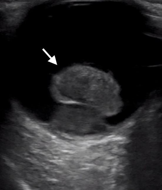
discharge, headache, or other focal neurological complaints. His past medical history included coronary artery disease, diabetes mellitus, hyperlipidemia, and hypertension. On physical exam, pupils were equal and reactive to light bilaterally. Extraocular movements were intact. Visual acuity was 20/100 in the right eye and 20/30 in the left eye. Slit lamp exam of the right eye revealed a deep, quiet anterior chamber, but a lack of pupillary reflection and inability to visualize the retina. Point-of-care ultrasound of the affected eye was performed in the sagittal and transverse anatomic planes using a high-frequency linear transducer. The prosthetic lens was visualized in the posterior chamber with the temporal side haptic still adherent to the lens capsule consistent with an IOL dislocation (Video, Image), whereas an appropriately positioned prosthetic lens would appear as a hyperechoic curvilinear structure within the lens capsule posterior to the iris. The patient was then asked to perform the six cardinal positions of gaze to maximize visualization of the ocular
332
Volume V, no. 3: August 2021