Terminology, Positioning, and Imaging Principles C H A P T E R 1
15
POSITIONING TERMINOLOGY Radiographic positioning refers to the study of patient positioning performed for radiographic demonstration or visualization of specific body parts on image receptors (IRs). Each person who plans to work as a radiologic technologist must clearly understand the correct use of positioning terminology. This section lists, describes, and illustrates the commonly used terms consistent with the positioning and projection terminology as approved and published by the American Registry of Radiologic Technologists (ARRT).* These terms, with the exception of the term “view,” are also generally consistent with the terms used in Canada, according to the Canadian Association of Medical Radiation Technologists (CAMRT). (See summary of potentially misused terms at the end of this section.) Throughout this text, named positions (i.e., with the proper name of the person who first described a specific position or procedure) are referred to as methods, such as the Towne, Waters, and Caldwell methods. The ARRT and the CAMRT concur regarding the use of the named method in parentheses after the projection or position term. The description of radiographic positions by the proper name method is becoming less common.
Viewing radiographs A general rule in viewing radiographs is to
display them so that the patient is facing the viewer, with the patient in the anatomic position.
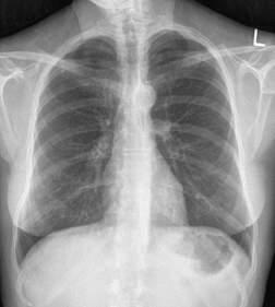
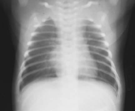
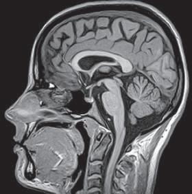
General Terms Radiograph (ra′-de-o-graf ): An image of a patient’s anatomic part(s), as produced by the action of x-rays on an image receptor (Fig. 1-33). If the radiograph is produced with the use of traditional film-screen technology, the image is stored and displayed on film; if the radiograph is produced via digital technology, the image is viewed and stored with the use of computers. Radiography (ra˝-de-og′-rah-fe): The process and procedures of producing a radiograph. Radiograph versus x-ray film: In practice, the terms radiograph and x-ray film (or just film) are often used interchangeably. However, x-ray film specifically refers to the physical piece of material on which a latent (nonprocessed) radiographic image is stored. The term radiograph includes the recording medium and the image. Image receptor (IR): The device that captures the radiographic image that exits the patient; refers to both film-screen cassettes and digital acquisition devices. Central ray (CR): Refers to the center-most portion of the x-ray beam emitted from the x-ray tube; the portion of the x-ray beam that has the least divergence. Radiographic examination or procedure
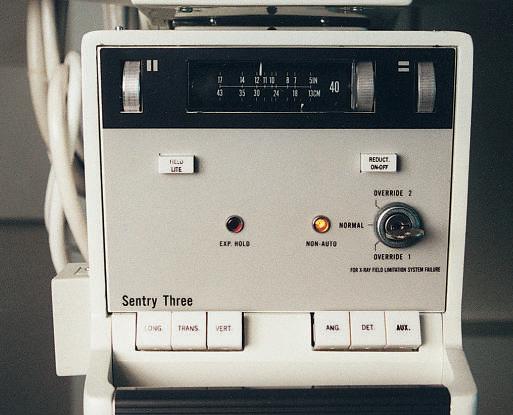
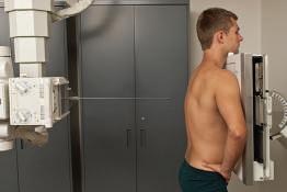
A radiologic technologist is shown positioning the patient for a routine chest examination or procedure (Fig. 1-34). A radiographic examination involves five general functions: 1. Positioning of body part and alignment with the IR and CR 2. Application of radiation protection measures and devices 3. Selection of exposure factors (radiographic technique) on the control panel 4. Instructions to the patient related to respiration (breathing) and initiation of the x-ray exposure 5. Processing of the IR (film-based [analog] and cassette-based [PSP] system)
L
Fig. 1-33 Chest radiograph.
Fig. 1-34 Radiographic examination.
Anatomic position
The anatomic (an˝-ah-tom′-ik) position is a reference position that defines specific surfaces and planes of the body. The anatomic position is an upright position with arms abducted slightly (down), palms forward, and head and feet directed straight ahead (Fig. 1-35). *ARRT educator’s handbook, ed 3, St. Paul, 1990, The American Registry of Radiologic Technologists; personal communication and correspondence with ARRT, November 1999.
Fig. 1-35 Anatomic position.
1