Terminology, Positioning, and Imaging Principles C H A P T E R 1
63
Specific Area Shielding Specific area shielding is essential when radiosensitive organs, such as the thyroid gland, breasts, and gonads, are in or near the useful beam and the use of such shielding does not interfere with the objectives of the examination. The most common and most important area shielding is gonadal shielding, which significantly lowers the dose to the reproductive organs. Gonadal shields, if placed correctly, reduce the gonadal dose by 50% to 90% if the gonads are in the primary x-ray field. Consider the following two examples. For a male, the AP unshielded hip delivers an ED of 0.43 mSv (43 mrem). This is primarily due to the dose to the testes, which can be greatly decreased to 0.07 mSv (7 mrem) with gonadal shielding. For a female, the AP thoracic spine without breast shields produces an ED of 0.63 mSv (63 mrem). This is primarily caused by dose to the breast, which can be reduced by breast shielding or collimation (ED is decreased to 0.35 mSv or 35 mrem). The two general types of specific area shielding are shadow shields and contact shields.
1
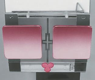
Fig. 1-179 Breast shadow shields designed to be attached to collimator exit surface with Velcro. Collimator Breast shields
Shadow shields As the name implies, shadow shields, which are
attached to the collimator, are placed between the x-ray tube and the patient and cast a shadow on the patient when the collimator light is turned on. The position of the shadow shield is adjusted to define the shielded area. One such type of shadow shield, as shown in Fig. 1-179, is affixed to the collimator exit surface with Velcro. Another type of shadow shield, as shown in Fig. 1-180, is mounted with magnets directly to the bottom of the collimator. These shields may be combined with clear lead compensating filters to provide more uniform exposure for body parts that vary in thickness or density, such as for a thoracic and lumbar spine scoliosis radiograph (Fig. 1-181).
Gonad shield
Fig. 1-180 Shadow shields in place under collimator (attached with magnets). (Courtesy Nuclear Associates, Carle, NY.)
Contact shields Flat gonadal contact shields are used most
commonly for patients in recumbent positions. Vinyl-covered lead shields are placed over the gonadal area to attenuate scatter or leakage radiation or both (Fig. 1-182). These shields usually are made from the same lead-impregnated vinyl materials that compose lead aprons. Gonadal contact shields, 1 mm lead equivalent, absorb 95% to 99% of primary rays in the 50- to 100-kV range. Examples of these include small vinyl-covered lead material cut into various shapes to be placed directly over the reproductive organs, as shown in Figs. 1-183 and 1-184. Male Gonadal shields should be placed distally to the symphysis pubis, covering the area of the testes and scrotum (Fig. 1-183). The upper margin of the shield should be at the symphysis pubis. These shields are tapered slightly at the top and are wider at the bottom to cover the testes and scrotum without obscuring pelvic and hip structures. Smaller sizes should be used for smaller males or children. Female Gonadal shielding is placed to cover the area of the ovaries, uterine tubes, and uterus but may be more difficult to achieve. A general guideline for women is to shield an area 4 12 to 5 inches (11 to 13 cm) proximal or superior to the symphysis pubis extending 3 to 3 12 inches (8 to 9 cm) each way from the pelvic midline. The lower border of the shield should be at or slightly above the symphysis pubis, with the upper border extending just above the level of the anterior superior iliac spines (ASIS) (Fig. 1-184). Various-shaped female ovarian shields may be used, but they should be wider in the upper region to cover the area of the ovaries and narrower toward the bottom to offer less obstruction of pelvic or hip structures. The shielded area should be proportionally smaller on children. For example, a 1-year-old girl would require a shield that is only about 2 to 3 inches (6 to 7 cm) wide and 2 inches (5 cm) high placed directly superior to the symphysis pubis.* *Godderidge C: Pediatric imaging, Philadelphia, 1995, Saunders.
Fig. 1-182 Vinyl-covered lead shield in place over pelvis for lateral mid and distal femur.
A
A –Male gonadal shield
B
Fig. 1-181 AP spine for scoliosis with compensating filter and breast and gonadal shields in place. (Courtesy Nuclear Associates, Carle, NY.)
Possible shapes
Fig. 1-183 A, AP pelvis with flat contact shield (1 mm lead equivalent). B, Male gonadal shield shapes.
–Female ovarian shield
B
Possible shapes
Fig. 1-184 A, AP right hip with flat contact shield (1 mm lead equivalent). B, Female ovarian shield shapes.