76 FORMACIÓN CONTINUA
del historial previo, serán los que nos permitirán llegar al diagnóstico definitivo. Otra prueba complementaria que puede ayudar al diagnóstico es la medición de troponinas, biomarcador que indica daño miocárdico. En ambas patologías los niveles estarán alterados, pero en caso de miocarditis están significativamente más altas que en pacientes con cardiomiopatía fenotipo hipertrófica, lo que nos puede encaminar hacia un diagnóstico u otro. En nuestro caso no fue posible la medición de las mismas por motivos económicos del propietario.
Con este caso se pretende destacar la importancia de incluir en nuestra lista de diagnósticos diferenciales el engrosamiento transitorio miocárdico y la cardiomiopatía fenotipo hipertrófica, sobre todo en gatos jóvenes que hayan afrontado previamente sucesos potencialmente predisponentes. Estos pacientes, al presentar pronóstico favorable e, incluso, no requerir tratamiento de manera crónica, pueden cambiar de manera significativa la manera de afrontar el caso, así como las decisiones clínicas que se tomen.
Bibliografía:
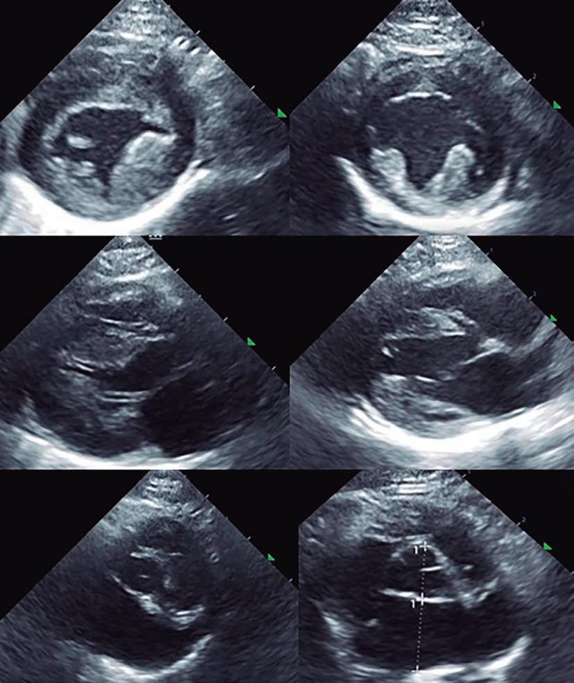
Figura 2. Comparativa de los cortes ecocardiográficos del paciente a la llegada (izquierda) y en el momento del alta definitiva, 30 días después (derecha).
Nº 239 • Junio 2022
1. J Novo Matos, N Pereira, T Glaus, L Wilkie, K Borgeat, J Loureiro, J Silva, V Law, A Kranjc, D J Connolly, V Luis Fuentes. Transient Myocardial Thickening in Cats Associated with Heart Failure J Vet Intern Med 2018 Jan;32(1):4856. 2. Fujiwara H, Hoshino T, Yamana K, et al. Number and size of myocytes and amount of interstitial space in the ventricular septum and in the left ventricular free wall in hypertrophic cardiomyopathy. Am J Cardiol 1983; 52:818–823 3. Campbell FE, Kittleson MD. The effect of hydration status on the echocardiographic measurements of normal cats. J Vet Intern Med 2007; 21:1008–1015. 4. Myers JA, Lunn KF, Bright JM. Echocardiographic findings in 11 cats with acromegaly. J Vet Intern Med 2014; 28:1235–1238. 12. 5. Payne J, Luis Fuentes V, Boswood A, et al. Population characteristics and survival in 127 referred cats with hypertrophic cardiomyopathy (1997 to 2005). J Small Anim Pract 2010; 51:540–547. 13. 6. Rush JE, Freeman LM, Fenollosa NK, et al. Population and survival characteristics of cats with hypertrophic cardiomyopathy: 260 cases (1990–1999). J Am Vet Med Assoc 2002; 220:202–207. 14. 7. Glaus T, Wess G. [Left ventricular hypertrophy in the cat - “when hypertrophic cardiomyopathy is not hypertrophic cardiomyopathy”]. Schweiz Arch Fur Tierheilkd 2010; 152:325 –330. 15. 8. Harris KM, Spirito P, Maron MS, et al. Prevalence, clinical profile, and significance of left ventricular remodeling in the endstage phase of hypertrophic cardiomyopathy. Circulation 2006; 114:216–225. 16. 9. Baty CJ, Malarkey DE, Atkins CE, et al. Natural history of hypertrophic cardiomyopathy and aortic thromboembolism in a family of domestic shorthair cats. J Vet Intern Med 2001; 15:595– 599. 17. 10.Kosutic J. Severe transient left ventricular hypertrophy in an infant with acute myocarditis and heart failure. Pediatr Cardiol 2004; 25:677–680. 18. 11.Riera Sagrera M, Fiol Sala M, Perez Barcena J, et al. Acute myocarditis and left ventricular “hypertrophy”. Echocardiogr 2000;17:567–570. 19. 12.Hauser AM, Gordon S, Cieszkowski J, et al. Severe transient left ventricular “hypertrophy” occurring during acute myocarditis. Chest 1983; 83:275–277. 13.Langhorn R, Tarnow I, Willesen JL, et al. Cardiac troponin I and T as prognostic markers in cats with hypertrophic cardiomyopathy. J Vet Intern Med 2014; 28:1485–1491. 32. 14.Payne JR, Brodbelt DC, Luis Fuentes V. Cardiomyopathy prevalence in 780 apparently healthy cats in rehoming centres (the CatScan study). J Vet Cardiol 2015;17(Suppl 1): S244–S257.